ODONTOGENIC CYSTS
49
-
Upload
yasminmoidin -
Category
Documents
-
view
24.143 -
download
6
description
ORAL AND MAXILLOFACIAL SURGERY
Transcript of ODONTOGENIC CYSTS


SEMINARODONTOGENIC CYSTS
CLINICAL FEATURES
RADIOGRAPHIC FEATURES
DIFFERENTIAL DIAGNOSIS
Yasmin Moidin
2008 Batch
Al Azhar Dental College
Thodupuzha

Introduction
Classification
Types of odontogenic cysts
Conclusion
Reference
CONTENTS

Cyst is defined as a pathological cavity filled with
fluid which is solid semisolid or gaseous form
which may or may not be lined by epithelium
Cyst can occur within bone or soft tissues
They may be asymptomatic or associated with
swelling and pain
INTRODUCTION

Cysts are generally slow growing, expansile
lesions
They grow by hydraulic expansion
Radiographically , they often appear
radiolucency surrounded by thin
radioopaque border

CLASSIFICATION BY ETIOLOGY
DEVELOPMENTAL: Unknown origin but are not the
result of an inflammatory reaction
Dentigerous cyst
Eruption cyst
Odontogenic keratocyst
Gingival cyst of newborn
CLASSIFICATION

Gingival cyst of adult
Lateral periodontal cyst
Calcifying odontogenic cyst
Glandular odontogenic cyst
INFLAMMATORY: result of inflammation
periapical cyst
Residual cyst
Paradental cyst

CLASSIFICATION BY TISSUE OF ORIGIN
DERIVED FROM RESTS OF MALASSEZ
Periapical cyst
Residual cyst
DERIVED FROM DENTAL LAMINA (RESTS OF
SERRES)
Odontogenic kertocyst
Gingival cyst of newborn
Gingival cyst of adult

Lateral periodontal cyst
Glandular odontogenic cyst
UNCLASSIFIED
Paradental cyst
Calcifying odontogenic cyst

A dentigerous cyst results because of the
enlargement of the follicular space of the whole
or part of the crown of an impacted or unerupted
tooth and is attached to the neck of the tooth
DENTIGEROUS CYST
CLINICAL FEATURES
The cyst is always associated initially with the crown
of an impacted, embedded or unerupted tooth
Sites: mandibular and maxillary third molar and
maxillary cuspid areas
Most dentigerous cysts are solitary
Expansion of bone with subsequent facial
asymmetry, extreme displacement of teeth, sever
root resorption of adjacent teeth and pain

RADIOGRAPHIC FEATURES
Reveal a radiolucent area associated with an
unerupted tooth crown
The radiolucent area is surrounded by a thin
sclerotic line representing bony reaction

Three radiological variations:-
Central variety, crown is enveloped symmetrically
Lateral type, results from dilatation of the follicle
on one aspect of the crown
Circumferential type, follicle expands and entire
tooth is enveloped by cyst
DIFFERENTIAL DIAGNOSIS
Ameloblastoma and ameloblastic fibroma
Adenomatoid odontogenic tumour
Calcifying odontogenic cyst

Developmental primordial and follicular
primordial cyst
Odontogenic keratocyst
Cystic ameloblastoma
Radicular cyst
Hyperplastic follicle

Eruption cyst is defined as an odontogenic cyst
that surrounds a tooth crown that has erupted
through bone but not soft tissue and is clinically
visible as a soft fluctuant mass on the alveolar
ridges
ERUPTION CYST

CLINICAL FEATURES
Found in children and occcasionally in adults
Common site – anterior to first molar
Lesion appear as circumscribed, fluctuant, translucent
swelling of the alveolar ridge over the site of the erupting
tooth
When the circumcoronal cystic cavity contains blood,
swelling appears purple or deep blue; hence the term
ERUPTION HAEMATOMA

RADIOGRAPHIC FEATURES
It may show soft tissue shadow since the cyst is
confined within it and there is usually no bone
involvement
DIFFERENTIAL DIAGNOSIS
Dentigerous cyst

A cyst derived from the remnants of the dental
lamina, with a distinctive lining of six to ten cells
in thickness and exhibits a basal cell layer of
palisaded cells and a surface of corrugated
parakeratin
ODONTOGENIC KERATOCYST

CLINICAL FEATURES The cyst may occur at any age
Peak incidence is in 2nd and 3rd decades of life
Predilection for occurrence in males
Common site:- third molar area
Other features: pain, soft-tissue swelling and
expansion of bone and neurological
manifestations – parasthesia of lips

RADIOGRAPHIC FEATURES
They are unilocular, presenting a well-defined
peripheral rim
Scalloping of the border represents variations in
the growth pattern of cyst

DIFFERENTIAL DIAGNOSIS
Dentigerous cyst
Ameloblastoma
Primordial cyst
Residual cyst
Traumatic cyst
Benign odontogenic tumor
Giant cell granuloma
Odontogenic myxoma

These are multiple , occasionally solitary,
superficial raised nodules on edentulous alveolar
ridges of infants that resolve without treatment,
derived from rests of the dental lamina and
consisting of keratin-producing epithelial lining
GINGIVAL CYST OF NEWBORN

CLINICAL FEATURES
Small discrete white swellings of the alveolar
ridge
Lesion appear to be asymptomatic and donot
produce discomfort in infants

A small developmental odontogenic cyst of the
gingival soft tissue derived from the rests of the
dental lamina, containing a lining of embryonic
epithelium of cuboidal cells and distinctive focal
thickenings
GINGIVAL CYST OF ADULT

CLINICAL FEATURES
Lesion is small, well-cirumscribed , painless,
swelling of the gingiva
Lesion is of same color as the adjacent normal
mucosa and 1cm in diameter
It occurs in free or attachment gingiva and
gingival papilla

RADIOGRAPHIC FEATURES
It is a soft tissue lesion and if it enlarges to
sufficient size, it causes superficial erosion of the
cortical plate of bone
DIFFERENTIAL DIAGNOSIS Lateral periodontal cyst

A slow growing, non expansile developmental
odontogenic cyst derived from one or more rests
of the dental lamina, containing an embryonic
lining of one to three cuboidal cells and
distinctive focal thickenings
LATERAL PERIODONTAL CYST

CLINICAL FEATURES
Predilection for occurrence in males
Site: mandibular bicuspid/cuspid/incisor area
When the cyst is located on labial surface of the
root, there is a slight mass

RADIOGRAPHIC FEATURES
Radiolucent area in apposition to the lateral
surface of a tooth root
Lesion is small, border is definitive and
surrounded by thin layer of sclerotic bone

DIFFERENTIAL DIAGNOSIS
Gingival cyst of adult Botryoid cyst Lateral radicular cyst Lateral periodontal abscess Lateral dentigerous cyst Residual cyst Primordial cyst Globullomaxillary cyst Median mandibular cyst Small OKC Mental foramen Small neurofibroma Radicular cyst

A rare, well circumscribed, solid or cystic lesion
derived from odontogenic epithelium that
contains ghost cells and spherical calcifications
CALCIFYING ODONTOGENIC CYST

CLINICAL FEATURES
It is more common in females
It occurs anterior to first molar
It is a slow growing , painless, non-tender
swelling which causes expansion and destruction
of cortical plates
The cystic mass may become palpable and
discharging

RADIOGRAPHIC FEATURES
The central lesion may appear as a cyst like
radiolucency with variable margins which may be
smooth well defined or irregular in shape with
poorly defined borders
Perforation of cortical plates can be seen

DIFFERENTIAL DIAGNOSIS
Fibrous dysplasia
Partially calcified odontoma
Adenomatoid odontogenic tumor
Ossifying fibroma
Odontogenic fibroma
Cementoblastoma
Dentigerous cyst
Ameloblastic fibroadenoma
Calcifying epithelial odontogenic tumor

A large solitary or multilocular odontogenic cyst
derived from rests of dental lamina, consisting of
a stratified squamous epithelium containing
numerous mucus-secreting cells
GLANDULAR ODONTOGENIC CYST

CLINICAL FEATURES
A slight male predilection
Common site is anterior mandible
Lesion shows slow progressive growth, painless
and locally destructive

RADIOGRAPHIC FEATURES
The lesions appear well defined with a multilocular pattern
DIFFERENTIAL DIAGNOSIS
Lateral periodontal cyst

Periapical cysts results when rests of malassez in
the periodontal ligament are stimulated to
proliferate and undergo cystic degeneration by
inflammatory products which are associated with
necrosis of the pulp
PERIAPICAL CYST

CLINICAL FEATURES
The tooth is painful and sensitive to percussion
It represents a chronic inflammatory process and
develops over a prolonged period of time
This acute exacerbation leads to abscess, then
proceed to a cellulitis and form a draining fistula

RADIOGRAPHIC FEATURES
A peri or para apical round or oval radiolucency of
variable size which is well-delineated and marked
with a radioopaque rim

DIFFERENTIAL DIAGNOSIS
Periapical granuloma
Periapical scar
Surgical defect
Periapical cementoma
Traumatic bone cyst
Periapical abscess
Mandibular infected buccal cyst
Odontogenic keratocyst
Benign tumor
Ossifying fibroma

Residual cyst are retained periapical cysts from
teeth that have been removed
CLINICAL FEATURES
Usually asymptomatic and found on routine
radiographic examinations
It is found in tooth bearing area
RESIDUAL CYST
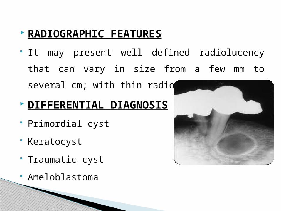
RADIOGRAPHIC FEATURES
It may present well defined radiolucency that can
vary in size from a few mm to several cm; with
thin radioopaque margins
DIFFERENTIAL DIAGNOSIS
Primordial cyst
Keratocyst
Traumatic cyst
Ameloblastoma

Paradental cyst is an inflammatory cyst which
develops on the lateral surface of a tooth root
PARADENTAL CYST

CLINICAL FEATURES
It occurs in younger age group
Seen in third decades of life
Male predilection
It is usually associated with third molar on buccal
surface and covers the bifurcation
The tooth is usually vital
It may occur bilaterally

RADIOGRAPHIC FEATURES
These are the non-widening of the periodontal
ligamentspace and the lesion was superimposed
on the buccal root surface
When there was a distal as well as a buccal
radiolucency, the distal element was separate
from distinct distal follicular space

DIFFERENTIAL DIAGNOSIS
Periodontal abscess
Langerhan’s cell histiocytosis
Dentigerous cyst

S M Balaji – textbook of oral and
maxillofacial surgery
Neelima Malik – textbook of oral and
maxillofacial surgery
Shafers – textbook of oral pathology
Freny Karjodhkar – textbook of radiology
REFERENCE


![Short Case Report · Calcifying odontogenic cysts (COCs), or Gorlin’s cysts, are rare lesions that account for less than 1% of all odontogenic cysts [1]. This article details two](https://static.fdocuments.in/doc/165x107/5fea60342725d22f6c4de3eb/short-case-report-calcifying-odontogenic-cysts-cocs-or-gorlinas-cysts-are.jpg)