clinicaljude-5thyear.yolasite.comclinicaljude-5thyear.yolasite.com/resources/RadiologyII... · Web...
16
Lecture 10 , 3-12-2014 Radiographic signs of cysts Cysts and benign odontogenic tumors are two cousin categories Definition :”cyst is a pathologic cavity filled with fluid, lined by epithelium, and surrounded by a definite connective tissue wall” Radiographic signs: Radiolucent lesion sometimes with dystrophic calcification and typically don’t have septatae well defined and corticated most of the time round or oval They expand by hydrostatic expansion like a balloon , translated radiographically as a uniform expansion typically unilocular not multilocular and they don’t usually have scalloped appearance Effects on surrounding structures: displacement(mass effect) ,resorption to the roots if they stay longer but they definitely would not destruct a cortex but they expand the cortex cause thinning of the cortex sometimes and remodel it another times, on extreme thinning may be converted to a simple perforation but that’s typically a really huge cyst that is long standing Cysts and cyst like lesions : Odontogenic cysts Nonodontogenic cysts Pseudo cysts Odontogenic cysts:
Transcript of clinicaljude-5thyear.yolasite.comclinicaljude-5thyear.yolasite.com/resources/RadiologyII... · Web...

Lecture 10 , 3-12-2014
Radiographic signs of cysts
Cysts and benign odontogenic tumors are two cousin categories
Definition :”cyst is a pathologic cavity filled with fluid, lined by epithelium, and surrounded by a definite connective tissue wall”
Radiographic signs:
Radiolucent lesion sometimes with dystrophic calcification and typically don’t have septatae
well defined and corticated most of the time round or oval They expand by hydrostatic expansion like a balloon , translated radiographically as a
uniform expansion typically unilocular not multilocular and they don’t usually have scalloped appearance
Effects on surrounding structures: displacement(mass effect) ,resorption to the roots if they stay longer but they definitely would not destruct a cortex but they expand the cortex cause thinning of the cortex sometimes and remodel it another times, on extreme thinning may be converted to a simple perforation but that’s typically a really huge cyst that is long standing
Cysts and cyst like lesions :
Odontogenic cysts
Nonodontogenic cysts
Pseudo cysts
Odontogenic cysts:
The most common one is radicular cyst Followd by residual cyst Dentegerous cyst Buccal bifurcation cyst Lat periodontal cyst Glandular odontogenic cyst KCOT cyst

Calcifying odontogenic cysts
Non odontogenic cysts :
nasopalatine duct cyst nasolabial cyst dermoid cyst
Pseodocysts:
simple bone cysts aneurismal bone cyst mucous retention phenomenon
The difference between those kinds : location , effect on surrounding structures or sometimes there is no difference unless it’s a dental difference (vital or non vital tooth)
Radicular cysts :
Its an Inflammatory cyst that is typically associated with a non vital tooth
How can I differentiate between a radicular cyst and a granuloma:
Size criteria : <1 cm granuloma , >1 cm : cyst Margin criteria: well defined corticated then it’s a cyst if I am not seeing a cortex I think
more of a granuloma According to Histopathology
Location : around an epicenter of a non vital tooth
Interestingly it comes More in the maxilla ( it has to do with the bone and vascularity of the bone
Just like any other cyst its Well defined corticated radiolucent and causes displacement and expansion
Superinfection will lead to loss of cortication( I opened a flap to take a biopsy or to do Marsupialization then I may introduce a superinfection on top of the existing cyst and that will make loss of cortication.
Treatment : RCT if the tooth structure is restorable and that’s it, but if it’s a remaining root I have nothing to do except extraction and curettage
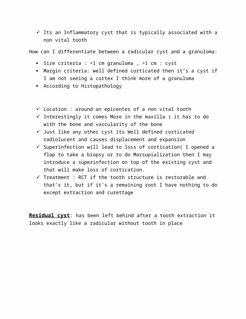
Residual cyst: has been left behind after a tooth extraction it looks exactly like a radicular without tooth in place
Dentigerous cyst : common
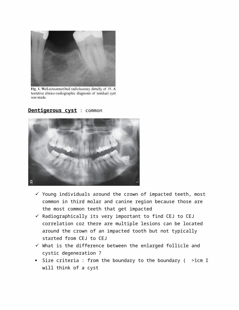
Young individuals around the crown of impacted teeth, most common in third molar and canine region because those are the most common teeth that get impacted
Radiographically its very important to find CEJ to CEJ correlation coz there are multiple lesions can be located around the crown of an impacted tooth but not typically started from CEJ to CEJ
What is the difference between the enlarged follicle and cystic degeneration ? Size criteria : from the boundary to the boundary ( >1cm I will think of a cyst But if I make a straight line from the center to the circumference of the cyst: >6 or 7mm I
will think of a cyst (coz its difficult to find the exact center) If there is an Effect on surrounding structures I will think of a cyst. for example I follow
up a third molar which was near the roots of the 7 then after 6 months it comes farther so it’s causing displacement just like the below images showing panoramic films of the same case taken several years apart demonstrate superior-posterior displacement of a maxillary third molar by a dentigerous cyst
Like any other cyst well defined, unilocular ,corticated, radiolucent and attached to the cervical region of an impacted crown
Differential diagnosis of any cyst around an impacted tooth: Unicystic ameloblastoma KCOT dentegarous cyst
those three are always high in the list of the differential diagnosis when we see a cyst around an impacted tooth

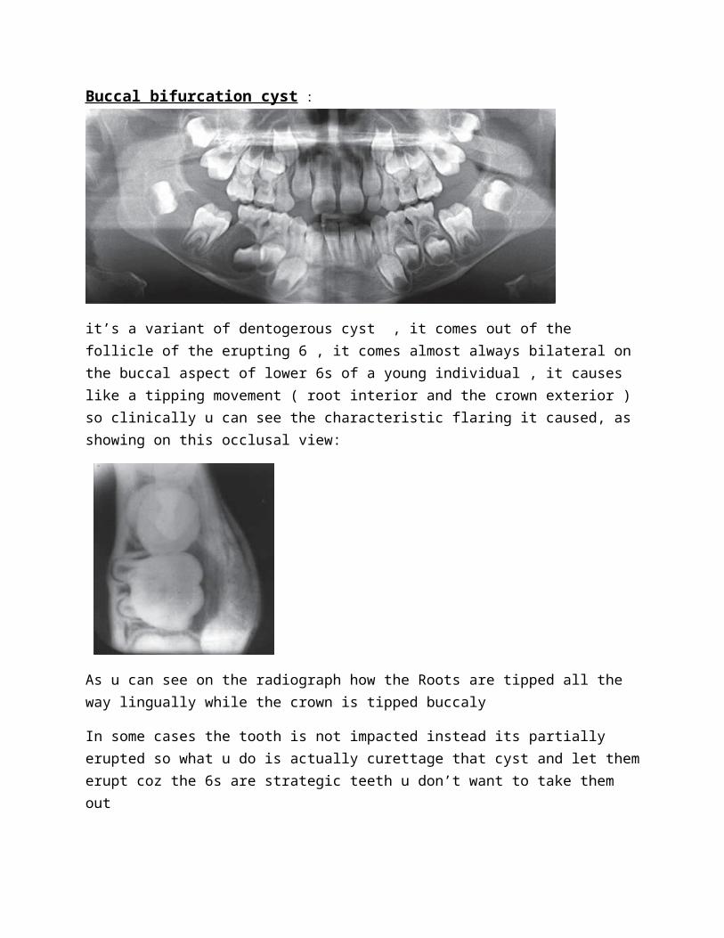
Buccal bifurcation cyst :
it’s a variant of dentogerous cyst , it comes out of the follicle of the erupting 6 , it comes almost always bilateral on the buccal aspect of lower 6s of a young individual , it causes like a tipping movement ( root interior and the crown exterior ) so clinically u can see the characteristic flaring it caused, as showing on this occlusal view:
As u can see on the radiograph how the Roots are tipped all the way lingually while the crown is tipped buccaly
In some cases the tooth is not impacted instead its partially erupted so what u do is actually curettage that cyst and let them erupt coz the 6s are strategic teeth u don’t want to take them out
Lateral periodontal cyst : typical location: on the coronal/middle third between the two premolars in the mandible but that doesn’t mean it doesn’t appear on other places but this is the typical location of it
Adjacent to Vital teeth and usually completely asymptomatic
This radiograph is very interesting I can see root canal treated tooth with a post and lat radiolucency it could be lateral radicular cyst because of a vertical root fracture (after the post) and this might need even more 3d imaging or even surgical exploration to see if the tooth is fractured, if not all I need to do is to curette this small lat periodontal cyst
It is much easier to diagnose if it’s a virgin tooth, I will make a vitality test to diagnose it , if non vital then it’s a lateral radicular cyst so RCT and follow up, if vital curette the lat perodontal cyst and that’s it
Also if I was lucky to see a lamina dura this will help me to differentiate between a lat periodontal cyst(originated from cell rest of serrez/developmental origin) and a lateral radicular cyst(cell rest of melessez/inflammatory origin)
Betryoid odontogenic cyst: grape like, from rest cell of serrez (it is a variant of lat periodontal cyst) but it’s multilocular and more aggressive than lat periodontal cyst and even some cases recurrence have been reported and it may cause swelling because its big

Glandular odontogenic cyst : rare entities first reported in the early 80s typical on the midline of the mandible , multilocular , very debatable origin because there are some salivary gland tissues in there
More aggressive could look like a very low grade malignancy benign lesion so treatment : not only curettage u have actually to go a little bit beyond and clean the margins
It goes down on the list for academic purposes coz it’s very rare Differential diagnosis : central mucoepidermoid carcinoma
When I see it in the radiographs, a cyst will not be high up in the list of my differential diagnosis it’s more of odontogenis locally aggressive benign tumors coz its more multilocular AND MORE EXPANSILE THAN ANY TYPICAL CYST.
KCOT :Keratocystic Odontogenic Tumor: it’s not about classification either it’s a tumor or a cyst it’s all about management , they found that KCOT is less aggressive from a clinical perspective than anybody was understanding so the WHO will return it in the classification of a cyst OKC ,so there are some lesions really not a clear cut
Young people , 2nd- 3rd decade , male predominant , posterior mandible it has its famous thick yellow aspiration” cheesy material”

It fits the radiographic logic of a tumor coz it has more of undulating border, some are multilocular but very limited expansion as opposed to the big expansion of amelobastoma Despite the large size in this panoramic image, the buccal and lingual cortical plates of the mandible have been expanded only slightly
if I found A KCOT the 2nd most important thing is to look for more, because we have to differentiate is it a solitary KCOT or multiple because of Gorlin-Gotz syndrome with multiple KCOTs and multiple nevoid basal cell carcinomas of the skin
Basal cell nevus syndrome (Gorlin Goltz) syndrome : multiple KCOTs ,
1st-3rd decades (comes earlier than solitary KCOT Basal cell carcinomas mostly on the head and neck area Higher recurrence rate than regular KCOT Basal cell carcinoma needs very good follow up , it comes on the skin so it’s easy to
follow it up unlike the KCOTs which is not easy to follow so it needs very strict radiographic follow up (panoramics , periapicals, low dose protocol CBCT of maxilla and mandible)coz the pt is a growing kid with teeth are trying to erupt so to take out a small part u will give him a chance for healthy growth of maxilla and mandible, proper eruption and proper occlusion if you took more then the results could be fibrosis ,adhesion ,growth issues , missing teeth problems,temporary prosthesis until the pt grow up to have an implant , so it’s very important for the quality of life of the pt
it needs to become very big in order to see enough asymmetry or enough expansion
CCOT : calcifying cystic odontogenic tumor (Gorlin cyst ) WHO now categorizes this entity as a tumor.

Very rare ,” any time, anywhere, any place” Slowly growing expansile It has a mixed density internal character Differential diagnosis of a mixed density lesion in the dentoalveolar area :
CCOT Adenomatoid Odontogenic Tumor Calcifying Epithelial Odontogenic tumor CEOT.
Non odontogenic cysts :
Naso palatine duct cyst: heart shaped between the two maxillary centrals
It can be a variant of anatomy as far as it’s a large incisive foramen) so we will return to Size criteria : >6-8 mm we will think more of a cyst but the doctor believes in displacement criteria : so if its big (=8 mm “border line” )and not creating any displacement of the adjacent central incisor then it’s probably not a cyst
Heart shape : The shadow of the nasal spine sometimes is superimposed on the cyst, giving it a heartshape. So typically the centrals have to be vital otherwise it could be radicular cyst related to one or both incisors
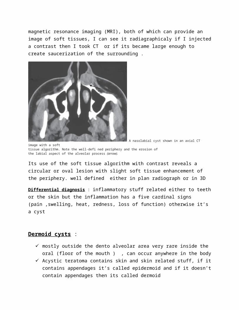
Naso labial cyst: mostly it’s a soft tissue thing Because this is a soft tissue lesion, plain radiographs may not show any detectable changes. The investigation could include either CT or magnetic resonance imaging (MRI), both of which can provide an image of soft tissues, I can see it radiagraphicaly if I injected a contrast then I took CT or if its became large enough to create saucerization of the surrounding .

A nasolabial cyst shown in an axial CT image with a softtissue algorithm. Note the well-defi ned periphery and the erosion ofthe labial aspect of the alveolar process (arrow).
Its use of the soft tissue algorithm with contrast reveals a circular or oval lesion with slight soft tissue enhancement of the periphery. well defined either in plan radiograph or in 3D
Differential diagnosis : inflammatory stuff related either to teeth or the skin but the inflammation has a five cardinal signs (pain ,swelling, heat, redness, loss of function) otherwise it’s a cyst
Dermoid cysts :
mostly outside the dento alveolar area very rare inside the oral (floor of the mouth ) , can occur anywhere in the body
Acystic teratoma contains skin and skin related stuff, if it contains appendages it’s called epidermoid and if it doesn’t contain appendages then its called dermoid
Neck is the most common place Soft tissue cyst definitely needs CT or MRI to see it Multiple densities according to its contents (if its fibrous, or more skin appendage, or
more fatty ) Developmental , rare
Treatment : excision

This radiograph is a Soft tissue window of a CT scan (the muscles are very clear and u can distinguish the fat between them , so its soft tissue axial cut on the level of C4 because the esophagus is clear there.
Cyst like conditions :
Simple bone cyst :
no epithelial lining , it doesn’t fit to be a cyst (hence the name :cyst like) Most of the time they are empty but sometimes it contains fluids Basically Unknown etiology they think it’s caused be trauma Effect on surrounding structures: No expansion or resorption it appears like undulating
border in between root surfaces(scalloped borders as u see in the radiograph below) because it’s just a simple cavity
Treatment : induce bleeding , but most of them heals by itself without any surgical or conservative intervention
Diff diagnosis:
Lat radicular cyst lat periodontal cyst KCOT but if I aspirate and nothing comes out(empty) then it’s a simple bone cyst
Aneurismal bone cyst : the most expansile cyst , so it’s very painful , blood on aspiration , coz its blood filled pools , most of those are in the posterior mandible (condyle area) very few comes anteriorly
It looks like CGCG since its granular cell origin but typical CGCG comes mostly in the anterior mandible so if I see this radiograph I will the start with the most common :
differential diagnosis : ameloblastoma Myxoma aneurismal bone cyst(if its painful and get large within 2 weeks extreme expansion)
Mucous retention phenomenon :

dome shape thickening of the mucosa at the floor of the maxillary sinuses , very dynamic disease ( related to allergies, seasons changes ) it disappears by itself and
reappears again I will worry about it if it becomes big enough to obstruct the Ostium or if I want to make
very major sinus lifting and I am worried if I lift the sinus floor that will place the cyst up to cause obstruction of the Ostium, otherwise its simple and nothing to worry about
I can see the floor of the sinus intact then up to it I see this dome shaped and no cortex
above it coz it’s a soft tissue lesion but if I see the cortical border(floor of the sinus) above it then I would think of other odontogenic stuff
Very important to differentiate it from antral polyps but antral polyps by definition comes multiple while mucous retention phenomenon comes as a single dome shaped