EnterohemorragischeEscherichia coli (O157:H7) (shared using ).

OCCURRENCE OF E COLI O157:H7 AND OTHER PATHOGENIC E COLI STRAINS IN
WATER SOURCES INTENDED FOR DIRECT AND
INDIRECT HUMAN CONSUMPTION by
W O K Grabow, E E Müller, M M Ehlers, C M E de Wet, M Uys and C G Clay
Department of Medical Virology, University of Pretoria and Rand Water Scientific Services, Vereeniging Final Report to the Water Research Commission WRC Report No 1068/1/03 ISBN No :1-86845-988-8
February 2003

Disclaimer This report emanates from a project financed by the Water Research Commission (WRC) and is approved for publication. Approval does not signify that the contents necessarily reflect the views and policies of the WRC or the members of the project steering committee, nor does mention of trade names or commercial products constitute endorsement or recommendation for use.

ii
EXECUTIVE SUMMARY 1. Background and Motivation Escherichia coli is a typical member of the bacterial flora of the gastrointestinal tract of humans and other warm-blooded animals. Generally they are not associated with adverse health effects. For these and related reasons they are widely used as indicators of faecal pollution in water quality assessment. However, under circumstances E coli may cause serious disease. Wild type E coli is notorious for infections with severe implications when it ends up in places where it does not belong, of which the urinary tract is a typical example. E coli may also cause severe disease in the gastrointestinal tract. This happens when wild type E coli bacteria are genetically converted by the integration of genes into the host genome which code for the production of virulence factors. These virulence factors turn the normally harmless bacteria into pathogens. A wide variety of virulence factors has been described for E coli. Depending on the virulence factors concerned, pathogenic strains of E coli have been classified into a number of groups. The origin and purpose of the genetic elements (short sections of nucleic acid which carry one or more genes responsible for the production of the virulence factors) is not clear. It is known that the genetic elements concerned are carried by specific phages which transfer the genetic elements from one E coli bacterium to another. Infection of an E coli bacterium by one of these phages will convert the bacterium into a pathogen. The phages are known to occur in the environment and they seem to serve as a reservoir for the genetic elements which code for the production of the virulence factors. Some of the genetic elements concerned may have been transferred form pathogenic bacteria to E coli by these phages. This theory is supported by the close similarity of the Shiga toxins produced by pathogenic E coli strains to the Shiga toxins of Shigella dysenteriae which causes dysentery with clinical symptoms resembling those caused by E coli O157:H7. This would seem to suggest that the genetic elements which code for the production of Shiga toxins in E coli were transmitted to E coli from S dysenteriae by phages. The phenomenon of phages carrying genetic elements which convert hosts into pathogens is not uncommon among bacteria, and a number of examples are well known. The above suggests that eventually research should actually focus on the key to the problem, namely the phages which convert harmless E coli bacteria into life-threatening pathogens. However, this project does not cover research on the phages which carry the genetic elements concerned. This project is restricted to the incidence of E coli pathogens in selected water resources. Enterotoxigenic strains of E coli (ETEC) are associated with two major clinical syndromes: weanling diarrhoea among children in the developing world, and traveller's diarrhoea typical among people of all ages visiting parts of the world with inadequate hygiene. Epidemiological investigations have implicated contaminated food and water as the most common vehicles for ETEC infection. Enteropathogenic strains of E coli (EPEC) are an import group of pathogens typically associated with infant diarrhoea in the developing world. Transmission seems to be predominantly by direct faecal-oral transfer via contaminated hands, contaminated weaning foods or contaminated

iii
fomites. Water and food have been implicated as the mode of transmission in outbreaks among adults. Enterohaemorrhagic strains of E coli (EHEC) cause life threatening haemorrhagic colitis and haemolytic uraemic syndrome. These strains of E coli produce potent cytotoxins similar to Shiga toxin produced by Shigella species responsible for dysentery. EHEC infections have also been associated with transmission by water and food. Waterborne outbreaks of diseases caused by pathogenic strains of E coli have been described in many parts of the world. One such outbreak which received world-wide publicity occurred in the small community of Walkerton in Canada during May 2000. The borehole drinking water supply got contaminated by rainwater runoff which contained cattle faeces. More than 2000 cases of severe illness and 6 deaths are on record. The outbreak serves as an example not only of the health risks constituted by pathogenic E coli in water sources and supplies, but also of the legal and financial implications to all parties concerned. The ensuing court cases involved the responsible water supply utility as well as public health and water affairs authorities concerned. Ministers of Parliament appeared in court to stand trial on charges of negligence and to take responsibility for the outbreak and claims by victims for large sums of money for compensation. Due to world-wide attention and in the interest of transparency, proceedings of the court case were presented on the internet. The pathogen involved in the Walkerton outbreak was an EHEC strain designated E coli O157:H7. This particular strain has a history of outbreaks in many parts of the world resulting in severe illness with a high mortality rate. It is therefore one of the most feared strains of E coli pathogens. The incidence of waterborne outbreaks of diseases associated with pathogenic strains of E coli would appear to increase. Contamination of water sources is possible in all parts of the world. One reason is that domestic animals, notable cattle and pigs, serve as reservoir for the pathogens. Practical methods for detecting and typing the pathogens are therefore essential. The techniques are required to control the diseases by monitoring the quality of raw and treated water sources, and to monitor the efficiency of treatment and disinfection processes. Efficient control would also require a better understanding of the epidemiology of the E coli pathogens, and the origin and transmission of the genetic elements which turn harmless wild type E coli bacteria into pathogens with serious health and related implications. Methods used in the past to identify and type pathogenic strains of E coli were labour intensive, expensive and time consuming. Some of the tests even required infection of laboratory animals. More recently molecular techniques have been developed which offer practical approaches to the identification and accurate typing of E coli pathogens. These techniques are based on the genetic identification of the genes which code for the virulence factors responsible for the pathogenicity of E coli. The detection techniques are based on either hybridisation of host nucleic acid with gene probes specific for the pathogenicity genes, or on physical detection of the virulence genes after amplification by specific primers in tests based on the polymerase chain reaction (PCR). There is no meaningful information on the incidence of pathogenic E coli in water resources in South Africa. To the best of our knowledge there is currently no other laboratory in the country engaged in comparable work. The pathogens are known to occur in South Africa among humans

iv
and animals like they do in the rest of the world. In other parts of the world the pathogens have been isolated from rivers as well as swimming pools and drinking water supplies. 2. Objectives $ Screen selected sewage and river water samples for the presence of E coli pathogens using
methods described in the literature $ Optimise and establish practical molecular techniques for the detection and typing of E coli
pathogens $ Develop a new procedure for the isolation of E coli O157:H7 from water $ Use the new more sensitive procedure to study the occurrence E coli O157:H7 in sewage and
river water A study of this kind has not previously been carried out in South Africa. The results were expected to have meaningful benefits for practical technology and expertise on water quality assessment with regard to potentially important pathogens. 3. Literature Review A literature review is presented in the Project Report. Further details have been recorded by Müller (2002) and also appear in publications which emanated from this project. 4. Results Two multiplex (triplex) PCR procedures were successfully established for the detection of the virulence factors of E coli pathogens in water. The new techniques were applied in a survey of water at 26 sites in the Vaal Barrage Reservoir drainage basin. Another multiplex PCR was established for the detection of the Stx1 and -2 toxin genes of EHEC E coli strains, notably E coli O157, after preliminary selection of these bacteria on a selective agar growth medium. The two triplex PCR procedures were designed to detect the E coli virulence genes ST and LT (ETEC strains), BFP (Bundle Forming Pilus gene of EPEC strains), eaeA, CNF (Cytotoxic Necrotising Factor gene) and the EIEC invasiveness plasmid gene. One or more of these genes were detected in all but 47 (26 %) of 180 E coli isolates from the Vaal Barrage. The eaeA gene was detected in 101 (56 %) of the isolates, the CNF gene in 96 (53 %), the BFP gene in 25 (13.8 %), the ST gene in 23 (12.7 %), the EIEC gene in 7 (3.8 %) and the LT gene in 3 (1.7 %). A total of 204 samples was analysed for the presence of the Stx1 and Stx2 genes, the haemolysin plasmid gene and E coli O157 specific genes. The haemolysin plasmid gene was detected in 7 (3.4 %), the Stx1 gene in 2 (0.98 %) and the Stx2 in 1 (0.49 %). One of the Stx1 isolates also carried the haemolysin plasmid. No E coli O157 specific genes were detected in any of the isolates. The multiplex PCR procedure established in this project for the detection and typing of E coli pathogens in water proved reliable, rapid (colonies are typed within 4-6 hours) and cost effective since two PCR reactions screen for six genes simultaneously. Since no E coli O157:H7 pathogens were detected in the survey, it was decided to improve the isolation method. The new procedure is based on preliminary enrichment of test samples by

v
means of peptone-saline containing three antibiotics to suppress background growth. The commercial immuno-magnetic (IMS) procedure is then used to specifically recover E coli O157:H7 from the enrichment cultures. The technique is based on magnetic beads covered with specific monoclonal antibodies which bind E coli O157:H7. The beads are then plated on growth media designed for the selective cultivation of E coli O157:H7 colonies. Suspect colonies are picked from these plates and tested for E coli O157:H7 by means of biochemical tests, agglutination with E coli O157 and H7 antisera, and molecular techniques to detect genes coding for toxicity factors. An assessment of the sensitivity of the new enrichment-IMS-selective agar procedure revealed that it was capable of detecting one E coli O157:H7 per ml of seeded samples of sewage, river water, grounded beef and milk. The results confirmed that this procedure was more sensitive than any method previously used for the isolation of E coli O157:H7 from water. The new enrichment-IMS-selective agar procedure was then applied in a survey for E coli O157:H7 in selected samples of sewage, river water, grounded beef and milk. At least one E coli O157:H7 organism was successfully isolated from a sewage sample. This is the first time these pathogens have been isolated from sewage in South Africa. This project confirms for the first time in South Africa the presence of a variety of E coli pathogens in sewage-contaminated river water, and E coli O157:H7 in sewage. The incidence of E coli O157:H7 in the water environments concerned, appeared lower than might be expected. The apparent low incidence of E coli O157:H7 may at least in part be due to observed shortcomings of the final selective cultivation procedure of the isolation procedure used. This warrants attempts to further improve the isolation procedure for E coli O157:H7. Unfortunately it was not possible to assess the findings by comparison to data elsewhere because no meaningful studies of this kind have as yet been reported from other parts of the world. If indeed the incidence of E coli O157:H7 is as low in sewage and river water as indicated by the results of this study, these pathogens would appear not to constitute a meaningful risk to consumers at least for purposes such as recreation and possibly even domestic use. However, results obtained and experience gained suggest the findings may under-estimate the situation (see Chapter 4). However, the finding that some 74 % of E coli bacteria in water of the Vaal River Barrage drainage basin carry one or more toxicity factors indicates reason for concern (see Chapter 3). The available information is inadequate for a meaningful assessment of the public health risk constituted by these pathogens. However, it is known that E coli which carry one or more of these toxicity factors are capable of causing gastro-intestinal disease in humans. The clinical intensity of the illness depends on a variety of factors. Even if the disease was not as severe as potentially caused by E coli O157:H7, it cannot be ignored. In an increasing component of consumers, notably the very young and very old as well as immunocompromised and undernourished individuals, these E coli pathogens may cause serious health implications. The high risk component of consumers referred to tends to increase in most parts of the world. South Africa is no exception. On the contrary, the immunocompromised component of consumers in many communities is exceptionally high due to the incidence of AIDS.

vi
A major benefit of the project is that expertise and technology have been established in an area of water quality analysis of particular public health importance. An infrastructure is in place for further research on the incidence of these pathogens in water resources and supplies. Further research is essential for meaningful assessment of the public health implications of these pathogens. This information is required to formulate control strategies and define quality specifications. 5. Cost estimates The isolation of E coli pathogens from water is a complex process which consists of a number of steps and tests. Not all steps and tests are required for the confirmation and typing of all E coli pathogens. It is therefore not possible to quote a fixed tariff for the isolation, confirmation and typing of E coli pathogens in water. On average the cost may amount to approximately R 700 for an E coli O157:H7 isolate. In addition, the running cost of the tests is probably not the most expensive part of the work. The tests require special laboratory facilities, particularly for the molecular work. The tests are complicated and require staff with high levels of training in advanced technology and expertise. This implies the tests are relatively expensive and not feasible for all laboratories engaged in routine water quality analysis. The tests are therefore best intended for specialist laboratories which render the work as a service to smaller laboratories as and when required. Large scale testing also reduces the cost of the tests. This would place specialist laboratories in a position to conduct research on aspects such as the incidence of the pathogens and their epidemiology, as well as improvement of techniques for the isolation of the pathogens from water and the typing of isolates. 6. Capacity building $ MSc student Mr EE Müller used part of the work he carried out for this project in his
dissertation. $ Technologists and research assistants in our laboratory got familiarised with the technology
concerned. 7. Technology transfer $ Staff at the Rand Water laboratory got familiarised with at least the preliminary steps of the
recovery and isolation of E coli pathogens from water. The characterisation of pathogenic E coli isolates by molecular techniques requires special facilities and advanced training the transfer of which was beyond the scope of this project.
8. Conclusions
vii
All the objectives of the project have been accomplished. New techniques for the detection and typing of pathogenic strains of E coli have been established and successfully applied in practice. Results of the first survey of its kind in South Africa revealed the presence of a substantial number of E coli strains carrying one or more virulence factors in water sources used for recreational purposes and the production of drinking water supplies. 9. Future Research $ This project has facilitated the establishment of an infrastructure for research on E coli
pathogens. Application of the technology and expertise which has been established made it possible to confirm that at least some E coli pathogens do occur in South African water resources and animal reservoirs. The infrastructure should now be used to expand the preliminary observations in order to obtain meaningful information on the situation in South Africa and to assess potential health risks in more detail.
$ The incidence and survival of E coli pathogens in water resources and in water treatment
processes should be investigated. This information is essential for the formulation of strategies to control their transmission by water and food.
$ Available information confirms that research on the phages which carry the genetic elements
for the production of toxins by E coli is essential. A better understanding of these phages is likely to hold the key to the control of E coli pathogens, and probably also a number of other pathogens.
10. Publications and presentations emanating from the Project 10.1. Conference presentations Müller, E.E., Clay, C.G. and Grabow, W.O.K. (2000) Detection and isolation Escherichia coli O157:H7 from sewage and environmental waters using immunomagnetic separation. Water Institute of Southern Africa. (WISA 2000 Conference). Sun City, South Africa 31 May-2 June 2000. Poster presentation. Müller, E.E., Clay, C.G. and Grabow, W.O.K. (2000) Improvement of the immunomagnetic separation method to detect Escherichia coli O157:H7 in sewage and environmental waters. The 1st World Congress of the International Water Association (IWA). Conference Preprints nr 7 (HRMP-A40). Health-Related Water Microbiology. Paris, France 3-7 July 2000. Poster presentation. Müller, E.E., Taylor, M.B., Grabow, W.O.K. and Ehlers, M.M. (2001) Isolation and Characterization of Escherichia coli O157:H7 and Shiga Toxin - converting Bacteriophages from Strains of Human, Bovine and Porcine Origin. The 2nd World Congress of the International Water Association (IWA). (B0308). Health-Related Water Microbiology. Berlin, Germany 15-19 October 2001. Oral presentation 10.2. Publications

viii
Müller, E.E., Ehlers, M.M. and Grabow, W.O.K. (2001) The Occurrence of E. coli O157:H7 in South African Water Sources Intended for Direct and Indirect Human Consumption. Water Research. 35, 3085-3088 Müller, E.E., Grabow W.O.K., and Ehlers, M.M. (2001) Application of the Immunomagnetic Separation Method for the Detection and Isolation of Escherichia coli O157:H7 from Grounded Beef, Milk, Sewage and Environmental Waters. Submitted for publication to: Journal of Medical Microbiology Müller, E.E., Taylor, M.B., Grabow, W.O.K. and Ehlers, M.M. (2001) Isolation and Characterization of Escherichia coli O157:H7 and Shiga Toxin - converting Bacteriophages from Strains of Human, Bovine and Porcine Origin. Submitted for publication to: Water, Science and Technology. Müller, E.E., Grabow W.O.K., and Ehlers, M.M. (2001) Host range susceptibility of toxin-converting bacteriophages infecting Escherichia coli O157:H7. Submitted for publication to: Journal of Medical Microbiology.

ix
ACKNOWLEDGEMENTS
Thanks are due to: The WRC Steering Committee: Mrs APM Moolman Water Research Commission (Chairperson) Dr N Mjoli Water Research Commission Dr A Kühn Department of Water Affairs Ms CME de Wet Rand Water Dr MC Steynberg Rand Water Ms P Coubrough CSIR Ms M du Preez CSIR Prof HJ Koornhof SAIMR Mr SN Venter University of Pretoria Dr CG Clay University of Pretoria Prof W O K Grabow University of Pretoria Dr P Gouws University of the Western Cape Mr P Jagals Technikon Free State Ms M van der Merwe Water Research Commission (Secretary) Collaborators: Prof J Jofre, University of Barcelona, Spain

1
CONTENTS EXECUTIVE SUMMARY ...................................................................................................... i 1. Background and motivation .................................................................................................. i 2. Objectives ............................................................................................................................ iii 3. Literature review ................................................................................................................. iii 4. Results ................................................................................................................................. iii 5. Cost estimates ........................................................................................................................ v 6. Capacity building .................................................................................................................. v 7. Technology transfer .............................................................................................................. v 8. Conclusions .......................................................................................................................... v 9. Future research .................................................................................................................... vi 10. Publications and presentations emanating from the project ........................................... vi Acknowledgements ................................................................................................................ vii CONTENTS ............................................................................................................................. 1 LIST OF FIGURES ................................................................................................................ 3 LIST OF TABLES .................................................................................................................. 4 ABBREVIATIONS ................................................................................................................. 5 1. INTRODUCTION .............................................................................................................. 7 1.1. Background and motivation ............................................................................................. 7 1.2. Conclusions ...................................................................................................................... 8 1.3. Objectives ........................................................................................................................ 9 1.4. References ........................................................................................................................ 9 2. LITERATURE REVIEW ................................................................................................ 11 2.1. Introduction .................................................................................................................... 11 2.2. Enterotoxigenic E coli .................................................................................................... 12 2.2.1. Heat-stable enterotoxins ................................................................................................ 12 2.2.2. Heat-labile enterotoxins .................................................................................................. 12 2.3. Enteropathogenic E coli .................................................................................................. 13 2.4. Enteroinvasive E coli ...................................................................................................... 14 2.5. Diffusely adherent E coli ................................................................................................ 14 2.6. Enterohaemorrhagic E coli ............................................................................................. 14 2.7. E coli O157:H7 .............................................................................................................. 15 2.7.1. Epidemiology ................................................................................................................. 16 2.7.2 Pathogenesis ................................................................................................................ 21 2.7.3. Clinical presentation ...................................................................................................... 26 2.7.4 Diagnosis and detection .............................................................................................. 29 2.7.5 Summary ..................................................................................................................... 34 3. SCREENING OF SEWAGE AND RIVER WATER

2
FOR E COLI PATHOGENS ........................................................................................... 36 3.1. Introduction .................................................................................................................... 36 3.2. Materials and Methods .................................................................................................. 36 3.2.1. Samples .......................................................................................................................... 36 3.2.2. Isolation of E coli pathogens ......................................................................................... 37 3.2.3. Molecular detection of E coli O157:H7 ......................................................................... 37 3.2.4. Isolation of Enteropathogenic E coli other than O157:H7 ............................................ 38 3.2.5. Detection of Enteropathogenic E coli strains by Molecular Techniques ...................... 39 3.3. Results ............................................................................................................................ 41 3.3.2. Enteropathogenic E coli other than EHEC .................................................................... 41 3.4. Discussion ...................................................................................................................... 49 3.5. Conclusions .................................................................................................................... 50 4. IMMUNOMAGNETIC ISOLATION OF E COLI O157:H7 ...................................... 52 4.1. Introduction .................................................................................................................... 52 4.2. Materials and Methods .................................................................................................. 53 4.2.1. Bacterial strains ............................................................................................................. 53 4.2.2. Sewage and river water .................................................................................................. 53 4.2.3. Beef and milk ................................................................................................................. 54 4.2.4. Immunomagnetic separation (IMS) of E coli O157:H7 ................................................ 54 4.2.5. Isolation of E coli O157 ................................................................................................. 54 4.3. Results ............................................................................................................................ 55 4.4. Discussion ...................................................................................................................... 57 4.5. Conclusions .................................................................................................................... 59 5. GENERAL DISCUSSION ............................................................................................... 60 5.1. Discussion ...................................................................................................................... 60 5.1.1. Costs ............................................................................................................................... 62 5.2. Conclusions .................................................................................................................... 63 5.3. Future research ............................................................................................................... 63 6. APPENDIX A .................................................................................................................... 65 6.1. Culture media ................................................................................................................. 65 6.2. SMAC Media Cefixime-Tellurite (CT) Supplement ..................................................... 66 LIST OF FIGURES

3
Due to prohibitive printing costs the following illustrative figures are not included in this report. The figures have been presented elsewhere (Müller, 2002) and are available on request. Figure
Title
2.1 Schematic representation of the enterotoxigenic E coli pathogenic scheme (Nataro and Kaper, 1998)
2.2 Schematic representation of the action mechanism of the STa class of heat-stabile enterotoxin of enterotoxigenic E coli (Nataro and Kaper, 1998)
2.3 The LT-I action mechanism of heat-labile enterotoxigenic E coli (Nataro and Kaper, 1998)
2.4 Three stage model of EPEC pathogenesis. The first stage represents the interaction of the BFP of the bacteria with the enterocyte layer. The second stage represents the eae and other genes that are activated, causing dissolution of the microvillar structure. During the third stage the bacterium binds to the epithelial membrane via the protein intimin (Donnenberg and Kaper; modified by Nataro and Kaper, 1998)
2.5 Schematic representation of the pathogenic scheme of enteroinvasive E coli (Nataro and Kaper, 1998)
2.6 Schematic representation of the pathogenic scheme of enterohaemorrhagicE. coli (Nataro and Kaper, 1998)
2.7 E. coli O157:H7 bound to anti-E. coli O157 antibodies on superparamagnetic, polystyrene Dynabeads7
2.8 E. coli O157:H7 incubated on CT-SMAC agar indicating its non-sorbitol fermenting properties
2.9 CHROMagar O157 with E. coli O157:H7 incubated on it to illustrate the chromogenic properties of this medium
2.1 Rainbow agar O157 with typical glucuronidase negative charcoal grey/ black colonies
4.1 The detection of Stx1(130 bp product) and Stx2 (346 bp product) amplicons of E. coli O157:H7 isolates from sewage at the Daspoort sewage treatment plant using Gel Electrophoresis.
LIST OF TABLES

4
Table
Title Page
2.1 Vehicles of transmission of E coli O157:H7 in 75 U.S. outbreaks between 1982 and 1995 (Centers for Disease Control and Prevention unpublished data)
18
2.2 Major worldwide outbreaks of enterohaemorrhagic E coli infection (Takeda, 1997)a
19
2.3 Outbreaks and sporadic disease by non-O157:H7 and non-O111 enterohaemorrhagic E coli (Goldwater and Bettelheim, 1998)
20
2.4 Nomenclature of members of the Shiga toxin family proposed by Calderwood et al. (1996).
23
2.5 Incidence of E coli O157:H7 infection in various conditions (Greenwald and Brandt, 1997)
27
2.6 Distinguishable features of HUS and TTP (Greenwald and Brandt, 1997) 29 3.1 Primer sequences and predicted sizes of PCR amplified products for
the detection of EHEC O157, the haemolysin plasmid and Stx (VT)-specific genes of E. coli O157:H7
38
3.2 Master Mix Preparation for Multiplex PCR: eaeA+BFP+CNF1 40 3.3 Master Mix Preparation for Multiplex PCR: ST+LT+EIEC 40 3.4 Nucleotide Sequences of Primer Pairs 41 3.5 Virulence Factors in E coli Isolates from River Water in the Vaal
Barrage Reservoir Drainage Basin 42
3.6 Pathogenic E coli in River Water in the Vaal Barrage Reservoir Drainage Basin (January to September 1998)
43
3.7 Pathogenic E coli in River Water in the Vaal Barrage Reservoir Drainage Basin (February to August 1999)
44
4.1 Assessment of the sensitivitiy of the enrichment-IMS-selective agar method using tests on seeded samples
56
4.2 Isolation of E coli O157:H7 from selected amples of sewage, river water, beef and milk
57
LIST OF ABBREVIATIONS
μl Microlitre μm Micrometre A Absorbancy AE Attaching and effacing

5
AFLP Amplified Fragment Length Polymorphism AMP Adenylate Monophosphate BFP Bundle forming pilus bp Base pairs CDC Centers for Disease Control and Prevention cfu Colony forming units CT Cefixime-tellurite DAEC Diffusely adherent Escherichia coli DMSO Dimethyl sulfoxide DNA Deoxyribonucleic acid dNTP diNucleotide Triphosphate E coli Escherichia coli EaeA E coli attaching and effacing EAEC Enteroaggregative E coli EAF EPEC adherence factor EAST Heat stabile enterotoxin EHEC Enterohaemorrhagic E coli EIEC Enteroinvasive E coli ELISA Enzyme linked immunosorbent assay EM Electron microscopy EPEC Enteropathogenic E coli ERIC Enterobacterial repetitive intergenic consensus Esp Extracellular serine protease ETEC Enterotoxigenic E coli GB3 Globotriaosylceramide GB4 Globotetraaosylceramide GC Guanylate cyclase GMP Guanylate monophosphate HAS Human serum albumin HC Haemorrhagic colitis HeLa Human cervical cancer cells Hly Enterohaemolysin HUS Haemolytic uraemic syndrome IL Interleuken IMS Immunomagnetic seperation ICTV (International Committee on Virus Taxonomy) IP Inositol phosphate kb Kilo Bases kDa Kilo Dalton LB Luria Bertani LEE Locus for enterocyte effacement LPS Lipopolysaccaride LT Heat-labile toxin Mda Mega dalton mg Milligram MgCl2 Magnisium Chloride ml Millilitre MM Millimolar MPC Magnetic Particle Concentrator MSA Modified Scholtens Agar

6
MSB Modified Scholtens Broth NaCl Sodiun Chloride NaN3 Sodium Azide ng Nanogram OD Optic Density OMP Outer Membrane Protien P Plasmid PAGE Polyacrylamide Gel Electrophoreses PBS Phosphate Buffered Saline PCR Polymerase Chain Reaction PFGE Pulse-field Gel Electrophoreses pfu Plaque Forming Unit Phage Bacteriophage PKC Phosphokinase C PTA Phosphotungstic Acid RAPD Random Amplified Polymorphic DNA RE Restriction Enzyme REP=s Repetitive Extragenic Polendromic Elements RFLP Restriction Fragment Length Polymorphism RNA Ribonucleic acid RPM Revolutions per Minute RSA Repetitive Sequence Analysis SMAC Sorbitol MacConkey Agar ST Heat Stable Toxin STEC Shigatoxigenic E coli Stx Shiga Toxin Tir Translocator Intimin Receptor TNF Tumor Necrosis Factor TTP Thrombotic Thrombocytopaenic Purpura UHQ Ultra High Quality UV Ultra Violet light VCC Vancomycin-Cefixime-Cefsulodin Vero Monkey Kidney Cells VT Verocytotoxin VTEC Verocytotoxigenic E coli WHO World Health Organization λ Lambda

7
1. INTRODUCTION 1.1. Background and motivation In 1982, investigation of two outbreaks of a bloody diarrheal syndrome led to the identification of a new bacterial pathogen, Escherichia coli 0157:H7. This pathogen, which belongs to the group of enterohaemorrhagic E coli (EHEC) bacteria, has emerged as an important cause of bloody diarrhea or haemorrhagic colitis (HC), which is characterised by severe crampy abdominal pain, watery diarrhoea followed by grossly bloody diarrhoea and little or no fever (Strockbine et al., 1986). This organism is responsible for the serious microangiopathic disorder of haemolytic uraemic syndrome (HUS) which can be defined by a triad of acute renal failure, thrombocyto-paenia, and microangiopathic haemolytic anaemia (Nataro and Kaper, 1998). The Shiga toxin family (Stx1 and Stx2) has been extensively reviewed (O=Brien et al, 1987, Okrend et al., 1990, Tesh et al., 1991; O=Brien et al., 1992; O=Brien et al., 1996) and identified as the major virulence factor involved in death and many other symptoms in patients infected with EHEC. It should be noted that EHEC also possess other virulence factors which together with Stx are required for pathogenesis, for example the eaeA gene (eae, for E coli attaching and effacing) and the enterohaemolysin plasmid. Genes for both toxins are phage-encoded, which suggests that E coli 0157:H7 may have acquired these toxin genes through phage-mediated transfer (Strockbine et al., 1986). It is hypothesized that at some point a bacterial virus (phage) acquired an Stx gene during infection of a Shigella bacterium and subsequently transferred the gene to an enteropathogenic E coli. This theory illustrates how acquisition of whole genes, sometimes from different bacterial species can markedly affect the pathogenic or epidemic potential of bacteria (Wachsmuth et al.,1997). O=Brien et al. (1984) reported that E coli 0157:H7 strain 933 harbours two different toxin converting phages designated 933J and 933W, and suggested that a family of shiga-like toxin converting phages exist in nature. Phage 933J is a lambdoid phage that codes for Stx1 while phage 933W codes for Stx2. Lysogenisation of E coli K-12 with a toxin converting phage resulted in a dramatic increase in the amount of shiga-like toxin produced (O=Brien et al., 1984). Therefore, converting phages could contain either the toxin structural genes or regulatory elements that act on structural genes already present in the host bacterium. Waterborne transmission of E coli O157:H7 has been reported for both recreational waters and drinking water (Swerdlow et al., 1992; Keene et al., 1994; Promed, 2000). A recent outbreak of E coli O157:H7 infection which attracted major international attention, occurred in the small farming community of Walkerton, Ontario in Canada in May 2000, with more than 2000 clinical cases and 6 deaths (ProMed, 2000). The only documented waterborne outbreak of E coli O157:H7 in Southern Africa was the 1992 outbreak in Swaziland (Effler et al., 2001) with some cases spreading to the adjacent provinces of South Africa. This outbreak was linked to contaminated surface water supplies. The high number of enterohaemorrhagic E coli organisms excreted by infected individuals supports concerns about the potential waterborne transmission of pathogens like E coli O157:H7. The incidence of E coli O157:H7 in sewage may also serve as an indicator for the prevalence of the pathogen in the community concerned. Contaminated foods, particularly meat products, would seem to be the most common source of E coli O157:H7 outbreaks (Griffin, 1995). Cattle are the main reservoirs of E coli O157:H7, although the pathogen has also been isolated from

8
other animals such as chickens, pigs and sheep (Griffin and Tauxe, 1991; Griffin, 1995). A variety of food sources other than meat products have been implicated in the transmission of E coli O157:H7, including raw cow=s milk and cheese, pasteurised milk, mayonnaise, apple cider, contaminated fruit and vegetables (Besser et al., 1993; Griffin, 1995; McCarthy, 1996; Nataro and Kaper, 1998). An additional concern about E coli O157:H7 is the potential for secondary transmission following introduction into a community by food or water (Paton and Paton, 1998). Secondary transmission is typically effected by personal contact with infected individuals as in day-care centres and nursing homes, or by contaminated food, while the primary source of the pathogen is no longer involved (Karmali, 1989; Griffin, 1995). The modes of transmission for sporadic E coli O157:H7 infections appear to be similar to those for outbreaks (Griffin, 1995). Major progress has been made in the development of laboratory techniques for the detection of E coli O157:H7 in patient stool specimens for reliable and practical diagnosis of infection. These techniques include immunomagnetic separation, selective culture techniques, cytotoxic activity tests, immunoassays and ELISA procedures, as well as molecular techniques based on DNA probe hybridisation and PCR tests for the detection of the genes coding for Stx, eaeA and pO157- haemolysin (Nataro and Kaper, 1998). Serological and molecular techniques for sub-typing strains proved valuable for purposes of accurate diagnosis and epidemiological studies (Nataro and Kaper, 1998). All these methods are designed for the detection of E coli O157:H7 in patient specimens which generally contain the pathogens in large numbers and not in water or food which typically contain the pathogens in very low numbers. In studies on the incidence of the pathogens in water sources and supplies detection was generally based on direct cultivation without recovery procedures (Swerdlow et al., 1992; Keene et al., 1994; Ackman et al., 1997). There is reason to believe that the sensitivity of these approaches to detect small numbers of E coli O157:H7 in water can be improved substantially. Epidemiological evidence available today confirms that E coli O157:H7 is globally an important cause of both epidemic and sporadic disease. Strategies aimed at controlling the spread of E coli pathogens by water and food require more reliable data on the incidence and behaviour of the pathogens in water sources and drinking water supplies. Meaningful progress in research along these lines heavily depends upon more sensitive recovery and detection techniques for pathogenic E coli strains in raw and treated water supplies. Effler et al. (2001) pointed out that the need for this research is particularly high in Africa because of close contact between humans and large numbers of domestic animals which may carry the pathogens. Details on the incidence of pathogenic E. coli strains in animal reservoirs and water resources are essential for meaningful assessment of health risks and for the formulation of strategies to control the risk. 1.2 Conclusions $ Pathogenic strains of E coli bacteria transmitted by food and water cause serious disease with
relatively high levels of mortality in many parts of the world. The pathogens are known to occur in South Africa but no meaningful information is available on their incidence in animal reservoirs and water supplies.
$ Available information confirms a need for information on E coli pathogens in order to assess potential health risks and to control their transmission by food and water in South Africa.
$ Research on E coli pathogens requires the establishment of the first meaningful infrastructure in South Africa for work on these pathogens in water. This includes the development of new

9
techniques for the detection and characterisation of the wide variety of pathogenic E coli strains in water. These techniques will then have to be applied in a survey of E coli pathogens in raw and treated water sources as well as potential animal reservoirs of the pathogens.
1.3. Objectives $ To evaluate and optimise techniques for the recovery and isolation of pathogenic E coli
strains. $ To assess the prevalence of E coli pathogens in waste water and polluted river water. $ To characterise pathogenic E coli isolates using molecular techniques. $ To assess potential health risks of E coli pathogens in South African water resources. 1.4. References Ackman D, Marks S, Mack P, Caldwell M, Root T, Birkhead (1997) Swimming associated haemorrhagic colitis due to Escherichia coli O157:H7 infection: evidence of prolonged contamination of a fresh water lake. Epidemiol Infect 119, 1-8 Besser RE, Lett SM, Weber JT, Doyle MP, Barret JT, Wells JG; Griffin PM. (1993) An outbreak of diarrhea and hemolytic uremic syndrome from Escherichia coli 0157:H7 in fresh-pressed apple cider. J Am Med Assoc 269, 2217-2220 Effler P, Isaacson M, Arntzen L, Heenan R, Canter P, Barret T, Lee L, Mambo C, Levine W, Zaidi A and Griffin PM. (2001) Factors contributing to the emergence of Escherichia coli O157 in Africa. CDC. Emerging Infectious Diseases. http://www.cdc.gov/ncidod/eid/vol7no5/efflerG1. htm. Griffin PM, Tauxe RV. (1991) The epidemiology of infections caused by Escherichia coli 0157:H7, and other enterohemorrhagic E. coli, and the associated hemolytic uremic syndrome. Epidemiol Rev 13, 60-98 Griffin PM. (1995) Escherichia coli 0157:H7 and other enterohaemorrhagic Escherichia coli, pp 739-761. In MJ Blaser, PD Smith, JI Ravdin, HB Greenberg, and RL Guerrant ed, Infections of the gastrointestinal tract Raven Press, New York, NY Karmali M. (1989) Infection by Verocytotoxin-producing Escherichia coli. Clin Microbiol Rev 2, 15-38 Keene WE, McAnulty JM, Hoesly FC, Williams LP, Hedberg K, Oxman GL, Barret TJ, Pfaller MA, Fleming DW. (1994) A swimming-associated outbreak of haemorrhagic colitis caused by Escherichia coli 0157:H7 and Shigella sonnei. N Engl J Med 331, 579-584 McCarthy M. (1996) E. coli 0157:H7 outbreak in USA traced to apple juice. Lancet 348, 1299 Nataro JP, Kaper JB. (1998) Diarrheagenic Escherichia coli. Clin Microbiol Rev 11, 142-201 O=Brien AD, Newland JW, Miller SF, Holmes RK, Smith HW and Formal SB. (1984). Shiga-

10
like toxin-converting phages from Escherichia coli strains that cause haemorrhagic colitis or infantile diarrhea. Science 226, 694-696 O=Brien AD, Holmes RK (1987) Shiga and Shiga-like toxins. Microbiol Rev 51, 206-220 O=Brien AD, Tesh VL, Donahue-Rolfe A, Jackson MP, Olsnes S, Sandvig K, Lindberg AA, Keusch GT. (1992). Shiga toxin: biochemistry, genetics, mode of action, and role in pathogenesis. Curr Top Microbiol Immunol 180, 65-94 O=Brien AD, Holmes RK. (1996) Protein toxins of Escherichia coli and Salmonella, p 2788-2802. In FC Neidhardt, R Curtiss III, JL Ingraham, ECC Lin, KB Low, B Magasanik, WS Reznikoff, M Riley, M Schaechter and HE Umbarger (ed.) Escherichia coli and Salmonella : cellular and molecular biology, 2nd edition, ASM Press, Washington, D.C. Okrend AJG, Rose BE, Bennet B. (1990) A screening method for the isolation of Escherichia coli 0157:H7 from ground beef. J Food Prot 53, 249-252 Paton JC, Paton AW. (1998) Pathogenesis and diagnosis of shiga toxin-producing Escherichia coli infections. Clin Microbiol Rev 11, 450-479 ProMed. (2000) www.promedmail.org . E. coli, EHEC - Canada (Ontario) (09) 16 Aug. Strockbine NA, Wells JG, Barret JT. (1986) Isolation of STEC from clinical specimens. In JB Kaper and AD O=Brien (ed.) Escherichia coli 0157:H7 and other Shiga toxin-producing E coli, in press. ASM Press, Washington, D.C. Swerdlow DL, Woodruff BA, Brady RC, Griffin PM, Tippen S, Donnel HD, Geldreich E, Payne BJ, Meyer A, Wells JG. (1992) A waterborne outbreak in Missouri of Escherichia coli 0157:H7 associated with bloody diarrhea and death. Ann Intern Med 117, 812-819 Tesh VL O=Brien AD. (1991). The pathogenic mechanisms of Shiga toxin and Shiga-like toxins. Mol Microbiol 5, 1817-1822 Wachmuth IK, Sparkling PH, Barret TJ, Potter ME. (1997). Enterohaemorrhagic Escherichia coli in the United States. FEMS Immun Med Microbiol 18, 233-239

11
2. LITERATURE REVIEW
2.1. Introduction Escherichia coli (E coli) is the most investigated bacterium to date and the predominant facultative anaerobe of the human colonic flora (Drasar and Hill, 1974). The organism colonizes the infant gastrointestinal tract at birth or immediately after birth and remains confined to the intestinal lumen without harmful interference (Drasar and Hill, 1974). In the past E coli was not considered to be a pathogen; however, it has become evident that E coli can cause a variety of diseases in humans such as diarrhoea, dysentery, renal infections, septicaemia, pneumonia and meningitis (Drasar and Hill, 1974). E coli is Gram negative, rod-shaped and about 2 μm long and 0.7 μm in diameter (Smith-Keary, 1988). Three types of appendages may be attached to the outer membrane (Smith-Keary, 1988): $ Flagellae: These are responsible for motility of the cell, each about 20 μm long and 20 nm in
diameter (Smith-Keary, 1988). $ Common pili or fimbriae: These are smaller than the flagellae (1 μm x 10 nm) and can be
numerous. The presence of surface adherence fimbriae is a property of virtually all E coli strains (Levine et al., 1984; Vial et al., 1988). Pathogenic E coli strains possess specific fimbrial antigens that enhance their intestinal colonizing ability and adherence to host cells situated in the small bowel mucosa (Levine et al., 1984; Vial et al., 1988).
$ The F- or sex-pilus: A single sex-pilus may be present on cells that harbour an F-factor or other conjugate plasmid. These pili are plasmid encoded. They have an unusual role of providing the specific receptor sites for certain phages known as male-specific or F-RNA phages (Smith-Keary, 1988).
There are more than 160 known serotypes of E coli (Salyers and Whitt, 1994). Classification of E coli strains based on serology that identifies the AO@ and AH@ antigens and the capsular or AK@ antigens, is still used (Riemann and Cliver, 1998). This is for practical reasons because serology is useful in tracing the source of disease outbreaks (Riemann and Cliver, 1998). The E coli genome versatility is conferred by two genetic configurations: virulence-related plasmids and chromosomal pathogenicity islands (Nataro and Kaper, 1998). E coli has been shown to carry at least one virulence-related property on a plasmid which could encode for factors such as host cell attachment, host invasion and toxin production (Levine et al., 1984; Vial et al., 1988; Nataro and Kaper, 1998). The chromosomal virulence genes of some strains of E coli are organized as a cluster referred to as a pathogenicity island (Nataro and Kaper, 1998). Phage DNA (additional virulence factors) carried into the host E coli by toxin-converting phages are incorporated in the host chromosome (O=Brien et al., 1984). The mechanism by which E coli causes disease varies between strains and these strains can be grouped depending on which mechanism is used by a particular strain (Nataro and Kaper, 1998). At present, six groups of pathogenic E coli have been identified (Nataro and Kaper, 1998): $ Enterotoxigenic E coli (ETEC) $ Enteropathogenic E coli (EPEC) $ Enteroinvasive E coli (EIEC) $ Enteroaggregative E coli (EAEC) $ Enterohaemorrhagic E coli (EHEC) $ Diffusely adherent E coli (DAEC) 2.2. Enterotoxigenic E coli

12
Enterotoxigenic E coli (ETEC) strains are one of the major causes of travelers= diarrhoea and dehydrating diarrhoeal illnesses in children, particularly in less-developed countries (Nataro and Kaper, 1998). ETEC belongs to a range of serogroups that is different to those associated with other groups of pathogenic E coli (Nataro and Kaper, 1998). These organisms adhere to the gut wall by colonization factors, where they produce heat-stable (ST) and heat-labile (LT) enterotoxins (Nataro and Kaper, 1998) (Fig. 2.1). 2.2.1. Heat-stable enterotoxins Heat-stable enterotoxins (ST) are almost identical to the Yersinia enterocolitica heat-stable enterotoxin and similar to ST produced by Vibrio cholerae non-O1 strains (Nataro and Kaper, 1998). These toxins are small, monomeric toxins that contain multiple cysteine residues, whose disulfide bonds account for the heat stability of these toxins (Nataro and Kaper, 1998). The two classes of STs (STa and STb) differ in structure and mechanism of action (de Sauvage et al., 1992; Vaandrager et al., 1994). 2.2.1.1. Heat-stable enterotoxin-a Heat-stable enterotoxin-a (STa) is associated with disease in both humans and animals (de Sauvage et al., 1992). STa consists of a 19-amino-acid peptide with a molecular mass of ca. 2 kDa (de Sauvage et al., 1992; Vaandrager et al., 1994). The major receptor for STa is a membrane-spanning enzyme called guanylate cyclase C (GC-C) (de Sauvage et al., 1992; Vaandrager et al., 1994). The binding of STa to GC-C stimulates guanylate cyclase (GC) activity, leading to increased intracellular cyclic guanylate monophosphate (cGMP) levels (de Sauvage et al., 1992; Vaandrager et al., 1994). The increased cGMP levels lead to stimulation of chloride secretion and inhibition of sodium chloride absorption, resulting in a net intestinal fluid secretion in the host (Crane et al., 1992; Mezoff et al., 1992; Sears and Kaper, 1996) (Fig 2.2). 2.2.1.2. Heat-stable enterotoxin-b Heat-stable enterotoxin-b (STb) is associated primarily with diarrhoea in piglets, although some human ETEC isolates expressing STb have been reported (Arriaga et al., 1995). STb is synthesized as a 71-amino-acid precursor protein, which is processed to a mature 48-amino-acid protein with a molecular weight of 5.1 kDa (Dreyfus et al., 1992; Arriaga et al., 1995). STb induces histological damage in the intestinal epithelium of the host, with loss of villus epithelial cells and partial villus atrophy (Dreyfus et al., 1992; Arriaga et al., 1995). STb stimulates the secretion of bicarbonate from intestinal cells in the host (Sears and Kaper, 1996). 2.2.2. Heat-labile enterotoxins The heat-labile enterotoxins (LTs) are related in structure and function to cholera enterotoxin which is expressed by Vibrio cholerae O1 strains (Sixma et al., 1993). There are two major serogroups of LT namely LT-I and LT-II (Sears and Kaper, 1996). LT-I is associated with disease in both humans and animals, while LT-II is found mainly in animals and not associated with disease in humans (Sears and Kaper, 1996). 2.2.2.1. Heat-labile enterotoxin-I Heat-labile enterotoxin-I (LT-I) is an oligomeric toxin composed of one 28 kDa A subunit and five identical 11.5 kDa B subunits (Lencer et al., 1995). The A subunit is primarily responsible for the enzymatic activity of the toxin (Lencer et al., 1995). LT-I shares some of the same characteristics as cholera toxin which includes structure, primary receptor identity and enzymatic activity (Lencer et al., 1995). After binding to the host cell membrane the toxin enters the cell by endocytosis and is translocated through the cell membrane (Lencer et al., 1995). LT targets the host enzyme

13
adenylate cyclase which is situated in the intestinal epithelial cells of the host (Lencer et al., 1995). This leads to an increase in the levels of intracellular cyclic AMP (cAMP) (Nataro and Kaper, 1998). cAMP-dependent protein kinase (A kinase) is activated, which leads to phosphorylation of chloride channels (Nataro and Kaper, 1998). The net result is the stimulation of Cl- secretion and inhibition of NaCl absorption, resulting in osmotic diarrhoea in the host (Nataro and Kaper, 1998) (Fig. 2.3). 2.2.2.2. Heat-labile enterotoxin-II There are several similarities between heat-labile enterotoxin-II (LT-II) and LT-I except for the fact that LT-II have been isolated primarily from animals and rarely from humans (Sears and Kaper, 1996). The LT-II serogroup showed 55% to 57% identity to LT-I in the A subunit (Sears and Kaper, 1996). The LT-II B subunit showed no homology to the B subunit of LT-I (Sears and Kaper, 1996; Guth et al., 1997). LT-II comprises of two antigenic variants, LT-IIa and LT-IIb, which share 71% and 66% identity in the A and B subunit sequences of the toxin, respectively (Sears and Kaper, 1996; Guth et al., 1997). LT-II increases intracellular cyclic-AMP levels similar to the mechanism of LT-I by activating adenylate cyclase in cell culture systems (Sears and Kaper, 1996). There is no evidence to suggest that LT-II is associated with human or animal disease even though it has been isolated from animals and some human individuals (Sears and Kaper, 1996). 2.3. Enteropathogenic E coli Epidemiological studies indicated that enteropathogenic E coli (EPEC) is one of the major causes of infantile diarrhoea in developing countries (Levine, 1987; Gross, 1990; Nataro and Kaper, 1998). EPEC strains are not associated with diarrhoeal illness in older children or adults (Levine, 1987). The classic histopathological lesion in the human intestine is caused by the close adherence of the bacteria to the enterocyte membrane, colonization of the epithelial cells of the small intestine and destruction of the microvilli. This lesion is termed the attaching-and-effacing (AE) lesion (Moon et al., 1983). The damage to the microvilli in the small intestine changes the balance between secretion and absorption, resulting in a watery diarrhoea with mucus (Moon et al., 1983). This lesion is quite different to the histopathology of V. cholerae and ETEC with regard to adherence to the epithelial cell membrane (Moon et al., 1983). EPEC adherence can be either localized or diffuse and is plasmid mediated (Moon et al., 1983). The plasmid is referred to as the EPEC adherence factor (EAF) plasmid (Baldini et al., 1983; Nataro et al., 1985). Girón and co-workers (1991), reported the identity of the mediated localized adherence factor. He described the 7 nm diameter fimbriae produced by EPEC strains (encoded by 13 genes on the EAF plasmid) which aggregated to form bundles, thereby naming it ABundle-forming pillus@ (BFP) (Girón et al., 1991) (Fig 2.4). BFP are involved in bacterium-bacterium adherence and there is no proof that BFP mediates adherence to epithelial cells (Baldwin et al., 1991). Adherence to the host epithelial is mediated by EAF plasmid (Baldini et al., 1983). Infection with EPEC increases the intracellular calcium levels in the epithelial cells and triggers the release of inositol phosphates (IP3 and IP4) in infected cells (Baldwin et al., 1991). Activation of phosphokinase C (PKC) (one of 2 kinases activated by EPEC adherence to epithelial cells) induces rapid changes in intestinal water and electrolyte secretion (Nataro and Kaper, 1998). The bacterial membrane protein, intimin, mediates the adherence of EPEC to epithelial cells (Nataro and Kaper, 1998). Jerse and co-workers (1990) first reported the gene which codes for intimin (eae, for E coli attaching and effacing). The eae gene is present in all EPEC, EHEC and other strains capable of producing the AE histopathology (Nataro and Kaper, 1998). All these mechanisms are involved in the onset of diarrhoea due to EPEC (Nataro and Kaper, 1998). The main features which define EPEC are the production of the AE lesion and the absences of shiga toxin. The latter toxin is found in all EHEC strains, many of which also produce AE

14
histopathology (Koornhof, 2001). 2.4. Enteroinvasive E coli Enteroinvasive E coli (EIEC) are similar to Shigella species in their ability to cause watery diarrhoea that develops into typical scanty dysenteric stools containing blood and mucus (Nataro and Kaper, 1998). EIEC possesses the biochemical profile of E coli and the phenotypic characteristics of Shigella spp.(Nataro and Kaper, 1998). The ability of EIEC to invade colonic mucosal cells is due to virulence genes present on the pInv plasmid (Nataro and Kaper, 1998). The mode of pathogenesis includes: (i) epithelial cell penetration by induced endocytosis, (ii) lysis of the endocytic vacuole, (iii) intracellular multiplication, (iv) directional movement through the cytoplasm and (v) extention into adjacent epithelial cells after lysis of the cells (Sansonetti, 1992; Goldberg and Sansonetti, 1993; Nataro and Deng, 1993) (Fig. 2.5). The incidence of EIEC in developed countries is low (Nataro and Kaper, 1998). Documented outbreaks of EIEC are foodborne or waterborne, although person-to-person transmission does occur (Harris et al., 1985). 2.5. Diffusely adherent E coli Little is known about diffusely adherent E coli (DAEC) which was initially used to refer to any HEp-2-adherent E coli strain that did not form EPEC-like microcolonies (Nataro and Kaper, 1998). This group of organisms is now recognized as an independent category of diarrhoeagenic E coli (Nataro and Kaper, 1998). Most epidemiological features of DAEC such as mode of transmission and acquisition are undetermined (Nataro and Kaper, 1998). 2.6. Enterohaemorrhagic E coli Enterohaemorrhagic E coli (EHEC), known as Shiga-like toxin-producing E coli or Vero cytotoxin-producing E coli, is characterized by its ability to produce a toxin that is cytotoxic to HeLa (human cervical cancer cells) and Vero (monkey kidney) cells (Ismaili et al.,1995). The primary site of infection in humans is the colon (Ismaili et al.,1995). In animal models, EHEC strains produce the same attachment-effacement (A/E) histopathology that is seen with EPEC strains (Ismaili et al.,1995). According to Ismaili et al. (1995), there are some differences between the human cellular response to EPEC and the response to EHEC. In addition to the AE lesion, EHEC strains produce cytotoxins known as Shiga toxins, which are among the most potent of all bacterial toxins (Takeda, 1993). Two distinct types of the toxin, Shiga toxin 1 (Stx1) and Shiga toxin 2 (Stx2) and several variants of both toxins have been reported (Takeda, 1993). A single EHEC strain may express Stx1, Stx2, both toxins or even multiple forms of Stx2 (Takeda, 1993). The amino acid sequences of Stx1 are identical to those of Shigella dysenteriae type 1 (Jackson et al., 1987). Stx1 and Stx2 have 55% and 57% sequence identity in the A and B subunits, respectively (Jackson et al., 1987). EHEC produces a variety of clinical syndromes including bloody and non-bloody diarrhoea, haemorrhagic colitis (HC) and haemolytic uraemic syndrome (HUS)(Nataro and Kaper, 1998). EHEC belongs to different O serogroups but those of serogroup O157 are the most important in human disease (Nataro and Kaper, 1998). The Escherichia coli O157:H7 strain has tended to dominate the world literature on EHEC. Infections caused by E coli O157:H7 are now recognized more frequently, which reflects increased interest in the incidence and detection of this organism (Nataro and Kaper, 1998). 2.7. E coli O157:H7 In the late 1970's and early 1980's there was an interest in the pathogenicity of verocytotoxins in

15
strains of enteropathogenic Escherichia coli (EPEC) (Konowalchuk et al., 1977; Karmali et al., 1983; Wells et al., 1983). These cytotoxins were first described in 1977 by Konowalchuk and colleagues in Canada (Konowalchuk et al., 1977). Subsequent studies have led to the recognition of a new class of E coli, the enterohaemorrhagic E coli (EHEC) (Nataro and Kaper, 1998). It was since 1982 that EHEC have been recognized as an important aetiological agent of diarrhoeal diseases in man and animals (Karmali et al., 1983; Wells et al., 1983; Karmali, 1989). E coli O157 was described as a Arare@ serotype (Karmali et al., 1983). Studies conducted between 1983 and 1985 in the United States and Canada, have linked EHEC infection to haemorrhagic colitis (HC) and it had a close relation with the classical form of haemolytic uraemic syndrome (HUS) (Karmali et al., 1985). As a result of these and other studies, Orskov et al. (1987) re-examined isolates of E coli belonging to the O157 serogroup that had been submitted to the International Escherichia and Klebsiella Centre. Three isolates were found that had the H7 antigen (Orskov et al., 1987). These three isolates were from the faeces of one animal out of a batch of 39 calves with colibacillosis in Argentina (Orskov et al., 1987). Orskov and colleagues (1987) speculated that cattle might be the reservoir for these organisms. From the first description of E coli O157:H7 in 1982, this serotype has tended to dominate the world literature on EHEC. More than 25 serotypes of EHEC have been isolated, with E coli O157:H7 the predominant EHEC serotype isolated in outbreaks of haemorrhagic colitis in countries world wide (Goldwater and Bettelheim, 1998). It was speculated that E coli O157:H7 were derived from one successful clone of E coli that has spread world wide (Goldwater and Bettelheim, 1998). Outbreaks associated with this strain were found to cause bloody diarrhoea which in some cases was described as Aall blood and no stool@, thus the symptom being named haemorrhagic colitis (HC) (Ostroff et al., 1989). The haemolytic uremic syndrome (HUS) was first described in 1955 (Gasser et al., 1955). In 1968, Kibel and Barnard suggested, (while describing HUS in South Africa), that a mutant strain of E coli (mutated by a bacteriophage) may be responsible for this syndrome. Major developments occurred in the 1980's when the clinical symptoms were linked by researchers to E coli O157:H7 infection (Nataro and Kaper, 1998). It was found that E coli O157:H7 did not produce any E coli toxins, such as heat-labile enterotoxin (LT) and heat-stabile enterotoxin (ST), nor was it an enteroinvasive E coli (Takeda, 1997). In the 1980's, it was first recognised by researchers that Stx is encoded by a bacteriophage in E coli (Karmali et al., 1983; Wells et al., 1983; Karmali, 1989). Thus, E coli O157:H7 and the diseases it causes have become prominent.

16
EHEC were originally defined as organisms that cause disease similar to E coli O157:H7 (Levine, 1987). Today EHEC are defined by: i) the ability to produce one or more cytotoxins called Shiga toxins (Stxs) and ii) the ability to cause attaching and effacing (AE) lesions (Nataro and Kaper, 1998). Our understanding of the mechanisms by which E coli O157:H7 cause disease has increased considerably in the last fifteen years. In view of its importance as a major public health risk all around the world, epidemiological, clinical and laboratory investigations have been carried out (Nataro and Kaper, 1998). This knowledge has led to the development of improved methods for the isolation and identification of these organisms. 2.7.1. Epidemiology The epidemiology of E coli O157:H7 infection which includes the incidence, modes of transmission and infectious dose are discussed in this section. All these factors are necessary when proper diagnosis of outbreaks or sporadic E coli O157:H7 infection are reported. 2.7.1.1. Incidence Sporadic infections comprise the major disease burden of EHEC even though outbreaks involving hundreds of individuals seem to attract the most attention. The failure of clinical laboratories to screen for this organism complicates estimates on the burden of disease caused by E coli O157:H7 and the incidence of this organism (Griffin and Tauxe, 1991). After E coli O157:H7 was identified as a major cause of haemorrhagic colitis, an international laboratory campaign in the United States, Canada and the United Kingdom arose to review their records to look for E coli O157:H7 (Riley et al., 1983). More than 3 000 E coli strains serotyped by the Centers for Disease Control (CDC) in the United States between 1973 and 1983 were screened (Riley et al., 1983). One O157:H7 was isolated in 1975 from a California woman with abdominal cramps and grossly bloody diarrhoea (Riley et al., 1983). The CDC laboratory in Canada reviewed more than 2 000 E coli strains isolated from patients with diarrhoea between1978 and 1982 (Johnson et al., 1983). They isolated the organism from six patients, two of whom had haemorrhagic colitis (Johnson et al., 1983). The Public Health Laboratory in the United Kingdom found one O157:H7 strain among 15 000 E coli that were serotyped between 1978 and 1982 (Day et al., 1983). This suggested that the rate of infection due to E coli O157:H7 was on the increase, although the data were limited and were difficult to interpret because of inadequate laboratory screening methods (Day et al., 1983). The reported incidence of these infections is likely to change over time as a result of changes in laboratory methods. The CDC estimated the annual disease burden of E coli O157:H7 in the United States to be more than 20 000 infections and with as many as 250 deaths (Boyce et al., 1995). According to Slutsker et al. (1997), E coli O157:H7 is the pathogen most frequently isolated from stool specimens that contain visible blood (Slutsker et al., 1997). The World Health Organization (WHO) is concerned about this organism because bloody diarrhoea is a major cause of morbidity and mortality among children in developing countries in the southern hemisphere, including South Africa (WHO, 1997). Infections caused by this organism are not notifiable in South Africa. There is limited information about E coli O157:H7 and other EHEC serotypes in humans and animals. According to the WHO Consultation report on the Prevention and Control of Enterohaemorrhagic Escherichia coli Infections (1997), there have been three cases of E coli O157 identified in Pretoria since 1988. The first case of E coli O157:H7 in South Africa associated with haemorrhagic colitis was reported by Browning et al. (1990). One case was associated with eating a hamburger from a fast-food outlet (WHO, 1997). A large number of haemorrhagic colitis cases, caused by non-motile E coli O157 has been identified in Swaziland and adjacent provinces such as Mpumalanga

17
and Kwa-Zulu Natal (WHO, 1997). Since 1982, 10 E coli O157 organisms were isolated from pigs with haemorrhagic colitis (WHO, 1997). The low incidence of this organism in many countries, including South Africa, are in accordance with studies including those of Cravioto et al. (1990) and Albert et al. (1995) where EHEC organisms had been actively sought. To reduce the incidence of this organism worldwide, the cooperative efforts of the scientific research community, public- and environmental health agencies and all other concerned parties are of crucial importance. 2.7.1.2. Modes of transmission of E coli O157:H7 Food-borne transmission of E coli O157:H7 is probably the most important source of infection in humans (Griffin and Tauxe, 1991). Transmission mostly occurs by the ingestion of faecally contaminated meat products (Griffin and Tauxe, 1991). Undercooked minced or ground beef used for the preparation of hamburgers, contaminated with this organism allow the survival of the pathogens (Nataro and Kaper, 1998). Cattle are the main reservoir for E coli O157:H7, although it has been isolated from other animals such as chickens, pigs and sheep (Griffin, 1995). Contaminated meat products from these animals might impose a health risk if ingested (Nataro and Kaper, 1998). An outbreak of diarrhoea due to strains of E coli O157:H7 among native Canadians in the Northern Territories of Canada (Orr et al., 1994) has implicated the ingestion of caribou meat, suggesting the possibility that these animals are carriers of E coli O157:H7. A variety of food sources other than meat products have been implicated in the transmission of E coli O157:H7 such as raw cow=s milk and cheese, pasteurized milk, apple cider and contaminated fruit and vegetables (Nataro and Kaper, 1998). Waterborne transmission of E coli O157:H7 has been reported from both recreational waters and contaminated drinking water supplies (Swerdlow et al., 1992; Keene et al., 1994). Another incident of water-borne transmission was reported in the United States in July 1998, with 200 suspect illnesses in 13 states (DeNileon, 1998). This water-borne outbreak constituted one of the largest US outbreak of E coli O157:H7 linked to municipal water (DeNileon, 1998). Another issue of concern with this organism is the potential for person-to-person transmission, by a direct faecal-oral route, once it is introduced into the community through a food or water vehicle. This mode of transmission usually occurs in day-care centres, nursing homes or places where there is close contact between individuals. In a large outbreak of E coli O157:H7 in a Canadian nursing home, both the patients and the personnel was affected (Griffin and Tauxe, 1991). Some of the residents consumed sandwiches contaminated with the organism (Griffin and Tauxe, 1991). The staff who cared for the sick got sick, which was indicative of person-to-person transmission (Griffin and Tauxe, 1991). The importance of person-to-person transmission in day-care centres should be stressed since small children are particularly susceptible to developing HUS, which is one of the complications of E coli O157:H7 (Griffin and Tauxe, 1991). The modes of transmission for sporadic E coli O157:H7 infections appear to be similar to those for outbreaks (Griffin and Tauxe, 1991). Persons with high exposure to cattle and ground beef due to their professions, may be at an increased risk. So far, 3 cases of laboratory-acquired infection have been reported (Ostroff et al., 1989; Booth and Rowe, 1993; Burnens et al., 1993). The vehicles of transmission of E coli O157:H7 in 75 US outbreaks are summarized in Table 2.1.

18
Table 2.1. Vehicles of transmission of E coli O157:H7 in 75 U.S. outbreaks between 1982 and 1995 (Centers for Disease Control and Prevention unpublished data)
Vehicles of transmission Number of outbreaks (%)
Ground beef 25 (50)
Other beef 5 (9)
Raw milk 2 (4)
Non-cattle foods 17 (30)
Water 3 (5)
Venison 1 (2)
2.7.1.3. Outbreaks E coli O157:H7 was first recognized as a pathogen in 1982 with the occurrence of two outbreaks in the United States and one in Canada (Takeda, 1997). Numerous outbreaks of E coli O157:H7 and non-O157:H7,including in the U.S.A, Scotland and Japan (Table 2.2+2.3) have been reported around the world (ProMed, 1999). Not included in Table 2.2 is an outbreak in 1996 in Sakai, Japan, where 5727 cases were reported. Smaller outbreaks during the same year occurred in Japan, totalling 9451 reported cases. No direct vehicle of transmission was identified although the organism was isolated from twelve samples of raw beef (WHO, 1997). Outbreaks were detected because of a cluster of haemolytic uraemic syndrome or thrombocytopaenic purpura cases or because a large number of people were hospitalized simultaneously for severe bloody diarrhoea (Griffin, 1995). Most of these outbreaks have been associated with the consumption of foods of bovine origin (Griffin, 1995). Undercooked hamburgers, roast beef, raw milk, other meat sources such as porcine, avian and sheep have directly been associated with outbreaks (Griffin and Tauxe, 1991; Griffin, 1995). Outbreaks of E coli associated with mayonnaise (Griffin, 1995), unpasteurized apple juice (Besser et al.,1993; McCarthy, 1996) and salami (Centers for Disease Control, 1995) illustrated the capability of the organism to adapt to low pH conditions. The organism can survive acidic conditions of pH 3.4 for several days (Zhao et al., 1993; Benjamin and Datta,1995; Leyer et al., 1995). The first outbreak of E coli O157:H7 in Great Britain was caused by workers packing potatoes in peat which was contaminated with calf manure (Griffin, 1995). One of the major outbreaks in the United States which brought the situation into public focus was the multistate outbreak in 1993 (Griffin, 1995). This outbreak which affected 583 persons was caused by the intake of hamburgers in a restaurant which was part of a popular hamburger restaurant chain (Griffin, 1995). Other outbreaks have occurred in various settings such as day-care centres, schools and nursing homes (Griffin, 1995). With modern food processing procedures and distribution methods used today, large-scale outbreaks become a meaningful possibility (Griffin, 1995). Such outbreaks include the outbreak in Sakai City in July 1996 in Japan which affected approximately 6 000 children and the November 1996 outbreak in central Scotland affecting 490 persons of whom 18 adults died (ProMed, 1999). Table 2.2: Major worldwide outbreaks of enterohaemorrhagic E coli infection (Takeda,

19
1997)a
Year Location Setting Vehicle of transmission
Serotype
1982
USA Community Hamburger
O157:H7
1982
USA Community Hamburger
O157:H7
1982
Canada Nursing home Hamburger
O157:H7
1983
Canada Community Non-food
O157:H7
1984
USA Nursing home Hamburger
O157:H7
1984
USA Day-care centre Non-food
O157:H7
1985
Canada Nursing home Cold sandwich
O157:H7
1985
UK Community Raw potato
O157:H7
1985
USA Community Ground beef
O157:H7
1986
Canada School Raw milk
O157:H7
1987
USA Mental Institute Ground beef
O157:H7
1988
USA School Precooked meat patties
O157:H7
1989
USA Community Municipal water
O157:H7
1990
USA Community Roast beef
O157:H7
1990
USA Psychogeriatric ward Non-food
O157:H7
1990
UK Restaurant Source unidentified
O157:H7
1991
USA Community Non-food
O157:H7
1991
USA Community Fresh apple cider
O157:H7
1991
Canada Community Non-food
O157:H7
1991
UK Community Yogurt
O157:H7
1992
Germany Day-care centre Non-food
O157:H7
1992
Italy Community Non-food
O111:H-
1993
USA Community Hamburger
O157:H7
1993
Italy Community Non-food
O158, O111, O86
1995
Australia Community Metwurst
O111:H-
1996
UK Community Cold cooked meat
O157
Table 2.3: Outbreaks and sporadic disease by non-O157:H7 and non-O111 enterohaemorrhagic E coli (Goldwater and Bettelheim, 1998)

20
Year
Location
Setting Cases
Serotype 1979
UK
BD:sporadic/community 3
O26
1989-91
UK
HC:HUS Unknown
O26:H-
1987-90
Germany
Unknown
O26:H11
1984-93
Japan
Sporadic/intrafamilial 16
O26:H11
1978-79
New Zealand
Diarrhoea Unknown
O26:H11
1992
Australia
HUS 1
O26:H11
1988-95
Czech Rep.
HUS 5
O26:H11
1987-90
Germany
HUS Unknown
O91:H21
1987-89
France
HUS 6
O103:H2
USA
Urinary infection 1
O103:H2
1994
USA
Outbreak/gastroenteritis 17
O104:H21
1997
Australia
BD TTP 1
O133:H21
1984
Japan
Diarrhoea 100
O145:H-
1991
Japan
School 89
Ont:H19
1984-93
Japan
? 1
Ont:H19
1995
Japan
? 4
O165
BD: Bloody diarrhoea; TTP: Thrombotic thrombocytopenic purpura; HUS: Haemolytic uraemic syndrome Water-borne infections and outbreaks due to EHEC infections are common. Water sources which include recreational waters (Keene et al., 1994), well water and municipal water systems have been associated with recent outbreaks (Geldreich et al., 1992; Swerdlow et al., 1992; Keene et al., 1994; Jones and Roworth, 1996; Ackman et al., 1997). The ability of these organisms to cause waterborne disease was demonstrated by two large outbreaks which were caused by people drinking unchlorinated municipal water (Swerdlow et al., 1992) and people swimming in a contaminated lake (Ackman et al., 1997). Apart from food- and waterborne transmission of E coli O157:H7, person-to-person transmission is well documented (Griffin, 1995). These outbreaks were reported in day-care centres, nursing homes and in institutions for the mentally retarded (Griffin, 1995). E coli O157:H7 may cause disease in all age groups although children are most affected (Griffin, 1995). 2.7.1.4. Infectious dose The general patterns of transmission in outbreaks (water-borne transmission and person-to-person spread), suggested that E coli O157:H7 has a low infectious dose (Griffin, 1995). The apparent
21
ease of person-to-person transmission together with water-borne outbreaks, where these organisms were diluted out (low number of organisms per volume of water) but still able to cause disease when ingested, supported the notion of a low infectious dose required to cause infection (Griffin and Tauxe, 1991). The low infective dose implied that the organisms had to survive exposure to gastric acidity (Griffin and Tauxe, 1991). E coli O157:H7 attached itself to surfaces in the stomach which made them resistant to inhibitors and inhibitory conditions (such as stomach acidity) and enabled the organisms to tolerate acid (Rowbury, 1997). Certain strains of E coli O157:H7 were inherently acid tolerant or have gained inducible acid tolerance prior to ingestion (Rowbury, 1997). Inducible acid tolerance responses present in the stomach were critical for E coli O157:H7 survival (Rowbury, 1997). Some strains were able to survive in pH conditions as low as 2.0 (Miller and Kaspar, 1994), in cold apple cider (pH 3.7 to 3.9) for up to 31 days (Besser et al., 1993; Zhao et al., 1993) and in mayonnaise with pH levels of 3.6 to 3.9 (Zhao and Doyle, 1994). The estimated infectious dose of E coli O157:H7 (taken in consideration a large number of outbreaks investigated) was in the order of 100 to 200 organisms to cause infection (Nataro and Kaper, 1998). 2.7.2. Pathogenesis The pathogenicity of E coli O157:H7 is a multistep process involving different complex interactions between bacteria and host. The organisms have to survive the acid environment of the stomach to colonize the intestines (Paton and Paton, 1998). Colonization of the colon and distal small intestine resulted in the formation of the typical attachment-effacement lesions accompanied by bloody diarrhoea (Nataro and Kaper, 1998). The events of pathogenesis can be summarized as follows: i) the colonization of the gut, ii) the effect of the virulence factors on the host and iii) disease caused by the virulence factors. 2.7.2.1. Colonization of the gut The initial event in the pathogenesis of E coli O157:H7 infection is the attachment or adherence of the organism to the wall of the gut (Karmali, 1989). In the case of E coli O157:H7, the main focus of attachment to the intestinal mucosa is the distal small intestine, colon and the rectum (Karmali, 1989). No information is available on the nature or mechanisms of intestinal colonization in humans. Studies on the colonization mechanisms of E coli O157:H7 have been performed in animal models such as gnotobiotic piglets ( Tzipori et al., 1989; Donnenberg et al., 1993; McKee et al., 1995; Tzipori et al., 1995), infant rabbits (Pai et al., 1986, while cultured epithelial cells have also been used (Knutton et al., 1989; Ismaili et al., 1995). After attachment to the intestinal mucosa, E coli O157:H7 colonized and multiplied in the human gut because natural peristaltic mechanisms were ineffective in removing the bacteria (Nataro and Kaper, 1998). This mechanism is called attaching and effacing adherence or A/E adherence (Nataro and Kaper, 1998). This A/E adherence causes a typical A/E lesion similar to that caused by enteropathogenic E coli in animal models (Paton and Paton, 1998). These A/E lesions have not yet been reported for human clinical specimens (Nataro and Kaper, 1998). The cellular responses due to EHEC have not been studied as intensively as they have been with EPEC (Paton and Paton, 1998). Attachment is critical to the survival of E coli O157:H7 because it derives its nutritional requirements from the intestinal mucosa (Beachley, 1981). Attachment to mucosal surfaces prevents the loss of organisms into the environment and it promotes the delivery of toxins to eukaryotic cell surfaces (Beachley, 1981). Differences between the A/E histopathology of EHEC and EPEC have been observed (Paton and Paton, 1998). High concentrations of polymerized actin are seen in EHEC mucosal lesions, with increased levels of IP3 and intracellular calcium (Knutton et al., 1989; Ismaili et al., 1995). In

22
contrast to EPEC, EHEC failed to induce tyrosine phosphorylation of epithelial cell proteins (Ismaili et al., 1995) as well as to efficiently invade HEp-2 and Henle 407 cells (Sherman et al., 1987; Oelschlaeger et al., 1994). E coli O157:H7 contains the same 35-kb Alocus for enterocyte effacement@ (LEE) pathogenicity island as the A/E phenotype for EPEC (Paton and Paton, 1998). This pathogenicity island includes a cluster of genes necessary for the pathogenicity of EHEC (Paton and Paton, 1998). Within this pathogenicity island are genes that code for intimin, which is a 97-kDa outer membrane protein (OMP), known as intiminO157 (Louie et al., 1993), the secretory proteins EspA, EspB, EspD and a type III secretion pathway (Jarvis and Kaper, 1996; McDaniel et al., 1995). The EHEC LEE encodes for a translocated intimin receptor (Tir) homolog similar to the Tir that is secreted by EPEC (Paton and Paton, 1998). E coli O157:H7 induces a host inflammatory response similar to that of EPEC, which is linked to the A/E histopathology (Nataro and Kaper, 1998). If this inflammatory response is inhibited, diarrhoea is reduced but not eliminated (Nataro and Kaper, 1998). The eaeA gene of E coli O157:H7, which is situated on the LEE pathogenicity island (the same as the eaeA gene of EPEC), is necessary for attaching and effacing to occur in the intestines (Paton and Paton, 1998). There is a strong association between carriage of eaeA and the ability of EHEC, especially E coli O157:H7 to cause severe human disease such as HC and HUS (Nataro and Kaper, 1998). A small number of EHEC strains that do not contain eaeA, were capable of causing HC and HUS (Nataro and Kaper, 1998). This suggested that these non-eaeA strains might produce additional virulence factors, not yet characterized, to compensate for the absence of eaeA (Nataro and Kaper, 1998). 2.7.2.2. Shiga toxins The precise role of Shiga toxins (Stx) in diarrhoeal illness, specifically haemorrhagic colitis and haemolytic uraemic syndrome has to be elucidated (Nataro and Kaper, 1998). These potent cytotoxins are major factors that lead to death of patients and are responsible for other symptoms in patients infected with E coli O157:H7 (Nataro and Kaper, 1998). 2.7.2.2.1. Nomenclature E coli strains that produce the Stx toxins have been referred to as VT-producing E coli (VTEC), shiga-toxigenic E coli (STEC) and enterohaemorrhagic E coli (EHEC) (Karmali, 1989). These three toxin nomenclatures have been used interchangeably in the literature which was further complicated by the existence of two major types of Stx (Stx1 and Stx2), with substantial sequence variation between them (Paton and Paton, 1998). Calderwood et al. (1996) proposed a justification of nomenclature among the Stx family and it is represented in Table 2.4.

23
Table 2.4. Nomenclature of members of the Shiga toxin family proposed by Calderwood et al. (1996).
Previous nomenclature
Proposed new nomenclature
Gene
Protein
Shiga toxin (Stx) Shiga-like toxin I (SLT-I) or verotoxin 1 (VT1)
SLT-II or VT2 SLT-IIc or VT2c SLT-IIe or VT2e
stx stx1
stx2 stx2c stx2e
Stx Stx1 Stx2 Stx2c Stx2e
2.7.2.2.2. Types and structure of Stx Shiga toxin is a subunit toxin made up of an A (active) subunit (32kDa) and a number of B (binding) subunits (7.7 kDa monomers) that form a pentameric ring around the C-terminal of the A subunit (Middlebrook and Dorland, 1984; O=Brien and Holmes, 1987; Fraser et al., 1994). The toxins bind to cellular receptors via the B subunits with internalization of the A subunit which interrupts cell function (Karmali, 1989). The molecular weight of the Shiga holotoxin is approximately 70kDa (Karmali, 1989). The Stx cytotoxins contain two distinct, immunologically non-cross-reactive groups called Stx1 (or VT1) and Stx2 (or VT2) (Nataro and Kaper, 1998). EHEC strains may express Stx1, Stx2, both toxins or multiple forms of Stx2 (Nataro and Kaper, 1998). The Stx1 toxin is essentially identical to Shiga toxin produced by Shigella dysenteriae type I (Nataro and Kaper, 1998). The two toxins, Stx1 and Stx2 have 55% and 57% sequence identity in the A and B subunits, respectively (Jackson et al., 1987). The eucaryotic cell surface receptor for Stx is globotriaosylceramide (Gb3) (Lingwood et al., 1987). The exception to this is the globotetraosylceramide (Gb4) receptor which is recognised by the Stx2e variant (DeGrandis et al., 1989). Stx is cytopathic for a restricted number of cultured cell lines in vitro and cytotoxic for endothelial and epithelial cells which includes human colonic and ileal epithelial cells (Sears and Kaper, 1996). Cell cultures susceptible to the toxins are HeLa and Vero cells (Sears and Kaper, 1996). The susceptibility of CaCo cells to Stx have not yet been described in the literature. 2.7.2.2.3. Stx-converting bacteriophages The first criteria for phage taxonomy were described by Bradley in 1967. He introduced six basic morphological types of phages which have been increased to eighteen (Ackermann and Eisenstark, 1974). All phage families and genera established by the International Committee on Virus Taxonomy (ICTV) were derived from their morphological features (Matthews, 1982). Phages with the same morphology may be found in conjunction with different bacterial genera (Ackermann, 1975). If morphological identical phages infect the same host organism they may be serologically related (Ackermann et al., 1978). Phages consist of a nucleic acid molecule surrounded by a protein coat (capsid), while some contain lipids and additional structures such as tails and spikes (Grabow et al., 2002). The tail may be contractile or non-contractile (Goyal et al., 1987). The morphology of the head may vary in length and width and may even be tubular (Goyal et al., 1987). The nucleic acid inside the capsid may be double-stranded DNA, single-stranded DNA, single-stranded RNA or double-stranded RNA (Freifelder, 1987). Toxin synthesis can be stimulated by the induction of integrated toxin-converting phages from the

24
host E coli O157:H7 organism (Kimmit et al., 2000). The most common way to induce/ release phages from E coli O157:H7 is by ultra-violet (UV) exposure of the organisms for a short period (Clay, 1988). The UV-mediated DNA damage of E coli O157:H7 triggers a bacterial SOS response resulting in high-level expression of previously silent bacteriophage genes, including Stx genes (Kimmit et al., 2000). Other SOS-inducing antimicrobial agents include quinolones, trimethoprim, furazolidone and some β-lactam antibiotics eg. Amoxycillin, cefuroxane and ceftriaxane, but not agents such as fosfomycin, aminoglycosides, tetracyclones, macrolides or imipeneum (Yoh and Honda, 1997; Kimmit et al., 2000). Phages infecting E coli O157:H7 described to date appear to be lambdoid, ie, morphologically similar to lambda (λ-type) phages (Muniesa et al., 1999). Rietra et al. (1989), described phages (strain 933 coding for both Stx1 and Stx2) infecting E coli O157:H7 having regular hexagonal heads and short tails. Phages of E coli O26 strains coding for Stx1 have elongated hexagonal heads and long tails (Rietra et al., 1989). In contrast to this report, O=Brien et al. (1984) reported an E coli strain 933 carrying the Stx1-encoding phage (933J) to be indistinguishable from Stx1 phage H19J in E coli O26 strain H19. The molecular size of the E coli O157:H7 phage DNA is between 66 kb and 68 kb (Rietra et al., 1989) whereas that of lambda phage is 49 kb (Jofre, personal communication, 1997). The shiga toxins are encoded by phages which are inserted into the chromosome of E coli O157:H7 (Nataro and Kaper, 1998) which was first suggested by Kibel and Barnard in 1968. Other potential virulence factors are encoded by the chromosome and a plasmid found in all E coli O157:H7 isolates (Nataro and Kaper, 1998). Additional pathogens carrying toxin-converting phages coding for specific virulence factors include species such as Vibrio cholerae (cholera toxin) (Waldor and Mekalanos, 1996), group A streptococci (exotoxin A) (Johnson et al., 1986) and EHEC strains (enterotoxin) (Scotland et al., 1983; O=Brien et al., 1984). There is a lack of knowledge regarding the role of phages infecting E coli O157:H7 in natural conditions (Muniesa and Jofre, 1998). Transduction and phage conversion between bacteria in the environment and in the human gut might prove feasible according to studies performed by Saye et al. (1990). The prevalence of phages carrying the gene coding for Stx2 in sewage from different countries has been investigated (Muniesa and Jofre, 1998; Muniesa and Jofre, 2000). It was found that naturally occurring phages that infect E coli O157:H7 and carry the Stx2 gene persisted more successfully in the natural environment than their host bacteria and may be the main reservoir of Stx2 in the environment (Muniesa et al., 1999). 2.7.2.2.4. Stx in haemorrhagic colitis The role or involvement of Stx in diarrhoeal illness such as haemorrhagic colitis and enterocolitis has been investigated in a number of studies, the first of which using experimental animals such as rabbits (Nataro and Kaper, 1998). The significance of Stx in intestinal disease can differ according to the animal model used (O=Brien and Holmes, 1987). Purified Stx caused fluid accumulation and histological damage when injected into rabbit ileal loops (O=Brien and Holmes, 1987).

25
Stx=s are Avasculotoxins@ targeting vascular endothelial cells with selective killing of adsorptive villus tip intestinal epithelial cells and preservation of secretory crypt cells (Kandel et al., 1989). Stx=s are responsible for damaging the microvasculature of the large intestine, damaging the capillary vessels in the colonic mucosa, causing edema, local ischaemia and an influx of inflammatory cells into the mucosa (Fontaine et al., 1988). Stx does not play a significant role in the levels of Na+ absorption and Cl- ion secretion (Nataro and Kaper, 1998). Apart from Stx, pathogenesis of intestinal secretion with EHEC strains involves expression of additional virulence proteins by these organisms which are situated on the LEE pathogenicity island (Nataro and Kaper, 1998). Thus, the formation of an A/E lesion is associated with disease such as the development of diarrhoea (Nataro and Kaper, 1998). The ability of EHEC to produce an A/E lesion without the presence of Stx is sufficient to cause non-bloody diarrhoea but Stx is essential in the development of bloody diarrhoea and haemorrhagic colitis (Nataro and Kaper, 1998). 2.7.2.2.5 Stx in Haemolytic Uraemic Syndrome It is assumed that through colonic vascular damage, shiga toxins produced in the intestine may be translocated to the bloodstream although Stxs have never been detected in the blood of patients suffering from HUS (Nataro and Kaper, 1998). Stx moves across the epithelial cell monolayer through a transcellular pathway (Acheson et al., 1992). Patients with bloody diarrhoea are more likely to develop HUS than patients with non-bloody diarrhoea (Griffin, 1995) which could be due to damage of the intestinal epithelium, bacterial lipopolysaccharide (LPS) or other inflammatory mediators aiding translocation of Stx to the bloodstream (Griffin, 1995; Nataro and Kaper,1998). The direct cytotoxic activity or action of Stx on renal endothelial cells with the aid of cytokines such as tumor necrosis factor alpha (TNF-a) and interleukin 6 (IL-6), is the basic mechanism for the development of HUS (Tesh et al., 1994). HUS is characterized by swollen glomerular endothelial cells with platelet and fibrin deposits (Louise and Obrig, 1995). Shiga toxin inhibits protein synthesis (O=Brien and Holmes, 1987) which causes the endothelial cells in the glomeruli to detach and expose platelets to initiate coagulation. Injured erythrocytes caused by the traversal of the occluded microvasculature and the decreased glomerular filtration rate causes acute renal failure characteristic of HUS (Nataro and Kaper, 1998). It is suggested that Stx2 is more important in the development of HUS than Stx1(Griffin, 1995). Shigella dysenteriae type 1, the only Shigella species producing Stx, is the only enteric pathogen other than the Stx-producing E coli serotypes that causes HUS (Griffin, 1995). 2.7.2.3. Enterohaemolysin Enterohaemolysin present in certain EHEC strains (subsequently designated EHEC-Hly), especially E coli O157:H7, is encoded by the 60-Mda Avirulence plasmid@(Schmidt et al., 1995). Nearly all E coli O157:H7 strains contained enterohaemolysin and which is distributed among non-O157 Stx-producing E coli strains (Nataro and Kaper, 1998). In a study performed by Beutin et al. (1994), 90% of all E coli Stx-positive strains isolated from patients contained genes that code for enterohaemolysin. In another study 88% of patients presenting with HUS contained E coli bacteria with genes coding for enterohaemolysin, while 22.2% of non-HUS patients contained enterohaemolysin-coding genes (Schmidt and Karch, 1996). The manner in which enterohaemolysin contributes to the pathogenicity of EHEC organisms is not fully understood and is subject to speculation. One of these speculations is that haemoglobin released by the action of enterohaemolysin may contribute to the stimulation of EHEC growth in the gut by means of providing a source of iron (Edelman and Levin, 1983). It was found that alpha-haemolysin was cytotoxic to endothelial cells in patients with HUS (Suttorp et al., 1990).

26
2.7.2.4. pO157 plasmid As the name suggests, E coli O157:H7 strains contain a plasmid which is called the pO157 plasmid (Nataro and Kaper, 1998). Its size varies between 93.6 to 104 kb (Schmidt et al., 1996). The pO157 plasmid can be found in almost all Stx-positive E coli strains including the O26:H11 strain (Beutin et al., 1994). The pO157 plasmid is found at high frequencies in human non-O157 Stx-positive isolates (Beutin et al., 1994). The fragment which was developed to aid as a diagnostic probe for EHEC and which encodes the enterohaemolysin, is located on the pO157 plasmid (Levine et al., 1987). Other fragments present on this plasmid include genes that code for potential adherence factors and catalase-peroxidase whose function is unknown (Brunder et al., 1996). The role of this plasmid in the adherence of the bacterium to epithelial cells is not defined. Evidence suggested that there was a strong correlation between the pO157 plasmid and the development of HUS rather than diarrhoea (Nataro and Kaper, 1998). It is uncertain what the significance of this pO157 plasmid is in pathogenesis of EHEC and disease caused by these organisms. 2.7.2.5. Other potential virulence factors The 104 kDa extracellular serine protease (EspP) encoded on the pO157 plasmid is considered to be a potential virulence factor of EHEC organisms (Brunder et al., 1997). EspP has 70% homology to the 110 kDa EPEC secreted protein, called EspC and is cytotoxic to Vero cells (Djafari et al., 1997). Brunder et al. (1997) suggested that secretion of EspP by the organism in the gut could cause haemorrhagic disease. Brundel et al. (1997) found EspP in four of six E coli O157 isolates, one of two E coli O26 isolates and none of two E coli O103 EHEC organisms tested. One can speculate that EspP is not an universal EHEC virulence factor but it could be involved in assisting other virulence factors in causing disease (Brundel et al., 1997). Another EHEC virulence factor worth mentioning, which might contribute to the pathogenesis of watery diarrhoea, is the 39-amino acid heat-stabile enterotoxin referred to as EAST1 (encoded by astA) seen in certain strains of EAEC (Nataro and Kaper, 1998). 2.7.3. Clinical presentation EHEC, especially E coli O157:H7, is responsible for a variety of clinical manifestations that vary from asymptomatic infection to mild uncomplicated non-bloody diarrhoea or gross bloody diarrhoea (Nataro and Kaper, 1998). The clinical syndrome which causes bloody diarrhoea is better known as haemorrhagic colitis (HC) and is characteristic of EHEC infections (Nataro and Kaper, 1998). The incubation period of EHEC diarrhoea is between 3 and 4 days but could be as long as 5 to 8 days or as short as 1 to 2 days in exceptional cases (Nataro and Kaper, 1998). Systemic complications of E coli O157:H7 include syndromes such as haemolytic uraemic syndrome (HUS) and thrombotic thrombocytopaenic purpura (TTP) (Nataro and Kaper, 1998). The onset of EHEC infection is firstly characterized by abdominal cramps with little or no diarrhoea, progressing to watery diarrhoea (a frequent early manifestation) and subsequently bloody diarrhoea (Karmali, 1989). Complications such as HUS and TTP are associated with children and the elderly infected with E coli O157:H7 (Karmali, 1989). The incidence of E coli O157:H7 in various conditions is summarized in Table 2.5. Table 2.5: Incidence of E coli O157:H7 infection in various conditions (Greenwald and
Brandt, 1997)

27
Condition Incidence of E coli O157:H7 (%) In all diarrhoea In bloody diarrhoea In haemorrhagic colitis In haemolytic uraemic syndrome
0.6-2.4 15-36 38-61 46-58
2.7.3.1. Asymptomatic infection and non-bloody diarrhoea Asymptomatic carriage of E coli O157:H7 has been detected in screening exercises of persons not affected in areas of E coli O157:H7 outbreaks (Greenwald and Brandt, 1997). These asymptomatic infections could lead to non-bloody diarrhoea without progression to haemorrhagic colitis (Greenwald and Brandt, 1997). These patients are more likely than HC patients to have a mild case of illness and are less likely to develop HUS or TTP (Greenwald and Brandt, 1997). More than 95% of all patients investigated with sporadic E coli O157:H7 infection had bloody diarrhoea (Ostroff et al.,1989). This very high percentage (95%) could be the result of under- reporting of non-bloody diarrhoeal cases (Ostroff et al.,1989). Most physicians do not perform stool cultures on patients presenting with non-bloody diarrhoea which could indicate asymptomatic E coli O157:H7 infection. The same goes for outbreak investigators who tend to focus on persons with bloody diarrhoea and consequently mis a number of asymptomatic E coli O157:H7 carriers. 2.7.3.2. Haemorrhagic colitis E coli O157:H7 is recognised as the most important aetiological agent of haemorrhagic colitis (HC) (Greenwald and Brandt, 1997). Common symptoms of HC caused by E coli O157:H7 are abdominal cramps and watery diarrhoea followed by a bloody discharge resembling lower gastrointestinal bleeding (Ostroff et al.,1989; Griffin and Tauxe, 1991; Greenwald and Brandt, 1997). This bleeding is sometimes accompanied by right lower quadrant pain, nausea, vomiting, little or no fever and chills (Greenwald and Brandt, 1997). There is evidence of colonic mucosal oedema in HC patients (Griffin and Tauxe, 1991). HC is distinguishable from inflammatory colitis by less severe fever and the lack of an inflammatory exudate (Karmali, 1989). Disease subsides within a week (3 to 7 days) of onset with no further complications (Greenwald and Brandt, 1997). In about 10% of children and elderly patients the HC would develop into complications such as HUS (Greenwald and Brandt, 1997). 2.7.3.3. Haemolytic uraemic syndrome Most patients infected with E coli O157:H7 will not develop any complications after infection (Greenwald and Brandt, 1997). However, some develop haemolytic uraemic syndrome with secondary complications like thrombocytopaenic thrombotic purpura (Greenwald and Brandt, 1997). Gasser et al. (1955) described the first case of haemolytic uraemic syndrome (HUS). He investigated five young children with a fatal disorder characterised by acquired haemolysis, thrombocytopaenia and renal failure (Gasser et al., 1955). Between 1957 and 1960, Dr. Renée Habib and her colleagues wrote five papers on HUS in France (Kaplan, 1998). One of her most important contributions was the recognition that the severity of the renal clinical findings could be correlated with the predominant histopathologic lesion and that typical and atypical HUS cases had different histopathological changes (Habib et al., 1982). In 1964 the Argentinian Carlos Gianantonio and colleagues defined the clinical features of diarrhoea-associated HUS (Gianantonio et al., 1964). The first reports on HUS in South Africa were from Griffiths and Irving (1961) and Senior and Javett (1962). Kibel and Barnard (1965) were the first to differentiate between typical and atypical

28
HUS. Other clinical manifestations of HUS include oligouria or anuria, oedema, pallor and in some cases, seizures (Nataro and Kaper, 1998). When HUS was first described, HUS and thrombotic thrombocytopaenic purpura (TTP) were considered to be the same disease (Proesmans, 1996). In the 1980's, HUS and TTP were considered to be two distinct entities (Proesmans, 1996). HUS was generally associated with infants and young children whereas TTP was mostly linked to adult patients, especially women (Proesmans, 1996). The mortality rate of patients with HUS used to be in the order of 50% but this number has dropped considerably over the past few years with the increase in knowledge regarding this syndrome and because of the availability of better treatment (Greenwald and Brandt, 1997). With the mortality rate today between 5% and 10%, infants, children and the elderly seem to be most affected by this syndrome and it is the common cause of acute renal failure in these patients (Greenwald and Brandt, 1997). Classical HUS presents a few days after the onset of acute bloody diarrhoea (Karmali, 1989). The rate of progression from E coli O157:H7 infection to HUS is between 2% and 7% (Greenwald et al., 1997). It is important to distinguish between diarrhoea-associated and non-diarrhoea-associated HUS. Diarrhoea-associated HUS (D(+)HUS) was shown to be an entity on its own with a specific aetiology, while non-diarrhoea-associated HUS (D(-)HUS) is being regarded as a mixed group of several entities including familial and idiopathic cases and TTP (Proesmans, 1996). The terminology of D(+)HUS and D(-)HUS (Trompeter et al., 1983) has been changed to typical HUS and atypical HUS, respectively. A number of other factors has been implicated in the development of this disease like drugs, chemicals, toxins, other microbes and even pregnancy (Greenwald and Brandt, 1997). The study performed by Kibel and Barnard (1968) in South Africa suggested that idiopathic HUS appeared to be substantially more common in white than in black children. In another study performed by Trompeter et al. (1983) in England, the HUS appeared to be more common in rural areas than in urban areas. It is not certain what the role of regional and socio-economical differences are in the prevalence of HUS. The recurrence of HUS is very uncommon (Siegler et al., 1993). In a US study that reported patients who had HUS more than once over a period of 20 years, 2.6% had a recurrent episode of the syndrome (Siegler et al., 1993). 2.7.3.4. Thrombotic thrombocytopenic purpura Thrombotic thrombocytopaenic purpura (TTP) is manifested by thrombocytopaenia, renal failure, microangiopathic haemolytic anaemia, fever and neurologic symptoms (Greenwald et al., 1997). Some cases are associated with fever (Griffin and Tauxe, 1991). TTP was first described by Moschcowich in 1924. This illness resembles HUS in some features but differs in that neurological signs and fever are more prominent in TTP and renal failure is less frequent (Karmali, 1989). Cases of TTP are characterized without any preceding illnesses whereas prodromal diarrhoea is an essential feature of classical HUS (Karmali, 1989). The presence of TTP points to the recognition of an E coli O157:H7 outbreak in a community. The progression of haemorrhagic colitis to TTP does not readily occur in children, but it has been reported in adults (Greenwald et al., 1997). The distinguishable features of HUS and TTP are summarised in Table 2.6.

29
Table 2.6. Distinguishable features of HUS and TTP (Greenwald and Brandt, 1997)
HUS TTP
Thrombocytopenia Thrombocytopenia
Renal failure Renal failure
Haemolytic anaemia Haemolytic anaemia
Renal thrombi Diffuse thrombi, Neurological changes, Fever
2.7.4. Diagnosis and detection Immunomagnetic separation, culture techniques, cytotoxic activity, immunoassays, ELISA methods, DNA probes and PCR are among the most common E coli O157:H7 detection methods used. Proper detection of E coli O157:H7 is crucial in diagnosing illness and in recognizing the aetiological agent in outbreak investigations. 2.7.4.1. Immunomagnetic separation Immunomagnetic separation (IMS) is the method of choice used for isolating E coli O157:H7 from clinical and food samples (Chapman and Siddons, 1996; Cubbon et al., 1996; Vernozy-Rozand et al., 1997; Tomoyasu, 1998). The effectiveness of the IMS method (using magnetic beads coated with antibodies against E coli O157) as a selective enrichment method for E coli O157 strains, has been well established (Wright et al.,1994 ; Bennet et al., 1996). Dynabeads7 anti-E coli was designed for selective enrichment of E coli O157 by IMS directly after a sample was pre-enriched in a culture broth. Dynabeads7 anti-E coli is made of uniform, superparamagnetic, polystyrene beads with adsorbed and affinity purified anti-E coli O157 antibodies covalently bound to the bead particle surfaces (Figure 2.7). These particles are suspended in phosphate buffered saline (PBS) with 0.1% human serum albumin (HSA) and 0.02% sodium azide (NaN3) (Dynal, 1995). The pH of this suspention is 7.4 (Dynal, 1995). These beads are incubated with the pre-enrichment culture and the magnetic antibody-coated beads bind to the target bacteria. Electrolytes present in the culture suspension could have an effect on the initial bacterial adsorption to the solid bead surfaces (Marshall et al., 1971; Uyen et al., 1988). These bead-bacteria complexes are separated with a magnetic particle concentrator (MPC), the Dynal MPC-M7. Presumptive results are obtained within 24 h by plating these bead-bacteria complexes onto selective E coli O157:H7 media such as Cefixime-Tellurite Sorbitol MacConkey agar (CT-SMAC), CHROMagar O157 and Rainbow agar O157. All these media are available commercially. 2.7.4.2. Culture techniques Several methods have been developed for the isolation of E coli O157:H7 from food and clinical samples based on the knowledge that sorbitol-fermenting enteric bacteria produce β-glucuronidase (Doyle et al., 1987). A variety of selective and differential plating media have been developed for the isolation of E coli O157:H7. The medium which is commonly used for experimental and routine screening of samples for E coli O157:H7, is Sorbitol MacConkey (SMAC) agar (Difco Laboratories, Detroit, Mich.) supplemented with an E coli O157-resistant cefixime-tellurite (CT) antibiotic solution (Figure 2.8.).

30
This medium is used for a number of reasons: i) E coli O157:H7 does not ferment sorbitol, and for this reason it is distinguishable as colourless colonies from other E coli organisms that do ferment sorbitol ii) it is inexpensive when compared to other E coli O157 selective media, and is thus suitable when screening large numbers of samples. However, as many as 20% of E coli strains may be sorbitol-negative and other species of enteric bacteria can grow as colourless colonies on SMAC (Biolog website, 1999). SMAC is not useful in screening for other EHEC strains. CHROMagar O157 (Dynal7, UK, Ltd) is another non-inhibitory E coli O157 selective medium specially designed to differentiate E coli O157 from other E coli organisms because of its specific chromogenic properties. This medium can differentiate E coli O157 by its pink-mauve colony colour, from sorbitol negative background micro-organisms such as Proteus and Pseudomonas found on SMAC (CHROMagar website, 2000). CHROMagar O157 was designed by Dynal7 to be used as plating medium after IMS with Dynabeads7 anti-E coli O157. The vast majority of other bacterial species are inhibited or give blue or colourless colonies (CHROMagar website, 2000) (Figure 2.9). Rainbow7 Agar O157 has both selective and chromogenic properties that make it particularly useful for isolating pathogenic E coli such as E coli O157:H7 and other enterohaemorrhagic or verocytotoxin-producing E coli (VTEC). The medium contains chromogenic substrates that are specific for two E coli- associated enzymes: β-galactosidase (blue-black substrate) and β-glucuronidase (red substrate). When plated onto this medium, E coli strains yield colonies ranging in colour from red, magenta, purple, violet, blue, grey to black. E coli O157:H7 is typically glucuronidase negative so it forms unique charcoal grey or black colonies on Rainbow7 Agar O157 (Figure 2.10). Other bacterial species are either inhibited on this medium or they grow as white or cream- coloured colonies. Non-O157-EHEC strains tend to overproduce β-galactosidase relative to β-glucuronidase on this medium and they are typically coloured purple, violet or blue. The selectivity of Rainbow7 Agar O157 can be increased by the addition of novobiocin to inhibit background growth of species found in stool such as enterococci . 2.7.4.3. Cytotoxic activity The tissue culture cytotoxicity assay is a sensitive method to detect the presence of Stx- producing organisms (Nataro and Kaper, 1998). This was first observed by Konowalchuk et al. (1977), using Vero cells to determine Stx=s cytotoxic effect. The high number of Gb receptors (Gb3 and Gb4) on Vero cell plasma membranes (the preferred receptors for Stx) can be used to detect all Stx variants (Paton and Paton, 1998). HeLa cells have been used to detect Stx-producing organisms, but because these cells lack the Gb4 receptor on its plasma membranes it is not as sensitive to some variants of Stx (Paton and Paton, 1998). Cytotoxic activity involves the treatment of cell monolayers with diluted stool filtrates and examining cells for cytopathogenic effect after 3 days of incubation (Karmali, 1989). Any cytotoxic activity is confirmed by neutralization with specific anti-Stx serum (Karmali, 1987). This method, although effective in detecting Stx-producing organisms, is labour-intensive and time-consuming. It is therefore not practical when rapid diagnosis of Stx-producing organisms is required (Paton and Paton, 1998). 2.7.4.4. Immunoassays Specific immunoassays used in detecting E coli O157:H7 are directed at the O157 lipopolysacharide (LPS) and H7 flagellar antigens of E coli O157:H7 (Nataro and Kaper, 1998). Suspect colonies are directly screened for the O157 antigen from the selective media but the H7 antigen requires passage through motility medium (Nataro and Kaper, 1998). Enzyme-linked

31
immunosorbent assay (ELISA) methods offered significant advantages over tissue culture-based methods for the detection of shiga toxins in patients with suspected Stx infection (Karmali, 1989). This included a shorter identification time than using DNA hybridization methods (Karmali, 1989). ELISA kits available to detect E coli O157:H7 antigen directly from stool samples are rapid, accurate and easy (Dylla et al., 1995). The ELISA detects the presence of shiga toxin-producing E coli (STEC) (or other Stx-producing bacteria) regardless of the serogroup (Paton and Paton, 1998). This method could be used to detect Stx-producing organisms where cell culture facilities are not available. ELISA=s are less sensitive than the verocytotoxicity assay (Downes et al., 1989). A number of commercial diagnostic kits are currently available on the market. This include kits for latex agglutination, ELISA and gold-labelled antibodies. Most of these kits seem to produce similar Stx-detection results when compared (Sowers et al., 1996). 2.7.4.5. DNA probes and PCR Specific DNA probes and PCR, directed to detect the Stx-encoding genes of E coli O157:H7 are available (Newland and Neill, 1988; Nataro and Kaper, 1998). These DNA probes have been used extensively in diagnostic procedures in different laboratories (Wilshaw et al., 1985; Wilshaw et al., 1987). Some of these oligonucleotide probes were based on highly conserved sequence regions among the various toxin genes, allowing the detection of all types of Stx (Paton and Paton, 1998). Other probes with less conserved regions allow distinction between different types of Stx genes (Stx1, Stx2 and Stx2e) (Brown et al., 1989). PCR has been extensively used to detect Stx, either Stx-only or in multiplex form catering for the detection of other genes like eae, ehx, uidA, or fliC (Nataro and Kaper, 1998). A few primer pairs designed for Stx2 can distinguish between its variants (Stx2 and Stx2e) (Ramotar et al., 1995). Although specific primers have been designed to distinguish between Stx2c and Stx2e, PCR combined with restriction fragment length polymorphism (RFLP) is normally used (Tyler et al., 1991; Rüssmann et al., 1994; Caprioli et al., 1995). The PCR technique proved to be more sensitive than SMAC agar for the detection of E coli O157:H7 but less sensitive than cell culture assays for the detection of free faecal cytotoxin (Nataro and Kaper, 1998). DNA probes and PCR can be used for the detection of other E coli O157:H7 virulence factors such as the eae gene, the pO157/haemolysin gene, fliC gene encoding the H7 antigen and other genes (Nataro and Kaper, 1998). 2.7.4.6. Serodiagnosis Serological techniques proved valuable in supplying diagnostic information on EHEC infections because many cases of HUS are not recognised until after faecal shedding of the organism has ceased (Nataro and Kaper, 1998). Because of the small numbers of EHEC present in faeces the diagnosis of EHEC-related disease by PCR can be problematic when patients present late in the course of disease (Paton and Paton, 1998). Antibodies to Stx and the LPS have been detected in the sera of patients with recent E coli O157:H7 infection (Chart et al., 1991; Paton and Paton, 1998). Variable results have been obtained by numerous researchers on the production of antibodies to Stx (Barret et al., 1991; Karmali et al., 1994; Yamada et al., 1994). It appeared that the serological responses to Stx were not sensitive enough to be used in diagnostic tests (Paton and Paton, 1998). The O157 LPS antibody response seemed to indicate a higher percentage of seropositive patients (Barret et al., 1991). Although the O157 LPS is useful for serodiagnostic purposes it is difficult to prepare and there may be cross reactions with other organisms because O157 LPS shares epitopes with E coli O44 LPS, O55 LPS and the LPS of some serogroups of Salmonella spp., Yersinia enterocolitica, Brucella abortus and Vibrio cholerae non-O1 strains (Yamada et al., 1994). 2.7.4.7. Strain subtyping

32
A number of different techniques have been used to differentiate between different strains of the E coli O157:H7 serotype and EHEC strains of serotypes other than E coli O157:H7 for epidemiological studies (Nataro and Kaper, 1998). The sequence variation that exists within Stx has been used to characterize strains of E coli O157:H7 by using specific Stx oligonucleotide probes and PCR techniques (Johnson et al., 1990; Hii et al., 1991; Tyler et al., 1991; Thomas et al., 1993; Rüssmann et al., 1994; Caprioli et al., 1995; Franke et al., 1995). 2.7.4.7.1. Phage typing Phage types are determined by the lysis pattern obtained when the isolate is subjected to a range of lytic phages (Strockbine et al., 1998). Phage typing can be used for subtyping E coli O157:H7 (Nataro and Kaper, 1998). According to the phage typing method E coli O157:H7 strains can be separated into 66 different phage types (Frost et al., 1993). A combination of Stx-specific gene probes and phage typing has been used successfully in epidemiological studies in the United Kingdom (Frost et al., 1993). Phage typing is relatively easy to perform and allows large numbers of isolates to be analysed (Strockbine et al., 1998). 2.7.4.7.2. Random amplified polymorphic DNA PCR Random amplified polymorphic DNA PCR (RAPD-PCR) has been used to differentiate between different O157:H7 strains by using low-stringency PCR amplification with randomly chosen oligonucleotide primers with small random core sequences (8-10 nucleotides) (Heuvelink et al., 1995; Birch et al., 1996; Larson et al., 1999). Relaxed PCR conditions allow multiple unspecific priming sites on each DNA strand (Larson et al., 1999). Primer sites are randomly distributed along the E coli O157:H7 genome and flank both conserved and highly variable regions (Birch et al., 1996). RAPD typing has significant advantages in its relative simplicity and the benefit of the rapid analysis of large sample numbers (Birch et al., 1996). This method eliminates the need for complex DNA extraction and purification techniques by simply boiling and freezing pure cultures (Birch et al., 1996). The major disadvantage of the RAPD method is the lack of reproducibility due to small primer core sequences that randomly bind to different locations on the genome, giving different patterns after each experiment (Tyler et al., 1997; Kock, 2000). If the reproducibility of RAPD between different laboratories can be improved, it could be implemented as a useful subtyping method (Strockbine et al, 1998). 2.7.4.7.3. Restriction fragment length polymorphism Restriction fragment length polymorphism (RFLP) can be used for strain subtyping of E coli O157:H7 using Stx probes (Samadpour, 1995). This method is based on the use of nucleic acid probes from Stx1 and Stx2 structural genes to generate restriction fragment length polymorphism patterns resulting from digestion of arbitrary isolated genomic DNA with different restriction enzymes (RE) (Samadpour, 1995). RE cut DNA at precise recognition sites within the bacterial genome defined by the specific sequence of nucleotides (4-6 bp) (University College, London website, 2000). Restriction enzymes used in RFLP include BamHI, EcoRI, HindIII and PvuII (Samadpour, 1995). RFLP is a sensitive method for inter-strain differentiation of E coli O157:H7 but yields complex patterns that can complicate the analysis of large numbers of strains (Samadpour, 1995). The disadvantages of RFLP is the labour-intensive requirement for blotting and probing the gel and the specialized reagents (Strockbine et al., 1998). 2.7.4.7.4. Pulse-field gel electrophoresis Pulse-field gel electrophoresis (PFGE) is a sensitive method for molecular E coli O157:H7 epidemiological studies but is labour- and equipment-intensive (Barret et al., 1994). PFGE is a derivative of the traditional DNA agarose gel electrophoresis in which the orientation of the

33
electric field is changed repeatedly (Meng et al., 1995). This change in electric field allows the resolution of large DNA fragments generated by digesting the E coli O157:H7 chromosome with restriction endonucleases that cleave the DNA infrequently (Meng et al., 1995). Interpretation of results with PFGE has been shown to be difficult when isolates differ by only a few fragments (Meng et al., 1995). Aside from the lack of sensitivity for inter-strain differentiation of E coli O157:H7, a major disadvantage of PFGE is the difficulty in accurate sizing, tabulation and comparing a large number of isolates (Samadpour, 1995). 2.7.4.7.5. Amplified fragment length polymorphism Amplified fragment length polymorphism (AFLP) is another PCR-based assay for bacterial and plant DNA fingerprinting which is easy to perform and can identify significant polymorphisms (Lin and Kuo, 2000). AFLP is used for digesting total genomic DNA randomly by restriction endonucleases such as EcoR I and Mse I, ligated to EcoR I and Mse I adapters and amplified by PCR using primers that contain the sequences of the adapters and one to three random nucleotides as selective sequences (Lin and Kuo, 2000). The high cost factor is the major drawback of this method. 2.7.4.7.6. Repetitive sequence analysis Repetitive sequence analysis can be used to detect short intergenic repeated sequences in E coli organisms (de Bruijn, 1992). These sequences contain highly conserved central inverted repeats which can be divided into three groups: i) Class I that consists of repetitive extragenic palindromic elements (REPs) (Stern et al., 1984), ii) class II consisting of the enterobacterial repetitive intergenic consensus (ERIC) sequences (Hulton et al., 1991) and iii) the recently discovered BOX element (Martin et al., 1994). These techniques allow fingerprinting and determining the phylogenetic relationships of individual genera, species and strains (de Bruijn, 1992). Amplification-based methods such as ERIC often suffer from the same lack of reproducibility (Tyler et al., 1997). The advantages of these methods include the absence of any probes and Southern blotting or DNA hybridization. A single set of primers and simple agarose gels for separation of PCR products are used (de Bruijn, 1992). 2.7.4.7.7. Toxin sequence analysis Sequencing of PCR products of E coli O157:H7 positive isolates and phages carrying the Stx2 genes allows the comparison of the nucleotide sequences between different strains of E coli O157:H7. With the sequence analysis of shiga toxins one can determine differences that may be present in the DNA of the bacterial or phage genome. The method can distinguish between the two major types of Stx (Stx1 and Stx2), with additional sequence variation between them (Paton and Paton, 1998). Toxin sequence analysis of an isolate has limited value by itself but can add useful information when combined with other testing (Strockbine et al., 1998). The restricted value of sequencing is due to the limited number of possible toxin types combined with the prevalence of a single toxin type in a specific geographical area (Strockbine et al., 1998). The presence of a variant Stx gene may be an extremely useful marker (Willshaw et al., 1997). 2.7.5. Summary The recognition of new virulence factors has led to the classification of E coli organisms into various groups according to their pathogenicity mechanisms. It has been 20 years since EHEC had been separated from the other E coli groups by their distinguished feature of producing shiga toxins. One serotype from the EHEC group, E coli O157:H7, has been the focus of this study. E

34
coli O157:H7 has been considered the most dangerous enteric pathogen that clinical microbiologists in developed countries were likely to encounter. The disease-causing mechanism of E coli O157:H7 was found to be mediated by shiga-like toxins which were introduced into the bacteria by lysogenic lambdoid toxin-converting bacteriophages which contained the structural genes for shiga toxins (Stx1 and Stx2) and were found to be chromosomally integrated (Karmali, 1989). Shiga toxins with the assistance of additional virulence factors such as enterohaemolysin, eaeA- and the EAST1 genes (which are situated on a pathogenicity island in the organism) are responsible for the clinical disease presentation of haemorrhagic colitis, haemolytic uraemic syndrome and thrombotic thrombocytopaenic purpura (Paton and Paton, 1998). Significant progress has been made in understanding the pathogenesis and epidemiology of disease entities caused by E coli O157:H7 and other enterohaemorrhagic E coli, as well as improving their laboratory diagnosis. It was concluded that pathogenicity was a multistep process involving different complex interactions between bacteria and host (Paton and Paton, 1998). Firstly, the organisms had to survive the harsh acid environment of the stomach and then compete with other organisms to colonize the lower gastrointestinal tract (Paton and Paton, 1998). Colonization of the colon and distal small intestine gave the bacteria the opportunity to target specific toxin receptors on the cell surfaces causing the typical attachment-effacement lesions accompanied by bloody diarrhoea (Karmali, 1989). Further complications such as HUS and TTP were common among children and the elderly (Nataro and Kaper, 1998). A low infectious dose for E coli O157:H7 infection has been estimated from data obtained from outbreak studies worldwide (Griffin and Tauxe, 1991). Foodborne transmission was the most common vehicle of infection among humans (Griffin and Tauxe, 1991; Griffin, 1995). Cattle are the principal reservoir of E coli O157:H7 organisms although they have been isolated from other animals such as pigs, sheep, goats, dogs and cats (Nataro and Kaper, 1998). E coli O157:H7 enters the human food chain through a number of sources such as contaminated meat products and milk infected with faeces, as well as other non-animal foods including apple cider, mayonnaise and radish sprouts contaminated with manure (Nataro and Kaper, 1998). Other transmission routes include waterborne- and person-to-person transmission (Griffin, 1995). Advances in the development of laboratory detection methods to detect E coli pathogens in human faeces have been crucial in the proper diagnosis of E coli O157:H7 infection. Immunomagnetic separation, culture techniques, cytotoxic activity, immunoassays, ELISA methods, DNA probes and PCR for the detection of genes coding for Stx, eaeA, and pO157/ haemolysin are among the most common E coli O157:H7 detection methods used currently (Nataro and Kaper, 1998). Serological and strain subtyping techniques proved valuable in E coli O157:H7 diagnostics and epidemiological studies (Nataro and Kaper, 1998). Most of these methods were not optimized to detect E coli O157:H7 in water, but exclusively for diagnosis of infection using patient specimens. All waterborne transmission studies used direct culture techniques for E coli O157:H7 isolation (Geldreich et al., 1992; Swerdlow et al., 1992; Keene et al., 1994; Jones and Roworth, 1996; Ackman et al., 1997). The above literature confirms that E coli O157:H7 and other pathogenic strains of E coli are globally recognised as an important cause of both epidemic and sporadic disease. More extensive studies are required to optimise the isolation of these pathogens from water environments. An important objective of this study was therefore to focus on immunomagnetic separation as a specific recovery method for E coli O157:H7.

36
3. SCREENING OF SEWAGE AND RIVER WATER FOR E COLI PATHOGENS 3.1. Introduction Evidence has been presented that strains of pathogenic E coli do occur in South Africa. According to a WHO Consultation Report on the Prevention and Control of Enterohaemorrhagic Escherichia coli infections (WHO, 1997), E coli O157:H7 infections have been diagnosed on three occasions in South Africa since 1988. The first case of E coli O157:H7 in South Africa associated with haemorrhagic colitis was reported by Browning et al. (1990). The second case was associated with eating a hamburger from a fast-food outlet (WHO, 1997). Thirdly, haemorrhagic colitis cases, caused by non-motile E coli O157 have been identified in provinces such as Mpumalanga, Kwa-Zulu Natal and the adjacent Swaziland (WHO, 1997). In addition 10 isolates of E coli O157 have been isolated from pigs with haemorrhagic colitis (WHO, 1997). Although the WHO Report confirm that pathogenic E coli O157:H7 do occur in South Africa, there is no statistically meaningful information on the incidence of the pathogens in the country. Reasons for this lack of information may include the fact that infections by these pathogens are not notifiable. In addition, no active research on these pathogens is currently in progress in the country. There is reason to believe that indications reflected by the above reports grossly underestimate the occurrence of the pathogens since the incidence of undiagnosed enteric diseases, many of which clinically resemble infections by E coli pathogens is as high as in other developing parts of the world (WHO, 1997). As far as can be established there is no meaningful information on record regarding the occurrence of pathogenic E coli in South African waste waters and water resources. The objectives of this part of the project were to screen selected sewage effluents and polluted river waters for the presence of E coli pathogens using established culture methods for the isolation of presumptive E coli pathogens. Molecular techniques would be used for the confirmation and typing of presumptive isolates. 3.2. Materials and Methods 3.2.1. Samples Samples were collected at regular intervals over a period of two years and transported to the laboratory according to established procedures. 3.2.1.1. Sewage Samples of raw sewage, predominantly domestic waste water, were supplied by the East Rand Water Board (ERWAT). 3.2.1.2. River water Samples of water were collected weekly at the sites below. These sites were selected because they represent river water polluted by wastes from a variety of communities. Some of the discharges were untreated and others treated to various levels of efficiency. Water in the Vaal River Barrage is used for direct and indirect contact recreation. The same applies to at least some extent to the other waters investigated. Water in the Vaal River Barrage is used for the production of drinking water supplies by conventional treatment and disinfection processes.

37
Water in the barrage and rivers concerned is without doubt also being used for domestic purposes by certain communities and individuals with little or no treatment. $ Klip River The Klip River contributed the largest volume of water to the Vaal River Barrage holding facility apart from water released from the Vaal Dam. The sampling point at the end of the Klip River was designated C-K19. Other sampling sites along the Klip River were designated C-N8, C-R5, C-R6, C-K.H2O, C-K25, C-K18, OCA and OCB. These sampling sites were selected to represent the length of a river which is exposed to surface run-off, waste discharges and partially treated sewage effluents from informal settlements and low socio-economic communities at various locations along the river. The pollution also includes wastes from substantial numbers of a spectrum of domestic animals kept in these communities. The river has a history of carrying high numbers of coliform bacteria. $ Vaal River Samples were collected at sampling point C-V2 on the Vaal River representing water released from the Vaal Dam into the Vaal River Barrage. The Vaal Dam is exposed to faecal pollution of human and animal origin from a very large catchment area. However, after prolonged storage water released from the dam contained relatively low counts of coliform bacteria. $ Suikerbosrand and Rietspruit Catchments Sampling points C-S2 and C-RV2 represented the Suikerbosrand and Rietspruit rivers just prior to their confluence with the Vaal River, in the Vaal River Barrage. C-B10, a sampling site in the Blesbokspruit which runs into the Suikerbosrand River, was included because wastes from a mine and treated sewage were known to be released into the river. 3.2.2. Isolation of E coli O157:H7 Volumes of 1 ml of serial tenfold dilutions of test samples were filtered through Gelman GN-6 Metricel* Grid membranes (pore size 0.45 μm). The membranes were incubated for 24 h at 37 C on Rainbow agar (RainbowTM Agar O157, Biolog, Inc., 3938 Trust Way, Hayward, California, U.S.A.) for the selective isolation of presumptive EHEC and E coli O157 colonies. This growth medium contained chromogenic substances specific for the enzymes ß-glucuronidase (a red chromogenic substrate) and ß-galactosidase (a blue-black chromogenic substrate) associated with E coli. Depending on the strain of E coli, colony colours ranged from pink through purple, violet, and blue to black. E coli O157:H7 is typically glucuronidase-negative resulting in unique and distinctive charcoal grey or black colonies. Suspect colonies were subcultured onto Sorbitol-MacConkey (SMAC) agar (Oxoid) for confirmation. Since E coli O157:H7 is sorbitol-negative it produces distinctive colourless colonies on this medium. Other E coli strains are glucuronidase-positive and produce distinguishable red or magenta colonies on Rainbow agar. It appeared that many other non-O157 EHEC strains overproduced ß-galactosidase relative to ß-glucuronidase on this medium and consequently the colonies were typically purple, violet or blue. Other bacterial species were either inhibited or grew as white or cream-coloured colonies. 3.2.3. Molecular detection of E coli O157:H7 The collection of samples and isolation of presumptive E coli pathogens were carried out by the Rand Water laboratory. Plates with presumptive colonies of E coli pathogens on Rainbow agar were supplied to the University laboratory to confirm and characterise E coli pathogens as follows: Six to eight presumptive E. coli O157:H7 colonies from each membrane were subcultured onto Sorbitol-MacConkey agar plates supplemented with a cifixime-tellurite (Dynal) antibiotic solution as described by the manufacturer. A loopful of non-sorbitol fermenting colonies was

38
dispersed in 500 μl of distilled water and boiled for 10 minutes at 99 C to release the bacterial DNA. The samples were centrifuged at 10 000 x g for 1 min (Eppendorf Centrifuge 5402) and 10 μl volumes were used for DNA amplification without further treatment. Oligonucleotide primers (Sigma-Genosys Ltd, London Road, Papisford, Cambridgeshire, CB2 4EF, UK) (Table 3.1) specific for Stx1 (VT1), Stx2 (VT2), EHEC 1 and 2 (E coli O157 specific primers), EHEC P1 and P2 (Enterohaemolysin plasmid primers) were used in the polymerase chain reaction (PCR) (Pollard et al., 1990; Gannon et al., 1993; Fratamico et al., 1995). The PCR cycle for all reactions consisted of an initial 5 min DNA denaturation cycle at 94 C followed by 35 cycles of 1 min at 94 C, 1 min at 55 C and 1 min at 72 C (Pharmacia LKB Thermocycler). The amplicons (20 μl aliquots from each amplification) were detected by gel electrophoresis using a 2 % agarose (SeaKem LE) gel suspension stained with ethidium bromide (Sigma). The amplified products were visualised by UV-transillumination (UVP -Transilluminator) and the image was captured using the UVP Image store 5000 gel documentation system with 100 bp DNA (Promega) as molecular size marker. Suspect E coli O157:H7 colonies were individually tested for agglutination using a commercial E coli O157 slide agglutination kit with antisera against E coli O157( MAST ASSURE, Mono factor O157, code M12030). Table 3.1. Primer sequences and predicted sizes of PCR amplified products for the
detection of EHEC O157, the haemolysin plasmid and Stx (VT)-specific genes of E coli O157:H7
Primer
Oligonucleotide sequence (5'-3') Target
Size of amplified
product (bp)
Reference
VT1a
GAAGAGTCCGTGGGATTACG Stx 1
130
Pollard et al
(1990) VT1b
AGCGATGCAGCTATTAATAA
VT2a
TTAACCACACCCACGGCAGT Stx2 346
Pollard et al
(1990) VT2b
GCTCTGGATGCATCTCTGGT
EHEC 1*
CAGGTCGTCGTGTCTGCTAAA eaeA 1087
Gannon
et al (1993) EHEC 2*
TCAGCGTGGTTGGATCAACCT
EHEC/P1#
ACGATGTGGTTTATTCTGGA 60-MDa
plasmid 166
Fratamico
et al (1995) EHEC/P2#
CTTCACGTCACCATACATAT
* = EHEC genes specific for E coli O157 # = Haemolysin plasmid 3.2.4. Isolation of Enteropathogenic E coli other than 0157:H7 3.2.4.1. Isolation of single E coli strains Weekly samples were supplied by Rand Water from various collection sites in the Vaal Barrage Reservoir drainage basin. Five to ten E coli colonies from each sample were plated onto MacConkey agar plates, and incubated overnight at 37 C. Single colonies were then transferred to 500μl sterile water and characterised by a multiplex PCR technique.

39
3.2.5. Detection of enteropathogenic E coli strains by Molecular Techniques 3.2.5.1. DNA extraction A loopful of all colonies on an overgrown MacConkey agar plate was suspended in 500 μl of distilled water and boiled for 10 minutes at 99C to release the bacterial DNA. The suspensions were then centrifuged at 14 000 x g for 1 min. The same procedure was applied to single colony isolates. Ten micro-litres of extracted DNA solution was pipetted into 90 μl of PCR cocktail. 3.2.5.2. Development of a multiplex PCR detection method for different E coli strains C Master mix Preparation (See Tables 3.2 and 3.3): Details on the preparation of master mixes for the combination eaeA+BFP+CNF1 is recorded in Table 3.2, and that for ST+LT+EIEC) in Table 3.3. Master mixes were stored in 90 μl aliquots. The PCR mixture for the optimal amplification of all three target genes in each triplex consisted of the following: 50 mM KCL, 10 mM Tris-HCL (pH 8.3) 2.0 mM MgCl2, 0.2mM of each deoxynucleotide triphosphate, 50 pmol of each primer and one unit Taq DNA polymerase. Large batches of master mixes were prepared without Taq enzyme and frozen at -20 C for purposes of convenience and quality control. Taq enzyme was then added as needed just before mixing with target DNA. The PCR programme for both multiplexes was 35 cycles of 94 C for 30 sec, 53 C for 1 min and 72 C for 2 min. The nucleotide sequences of primer pairs used are listed in Table 3.4.
40
Table 3.2. Master Mix Preparation for Multiplex PCR: eaeA+BFP+CNF1 Master Mix Components Volume (μl) per sample Distilled water 4 887 10x Buffer (Promega) 1 000 10x MgCl2 buffer (2.0 nM)(Promega) 800 dNTPs (Promega)(200 μM) 200 Primers: eaeA B52 (Boehringer) 1 000
eaeA B53 (Boehringer) 40
BFP EP1 (Boehringer) 40
BFP EP2 (Boehringer) 4.6
CNF 602 (Boehringer) 4.6
CNF 603 (Boehringer) 20 Promega Taq 20
Table 3.3. Master Mix Preparation for Multiplex PCR: ST+LT+EIEC
Master mix components Volume (μl) per sample
Distilled water 6 983 10x Buffer (Promega) 1 000 10x MgCl2 buffer (2.0 mM)(Promega) 600 dNTPs (200 μM)(Promega) 200 Primers: E1 (Boehringer)(50 pmol) 40
E2 (Boehringer)(50 pmol) 21 ST1(Boehringer)(50 pmol) 18 ST2(Boehringer)(50 pmol) 12 LT1(Boehringer)(50 pmol) 71 LT2(Boehringer)(50 pmol) 35 Promega Taq 20

41
Table 3.4. Nucleotide Sequences of Primer Pairs
Gene
Primer and Sequence (5-3 Prime) Size of PCR Product eaeA
B52: AGGCTTCGTCACAGTTG B53: CCATCGTCACCAGAGGA
570
BFP
EP1: AATGGTGCTTGCGCTTGCTGC EP2: GCCGCTTTATCCAACCTGGTA
326
CNF
602: GAACTTATTAAGGATAGT 603: CATTATATTTATAACGCTG
278
ST
ST1: CTG TAT TGT CTT TTT CAC CT ST: GCA CCC GGT ACA AGC AGG AT
182
LT
LT1: GCG ACA AAT TAT ACC GTG CT LT2: CCG AAT TCT GTT ATA TAT GT
708
EIEC
E1: GCT GGA AAA ACT CAG TGC CT E2: CCA GTC CGT AAA TTC ATT CT
424
3.3. Results 3.3.1. Detection of E coli O157:H7 A total of 204 water samples was analysed. Colonies recovered from 8 of these samples (4 %) revealed the presence of E. coli O157 virulence factors (Table 3.5). The haemolysin plasmid was detected in 7 of the 8 presumptive isolates, Stx1 sequences in 2 isolates and Stx2 sequences in one isolate. Sampling site C-V2 yielded an isolate which was positive for Stx1 and another isolate positive for haemolysin. Sampling site C-R5 yielded an isolate positive for Stx2 and another isolate positive for haemolysin. However, none of the 8 isolates was agglutinated by antiserum specific for E coli O157, and none of them contained the full range of E coli O175 virulence factors, which disqualifies all of them as E coli O157. Incubation of membranes on Rainbow agar yielded a wide range of interfering background growth which made it difficult to select potential E coli O157 colonies. The variation in colony colour, in particular, complicated the choice of candidate colonies. This proved a major shortcoming of the medium for routine screening of water samples for the presence of the pathogens. 3.3.2. Enteropathogenic E coli other than EHEC A total of 354 samples collected from January 1998 to September 1998 was analysed for the presence of E coli organisms carrying virulence factors by means of the multiplex PCR method (Table3.6). Details on virulence factors detected in E coli bacteria isolated from 180 river water samples collected in the same area from February to August 1999 are listed in Table 3.7.
42
Table 3.5. Virulence Factors in E coli Isolates from River Water in the Vaal Barrage
Reservoir Drainage Basin
Site
Number of Number of isolates (per sample) with virulence factor
Samples
Isolates Stx1 Stx2 Haemolysin
EHEC O157
C-K19
6
42 1(1) 0 0
0
C-N8
15
108 0 0 0
0
C-R5
18
135 0 1(1) 1(1)
0
C-R6
17
109 0 0 0
0
C-K21
14
98 0 0 1(1)
0
C-KH2O
15
104 0 0 2(2)
0
C-K25
14
93 0 0 0
0
C-K18
17
129 0 0 2(2)
0
C-V2
14
95 1(1) 0 1(1)
0
C-S2
18
115 0 0 0
0
C-RV2
16
99 0 0 0
0
C-B10
17
108 0 0 0
0
C-ERWAT
19
102 0 0 0
0
OCA
2
14 0 0 0
0
OCB
2
12 0 0 0
0
Total
204
1363 1 1 7
0
None of these isolates had the full complement of virulence factors of E coli O157:H7

43
Table 3.6. Pathogenic E coli in River Water in the Vaal Barrage Reservoir Drainage Basin
(January to September 1998)
Sampling point (no of samples)
Percentage of samples from which E coli carrying the following virulence factors
was isolated
eaeA
BFP CNF1 ST LT
EIEC C-K19 (27)
41
7 56 7 0
7
C-T1 (15)
27
7 33 7 7
0
C-N8 (25)
36
12 60 12 0
0
C-B10 (24)
42
8 54 4 8
0
C-V2 (26)
39
8 12 0 0
0
C-S2 (27)
37
15 26 0 0
0
C-K Water (27)
37
4 59 11 0
0
C-R5 (24)
42
4 54 4 4
0
C-R6 (28)
61
21 57 14 4
0
C-K18 (25)
48
8 44 12 4
0
C-RV2 (27)
44
4 63 11 4
0
C-K21 (28)
54
0 46 4 4
0
C-K25 (28)
48
11 70 11 4
0
C-Erwat (23)
26
0 65 13 0
4
Total number of samples analysed = 354

44
Table 3.7. Pathogenic E coli in River Water in the Vaal Barrage Reservoir Drainage Basin
(February to August 1999)
Date
Sample
Volume tested
Count per ml
No of colonies picked
Organism Virulence Factor
99-02-11 CK19
0.4 5 5 -
CN8
0.1 7 7 eaeA + 2xST
CB10
5 51 7 + sweep sweep = eaeA
CV2
25 13 6 -
CS2
10 >80 sweep eaeA
CK Water
0.1 10 10 -
CR5
1 27 6 -
CR6
3 >80 sweep eaeA + CNF + ST
CK18
0.1 10 10 -
CRV2
0.1 4 4 -
CK21
3 >80 sweep eaeA + CNF + ST
CK25
1 >80 sweep eaeA + CNF + ST
C Erwat
0.1 >80 sweep -
99-02-23 CT1
- - 10 -
CN8
- - 10 + sweep eaeA + ST
CB10
- - 10 + sweep CNF
CV2
- - 10 + sweep -
CS2
- - 10 + sweep eaeA
CK Water
- - 10 + sweep -
CR5
- - sweep eaeA + CNF
CR6
- - 10 + sweep eaeA+CNF+ST+BFP
CK18
- - 10 -
CK21
- - 10 -
CK25
- - sweep CNF + ST
C Erwat
- - 10 -
99-03-01 CK19
1 46 10 + sweep eaeA
CN8
3 39 10 + sweep eaeA + ST
CB10
25 77 10 + sweep eaeA + BFP
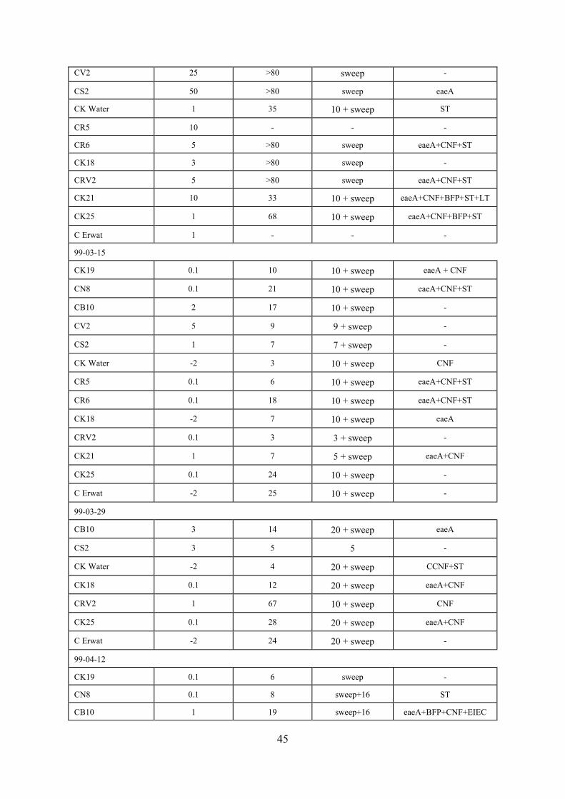
45
CV2 25 >80 sweep - CS2
50 >80 sweep eaeA
CK Water
1 35 10 + sweep ST
CR5
10 - - -
CR6
5 >80 sweep eaeA+CNF+ST
CK18
3 >80 sweep -
CRV2
5 >80 sweep eaeA+CNF+ST
CK21
10 33 10 + sweep eaeA+CNF+BFP+ST+LT
CK25
1 68 10 + sweep eaeA+CNF+BFP+ST
C Erwat
1 - - -
99-03-15 CK19
0.1 10 10 + sweep eaeA + CNF
CN8
0.1 21 10 + sweep eaeA+CNF+ST
CB10
2 17 10 + sweep -
CV2
5 9 9 + sweep -
CS2
1 7 7 + sweep -
CK Water
-2 3 10 + sweep CNF
CR5
0.1 6 10 + sweep eaeA+CNF+ST
CR6
0.1 18 10 + sweep eaeA+CNF+ST
CK18
-2 7 10 + sweep eaeA
CRV2
0.1 3 3 + sweep -
CK21
1 7 5 + sweep eaeA+CNF
CK25
0.1 24 10 + sweep -
C Erwat
-2 25 10 + sweep -
99-03-29 CB10
3 14 20 + sweep eaeA
CS2
3 5 5 -
CK Water
-2 4 20 + sweep CCNF+ST
CK18
0.1 12 20 + sweep eaeA+CNF
CRV2
1 67 10 + sweep CNF
CK25
0.1 28 20 + sweep eaeA+CNF
C Erwat
-2 24 20 + sweep -
99-04-12 CK19
0.1 6 sweep -
CN8
0.1 8 sweep+16 ST
CB10
1 19 sweep+16 eaeA+BFP+CNF+EIEC

46
CV2
10 14 sweep+16 -
CS2
5 11 sweep+16 eaeA+BFP
CK Water
-4 68 sweep+16 CNF+ST
CR5
0.1 14 sweep+16 eaeA+BFP+CNF+ST
CR6
1 7 sweep+16 eaeA+CNF
CK18
0.1 13 sweep+16 eaeA+CNF
CRV2
0.1 5 sweep+16 eaeA+CNF
CK21
1 10 sweep+16 -
CK25
-2 8 sweep+16 CNF
C Erwat
-3 7 sweep+16 CNF
99-05-31 CK19
-1 15 sweep+16 eaeA+CNF(2)
CN8
3 19 sweep+16 eaeA(2)+CNF+EIEC
CB10
10 31 sweep+16 eaeA+ST
CS2
- - - -
CK Water
1 87 sweep+16 eaeA+CNF
CR5
-2 7 sweep+16 eaeA+CNF
CR6
1 21 sweep+16 eaeA+CNF
CK18
1 48 sweep+16 eaeA+VFP+EIEC
CRV2
- - - -
CK21
3 22 sweep+16 eaeA+CNF(2)
CK25
1 39 sweep+16 CNF(2)+eaeA(2)
C Erwat
-2 31 sweep+16 eaeA+BFP+CNF
99-06-07 CK19
1 12 - -
CN8
1 27 16 eaeA+BFP_EIEC
CB10
10 17 16 eaeA+BFP
CV2
25 8 - -
CS2
25 25 16 eaeA
CK Water
5 14 - -
CR5
-1 33 16 CNF(2)+EIEC
CR6
1 23 16 eaeA+CNF+LT
CK18
5 39 16 eaeA+BFP
CRV2
3 22 16 eaeA+CNF+LT
CK21
5 26 16 eaeA
CK25
3 23 16 eaeA
CErwat
1 12 16 CNF

47
99-06-21 CK19
-1 54 16 eaeA+CNF
CN8
1 37 16 eaeA+CNF(2)+EIEC
CB10
10 19 16 eaeA+CNF
CV2
50 10 - -
CS2
25 17 -16 -
CK Water
-1 83 16 eaeA+CNF
CR5
-1 31 16 eaeA+CNF
CR6
1 24 16 eaeA+CNF
CK18
-1 56 16 eaeA+CNF
CRV2
10 34 16 eaeA+CNF
CK21
3 23 16 eaeA+CNF
CK25
-1 97 16 CNF
99-06-28 CK19
1 11 16 eaeA+CNF
CN8
1 73 16 eaeA+BFP+CNF
CB10
5 12 - -
CV2
25 10 16 eaeA
CS2
50 37 - -
CK Water
5 138 16 CNF
CR5
-1 17 16 eaeA
CR6
3 59 16 eaeA
CK18
1 17 16 eaeA+BFP
CRV2
10 56 - -
CK21
5 13 16 eaeA+BFP
CK25
3 49 16 BFP
C Erwat
-3 3 - -
99-07-05 CK19
1 31 16 eaeA+CNF
CN8
1 62 16 eaeA+CNF
CB10
25 21 16 eaeA+CNF
CV2
- - - -
CS2
25 17 16 eaeA+CNF+BFP
CK Water
-1 48 16 CNF
CR5
-1 39 16 eaeA+CNF
CR6
1 54 16 eaeA+CNF(3x)
CK18
1 43 16 CNF(2x)

48
CK21
- - - -
CK25
1 64 16 eaeA+CNF+BFP
C Erwat
- - - -
99-07-12 CN8
-2 3 16 eaeA+CNF+BFP+ST
CB10
5 18 16 CNF
CV2
5 5 - -
CS2
5 22 - -
CR5
-2 12 16 CNF
CR6
1 57 16 eaeA+CNF
CK18
1 32 16 eaeA+CNF+BFP
CRV2
-1 29 16 CNF
CK21
10 34 16 eaeA+CNF+BFP+ST
CK25
1 15 16 eaeA+CNF+BFP
99-08-02 CK19
1 29 16 eaeA
CN8
1 27 16 eaeA+CNF+BFP
CB10
1 52 16 CNF
CV2
50 24 - -
CS2
50 41 16 eaeA
CK Water
-1 57 16 eaeA+CNF+BFP
CR5
-2 14 16 eaeA+CNF
CR6
1 15 16 eaeA+CNF+EIEC
CK18
1 61 16 CNF
CRV2
-1 33 16 eaeA
CK21
5 18 16 eaeA+CNF
CK25
1 94 16 eaeA+CNF
C Erwat
-2 43 16 CNF
99-08-16 CK19
1 11 16 CNF+eaeA
CN8
1 41 16 CNF+eaeA+ST
CV2
100 31 16 CNF
CS2
25 24 16 eaeA
CK Water
-1 69 16 CNF+eaeA
CR5
-1 47 16 CNF+eaeA
CR6
1 34 16 CNF+eaeA+BFP
CK18
1 27 - -

49
CRV2
1 19 16 CNF(2x)
CK25
1 51 16 eaeA
C Erwat
-2 23 - -
99-08-23 CK19
1 44 16 CNF+eaeA
CN8
1 31 16 CNF
CB10
1 27 - -
CV2
50 24 16 eaeA+ST
CS2
25 17 16 CNF+eaeA
CK Water
-1 73 16 CNF
CR5
-1 53 16 eaeA(2x)+CNF
CR6
1 26 16 CNF
CK18
1 39 16 CNF+eaeA
CRV2
-1 27 16 CNF+eaeA
CK21
25 9 - -
CK25
1 58 16 CNF+eaeA+BFP
C Erwat
-2 24 16 CNF
Total number of samples analysed = 180 3.4. Discussion The failure to detect E coli O157:H7 among 1363 colonies isolated from 204 samples taken at 15 different sites in the Vaal Barrage drainage basin (Table 3.5) suggests that the incidence of the pathogens is low in the water environments concerned. Since the waters concerned represent sewage and wastes from many communities over a large area, the same may apply to many other parts of the country. However, the reliability of the results is not altogether clear from the findings at hand. Shortcomings of the Rainbow agar for the selective recovery of candidate E coli O157:H7 organisms may imply that the pathogens were missed by the screening procedure used. Unfortunately there are no data on the incidence of clinical cases of E coli O157:H7 infections in the communities concerned because this might give some indication of the expected incidence of the pathogens in their wastes. There is also very little if any meaningful information on the incidence of these pathogens in comparable water environments in the rest of the world. However, since waterborne transmission of these pathogens has been confirmed, they must occur in substantial numbers in water sources and supplies at least under certain circumstances. The general incidence of clinical cases of E coli O157 infections would also seem to suggest that the pathogens should be more readily detectable in sewage and polluted water resources. In sharp contrast to the findings on E coli O157:H7 the incidence of other pathogenic strains of E coli was high (Tables 3.6 and 3.7). One or more of 6 virulence factors was detected in 53 of 180

50
(74 %) E coli bacteria isolated from 26 sites in the Vaal River Barrage over a period of one year. The eaeA gene was detected in 56 % (101/180) of the isolates, the cytotoxic necrotizing factor (CNF) genes in 53 % (96/180), and the bundle forming pilus gene characteristic of Enteropathogenic E coli in 13.8 % (25/180) of collections. This high incidence is supported by another survey actually aimed at isolating E coli O157:H7 in which 4 % of 204 suspect E coli O157:H7 isolates were found to carry genetic elements coding for one or more toxicity factors (Table 3.5). The high incidence of eaeA and CNF toxicity factors may to meaningful extent be due to pollution by wastes from cattle and other animals which also excrete E coli strains carrying the genetic elements concerned. These typically zoonotic pathogens cause disease in humans. E coli strains which only carry the eaeA gene are described as Atypical EPEC@. Outbreaks caused by these pathogens have been described. CNF producing strains of E coli have been strongly associated with extra-intestinal infections in humans, notably urinary tract infections and bacteraemia. The relatively high incidence of BFP, ST, LT toxicity factors, and even the presence of EIEC, strongly indicates pollution by human wastes. All these virulence markers are associated with E coli that cause diarrhoea. Apart from details on the incidence of pathogenic bacteria, the results reported here do therefore also cast light on the origin of faecal pollution. For instance, almost 25 % of isolates carrying the ST virulence factor, 16 % of isolates carrying the BFP factor and 43 % of isolates carrying the EIEC factor were detected at Site CN8, which strongly suggests human faecal pollution. The finding that 74 % of E coli bacteria in the Vaal River Barrage carry one or more toxicity factors indicates a potential health risk since all these strains are associated with disease in humans. Even if the disease was not as severe as potentially caused by E coli O157:H7, it cannot be ignored. In an increasing component of consumers, notably the very young and very old as well as immunocompromised and undernourished individuals, these E coli pathogens may cause serious health implications. Meaningful assessment of the health risk concerned requires substantially more information. However, the results at hand convey a clear message which warrants further investigation of the situation in the study area concerned as well as similar situations in other parts of the country. 3.5. Conclusions C In a survey of sewage and a spectrum of polluted river waters, not one of 204 presumptive
colonies isolated on a selective growth medium proved to be E coli O157:H7. However, 4 % of these isolates carried genetic elements which code for one or more toxicity factors. These isolates are pathogenic.
C The incidence of E coli O157:H7 reflected by the results reported here appears to be lower
than might be expected. One explanation for the apparent low incidence may be related to difficulties with the identification of presumptive colonies on the Rainbow agar used for the selective cultivation of E coli O157. The implication is that E coli O157:H7 colonies may have been missed. Improvement of the isolation method is therefore required.

51
C Some 74 % of 180 E coli colonies isolated from river water carried one or more genetic elements which code for toxicity factors. Since these organisms constitute a health risk to at least some consumers, their presence in water resources warrants attention.
C The high incidence of pathogenic E coli strains in the river water under investigation calls for
further investigation. For instance, the accuracy of the data need to be confirmed, and it is important to determine whether E coli bacteria in water resources and supplies elsewhere also carry toxicity factors at these levels.
C Meaningful assessment of the risk of infection constituted by these E coli pathogens would
require substantially more information on the toxic properties of these bacteria, their incidence and behaviour in water resources and supplies, and the human and animal utilisation of the waters concerned.
C Molecular techniques suitable for research on E coli pathogens have been established. The
multiplex principle proved practical, cost effective and relatively rapid. The typing of colonies in which two PCR reactions are used to screen for 6 genes simultaneously, takes about six hours. However, further optimisation for a variety of purposes is possible.

52
4. IMMUNOMAGNETIC ISOLATION OF E COLI 0157:H7 4.1. Introduction The transmission of E coli O157:H7 by recreational water and drinking water has been confirmed in a number of studies (Swerdlow et al., 1992; Keene et al., 1994; ProMed, 2000). The even more common vehicle of transmission would appear to be a variety of foods and food products, including meat products, raw and pasteurised cow=s milk and cheese, mayonnaise, apple cider, fruit and vegetables (Besser et al., 1993; Griffin, 1995; McCarthy, 1996; Nataro et al., 1998). Cattle are the main reservoir of E coli O157:H7, and cattle meat products are the predominant single source of infections (Griffin, 1995). However, E coli O157:H7 has also been isolated from other animals such as chickens, pigs and sheep (Griffin and Tauxe, 1991; Griffin, 1995; Müller et al., 2002). Three cases of laboratory-acquired E coli O157:H7 infection have also been reported (Ostroff et al., 1989; Booth and Rowe, 1993; Burnens et al., 1993). The above epidemiological data confirm that E coli O157:H7 and other pathogenic strains of E coli are typically transmitted by the faecal-oral route like other enteric pathogens. This feature implies certain basic properties. Among others, the pathogens must be excreted in substantial numbers by infected individuals. Furthermore, they must be able to survive successfully in unfavourable conditions. These would include conditions in water resources, and water treatment and disinfection processes. Particularly important is survival in the hostile environment in the stomach of the new host, which includes extremely low pH levels and a variety of proteolytic enzymes, to the extent that the duodenum is reached in a condition fit for multiplication. There is no indication that the additional load of genetic material which codes for toxicity factors affects the survival of E coli pathogens. These pathogens may therefore be expected to survive as successful in the environment as wild type E coli organisms. Epidemiological data on E coli pathogens support these considerations. Largely due to lack of practical methods, clinical diagnostic laboratories often fail to report the infections. In South Africa these infections are not notifiable. In addition there would not appear to be any meaningful research in progress on the epidemiology or incidence of these infections in South Africa. The lack of meaningful data on the incidence of the pathogens restricts estimates of the burden of disease associated with pathogenic E coli. Despite these shortcomings, the US Center for Disease Control and Prevention (CDC) estimated the annual disease burden of E coli O157:H7 in the USA to be more than 73 000 cases and as many as 61 deaths (CDC, 2001). E coli O157:H7 is the pathogen most frequently isolated from stool specimens that contain visible blood (Slutsker et al., 1997). The World Health Organization (WHO) is particularly concerned about this because bloody diarrhoea is a major cause of morbidity and mortality among children in developing countries in the southern hemisphere, including South Africa (WHO, 1997). A recent outbreak of E coli O157:H7 in the small farming community of Walkerton in Ontario, Canada, attracted world-wide attention. More than 2000 people fell ill and six died when the borehole raw water source of the water supply system got contaminated by stormwater run-off containing cattle manure. The treatment and chlorination processes for the water supply failed to control the number of E coli O157:H7 in the final water. The international attention was not only about the health implications and failure of conventional treatment to control E coli O157:H7. Interest was largely in the legal implications and consequences of the outbreak. Court cases followed with claims for large sums of money for compensation to victims. Cabinet ministers had to stand trial in court on charges of negligence and failures of strategies to protect the health

53
of consumers who purchase domestic water supplies from a public utility (ProMed, 2000). This incident was seen as an example of new attitudes towards responsibility and accountability with major financial implications for water utilities and authorities concerned. The above information confirms the need for sensitive and reliable techniques to detect E coli O157:H7 in water. In previous attempts to isolate these pathogens from water test samples were directly plated on agar media for the selective cultivation of E coli O157:H7 strains, as described in Chapter 3, and as recommended by Westwood (1996). In view of the conclusions derived from the work described in Chapter 3 which indicated that this approach should be improved, this Chapter deals with efforts to develop a new more sensitive procedure for the isolation of E coli O157:H7 from water. The new approach developed and applied in work described in this Chapter is based on selective enrichment of E coli bacteria in test water samples using peptone saline broth supplemented with three selective antibiotics, followed by selective recovery of E coli O157:H7 from these cultures by means of immunomagnetic separation, and inoculating the immunomagnetic beads on appropriate agar media for the selective cultivation of E coli O157:H7. The efficiency of the immunomagnetic separation (IMS) method for the selective recovery of E coli O157:H7 from food and stool specimens has been well established (Wright et al., 1994; Tomoyasu, 1998). Dynabeads (Dynabeads7 anti-E coli O157; Dynal, Oslo) are uniform, superparamagnetic, polystyrene beads covered with covalently bound adsorbed and affinity purified anti-E coli O157 monoclonal antibodies. In the isolation procedure developed in this study Dynabeads are incubated with pre-enrichment cultures for the antibodies to capture and bind E coli O157:H7 bacteria to the beads. The beads are then recovered from the cultures by means of a magnetic device, washed to remove unbound bacteria, and plated onto appropriate media for the selective cultivation of E coli O157:H7. Suspect colonies are then picked from these plates and E coli O157:H7 is confirmed by biochemical tests, agglutination with antibodies specific for E coli O157:H7, and molecular techniques to type toxicity factors and genetic characteristics. This Chapter describes the optimisation of the new enrichment-IMS-selective agar procedure, assessment of the efficiency of the procedure, and the first application of the procedure for the isolation of E coli O157:H7 from sewage, river water, grounded beef and milk. 4.2. Materials and Methods 4.2.1. Bacterial strains Shiga toxin 2 (Stx2)-positive strain E coli O157:H7 (ATCC 43889) and Stx2-negative strain E coli O157:H7 (ATCC 43888) were used as Stx2 positive and negative controls respectively (Muniesa and Jofre, 1998). Stx1-positive E coli C600 and Stx1-negative E coli C600 were used as Stx1 positive and negative controls. All E coli control cultures were kindly supplied by Prof J Jofre, University of Barcelona, Spain. 4.2.2. Sewage and river water Weekly 500 ml sewage and river water samples were collected from September 1998 to August 1999. Settled sewage was collected at the Daspoort-, Zeekoegat- and Baviaanspoort sewage treatment plants near Pretoria. River water samples were collected from the Levuvhu River in the Northern Province, Pienaars- and Apies Rivers near Pretoria, and the Klip River near

54
Johannesburg. Samples were kept at 4 to 10 C and examined within 24 h after collection. 4.2.3. Beef and milk The grounded beef (200 g) and milk (250 ml) samples were randomly obtained from different retail outlets. Upon receipt at the laboratory, the samples were analysed immediately or held at 4 C for no longer than 48 h before analysis. 4.2.4. Immunomagnetic separation (IMS) of E coli O157:H7 Municipal sewage: Settled sewage samples (100 μl) and samples seeded with E coli O157:H7 (1.2 B 1.2 x 103.ml-1) were inoculated into 50 ml of buffered peptone-saline water (Oxoid, CM509) supplemented with Vancomycin (8 mg.l-1), Cefixime (0.05 mg.l-1) and Cefsulodin (10 mg.l-1) (VCC) antibiotic solution (MAST7 Diagnostics) to inhibit the growth of gram-positive organisms as well as Aeromonas and Proteus spp. The sewage enrichment broth suspensions were incubated in a shaker incubator (Hub-O-Mat) for 6 h at 37 C while rotating at 100 rpm. River water: River water samples (100 ml) and samples seeded with E coli O157:H7 (1.2 B 1.2 x 103.ml-1) were filtered through 0.45 μm Gelman GN-6 Metricel filter membranes (Prod No. 66191) and incubated in VCC-supplemented buffered peptone-saline water (Difco) for 6 h at 37 C. Beef: Ten grams of grounded beef samples and samples seeded with E coli O157:H7 (9.7 B 9.7 x 103.ml-1) were mixed with 100 ml of VCC-supplemented buffered peptone-saline water and incubated for 6 h at 37 C. Milk: Milk samples (1 ml) and samples seeded with E coli O157:H7 (9.7 B 9.7 x 103.ml-1) were directly inoculated into 50 ml of VCC-buffered peptone-saline broth. The milk enrichment broth suspensions were incubated in a shaker incubator (Hub-O-Mat) for 6 h at 37 C while rotating at 100 rpm. Aliquots (1 ml) of the pre-enriched samples with the addition of 20 μl of Dynabead suspension were incubated at room temperature for 10 min with continuous mixing. This step was carried out to allow the O157-specific antibodies to capture E coli O157:H7 bacteria and bind them to the beads. The bead-bacteria complexes were separated using a magnetic particle concentrator, Dynal7 MPC-M for 3 min (Dynal, Oslo). After discarding the supernatant and resuspending the bead-particles in PBS-Tween (Sigma), the process was repeated 4 times (Dynal7 product brochure, 1995). The final bead-bacteria complexes were resuspended in 100 μl washing buffer (PBS-Tween) to remove bacteria not bound to the beads by the monoclonal antibodies. 4.2.5. Isolation of E coli O157 After immunomagnetic separation, 10 μl and 50 μl volumes of bead concentrates were transferred to E coli O157 selective media. The E coli O157:H7 selective media used in this study were Cefixime-tellurite Sorbitol-MacConkey agar (CT-SMAC) (Oxoid), CHROMagar O157 (Dynal7) and Rainbow agar O157 (Biolog). E coli O157:H7 strains produced typical colourless colonies on CT-SMAC, red/pink colonies on CHROMagar O157 and grey/black colonies on Rainbow O157 agar after 24 h of incubation at 37 C. All suspect colonies were subcultured on CT-SMAC. Non-sorbitol fermenting colonies were examined for the presence of the genes coding for Stx1, Stx2, enterohaemolysin and eaeA. A loopful of these colonies was dispersed in 500 μl ultra high quality (UHQ) water and boiled at 99 C for 10 min without further treatment.

55
Oligonucleotide primers (Sigma-Genosys Ltd) specific for Stx1 (VT1), Stx2 (VT2), EHEC 1 and 2 (eaeA primers) and EHEC P1 and P2 (Enterohaemolysin plasmid primers) were used in the polymerase chain reaction (PCR) (Pollard et al., 1990; Gannon et al., 1992; Fratamico et al., 1995) (Table 3.1). The PCR cycle consisted of an initial 5 min DNA denaturation cycle at 94 C followed by 35 cycles of 1 min at 94 C, 1 min at 55 C and 1 min at 72 C (Pharmacia LKB Thermocycler). The amplicons (20 μl aliquots from each amplification) were detected by gel electrophoresis using a 2 % agarose (SeaKem7 LE) gel suspension stained with ethidium bromide (Sigma). A 100 base pair DNA molecular size marker (Promega) was used. The amplified products were visualised by UV-transillumination (UVP -Transilluminator) and the image was captured using the UVP Image store 5000 gel documentation system. Suspect E coli O157:H7 colonies were individually tested for agglutination using a commercial E coli O157 slide agglutination kit with antisera against E coli O157 (Mast Assure, Mono Factor O157, code:M12030). All colonies were biochemically confirmed as E coli by their ability to produce indole from tryptophan using Kovac=s reagent (ISO, 2001). 4.3. Results Assessment of the sensitivity of the enrichment-IMS-selective agar procedure revealed that E coli O157:H7 colonies were recovered from sewage and river water samples with average counts of seeded E coli O157:H7 as low as 1.2 ml-1. In the case of both seeded beef and milk samples the lowest average count of E coli O157:H7 in test samples from which the organisms were recovered was 9.7 ml-1. The enrichment procedure increased average counts of E coli O157:H7 in seeded sewage samples from 1.2 to 45 ml-1 (3650 %), in seeded river water samples from 1.2 to 72 ml-1 (5900 %), in seeded grounded beef samples from 9.7 to 54 ml-1 (457 %), and seeded milk samples from 9.7 to 67 ml-1 (591 %). In the case of samples seeded with higher numbers of E coli O157:H7 the percentage increase in counts of E coli O157:H7 accomplished by enrichment was substantially lower (Table 4.1). For instance, in the case of sewage seeded with E coli O157:H7 to average counts of 1200 ml-1, the percentage increase was only 45 %. After the above establishment of the sensitivity of the enrichment-IMS-selective agar method, the procedure was applied in studies on the incidence of naturally occurring E coli O157:H7 in selected samples. A total of 91 sewage, 40 river water, 25 beef and 25 milk samples was examined. Suspect E coli O157:H7 colonies were isolated from 16 sewage (17.6 %) and 8 river water (20 %) samples on the 3 selective media used. No such colonies were isolated when sewage, river water, beef and milk samples were directly plated on the selective media due to interfering growth and overwhelming numbers of non-pathogenic strains of E coli. Three colonies isolated on CT-SMAC from one sewage sample of the Daspoort West Intake agglutinated with the anti-E coli O157 and H7 antisera, tested positive for indole using Kovac=s reagent and contained the genes coding for Stx2, hly and eaeA (Table 4.2). These three isolates imply that 6.25 % of suspect E coli O157:H7 colonies isolated from sewage were confirmed to be E coli O157:H7, and that E coli O157:H7 colonies were isolated from 1.1 % of sewage samples analysed. No suspect E coli O157:H7 colonies were isolated from any of the 40 river water samples, 25 grounded beef samples and 25 milk samples analysed. Although not confirmed by statistically meaningful results, CT-SMAC proved the agar medium of choice for the selective cultivation of E coli O157:H7. The choice is predominantly based on the morphology and colour of suspect colonies, and the extent to which their detection was obscured by background growth.

56
Table 4.1. Assessment of the sensitivity of the enrichment-IMS-selective agar method using
tests on seeded samples
Seeded
Count of seeded E coli O157:H7 per ml
Samples
Before Enrichment After Enrichment Percentage Increase
Sewage
1 200 1 740 45
120 170 42
12 52 333
1.2 45 3650
River water
1 200 2 950 146
120 890 641
12 140 1 067
1.2 72 5900
Grounded beef
9 700 17 400 79
970 1 700 75
97 520 436
9.7 54 457
Milk
9 700 25 000 158
970 1 100 13
97 520 436
9.7 67 590
Selective cultivation of candidate E coli O157:H7 isolates was on CT-SMAC Agar. Counts are averages of tests carried out in triplicate. E coli O157:H7 was detected in all seeded samples.

57
Table 4.2. Isolation of E coli O157:H7 from selected samples of sewage, river water, beef
and milk
Number of samples
Percentage of Total in brackets
Toxicity Factors
Samples Total
Suspect Isolates Confirmed Isolates Stx2
Hly
eaeA
Sewage
91
16 (17.6 %) 1 (1.1 %) 1
1
1
River water
40
0 0 0
0
0
Beef
25
0 0 0
0
0
Milk
25
0 0 0
0
0
Total
181
16 (8.8 %) 1 (0.6 %) 1
1
1
Three colonies were isolated from the single sewage sample in which the presence of E coli O157:H7 was confirmed. All three isolates were agglutinated by E coli O157 and H7 antiserum and they carried the same three toxicity factors. Toxicity factor Stx1 was not detected. 4.4. Discussion Evidence has been presented that the enrichment-IMS-selective agar procedure substantially increased the sensitivity of E coli O157:H7 isolation compared to direct plating of test samples onto selective agar generally practised in the past. Comparative tests revealed that the enrichment step increased counts of E coli O157:H7 seeded into samples of sewage, river water, grounded beef and milk by up to 5900 % (Table 4.1). The higher percentage increase in counts of E coli O157:H7 in samples seeded with low numbers of the organism, is probably due to normal population dynamics and maximum numbers of organisms attainable in a steady state culture. This phenomenon serves the objectives of the enrichment procedure well because low numbers of E coli O157:H7 require maximum levels of enrichment for detection. Since E coli O157:H7 colonies were recovered from all seeded samples, the lowest numbers of the organisms detectable in the samples concerned has unfortunately not been established. Determination of the lowest number of E coli O157:H7 detectable would require tests in which samples are seeded with numbers of E coli O157:H7 even lower than those in Table 4.1. However, the results indicate that E coli O157:H7 would be detectable when present in numbers at least as low as 1.2 ml-1. Although not confirmed by statistically meaningful results, the CT-SMAC Agar medium seemed to yield the best results of the three media used for the selective cultivation of E coli O157:H7 bacteria. In addition, CT-SMAC agar was less expensive than Rainbow Agar O157 and CHROMagar O157. The higher efficiency of the enrichment-IMS-selective agar procedure for the recovery of naturally occurring E coli O157:H7 in sewage and river water would appear to be supported by the isolation of at least one E coli O157:H7 organism from a sewage sample in this study. In

58
comparison, the survey described in Chapter 3 using conventional plating of test samples on selective media, failed to recover the pathogen from any of the samples analysed. Enterohaemorrhagic E coli bacteria have been isolated from sewage in Germany by direct plating of test samples onto selective media (Höller et al., 1999). E coli O157:H7 has been isolated from patient stool specimens as well as samples of meat products and milk associated with infections (Nataro and Kaper, 1998). However, as far as can be established we report here for the first time the isolation of E coli O157:H7 from sewage in South Africa, and the first application of the enrichment-IMS-selective agar procedure for the isolation of E coli O157:H7 from water anywhere. The finding that isolates from only one of 16 samples (6,3 %) from which suspect E coli O157:H7 were isolated turned out to actually be E coli O157:H7, confirms shortcomings of the agar media for the selective cultivation of E coli O157:H7. The three E coli O157:H7 colonies isolated from the same sewage sample were probably the offspring from the same original organism which multiplied during the initial enrichment stage. This is confirmed by identical features such as the toxicity factors they carried (Table 4.2). Shortcomings of media for the selective cultivation of E coli O157:H7 in test samples with heavy background growth such as sewage and river water has also been reported by others. Bettelheim (1998) pointed out that the black colonies of E coli O157:H7 were difficult to distinguish on Rainbow Agar O157 in the presence of large numbers of other E coli colonies of different colours. These observation call for further improvement of methods for the selective cultivation of E coli O157:H7 in the presence of large numbers of wild type E coli and other bacteria capable of growing on the selective media. Resistance to the antibiotics used for the suppression of bacteria other than E coli O157:H7 may largely be accountable for the problem. One solution may therefore be to find antimicrobial agents which more efficiently suppress background growth and strains of E coli other than E coli O157:H7. Despite shortcomings of the growth media for the selective cultivation of E coli O157:H7, evaluation of the sensitivity of the enrichment-IMS-selective agar procedure in seeding experiments actually confirmed that it was capable of detecting numbers of E coli O157:H7 as low as one per ml in any of the sewage, river water, grounded beef and milk samples analysed (Table 4.1). This indicates that E coli O157:H7 bacteria did actually occur in low numbers in the samples under investigation. Data reported here on the isolation of at least one E coli O157:H7 organism from sewage and results of the seeding experiments, confirm that a procedure is now available for the relatively sensitive isolation of the pathogen from a variety of environmental samples, even in the presence of heavy background growth and large numbers of wild type E coli and related bacteria. It would be most useful to apply the procedure for purposes such as analysis of samples related to E coli O157:H7 infections. 4.5. Conclusions $ An enrichment-IMS-selective agar procedure for the isolation of E coli O157:H7 pathogens
from a variety of water and food samples has been developed. The procedure is based on enrichment of the pathogens in test samples by propagation in a peptone growth medium,

59
recovery of the pathogens from these cultures by means of immunomagnetic separation (IMS) beads covered with monoclonal antibodies for the selective recovery of E coli O157:H7, and plating of the beads on agar media for the selective cultivation of E coli O157:H7. A number of tests, including molecular techniques, are then used to identify and type E coli O157:H7 isolates.
$ An assessment of the sensitivity of the enrichment-IMS-selective agar procedure proved that it
was sensitive enough to isolate E coli O157:H7 in numbers as low as one per ml from seeded samples of sewage, river water, grounded beef and milk.
$ The new isolation technique was used to screen sewage, river water, grounded beef and milk
samples for the presence of E coli O157:H7 organisms. The pathogen was successfully isolated from one sewage sample.
$ Evidence has been presented that a relatively sensitive and reliable technique for the isolation
of E coli O157:H7 from a variety of environmental samples has been established. $ The failure to detect E coli O157:H7 in river water samples suggested that the pathogens may
not be present in these waters in sufficient numbers to constitute a meaningful risk of infection to consumers.

60
5. GENERAL DISCUSSION 5.1. Discussion The literature review revealed that pathogenic strains of E coli are frequently associated with water-borne transmission. A number of water-borne outbreaks are on record. The incidence of water-borne infections would appear to increase. A recent outbreak in the small farming community of Walkerton in Ontario, Canada, attracted world-wide attention. More than 2000 people fell ill and six died when the borehole raw water source of the water supply system got contaminated by stormwater run-off containing cattle manure. The treatment and chlorination processes for the water supply failed to control the number of E coli O157:H7 in the final water. The international attention was not only about the health implications and failure of conventional treatment to control E coli O157:H7. Interest was largely in the legal implications and consequences of the outbreak. Court cases followed with claims for large sums of money for compensation to victims. Cabinet ministers had to stand trial in court on charges of negligence and failures of strategies to protect the health of consumers who purchase domestic water supplies from a public utility (ProMed, 2000). This incident was seen as an example of new attitudes towards responsibility and accountability with major financial implications for water utilities and authorities concerned. Clinical cases of pathogenic E coli infections have been described in South Africa. Although none of these were associated with water-borne transmission, the potential for water-borne transmission as described in other parts of the world must exist. In view of these considerations the objective of this project was to investigate the incidence of pathogenic E coli strains in sewage and selected river waters. It has been decided to focus on water in the Vaal River Barrage and its catchment. This water source is used for the production of drinking water by conventional treatment processes. In addition, the water is also extensively being used for recreation such as boating, fishing and swimming, and some individuals may even use the water for domestic purposes with little if any treatment. In a first survey (Chapter 3) pathogenic E coli strains were isolated from water samples by direct plating on selective growth media. This method was used in similar studies carried out elsewhere in the world according to recommendations by Westwood (1996). No E coli O157:H7 were isolated from a large number of samples. However, some 74 % of E coli isolates carried one or more toxicity factors associated with enteric disease in humans. These E coli organisms constitute a health risk. This is the first evidence of potentially pathogenic features of E coli organisms in South African water resources. No comparable data are on record from other parts of the world. The extent to which these E coli pathogens constitute a public health risk is not evident from the results obtained in this project. A meaningful risk assessment would require substantially more information on the incidence and pathogenic properties of the organisms, as well as the utilisation of the waters concerned. However, a potential health risk has been identified, and the results warrant further investigation. The incidence of E coli O157:H7 in the waters under investigation was lower than expected, and shortcomings in the isolation procedure were identified. Consequently it was decided to improve the efficiency of the isolation technique for E coli O157:H7. The approach was based on initial selective enrichment by cultivation of test samples in peptone-saline supplemented with three

61
antibiotics to suppress the multiplication of bacteria other than E coli. E coli O157:H7 was then selectively recovered from this enrichment culture by means of immunomagnetic separation (IMS) based on magnetic beads covered with monoclonal antibodies specific for E coli O157:H7. The beads were then plated onto agar media for the selective cultivation of E coli O157:H7. Suspect colonies were picked and E coli O157:H7 was confirmed by biochemical tests, agglutination with antibodies specific for E coli O157 and H7, and molecular techniques to identify genes coding for toxicity factors. An assessment of the efficiency of the new enrichment-IMS-selective agar procedure revealed that it was sensitive enough to isolate seeded E coli O157:H7 from sewage, river water, grounded beef and milk samples at levels as low as average counts of one per ml (Table 4.1). The new enrichment-IMS-selective agar procedure was then used in a survey for the presence of E coli O157:H7 in selected samples of sewage and river water collected over a period of one year, as well as representative samples of grounded beef and milk. E coli O157:H7 was successfully recovered from a sewage sample (Table 4.2). The incidence of E coli O157:H7 reflected by the results in this report would still appear low. The procedure used had certain shortcomings, particularly with regard to the cultivation of E coli bacteria on selective agar media in the final step. E coli O157:H7 colonies proved difficult to distinguish from background growth and other E coli colonies. Consequently the incidence of E coli O157:H7 among suspect colonies was rather low (Table 4.2). This implies that E coli O157:H7 colonies were possibly missed, and that the actual incidence is higher than reflected by the single isolate from a sewage sample. It is however possible that the incidence of E coli O157:H7 in the waters under investigation is indeed low. Unfortunately it is difficult to assess the potential presence of these pathogens in the waste waters concerned because no meaningful data are available on the incidence of E coli O157:H7 infections in the communities from which these wastes originate. In addition, there are no meaningful data in the rest of the world to which these results can be compared. Although E coli O157:H7 have been recovered from waste waters in countries such as Germany (Höller et al., 1999), no quantitative data on the incidence of the pathogens in the waters under investigation are available. The apparently low incidence of E coli O157:H7 in the South African waste waters investigated may perhaps be explained by data which suggest that the incidence of E coli O157:H7 infections is lower among humans in South Africa than in countries of the northern hemisphere (WHO, 1997). Based on indications that the minimal infectious dose of E coli O157:H7 may be more than 100 organisms, the results of the survey described in Chapter 4 would seem to indicate that the waters under investigation do not constitute a meaningful risk of E coli O157:H7 infection. This would apply to exposure to water in the Vaal River during recreational activities such as boating and fishing, and probably even swimming. The water may not even constitute a meaningful risk of infection when used for domestic purposes. Utilisation of the water in the Vaal River barrage for the production of drinking water supplies by appropriately applied conventional treatment and disinfection procedures should adequately reduce risks of infection to acceptable limits. However, data on water-borne outbreaks of E coli O157:H7, and the isolation of these pathogens from sewage in South Africa and other parts of the world, warrants attention and caution. This refers in particular to high risk communities which include the very young and very old, under-

62
nourished people, and immuno-compromised patients. The latter include AIDS patients and patients under immuno-suppression for conditions such as cancer and organ transplants. E coli pathogens, like most other pathogens, constitute risks of life-threatening complications and implications in these individuals which are much more severe than in normal people. The high risk component of many communities in South Africa would appear to be substantial and increasing as it does in many other parts of the world. A major goal of this project has been successfully accomplished. This is the development of technology and expertise on a potentially important water-borne pathogen. The enrichment-IMS-selective agar procedure developed in this project proved relatively sensitive and efficient. The same applies to molecular techniques established for the characterisation of E coli O157:H7 and other E coli pathogens. An infrastructure for services and further research on these pathogens has been established. The work resulted in the first isolation of E coli O157:H7 from water in South Africa. The new enrichment-IMS-selective agar procedure for the isolation of E coli O157:H7 is the first of its kind in the world and according to data presented in this project more sensitive than any methods used for the purpose to date. Important areas for further research have been identified. These include further improvement of the isolation procedure. Shortcomings in this procedure have been recognised. Particularly important is research on the phages which transfer the genetic elements coding for toxicity factors to normally harmless E coli bacteria. Details on these phages are likely to hold the key to meaningful strategies for the control of E coli pathogens which constitute major public health risks. 5.1.1. Costs The cost of isolating E coli bacteria from water by means of membrane filtration amounts to about R 60 per sample. The cost of isolating E coli O157:H7 from water by means of the enrichment-IMS-selective agar procedure amounts to about R 250,00 per sample. The subsequent confirmation of E coli O157:H7 among suspect colonies amounts to about R 100,00 per colony. The genetic typing of E coli O157:H7 isolates by means of molecular techniques amounts to about R 350,00 per isolate. This implies that the running costs for the isolation and characterisation of E coli pathogens from water is relatively expensive. The possibly even larger cost component may be the facilities and capital outlay required for the work, and the availability of duly trained laboratory staff for this relatively complicated work. Consequently the intention is not for this work to be included in the routine programme of every laboratory engaged in water quality monitoring. It would be more economic and practical in terms of capital outlay, sophisticated equipment and highly trained staff if this work were to be restricted to specialist laboratories. These laboratories could then render services on E coli pathogens. Another benefit of this policy would be that specialist laboratories could also focus on research regarding E coli pathogens and the processing of data on these pathogens. 5.2. Conclusions $ The literature survey outlined public health risks constituted by E coli pathogens. The

63
pathogens are known to occur in South Africa as they do in the rest of the world. Water-borne transmission has not yet been recorded in South Africa, and isolation of the pathogens from water resources in the country has not yet been described.
$ In a preliminary survey a spectrum of pathogenic strains of E coli was isolated from sewage
and river water. The absence of E coli O157:H7 from these isolates prompted the development of a more sensitive procedure for the isolation of E coli O157:H7 from water.
$ A new procedure for the isolation of E coli O157:H7 from water has been developed. The
evaluation of the enrichment-IMS-selective agar procedure using seeding experiments confirmed that it was more sensitive for the isolation of E coli O157:H7 from water than techniques used in the past.
$ Application of the new enrichment-IMS-selective agar procedure resulted in the isolation of E
coli O157:H7 from a sewage sample. $ Expertise and technology on E coli pathogens has been established, and a practical procedure
for the isolation of E coli pathogens from water is in place. 5.3. Future research $ Improvement of procedures for the recovery of E coli O157:H7 pathogens from water. The
enrichment-IMS-selective agar procedure developed in this project is superior to methods used in the past, but there is potential for further improvement. This refers in particular to the final step for the selective cultivation of E coli O157:H7 isolates. Selection may perhaps be improved by using alternative antimicrobial agents which more efficiently suppresses interfering background growth and wild type E coli.
$ More data are required on the incidence and behaviour of E coli pathogens in sewage and in
raw and treated water supplies in order to assess health risks to meaningful extent which is required for the formulation of control strategies.
$ More data are required on the incidence of E coli pathogens in animal reservoirs, notably
cattle, but also other animals such as pigs. This information is essential for research on the epidemiology of the pathogens, and will cast light on the role of animals in human infections.
$ More data are required on the incidence of pathogenic E coli infections among humans in
South Africa. These details are required for calculations of the burden of disease, which is essential for the formulation of control strategies. Data on the incidence of infections in humans and animals will also facilitate estimation of the numbers of these pathogens that can be expected in waste water. This information is required to control water-borne transmission.
$ Research on the phages which transmit the genetic elements coding for toxicity factors to
harmless E coli bacteria is of particular importance. These phages serve as reservoirs for the genetic elements which convert E coli bacteria into serious pathogens. A preliminary survey carried out in collaboration with co-workers at the University of Barcelona revealed the presence of these phages in sewage samples from a number of sources in and around Pretoria. Information is required on the incidence and behaviour of these phages in water environments, and conditions under which they transfer the toxicity genes to E coli bacteria need to be

64
elucidated.
65
6. APPENDIX
6.1. Culture media 6.1.1. Sorbitol MacConkey agar
Agar 51.5 g Distilled water 1 000 ml Bring to the boil to dissolve completely. Sterilize by autoclaving at 121�C for 15 min.
6.1.2. Rainbow agar O157
Agar 60 g Distilled water 1 000 ml
Autoclave at 121�C for 10 min (Medium is heat sensitive).
6.1.3. CHROMagar O157
Agar 1 ampoule Distilled water 250 ml
Bring to the boil to dissolve completely. Do not autoclave.
6.1.4. Peptone buffered water
Peptone 10 g NaCl 5 g di-Sodium hydrogen phosphate 3.5 g Potassium dihydrogen phosphate 1.5 g Distilled water 1 000 ml Adjust pH to 7.0 Autoclave at 121 �C for 15 min
6.2 SMAC Media Cefixime-Tellurite (CT) Supplement (Product # 740.01)

66
6.2.1 Preparation of stock solutions of Cefixime and Potassium Tellurite.
Cefixime Stock solution (final concentration 1 mg/ml) in absolute ethanol
6.2.1.1. Add 1 ml of absolute ethanol to the vial of Cefixime 6.2.1.2. Dissolve completely 6.2.1.3. Gently vortex to ensure complete mixing
The prepared stock Cefixime is stable for 14 days under refrigeration (4 �C).
6.2.2 Potassium Tellurite Stock solution (final concentration 50 mg/ml)
6.2.2.1 Add 1 ml of sterile distilled water to the vial containing Potassium Tellurite.
6.2.2.2. Dissolve completely. 6.2.2.3. Gently vortex to insure comple mixing. 6.2.2.4. Store at room temperature.

67
7. REFERENCES Acheson DWK, Moore R, DeBreucker S, Lincicome LL, Jacewicz M, Skutelsky E, Keusch GT. (1992) Translocation of Shiga toxin across polarized intestinal cells in tissue culture. Infect Immun 64, 3294-3000. Ackermann H.-W. (1975) La classification des bacteriophages des cocci gram-positifs: Micrococcus, Staphylococcus et Streptococcus. Path Biol 23, 247-253. Ackermann H.-W, Audurier A, Berthiaume L, Jones LA, Mayo JA, Vidaver AK. (1978) Guidelines for bacteriophage characterization. Adv Virus Res 23, 1-24. Ackermann, H-W, and A Eisenstark. (1974) The present state of phage taxonomy Intervirol 3, 201-219 Ackman D, Marks S, Mack P, Caldwell M, Root T, Birkhead G. (1974) Swimming associated haemorrhagic colitis due to Escherichia coli O157:H7 infection: evidence of prolonged contamination of a fresh water lake. Epidemiol Infect 119, 1-8 Albert MJ, Faruque SM, Faruque AS, Neogi PK, Ansaruzzaman M, Bhuiyan NA, Alam K, Akbar MS. (1995) Controlled study of Escherichia coli diarrheal infections in Bangladeshi children J Clin Microbiol 33, 973-977. Arriaga YL, Harville BA, Dreyfus LA. (1995) Contribution of individual disulfide bonds to biological action of Escherichia coli heat-stabile enterotoxin B. Infect Immun 63, 4715-4720. Baldini MM, Kaper JB, Levine MM, Candy DC, Moon HW. (1983) Plasmid-mediated adhesion in enteropathogenic Escherichia coli. J Pediatr Gastroenterol Nutr 2, 534-538. Baldwin TJ, Ward W, Aitken A, Knutton S, Williams PH. (1991) Elevation of intracellullar free calcium levels in HEp-2 cells infected with enteropathogenic Escherichia coli. Infect Immun 59, 1599-1604. Barnard PJ, Kibel M. (1965) The haemolytic-uraemic syndrome of infancy and childhood. A report of eleven cases Cent. Afr J Med 11, 4-11;31-34 Barret TJ, Lior H, Green JH, Khakhria R, Wells JG, Bell BP, Greene KD, Lewis J, Griffin PM. (1994) Laboratory investigation of a multistate food-borne outbreak of Escherichia coli 0157:H7 by using pulse-field gel electrophoresis and phage typing. J Clin Microbiol 32, 3013-3017 Barret TJ, Green JH, Griffin PM, Pavia AT, Ostroff SM, Wachmuth IK. (1991) Enzyme-linked immunosorbent assays for detecting antibodies to Shiga-like toxin I, Shiga-like toxin II, and Escherichia coli 0157:H7 lipopolysaccharide in human serum. Curr Microbiol 23, 189-195 Beachley EH. (1981) Bacterial adherence: adhesin-receptor interactions mediating the attachment of bacteria to mucosal surfaces. J Infect Dis 143, 325-345 Benjamin MM, Datta AR. (1995) Acid tolerance of enterohaemorrhagic Escherichia coli. Appl Environ Microbiol 61, 1669-1672

68
Bennet AR, MacPhee S, Betts RP. (1996) The isolation and detection of Escherichia coli O157 by use of immunomagnetic separation and immunoassay procedures. Lett Appl Microbiol 22, 237-243 Besser RE, Lett SM, Weber JT, Doyle MP, Barret JT, Wells JG; Griffin PM. (1996) An outbreak of diarrhea and hemolytic uremic syndrome from Escherichia coli 0157:H7 in fresh-pressed apple cider. J Am Med Assoc 269, 2217-2220 Bettelheim KA. Studies of Escherichia coli cultured on RainbowTM Agar O157 with particular reference to enterohaemorrhagic Escherichia coli (EHEC). Microbiol Immunol 1998; 42: 265-269. Beutin L, Aleksic S, Zimmerman S, Gleier K. (1994) Virulence factors and phenotypic traits of verotoxigenic strains of Escherichia coli isolated from human patients in Germany. Med Microbiol Immunol (Berlin) 183, 13-21 Biolog internet website 1999 http: //wwwbiologcom/ro157html Birch M, Denning DW, Law D. (1996) Rapid genotyping of Escherichia coli O157 isolates by Random Amplification of Polymorphic DNA. Eur J Clin Microbiol Infect Dis 15, 297-302 Booth L, Rowe B. (1993) Possible occupational acquisition of Escherichia coli O157 infection. Lancet 342, 1298-1299 Bradley DE. (1967) Ultrastructure of bacteriophages and bacteriocins. Bact Rev 31, 230-314 Brown JE, Sethabutr O, Jackson MP, Lolekha S, Echeverria P. (1989) Hybridization of Escherichia coli producing Shiga-like toxin I, Shiga-like toxin II, and variant Shiga-like toxin II with synthetic oligonucleotide probes. Infect Immun 57, 2811-2814 Browning NG, Botha JR, Sacho H, Moore PJ. (1990) Escherichia coli 0157:H7 haemorrhagic colitis. Report of the first South African case. S Afr J Surg 28, 28-29 Brunder W, Schmidt H, Karch H. KatP, (1996) a novel catalase-peroxidase encoded by the large plasmid of enterohaemorrhagic Escherichia coli 0157:H7. Microbiol 142, 3305-3315 Brunder W, Schmidt H, Karch H. EspP, (1997) a novel extracellular serine protease of enterohaemorrhagic Escherichia coli 0157:H7 cleaves human coagulation factor V. Mol Microbiol 24, 767-778 Burnens AP, Zbinden R, Kaempf L, Heinzer I, Nicolet J. (1993) A case of laboratory acquired infection with Escherichia coli 0157:H7. Zentralbl Bakteriol 279, 512-517 Calderwood SB, Acheson DWK, Keusch GT, Barret TJ, Griffin PM, Strockbine NA, Swaminathan B, Kaper JB, Levine MM, Kaplan BS, Karch H, O=Brien AD, Obrig TG, Takeda Y, Tarr PI, Wachsmuth IK. (1996) Proposed new nomenclature for SLT VT family. Am Soc Microbiol News 62, 118-119

69
Caprioli A, Luzzi I, Gianviti A, Rüssmann H, Karch H. (1995) Pheno-genotyping of verotoxin 2 VT2-producing Escherichia coli causing haemorrhagic colitis and haemolytic uraemic syndrome by direct analysis of patient=s stools. J Med Microbiol 43, 348-353 CDC (1995) Escherichia coli 0157:H7 outbreak linked to commercially distributed dry-cured salami- Washington and California. Morbid Mortal Weekly Rep 44, 157-160 CDC (2001) Escherichia coli O157:H7. Disease Information: Division of Bacterial and Mycotic Diseases, Centers for Disease Control and Prevention, USA. Http://www.cdc.gov/ncidod/dbmd/ diseaseinfo/escherichiacoli_g.htm Chapman PA, Siddons CA. (1996) A comparison of immunomagnetic separation and direct culture for the isolation of verocytotoxin-producing Escherichia coli O157 from cases of bloody diarrhoea, non-bloody diarrhoea and asymptomatic contacts. J Med Microbiol 44, 267-271 Chart H, Smith HR, Scotland SM. (1991) Serological identification of Escherichia coli 0157:H7 infection in haemolytic uraemic syndrome. Lancet 337, 138-140 CHROM agar website 2000 http: //wwwgiga-planetfr/chromagar Clay CG. (1988) Characterization of Escherichia coli strains which produce shiga-like toxins. MSc-thesis. Medical University of South Africa Crane JK, Wehner MS, Bolen EJ, Sando JJ, Linden I, Guerrant RL, Sears CL. (1992) Regulation of intestinal guanylate cyclase by the heat-stable enterotoxin of Escherichia coli STa and protein kinase C. Infect Immun 60, 5004-5012 Cravioto A, Reyes RE, Trujillo F, Uribe F, Navarro A, de la Roca JM, Hernandez JM, Perez G, Vazquez V.(1990) Risk of diarrhea during the first year of life associated with initial and subsequent colonization by specific enteropathogens. Am J Epidemiol 131, 886-904 Cubbon, MD, Coia JE, Hanson MF, Thomson-Carter FM. (1996) A comparison of immunomagnetic separation, direct culture and polymerase chain reaction for the detection of verocytotoxin-producing Escherichia coli O157 in human faeces. J Med Microbiol 44, 219-222 Day NP, Scotland SM, Cheasty T, et al. (1983) Escherichia coli 0157:H7 associated with human infections in the United Kingdom (Letter). Lancet 1, 825 de Bruijn FJ. (1992) Use of repetitive (repetitive extragenic palindromic and enterobacterial repetitive intergenic consensus) sequences and the polymerase chain reaction to fingerprint the genomes of Rhizobium meliloti isolates and other soil bacteria. Appl Environ Microbiol 58, 2180-2187 de Sauvage FJ, Horuk R, Bennet G, Quan C, Burnier JP, Goeddel DV. (1992) Characterization of the recombinant human receptor for Escherichia coli heat-stable enterotoxin. J Biol Chem 267, 6479-6482 DeGrandis S, Law H, Brunton J, Gyles C, Lingwood CA. (1989) Globotetraosyl ceramide is recognised by the pig edema disease toxin. J Biol Chem 264, 12520-12525

70
DeNileon GP. (1998) Water supply implicated in E. coli outbreak. Am Water Works Assoc Mainstream 42, 1 Djafari S, Ebel F, Deibel C, Kramer S, Hudel M, Chakraborty T. (1997) Characterization of an exported protease from Shiga toxin-producing Escherichia coli. Mol Microbiol 25, 771-784 Donnenberg MS, Tzipori S, McKee ML, O=Brien AD, Alroy J, Kaper JB. (1993) The role of the eae gene of enterohaemorrhagic Escherichia coli in intimate attachment in vitro and in a porcine model. J Clin Invest 92, 1418-1424 Downes FP, Green JH, Greene K, Strockbine N, Wells JG, Wachsmuth IK.(1989) Development and evaluation of enzyme-linked immunosorbent assays for detection of Shiga-like toxin I and Shiga-like toxin II. J Clin Microbiol. 27, 1292-1297 Doyle MP, Schoeni JL. (1987) Isolation of Escherichia coli 0157:H7 from retail fresh meats and poultry. Appl Environ Microbiol 53, 2394-2396 Drasar BS, Hill MJ. (1974) Human intestinal flora, p 36-43 Academic Press, Ltd, London, United Kingdom Dreyfus LA, Harville B, Howard DE, Shaban R, Beatty DM, Morris SJ. (1993) Calcium influx mediated by the Escherichia coli heat-stabile enterotoxin B (STB). 90, 3202-3206 Dynal Product Brocure. Dynabeads7 anti-E. coli. For rapid selective enrichment of Escherichia coli. Dynal AS Oslo, Norway 1995 Edelman R, Levine MM. (1983) Summary of a workshop on enteropathogenic Escherichia coli. J Infect Dis 147, 1108-1118 Fontaine A, Arondel J, Sansonetti PJ. (1988) Role of Shiga toxin in the pathogenesis of bacillary dysentry, studied by using a tox-negative mutant of Shigella dysenteriae 1. Infect Immun 56, 3099-3109 Franke S, Harmsen D, Caprioli A, Pierard D, Wieler LH, Karch H. (1995) Clonal relatedness of Shiga-like toxin producing Escherichia coli O101 strains of human and porcine origin. J Clin Microbiol 33, 3174-3178 Fraser ME, Chernaia MM, Koslov YV, James MN. (1994) Crystal structure of the holotoxin from Shigella dysenteriae at 25 A resolution. Nat Struct Biol 1, 59-64 Fratamico PM, Sackitey SK, Wiedmann M, Deng MY. (1995) Detection of Escherichia coli O157:H7 by multiplex PCR. Journal of Clinical Microbiology 33, 2188-2191 Freifelder D. (1987) In Molecular Biology Second Edition Ed: Jones and Bartlett Publishers, Inc Boston, Portola Valley pp 551-593 Frost JA, Cheasty T, ThomasA, Rowe B. (1993) Phage typing of Vero cytotoxin-producing Escherichia coli O157 isolated in the United Kingdom: 1989-91. Epidemiol Infect 110, 469-475 Gannon VPJ, Rashed M, King RK, Thomas EJG. (1992) Detection and characterization of the

71
eae gene of shiga-like toxin-producing Escherichia coli using polymerase chain reaction. Journal of Clinical Microbiology 31, 1268-1274. Gasser C, Gautier E, Steck A, Siebenmann RE, Oechslin R. (1955) Hamolytish-uramische syndrome: Bilaterale nierenrindennekrosen bei akuten erworbenen haemolytishen anaemien. Schweiz Med Wochenschr 85, 205-209 Geldreich E, Fox KR, Goodrich JA, Rice EW, Clark RM, Swerdlow DM. (1992) Searching for a water supply connection in the Cabool, Missouri disease outbreak of Escherichia coli O157:H7. Water Research 26, 1127-1137. Gianantonio C, Vitacco M, Mendilaharzu F, Rutty A, Mendilaharzu J. (1964) The hemolytic-uremic syndrome. J Pediatr 64, 478-491 Girón JA, Ho ASY, Schoolnik GK. (1991) An inducable bundle-forming pilus of enteropathogenic Escherichia coli. Science 254, 710-713 Goldberg MB, Sansonetti PJ. (1993) Shigella subversion of the cellular cytoskeleton: a strategy for epithelial colonization. Infect Immun 61, 4941-4946 Goldwater PN, Bettelheim KA. (1998) New perspectives on the role of Escherichia coli 0157:H7 and other enterohaemorrhagic E. coli serotypes in human disease. J Med Microbiol 47, 1039-1045 Goyal SM, Gerba CP, Bitton G. (1987) In Phage Ecology Ed: John Wiley and Sons, New York pp 45-74 Grabow, WOK, Uys M, Clay CG (2002) Molecular characterization of F-RNA coliphages in South African water sources. WRC Research Report No 928. Water Research Commission, Pretoria Greenwald DA, Brandt LJ. (1997) Recognising E. coli 0157:H7 infection. Hospital Practice Apr 15, pp 123-140 Griffin PM, Tauxe RV. (1991) The epidemiology of infections caused by Escherichia coli 0157:H7, and other enterohemorrhagic E. coli, and the associated hemolytic uremic syndrome. Epidemiol Rev 13, 60-98 Griffin PM. (1995) Escherichia coli 0157:H7 and other enterohaemorrhagic Escherichia coli, pp 739-761. In MJ Blaser, PD Smith, JI Ravdin, HB Greenberg, and RL Guerrant ed, Infections of the gastrointestinal tract Raven Press, New York, NY Griffiths J, Irving KG. (1961) A haemolytic-uraemic syndrome in infancy. Arch Dis Child 36, 500-506 Gross RJ. (1990) Escherichia coli diarrhoea In: Smith GR, Easmon CSF, eds Topley and Wilson=s principles of bacteriology, virology and immunity, 8th ed, vol 3 London: Hodder & Stroughton, pp 469-87
72
Guth BE, Twiddy EM, Trabulsi LR, Holmes RK. (1997) Variation in chemical properties and antigenic determinants among type II heat-labile enterotoxins of Escherichia coli. Infect. Immun 54, 529-536 Habib R, Levy M, Gagnadoux MF, Broyer M. (1982) Prognosis of the hemolytic uremic syndrome in children. Adv Nephrol 11, 109-128 Harris JR, Mariano J, Wells JG, Paine BS, Donnel HD, Cohen ML. (1985) Person-to-person transmission in an outbreak of enteroinvasive Escherichia coli. Am J Epidemiol 122, 245-252 Heuvelink AE, Van de Kar NCAJ, Meis JFGM, Monnens LAH, Melchers WJG. (1995) Characterization of verocytotoxin-producing Escherichia coli O157 isolates from patients with haemolytic uraemic syndrome in Western Europe. Epidemiol Infect 115, 1-14 Hii JH, Gyles C, Morooka T, Karmali MA, Clarke R, DeGrandis S, Brunton JL (1991) Development of verotoxin 2- and verotoxin 2 variant VT2v-specific oligonucleotide probes on the basis of the nucleotide sequence of the B cistron of VT2v from Escherichia coli E32511 and B2F1. J Clin Microbiol 29, 2704-2709 Höller C, Koshinsky S, Witthuhn D. (1999) Isolation of enterohaemorrhagic E. coli from municipal sewage. Lancet 1999; 353: 2039. Holtzhausen, CS. (1992) An evaluation of Bacteroides fragilis phages as indicators of human enteric viruses. MSc thesis. Dept Medical Virology, University of Pretoria, Pretoria Hulton CSJ, Higgins CF, Sharp PM. (1991) ERIC sequences: a novel family of repetitive elements in the genomes of Escherichia coli, Salmonella typhimurium and other enteric bacteria. Mol Microbiol 5, 825-834 Ismaili A, Philpott DJ, Dytoc MT, Sherman PM. (1995) Signal transduction responses following adhesion of verocytotoxin-producing Escherichia coli. Infect Immun 63, 3316-3326 ISO. (2001) Microbiology of food and animal feeding stuffs - Horizontal method for the detection of Escherichia coli O157. ISO/FDIS 16654. 2001. Jackson MP, Neill RJ, O=Brien AD, Holmes RK, Newland JW. (1987) Nucleotide sequence analysis and comparison of the structural genes from Shiga-like toxin I and Shiga-like toxin II encoded by bacteriophages from Escherichia coli 933. FEMS Microbiol Lett 44, 109-114 Jarvis KG, Kaper JB. (1996) Excretion of extracellular proteins by enterohaemorrhagic Escherichia coli via a putative type III secretion system. Infect Immun 64, 4826-4829 Javett SN, Senior B. (1962) Syndrome of hemolysis, thrombopenia annd nephropathy in infancy. Pediatrics 29, 209-223 Jerse AE, Yu J, Tall BD, Kaper JB. (1990) A genetic locus of enteropathogenic Escherichia coli necessary for the production of attaching and effacing lesions on tissue culture cells. Proc Natl Acad Sci USA. 87, 7839-7843

73
Johnson WM, Pollard DR, Lior H, Tyler SD, Rozee KR. (1990) Differentiation of genes coding for Escherichia coli verotoxin-2 and the verotoxin associated with porcine edema disease VTE by the polymerase chain reaction. J Clin Microbiol 28, 2351-2353 Johnson LP, Tomai MA, Schlievert PM. (1986) Bacteriophage involvement in group A streptococcal pyrogenic exotoxin A production. J Bacteriol 166, 623-627 Johnson WM, Lior H, Bezanson GS. (1983) Cytotoxic Escherichia coli 0157:H7 associated with haemorrhagic colitis in Canada (Letter). Lancet 1, 76 Kandel G, Donohue-Rolfe A, Donowitz M, Keusch GT. (1989) Pathogenesis of Shigella diarrhea XVI Selective targetting of shiga toxin to villus cells of rabbit jejunum explains the effect of the toxin on intestinal electrolyte transport. J Clin Rev 84, 1509-1517 Karmali M. (1989) Infection by Verocytotoxin-producing Escherichia coli. Clin Microbiol Rev 2, 15-38 Karmali MA, Petric M, Lim C, Fleming PC, Arbus GS, Lior H. (1985) The association between ideopathic haemolytic uraemic syndrome and infection by verotoxin-producing Escherichia coli. J Dis Imm 151, 775-782 Karmali MA, Petric M, Winkler M, Bielaszewska M, Brunton J, van de Kar N, Morooka T, Nair GB, Richardson SE, Arbus GS. (1994) Enzyme-liked immunosorbent assay for detection of immunoglobulin G antibodies to Escherichia coli Vero cytotoxin 1. J Clin Microbiol 32, 1457-1463 Karmali MA, Steele BT, Petric M, Lim C. (1983) Sporadic cases of haemolytic uraemic syndrome associated with faecal cytotoxin and cytotoxin-producing Escherichia coli in stools. Lancet 1, 619-20 Keene WE, McAnulty JM, Hoesly FC, Williams LP, Hedberg K, Oxman GL, Barret TJ, Pfaller MA, Fleming DW. (1994) A swimming-associated outbreak of haemorrhagic colitis caused by Escherichia coli 0157:H7 and Shigella sonnei. N Engl J Med 331, 579-584 Kibel MA, Barnard PJ. (1968) The hemolytic uremic syndrome: a survey in Southern Africa. S Afr Med J 42, 692-698 Kimmit PT, Harwood CR, Barer MR. (2000) Toxin gene expression by Shiga toxin-producing Escherichia coli: the role of antibiotics and the bacterial SOS response. Emerg Infect Dis 6, 5 Knutton S, Baldwin T, Williams PH, McNeish AS. (1989) Actin accumulation at sites of bacterial adhesion to tissue culture cells: basis of a new diagnostic test for enteropathogenic and enterohaemorrhagic Escherichia coli. Infect Immun 57, 1290-1298 Kock M. (2000) Dept. Microbiology and Plant Pathology, University of Pretoria. Personal communication Konowalchuk J, Speirs JI, Stovric S. (1977) Vero response to a cytotoxin of Escherichia coli.

74
Infect Immun 18, 775-9 Koornhof HJ.. (2001) South African Institute for Medical Research.. Personal communication Larson P, Wikstöm P, Forsman M. (1999) Quantitative properties of random amplified polymorphic DNA (RAPD). Quant Microbiol 1, 171-181 Lencer WI, Constable C, Moe S, Jobling MG, Webb HM, Ruston S, Madara JL, Hurst TR, Holmes RK. (1995) Targeting of cholera toxin and Escherichia coli heat-labile toxin in polarized epithelia: role of COOH-terminal KDEL. J Cell Biol 131, 951-962 Levine MM. (1987) Escherichia coli that cause diarrhea: enterotoxigenic, enteropathogenic, enteroinvasive, enterohemorrhagic, and enteroadherent. J Infect Dis 155, 377-389 Levine MM, Ristaino P, Marley G, Smyth C, Knutton S, Boedeker E, Black R, Young C, Clements ML, Cheney C, Patnaik R. (1984) Coli surface antigens 1 and 3 of colonization factor antigen II-positive enterotoxigenic Escherichia coli : morphology, purification, and immune responses in humans. Infect Immun 44, 409-420 Leyer GJ, Wang LL, Johnson EA. (1995) Acid adaption of Escherichia coli 0157:H7 increases survival in acidic foods. Appl Environ Microbiol 61, 3752-3755 Lin J-J, Kuo J. (2000) AFLP: A novel PCR-based Assay for plant and bacterial DNA fingerprinting. Focus 17, Number 2, pp 66-70. Lingwood CA, Law H, Richardson S, Petric M, Brunton JL, DeGrandis S, Karmali M. (1987) Glycolipid binding of natural and cloned Escherichia coli produced verotoxin in vitro. J Biol Chem 262, 8834-8839 Louie M, Azavedo JCS, Handelman MYC. (1993) Expression and characterization of the eaeA gene product of Escherichia coli serotype 0157:H7. Infect Immun 61, 4085-4092 McCarthy M. (1996) E. coli 0157:H7 outbreak in USA traced to apple juice. Lancet 348, 1299 Marshall KC, Stout R, Mitchell R. (1971) Mechanism of the initial events in the sorption of marine bacteria to surfaces. J Gen Microbiol 68, 337-348 Martin B, Humbert O, Camara M, Guenzi E, Walker J, Mitchell T, Andrew P, Prudhomme M, Alloing G, Hakenbeck R, Morrison DA, Boulnois GJ, Claverys P-J. (1992) A highly conserved repeated DNA element located in the chromosome of Streptococcus pneumoniae. Nucleic Acid Res 20, 3479-3483 Matthews REF. (1982) Fourth report of the International Committee on Taxonomy of Viruses In Classification and nomenclature of viruses. Kager, Basel McDaniel TK, Jarvis KG, Donnenberg MS, Kaper JB (1995) A genetic locus of enterocyte effacement conserved among diverse enterobacterial pathogens. Proc Natl Acad Sci USA 92, 1664-1668

75
McKee ML, Melton-Celsa AR, Moxley RA, Francis DH, O=Brien AD. (1995) Enterohaemorrhagic Escherichia coli 0157:H7 requires intimin to colonize the gnotobiotic pig intestine and to adhere to Hep-2 cells. Infect Immun 63, 3739-3744 Meng J, Zhao S, Doyle MP. (1995) Molecular characterization of Escherichia coli O157:H7 isolates by pulse-field gel electrophoresis and plasmid DNA analysis. J Med Microbiol 42, 258-263 Mezoff AG, Giannella RA, Eade MN, Cohen MB. (1992) Escherichia coli enterotoxin STa binds to receptors, stimulates guanyl cyclase, and impairs adsorption in rat colon. Gastroenterol 102, 816-822 Middlebrook JL, Dorland RB. (1984) Bacterial toxins: cellular mechanisms of action. Microbiol Rev 48, 199-221 Miller LG, Kaspar CW. (1994) Escherichia coli 0157:H7 acid tolerance and survival in apple cider. J Food Prot 57, 460-4 Moon HW, Whipp SC, Argenzio RA, Levine MM, Gianella RA. (1983) Attaching and effacing activities of rabbit and human enteropathogenic Escherichia coli in pig and rabbit intestines. Infect Immun 41, 1340-1351 Moschcowich E. (1924) Hyaline thrombosis of the terminal arterioles and capillaries: a hitherto undescribed disease. Proc NY Pathol Soc 24, 21-24 Müller EE, Ehlers MM., Grabow WOK. (2001b) The Occurrence of E. coli O157:H7 in South African Water Sources Intended for Direct and Indirect Human Consumption. Wat Res 2001b; 35: 3085-3088. Müller EE, Taylor MB, Grabow WOK, Ehlers, MM. (2001a) Isolation and Characterization of Escherichia coli O157:H7 and Shiga Toxin - converting Bacteriophages from Strains of Human, Bovine and Porcine Origin. 2001a. Submitted for publication in Wat Sci Tech. Muniesa M, Jofre J. (1998) Abundance in sewage of bacteriophages that infect Escherichia coli 0157:H7 and that carry the shiga toxin 2 gene. Appl Environ Microbiol 64, 2443-2448 Muniesa M, Lucena F, Jofre J. (1999) Comparative survival of free Shiga Toxin 2-encoding phages and Escherichia coli strains outside the gut. Appl Environ Microbiol 65, 5612-5618 Muniesa M, Jofre J. (2000) Occurrence of phages infecting Escherichia coli 0157:H7 carrying the Stx 2 gene in sewage from different countries. FEMS Microbiol Lett 183, 197-200 Nataro JP, Baldini MM, Kaper JB, Black RE, Bravo N, Levine MM.(1985) Detection of an adherence factor of enteropathogenic Escherichia coli with a DNA probe. J Infect Dis 152, 560-565 Nataro JP, Deng Y. (1993) Unpublished data

76
Nataro JP, Kaper JB.(1998) Diarrheagenic Escherichia coli. Clin Microbiol Rev 11, 142-201 Newland JW, Neill RJ.(1988) DNA probes for Shiga-like toxins I and II and for toxin-converting bacteriophages. J Clin Microbiol 26, 1292-1297 O=Brien AD, Holmes RK. (1987) Shiga and Shiga-like toxins. Microbiol Rev 51, 206-220 O=Brien AD, Newland JW, Miller SF, Holmes RK, Smith HW, and Formal SB. (1984) Shiga-like toxin-converting phages from E. coli strains that cause hemorrhagic colitis or infantile diarrhea. Science 226, 694-696 Oelschlaeger, TA, Barret TJ, Kopecko DJ. (1994) Some structures and processes of human epithelial cells innvolved in uptake of enterohaemorrhagic Escherichia coli 0157:H7 strains. Infect Immun 62, 5142-5150 Orr P, Lorencz B, Brown R, Kielly R, Tan B, Holton D, Clugstone H, Lugtig L, Pim C, MacDonald S. (1994) An outbreak of diarrhea due to verotoxin-producing Escherichia coli in the Canadian Northwest Territories. Scand J Infect Dis 26, 675-684 Ostroff SM, Kobayashi JM, Lewis JH. (1989) Infections with Escherichia coli 0157:H7 in Washington State: the first year of state-wide disease surveillance. J Am Med Assoc 262, 355-359 Pai CH, Kelly JK, Meyers GL. (1986) Experimental infection of infant rabbits with verotoxin-producing Escherichia coli. Infect Immun 51, 16-23 Paton JC, Paton AW. (1998) Pathogenesis and diagnosis of shiga toxin-producing Escherichia coli infections. Clin Microbiol Rev 11, 450-479 Pollard DR, Johnson WM, Lior H, Tyler SD, Rozee KR. (1990) Rapid and specific detection of verotoxin genes in Escherichia coli by the polymerase chain reaction. J. Clin.Microbiol. 28, 540-545. Proesmans W. (1996) Typical and atypical hemolytic uremic syndrome. Kidney and Blood Press Res 19, 205-208 ProMed website. (2000) www.promedmail.org Ecoli, EHEC - Canada (Ontario) (09) 16 Aug. Riemann HP, Cliver DO. (1998) Escherichia coli 0157:H7. Microb Food Borne Path 14, 41-48 Rietra PJGM, Wilshaw GA, Smith HR, Field AM, Scotland SM, Rowe B. (1989) Comparison of Vero-cytotoxin-encoding phages from Escherichia coli of human and bovine Origin. J Gen Microbiol 135, 2307-2318 Riley LW, Remis RS, Helgerson SD, McGee HB, Wells JG, Davis BR, Hebert RJ, Olcott ES, Johnson LM, Hargrett NT, Blake PA, Cohen ML (1983) Hemorrhagic colitis associated with a rare Escherichia coli serotype. N Engl J Med 308, 681-5

77
Rowbury RJ. (1997) More chaos on the microbiology front: the E. coli O157 outbreaks. Science Progress 80, 3-4 Rüssmann H, Schmidt H, Heesemann J, Caprioli A, Karch H. (1994) Variants of Shiga-like toxin II constitute a major toxin component in Escherichia coli O157 strains from patients with haemolytic uraemic syndrome. J Med Microbiol 40, 338-343 Salyers AA, Whitt DD. (1994) Bacterial Pathogenesis, a Molecular Approach Washington, DC, ASM Press. Samadpour M. (1995) Molecular epidemiology of Escherichia coli 0157:H7 by restriction fragment length polymorphism using Shiga-like toxin genes. J Clin Microbiol 33, 2150-2154 Sansonetti PJ. (1992) Molecular and cellular biology of Shigella flexneri invasiveness : from cell assay systems to shigellosis. Curr Top Microbiol Immunol 180, 1-19 Saye DJ, Ogunseitan O, Sayler GS, Miller RV. (1990) Transduction of linked chromosomal genes between Pseudomonas aeruginosa during incubation in situ in a freshwater habitat. Appl Environ Microbiol 56, 140-145 Schmidt H, Beutin L, Karch H. (1995) Molecular analysis of the plasmid-encoded hemolysin of Escherichia coli 0157:H7 strain EDL 933. Infect Immun 63, 1055-1061 Schmidt H, Karch H. (1996) Enterohaemolytic phenotypes and genotypes of shiga toxin-producing Escherichia coli O111 strains from patients with diarrhea and hemolytic uremic syndrome. J Clin Microbiol 34, 2364-2367 Schmidt H, Kernbach C, Karch H. (1996) Analysis of the EHEC hly operon and its location in the physical map of the large plasmid of enterohaemorrhagic Escherichia coli 0157:H7. Microbiol 142, 907-914 Scotland SM, Smith HR, Wilshaw GA, Rowe B. (1983) Verocytotoxin production in strain of Escherichia coli is determined by genes carried on bacteriophage. Lancet 2, 216 Sears CL, Kaper JB. (1996) Enteric bacterial toxins: mechanism of action and linkage to intestinal secretion. Microbiol Rev 60, 167-215 Sherman P, Soni R, Petric M, Karmali M. (1987) Surface properties of the Vero cytotoxin-producing Escherichia coli 0157:H7. Infect Immun 55, 1842-1829 Siegler RL, Griffin PM, Barret TJ, Strockbine NA. (1993) Recurrent hemolytic uremic syndrome secondary to Escherichia coli 0157:H7 infection. Pediatrics 91, 666-668 Sixma TK, Kalk KH, van Zanten BA, Dauter Z, Kingma J, Witholt B, Hol WG. (1993) Refined structure of Escherichia coli heat-labile enterotoxin, a close relative of cholera toxin. J Mol Biol 230, 890-918 Slutsker L, Ries AA, Greene KD, Wells JG, Hutwagener L, Griffin PM. (1997) Escherichia coli

78
0157:H7 diarrhea in the United States: clinical and epidemiological features. Ann Intern Med 126, 505-513 Smith-Keary PF. (1988) Genetic elements in Escherichia coli. Macmillan Molecular Biology. First edition. Macmillan Education Ltd Sowers EG, Wells JG, Strockbrine NA. (1996) Evaluation of commercial latex reagents for identification of O157 and H7 antigens of Escherichia coli. J Clin Microbiol 34, 1286-1289 Stern MJ, Ames GF-L, Smith NH, Robinson EC, Higgins CF. (1984) Repetitive extragenic palindromic sequences: a major component of the bacterial genome. Cell 37, 1015-1026 Strockbine NA, Wells JG, Bopp CA, Barret TJ. (1998) Overview of detection and subtyping methods. In Escherichia coli O157:H7 and other Shiga toxin- producing E. coli strains. Edited by JB Kaper and AD O=Brien. ASM Press. Washington DC Suttorp N, Flöer B, Schnittler H, Seeger W, Bhakdi S. (1990) Effects of Escherichia coli hemolysin on endothelial cell function. Infect Immun 58, 3796-3801 Swerdlow DL, Woodruff BA, Brady RC, Griffin PM, Tippen S, Donnel HD, Geldreich E, Payne BJ, Meyer A, Wells JG. (1992) A waterborne outbreak in Missouri of Escherichia coli 0157:H7 associated with bloody diarrhea and death. Ann Intern Med 117, 812-819 Takeda Y. (1997) Enterohaemorrhagic Escherichia coli. Rapp Trimest Statist Sanit Mond 50, 74-80 Takeda Y, Kurazono H, Yamasaki, S. (1993) Vero toxins (Shiga-like toxins) produced by enterohaemorrhagic Escherichia coli (verocytotoxin-producing E. coli). Microbiol Immun 37, 591-599 Thomas A, Smith HR, Rowe B. (1993) Use of dioxigenin-labelled oligonucleotide DNA probes for VT2 and VT2 human variant genes to differentiate Vero cytotoxin-producing Escherichia coli strains of serogroup O157. J Clin Microbiol 31, 1700-1703 Tomoyasu T. (1998) Improvement of the immunomagnetic separation method selective for Escherichia coli O157 strains. Appl Environ Microbiol 64, 376-382 Trompeter RS, Schwartz R, Chantler C, Dhillon MJ, Haycock GB, Kay R, Barratt TM. (1983) Haemolytic uraemic syndrome: an analysis of prognostic features. Arch Dis Child 58, 101-105 Tyler SD, Johnson WM, Lior H, Wang G, Rozee KR. (1991) Identification of verotoxin type 2 variant B subunit genes in Escherichia coli by the polymerase chain reaction and restriction fragment length polymorphism analysis. J Clin Microbiol 29, 1339-1343 Tyler KD, Wang G, Tyler SD, Johnson WM. (1997) Factors affecting reliability and reproducibility of amplification-based DNA fingerprinting of representative bacterial pathogens. J Clin Microbiol 35, 339-346 Tzipori S, Gunzer F, Donnenberg MS, deMontegny L, Kaper JB, Donohue-Rolfe A. (1995) The

79
role of the eaeA gene inn diarrhoea and neurological complications in a gnotobiotic piglet model of enterohaemorrhagic Escherichia coli O0157:H7 infection. Infect Immun 63, 3621-3627 Tzipori S, Gibson R, Montanaro J. (1989) Nature and distribution of mucosal lesions associated with enteropathogenic and enterohaemorrhagic Escherichia coli in piglets and the role of plasmid-mediated factors. Infect Immun 57, 1142-1150 Uyen HM, van der Mei HC, Weerkamp AH, Busscher HJ. (1988) Comparison between the adhesion to solid substrata of Streptococcus mitis and that of polystyrene particles. Appl Environ Microbiol 54, 837-838 Vaandrager AB, van der Wiel E, Hom ML, Luthjens LH, de Jonge HR. (1994) Heat-stabile enterotoxin receptor/guanylyl cyclase C is an oligomer consisting of functionally distinct subunits, which are non-covalently linked in the intestine. J Biol Chem 269, 16409-16415 Vernozy-Rozand C, Mazuy C, Ray-Gueniot S, Boutrand-Loeï S, Meyrand A, Richard Y.(1997) Detection of Escherichia coli O157 in French food samples using an immunomagnetic separation method and the VIDASTM E. coli O157. Lett Appl Microbiol 25, 442-446 Vial PA, Robins Browne R, Lior H, Prado V, Kaper JB, Nataro JP, Maneval D, Elsayed A, Levine MM. (1988) Characterization of enteroadherent-aggregative Escherichia coli, a putative agent of diarrheal disease. J Infect Dis 158, 70-79 Waldor MK, Mekalanos JJ. (1996) Lysogenic convertion by a filamentous phage encoding cholera toxin. Science 272, 1910-1914 Wells JG, Davis BR, Wachsmuth IK, Riley LW, Remis RS, Sowolow R, Morris GK. (1983) Laboratory investigation of haemorrhagic colitis outbreaks associated with a rare Escherichia coli serotype. J Clin Microbiol 18, 512-20 Westwood D. Method for the isolation and identification of Escherichia coli O157:H7 from waters. 1996. Standing committee of analysts. Published by HMSO (ISBN 0 11 752669 X). WHO (1997) Prevention and control of enterohaemorrhagic Escherichia coli EHEC infections. Report of a WHO Consultation. Consultations and Workshops, 8 April to 1 May. World Health Organization, Geneva, Switzerland Willshaw GA, Smith HR, Scotland SM, Rowe B. (1985) Cloning of genes determining the production of Vero cytotoxin by Escherichia coli. J Gen Microbiol 131, 3047-3053 Willshaw GA, Smith HR, Scotland SM, Field AM, Rowe B. (1987) Heterogeneity of Escherichia coli phages encoding Vero cytotoxins: comparison of cloned sequences determining VT1 and VT2 and development of specific gene probes. J Gen Microbiol 133, 1309-1317 Willshaw GA, Smith HR, Cheasty T, Wall PG, Rowe B. (1997) Vero cytotoxin-producing Escherichia coli O157 outbreaks in England and Wales, 1995: phenotypic methods and genotypic subtyping. Emerging Infect Dis 3, 561-565

80
Wright DJ, Chapman PA, Siddons CA . (1994) Immunomagnetic separation as a sensitive method for isolating Escherichia coli O157 from food samples. Epidemiol Infect 113, 31-39 Yamada S, Kai A, Kudoh Y. (1994) Serodiagnosis by passive hemagglutination test and verotoxin enzyme -linked immunosorbent assay of toxin-producing Escherichia coli infections in patients with hemolytic-uremic syndrome. J Clin Microbiol 32, 955-959 Yoh M, Honda, T. (1997) The stimulating effect of fosfomycin, an antibiotic in common use in Japan, on the production/release of verotoxin-1 from enterohaemorrhageic Escherichia coli O157:H7 in vitro. Epidemiol Infect 119, 101-103 Zhao T, Doyle MP, Besser RE. (1993) Fate of enterohaemorrhagic Escherichia coli 0157:H7 in apple cider with and without preservatives. Appl Environ Microbiol 59, 2526-2530 Zhao T, Doyle MP. (1994) Fate of enterohaemorrhagic Escherichia coli 0157:H7 in commercial mayonnaise. J Food Prot 57, 780-3