Obstructive Jaundice and Gall stones19thbatch.weebly.com/uploads/2/3/9/4/23941270/...Objectives To...
88
Obstructive Jaundice and Gall stones Prof Mohan de Silva 17.10.12
Transcript of Obstructive Jaundice and Gall stones19thbatch.weebly.com/uploads/2/3/9/4/23941270/...Objectives To...

Obstructive Jaundice and Gall stones
Prof Mohan de Silva
17.10.12

Objectives
To learn the clinical approach to workout the cause and the level of obstruction in a patient who has evidence of extra hepatic biliary obstruction
To learn the different investigative modalities
To workout the best management plan based on pathological and radiological evidence

A 72-year-old male presents with a one month history of
generalised weakness, loss of appetite and jaundice. his urine
is dark and stools are pale. He has pruritus especially at
night. He has no abdominal pain. He is a no alcohholic and
has no other co-morbidities
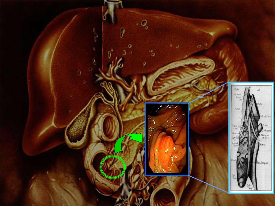
On examination he is thin and icteric. No lymphadenopathy
or stigmata of chronic liver disease. Abdominal examination
reveals a enlarged liver and a tensely cystic mass in the right
hypochondrium. No ascites and the digital rectal
examination is normal

Summary
An elderly male presenting with
painless jaundice, dark urine, pale
stools and pruritus. He has a palpable
gall bladder. The clinical picture is
suggestive of obstructive jaundice


What are the cardinal features of obstructive Jaundice?

Cardinal features of obstructive jaundice
JAUNDICE
PALE STOOLS
DARK URINE
PRURITUS
Obstruction to the extra hepatic
biliary tree
When do you suspect cancer?
PAINLESS = CANCER

Jaundice becomes clinically apparent when
the bilirubin level reaches 40mmol/l
The scleral elastin has a high affinity for
bilirubin, This is the reason why jaundice is
easily detectable in the eyes

Dark urine confirms conjugated
hyperbilirubinaemia which is filtered
via the kidneys. Unconjugated
bilirubin is tightly bound to albumin
which prevents glomerular filtration

Stools are pale because the bile is not reaching the duodenum
Deposition of bile salts irritate the skin and cause pruritus

The most likely CAUSE of jaundice in this patient is
Malignant obstruction of the extra
hepatic biliary tree
Painless progressive obstructive
jaundice in the elderly is highly
suggestive of a malignant obstruction

Could the cause of the obstruction be a stone?
Unlikely, because he has
painless obstructive
jaundice and has a palpable
gallbladder
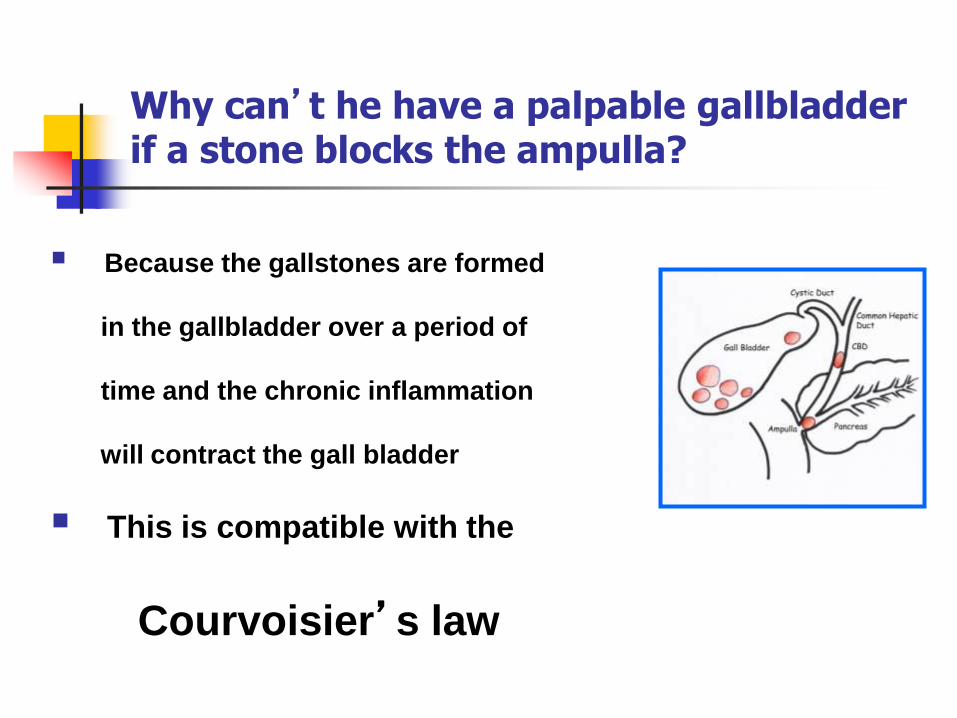
Why can’t he have a palpable gallbladder if a stone blocks the ampulla?
Because the gallstones are formed
in the gallbladder over a period of
time and the chronic inflammation
will contract the gall bladder
This is compatible with the
Courvoisier’s law
What is Courvoisier’s law?
In a patient with jaundice and a
palpable gallbladder the cause of
jaundice is unlikely to be due to a
stone

Most likely LEVEL of obstruction
Below the insertion
of the cystic duct

Could the level be higher such as at the porta hepatis?
No
because the gallbladder is palpable

What is the differential diagnosis?
Carcinoma of the head of pancreas
Carcinoma of the ampulla of vator
Carcinoma of the distal common
bile duct
Could it be Klatskin’s tumor?

What is a Klatskin’s tumour?
Katskin’s tumour is a cancer involving the common hepatic duct
The gallbladder is not distended in these patients as the bile cannot reach the gallbladder

What leading questions would help to support the diagnosis?
Presence of melaena Ampullary cancers bleed and cause intermittent melaena
because stagnation of bile cause pressure on the tumour
to break and bleed
Weight loss
Back pain

Progress of the patient - What investigations?
FBC
Liver profile (Alkaline Phosphatase)
Renal profile
PT/INR
Tumour markers
Ultrasound scan
Dilated intrahepatic biliary tree and distended gallbladder and Pancreatic head mass
Contrast CT Scan
2.5X 2.5 cm mass involving the Head of pancreas
not involving Superior mesenteric vein

Progress of the patient- What is the next step?
To obtain a histological diagnosis of the pancreatic head mass
Endoscopic Ultrasound guided core biopsy performed
Histology report
Adenocarcinoma
Progress of the patient
MDT discussion
Pre operative laparoscopy to exclude small peritoneal deposits
If negative- WHIPPLES OPERATION

What is WHIPPLES OPERATION ?
Major complex operation involving
removal of distal stomach,distal bile
duct,gall bladder ,entire duodenum and
head and neck of pancreas

Cancer of the pancreas - Core
Extremely poor prognosis –Survival after curative intent resection <5%
Significant post operative morbidity and mortality Most are not suitable for radical surgery at the diagnosis
70% occur in the Head of pancreas
Body and tail present late- rarely suitable for radical surgery
Radiotherpy is not used
Gemcytabine as adjuvant therapy has shown some survival
benefit
Palliative stenting using metal is the treatment of choice to relieve obstruction
OUTCOME - Bile duct and Head of Pancreas cancers
Peri-operative Mortality for Whipples operation is
0-8% in established units for pancreatic
surgery
5 year survival for Peri ampullary cancers is
40-70%
5 year survival for adenocarcinoma of the Head of
the pancreas is 5- 11% in best of centres
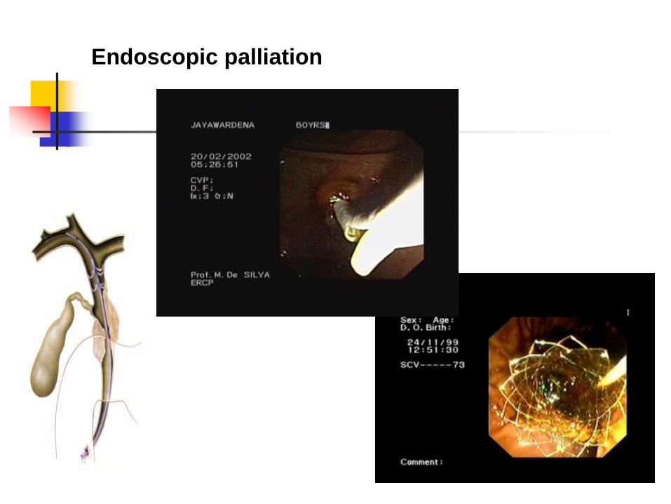
Endoscopic palliation

Questions

When do we say that a patient has obstructive Jaundice?
What is the cardinal clinical feature that differentiates between stone obstruction and cancer obstruction?
What is the physical sign that helps to identify the level of obstruction?
What is Courvoisier’s law?

What investigations will help to confirm the level of the obstruction?
What investigations help to confirm the cause of obstruction?

How to obtain a histology in biliary and pancreatic head cancers?
Peri ampullary cancer - Direct biopsy
Distal or proximal cholangiocarcinoma –Brush cytology
Pancreatic head cancer- Endoscopic ultrasound guided core biopsy

What are the complications anticipated in patients following surgery for Obstructive Jaundice?
1. Bleeding problems
( Solution - get INR corrected, Vit K, FFP)
2. Hepato renal syndrome
3. Wound healing problems
4. Sepsis

MCQ
Possible sites of obstruction in a patient with painless
obstructive jaundice, a palpable gallbladder and dilation of
the intrahepatic biliary tree on ultrasound would be
a) Common bile duct
b) Cystic duct
c) Common hepatic duct
d) Porta hepatis
e) Ampulla of vator

An US Scan performed on a deeply icteric elderly male with
no abdominal pain, reveals a dilated intrahepatic biliary
tree. Gall bladder and the bile ducts are not visualised
Likely causes of the obstruction include
a. Carcinoma of the head of pancreas
b. Klatskin’s tumor
c. Carcinoma of ampulla of vator
d. Cholangiocarcinoma of the common bile duct
e. Enlarged lymph nodes at the porta causing external compression
A 65 year-old man presents with painlss progressive
obstructive type jaundice with a palpable gall gall bladder.
Ultrasound is suggestive of pancreatic head mass and
distended gallbladder and dilated intra hepatic bile ducts
Most useful investigation to establish a diagnosis is
a) ERCP
b) MRCP
c) Ca 19.9
d) Abdominal CT scan and biopsy
e) Endoscopic ultrasound and biopsy

True/false
Progressive painless jaundice in elderly is indicative of a gallstone obstructing the bile duct
Courvoisier’s law is applied only to patients who are jaundiced
Klatskin tumour is a cholagiocarcinoma in the distal bile duct
Intermittent painless jaundice in elderly is suggestive of periampullary cancer
ERCP is the investigation of choice to diagnose pancreatic head cancer

BILIARY COLIC

Biliary colic
Objectives
To learn the varying clinical presentations of gallstones
To recollect the pathogenesis of gallstone disease
To learn the process of clinical judgment and decision making in a patient with symptomatic gallstones

A 60-old female presents with epigastric pain
radiating to right upper abdomen of 2 hour duration
Pain commenced as a dull ache but became so
severe that she had to be rushed to the hospital at
night
She looked unwell and the temperature is 38.4 c.
Vital signs are normal. Mild right hypochondrial
tenderness is noted

She is given 50 mg of Pethidine intravenously
Pain has disappeared when she work up in the morning but a dull ache in the right hypochondrium is persisting
What investigations would you perform?
WCC 14,000/cubic mm
Liver profile – Normal
Serum Amylase- Normal
Renal profile – Normal
Ultrasound scan

Ultrasound scan findings
Multiple gallbladder stones, thickened
gallbladder wall and a bile duct diameter of
5 mm
What is the normal diameter of the bile duct
on US?
6-7 MM in an adult

Most likely clinical diagnosis
Biliary colic leading to early acute
cholecystitis
WHY NOT BILIARY COLIC?

Why not biliary colic?
Pain is persisting as a dull ache
Slight pyrexia
High WCC
Oedematous gallbladder wall on
US

Biliary colic is a misnomer
colic
Biliary colic

She has epigastric pain and pyrexia? Could this be Cholangitis?
What is Cholangitis?
Could this be Cholangitis?
• No obstructive Jaundice
• No fever with chills and rigors
• No US evidence of dilated bile duct
• Liver profile is normal
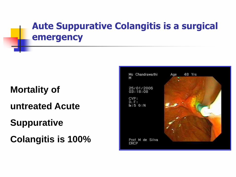
Aute Suppurative Colangitis is a surgical emergency
Mortality of
untreated Acute
Suppurative
Colangitis is 100%

Could she have Empyema of the Gallbladder?
What is Empyema of the Gallbladder?
Duration is short
No significant co-morbid factors
General condition is not very poor
No physical signs of peri-cholecystitis
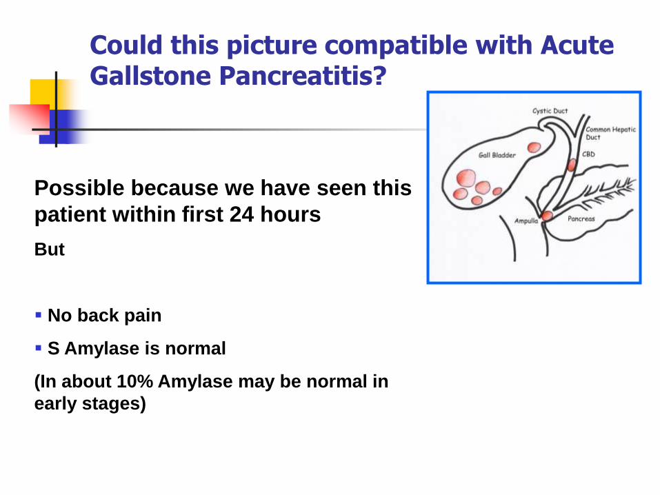
Could this picture compatible with Acute Gallstone Pancreatitis?
Possible because we have seen this
patient within first 24 hours
But
No back pain
S Amylase is normal
(In about 10% Amylase may be normal in
early stages)

Could she have Chronic Cholecystitis ?
Unlikely
Forty, fertile, fatty female with chronic
persistent right hypochondrial discomfort and
fat intolerance
Galllbladder dyspepsia = Chronic cholycystitis
This patient did not have dyspepsia

Progress of the patient
How would you manage this patient as a house officer?
Nil by mouth/IV fluids
IV Antibiotics Cefuroxime or Ciprofloxicin
Review to assess the response
Definitive treatment
Laparoscopic or open cholecystectomy?
Immediately or in 4 - 6 weeks?
Present consensus
Emergency Cholecystectomy is safe if performed within
48-72 hours of the onset of Acute Cholecystitis

Progress of the patient
Patient underwent an uneventful laparsocopic cholcystectomy
Operative cholangiogram was not performed

What is operative Cholangiogram? What other cholangiograms you know of?
Percutaneous Trans-hepatic
Cholangiogram PTC
Endoscopic Rerograde
Cholangiogram ERC
T tube Cholangiogram

Questions
Essential core - Gallstones
10% has it
10% has both gallbladder and bile duct stones
¾ are asymptomatic
¼ are symptomatic
Produce abnormal bile – Lithogenic bile
In planning strategy, bile duct stones takes priority over gallbladder stones
Can we dissolve gallstones?
YES
Oral bile acids effectively dissolve small gall stones made of cholesterol
Duration of therapy 2 years
50 % recur
Not used as a acceptable form of therapy

Asymptomatic Gall stones - Evidence for
decision making
“ Middle aged patient was found to have gall stones during a routine medical. He is seeking your guidance”
Does he need surgery?
Majority (70%) remain asymptomatic through out the life
Risk of developing complications – 1 % per year
When symptoms occur ,is usually biliary colic than a life threatening complication
HE DOES NOT NEED SURGERY
Picci R. Chir Ital 2005

I have heard that long term presence of
gallstones in the gallbladder predisposes to the
development of gallbladder cancer?
So is it safe for me not to have a
cholecystectomy just because I have not had
any symptoms from it?
Gall bladder cancer is a aggressive and lethal disease
Gall stones are associated with gall bladder cancer
No evidence to suggest prophylactic cholecystectomy reduces deaths related to gall bladder cancer
Most decision analysis studies do not favor prophylactic cholecystectomy for silent gall stones

Supplementary reading
Mirrizzi syndrome
Mucocele of Gallbladder
Gallbladder cancers

Obstructive Jaundice and Gall stones
Prof Mohan de Silva

What is obstructive Jaundice?
Obstruction to the extra hepatic biliary tree

What are our learning objectives?
How to workout the cause of obstruction?
How to work out the level of obstruction?
on history and examination
How to investigate?
How to work out the management based on the cause and level of obstruction?
What are the cardinal features of obstructive Jaundice?

Cardinal features of obstructive jaundice
JAUNDICE
PALE STOOLS
DARK URINE
PRURITUS
Obstruction to the extra hepatic
biliary tree

Jaundice becomes clinically apparent when
the bilirubin level reaches 40mmol/l
The scleral elastin has a high affinity for
bilirubin, This is the reason why jaundice is
easily detectable in the eyes

Dark urine confirms conjugated
hyperbilirubinaemia which is filtered
via the kidneys. Unconjugated
bilirubin is tightly bound to albumin
which prevents glomerular filtration

Stools are pale because the bile is not reaching the duodenum
Deposition of bile salts irritate the skin and cause pruritus

Cardinal features of obstructive jaundice
JAUNDICE
PALE STOOLS
DARK URINE
PRURITUS
Obstruction to the extra hepatic
biliary tree
When do you suspect cancer?
PAINLESS = CANCER
PAINFUL = STONES
BILE DUCT STONE or CANCER
Benign or Malignant

STONE IN THE GALLBLADDER WILL NOT CAUSE JAUNDICE
Painful obstructive jaundice means A
STONE IN THE BILE DUCT
EXCEPTION?

Painful obstructive jaundice means A STONE IN THE BILE DUCT
Exception
MIRIZZI’S SYNDROME

Is, bile duct stones the only benign condition?
BILE DUCT STONE or CANCER
Benign or Malignant

• Benign bile duct strictures due to iatrogenic bile duct injury
• Chronic Pancreatitis
Benign causes of Obstructive jaundice other than stones

Painless progressive obstructive
jaundice in the elderly is highly
suggestive of a malignant obstruction

What are the cancers?
Carcinoma of the head of pancreas
Carcinoma of the ampulla of vator
Carcinoma of the common bile duct
Carcinima of the common hepatic duct
(Klatskin’s tumour)
Gallbladder cancer


What are our learning objectives?
How to workout the cause of obstruction?
How to work out the level of obstruction?
on history and examination
How to investigate?
How to work out the management based on the cause and level of obstruction?

TO LOOK WHETHER THE GALLBLADDER IS
PALPABLE OR NOT?
How to workout the level of obstruction?


What is Courvoisier’s law?
In a patient with jaundice and a
palpable gallbladder the cause of
jaundice is unlikely to be due to a
stone

What investigations?
FBC
Liver profile (Alkaline Phosphatase)
Renal profile
PT/INR
Tumour markers
Ultrasound scan
Dilated intrahepatic biliary tree,distended gallbladder, Pancreatic head mass
Contrast CT Scan
Mass characteristics? – Is Superior mesenteric vein involved?
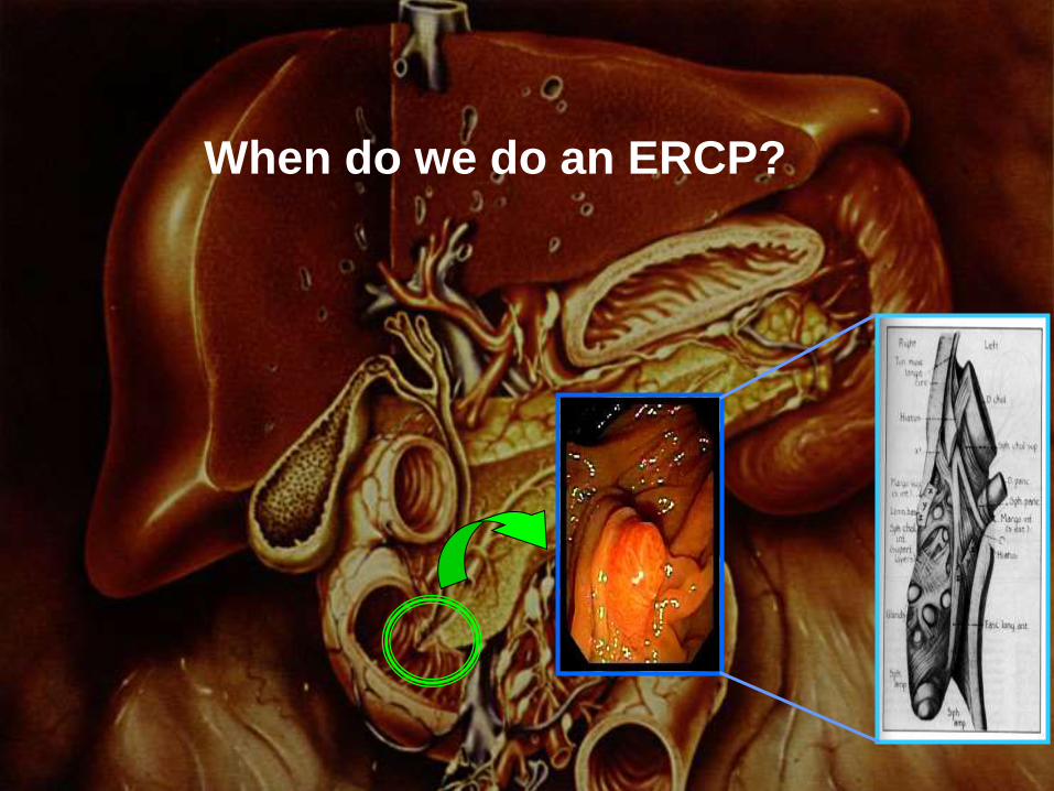
When do we do an ERCP?

When do we do an ERCP?
1. To confirm presence of bile duct stones
2. To remove bile duct stones
3. To obtain cytology
4. To palliate malignant obstruction by
placement of stents

How to obtain a histology in biliary and pancreatic head cancers?
Peri ampullary cancer - Direct biopsy
Distal or proximal cholangiocarcinoma –Brush cytology
Pancreatic head cancer- Endoscopic ultrasound guided core biopsy

What are our learning objectives?
How to workout the cause of obstruction?
How to work out the level of obstruction?
on history and examination
How to investigate?
How to work out the management based on the cause and level of obstruction?