Novel Aspects of Cytokine Action in Porcine Uterus
9
PL-ISSN 0015-5497 (print), ISSN 1734-9168 (online) Folia Biologica (Kraków), vol. 61 (2013), No 3-4 Ó Institute of Systematics and Evolution of Animals, PAS, Kraków, 2013 doi:10.3409/fb61_3-4.253 Novel Aspects of Cytokine Action in Porcine Uterus – Endometrial and Myometrial Production of Estrone (E 1 ) in the Presence of Interleukin 1â (IL1â), Interleukin 6 (IL6) and Tumor Necrosis Factor (TNFá) – in Vitro Study* Anita FRANCZAK, Bartosz WOJCIECHOWICZ and Genowefa KOTWICA Accepted May 15, 2013 F A., W B., K G. 2013. Novel aspects of cytokine action in porcine uterus - endometrial and myometrial production of estrone (E ) in the presence of interleukin 1â (IL1â), interleukin 6 (IL6) and tumor necrosis factor (TNFá) – in vitro study. Folia Biologica (Kraków) 61: 253-261. Interleukin 1ß (IL-1ß), interleukin 6 (IL-6) and tumor necrosis factor á (TNFá) increased (P<0.05) estrone (E ) release from endometrial explants of pregnant pigs on days 10 to 11 after 12 h of tissue incubation in vitro with cytokines and on days 12 to 13 after 6 h of incubation. After 12 h of incubation on days 12 to 13 and 15 to 16 of pregnancy only IL6 increased E release. In non-gravid pigs IL1â, IL6 and TNFá increased endometrial E release only on days 12 to 13 of the estrous cycle. The cytokines did not affect myometrial E release on days 10 to 11 and 15 to 16 of pregnancy. On days 12 to 13 of pregnancy myometrial release of E was markedly increased in response to IL1â and IL6. In cyclic pigs only IL6 after 6 h of in vitro incubation increased myometrial E release on days 12 to 13 and 15 to 16. Progesterone (P ) increased both endometrial and myometrial release of E during the studied days of pregnancy and the estrous cycle, except for endometrial release on days 10 to 11 and 15 to 16 of the estrous cycle after 6 h of in vitro incubation. The results demonstrated that these cytokines may regulate the release of E both from the endometrium and myometrium harvested from gravid and non-gravid pigs. The results showed a pivotal role of IL1â, IL6 and TNFá in the regulation of E release in the porcine uterus in vitro. Key words: Interleukin 1â, Interleukin 6, Tumor necrosis factor á, Estrone, Endometrium, Myometrium, Pregnancy, Pigs. Anita F , Bartosz W , Genowefa K Department of Animal Physiology, Faculty of Biology and Biotechnology, University of Warmia and Mazury in Olsztyn, Oczapowski 1A, 10-718 Olsztyn, Poland. Email: [email protected] In pigs estrone (E ) and estradiol (E ) produced by porcine embryos were shown to be signals for maternal recognition of pregnancy (BAZER & TCHATCHER 1977). Previously we have demon- strated that tissues of porcine uterus can produce estrogens (FRANCZAK 2008; FRANCZAK &KOT- WICA 2008; FRANCZAK &KOTWICA 2010) and the endometrium together with the myometrium on days 14 to 16 of early pregnancy are an impor- tant source of estrone (E ) (FRANCZAK 2008; FRANCZAK &KOTWICA 2008). E was found to be a main contributor in endometrial and myometrial total steroid secretion (FRANCZAK 2008). On days 14 to 16 of pregnancy the endometrium releases more E than on days 14 to 16 of the estrous cycle, while the myometrium apparently equally partici- pated in the total uterine basal E secretion. Thus, in pigs during early pregnancy the endometrium and the myometrium, while during luteolysis only the myometrium, may mostly contribute to the ba- sal total secretion of E (FRANCZAK 2008). The uterus of pigs is known to be a source of es- trogens, however the factors involved in the regu- lation of myometrial E production remain unknown. In general, P was shown to be a substrate for ster- oid synthesis in the porcine uterus (FRANCZAK 2008; FRANCZAK &KOTWICA 2008; FRANCZAK &KOTWICA 2010), but the hormone did not stimu- late E secretion on days 14 to 16 of pregnancy (FRANCZAK 2008). This phenomenon was not ex- plained. Little is known about the hormones or other factors that influence uterine steroidogenesis. _______________________________________ *This research was funded by grant N N311 0685 33 (2007-2010) from the Ministry of Science and Higher Education, Poland and by grant N N311 526940 (2011 to 2013) from the National Science Centre, Poland.
Transcript of Novel Aspects of Cytokine Action in Porcine Uterus
PL-ISSN 0015-5497 (print), ISSN 1734-9168 (online) Folia Biologica (Kraków), vol. 61 (2013), No 3-4Ó Institute of Systematics and Evolution of Animals, PAS, Kraków, 2013 doi:10.3409/fb61_3-4.253
Novel Aspects of Cytokine Action in Porcine Uterus – Endometrial and
Myometrial Production of Estrone (E1) in the Presence of Interleukin 1â (IL1â),
Interleukin 6 (IL6) and Tumor Necrosis Factor (TNFá) – in Vitro Study*
Anita FRANCZAK, Bartosz WOJCIECHOWICZ and Genowefa KOTWICA
Accepted May 15, 2013
FRANCZAK A., WOJCIECHOWICZ B., KOTWICA G. 2013. Novel aspects of cytokine action inporcine uterus - endometrial and myometrial production of estrone (E1) in the presence ofinterleukin 1â (IL1â), interleukin 6 (IL6) and tumor necrosis factor (TNFá) – in vitro study.Folia Biologica (Kraków) 61: 253-261.
Interleukin 1ß (IL-1ß), interleukin 6 (IL-6) and tumor necrosis factor á (TNFá) increased(P<0.05) estrone (E1) release from endometrial explants of pregnant pigs on days 10 to 11after 12 h of tissue incubation in vitro with cytokines and on days 12 to 13 after 6 h ofincubation. After 12 h of incubation on days 12 to 13 and 15 to 16 of pregnancy only IL6increased E1 release. In non-gravid pigs IL1â, IL6 and TNFá increased endometrial E1release only on days 12 to 13 of the estrous cycle. The cytokines did not affect myometrial E1release on days 10 to 11 and 15 to 16 of pregnancy. On days 12 to 13 of pregnancy myometrialrelease of E1was markedly increased in response to IL1â and IL6. In cyclic pigs only IL6 after6 h of in vitro incubation increased myometrial E1 release on days 12 to 13 and 15 to 16.Progesterone (P4) increased both endometrial and myometrial release of E1 during the studieddays of pregnancy and the estrous cycle, except for endometrial release on days 10 to 11 and15 to 16 of the estrous cycle after 6 h of in vitro incubation. The results demonstrated thatthese cytokines may regulate the release of E1 both from the endometrium and myometriumharvested from gravid and non-gravid pigs. The results showed a pivotal role of IL1â, IL6 andTNFá in the regulation of E1 release in the porcine uterus in vitro.
Key words: Interleukin 1â, Interleukin 6, Tumor necrosis factor á, Estrone, Endometrium,Myometrium, Pregnancy, Pigs.
Anita FRANCZAK, Bartosz WOJCIECHOWICZ, Genowefa KOTWICA, Department of AnimalPhysiology, Faculty of Biology and Biotechnology, University of Warmia and Mazury inOlsztyn, Oczapowski 1A, 10-718 Olsztyn, Poland.Email: [email protected]
In pigs estrone (E1) and estradiol (E2) producedby porcine embryos were shown to be signals formaternal recognition of pregnancy (BAZER &TCHATCHER 1977). Previously we have demon-strated that tissues of porcine uterus can produceestrogens (FRANCZAK 2008; FRANCZAK & KOT-
WICA 2008; FRANCZAK & KOTWICA 2010) andthe endometrium together with the myometriumon days 14 to 16 of early pregnancy are an impor-tant source of estrone (E1) (FRANCZAK 2008;FRANCZAK & KOTWICA 2008). E1 was found to bea main contributor in endometrial and myometrialtotal steroid secretion (FRANCZAK 2008). On days14 to 16 of pregnancy the endometrium releasesmore E1 than on days 14 to 16 of the estrous cycle,while the myometrium apparently equally partici-
pated in the total uterine basal E1 secretion. Thus,in pigs during early pregnancy the endometriumand the myometrium, while during luteolysis onlythe myometrium, may mostly contribute to the ba-sal total secretion of E1 (FRANCZAK 2008).
The uterus of pigs is known to be a source of es-trogens, however the factors involved in the regu-lation of myometrial E1 production remain unknown.In general, P4 was shown to be a substrate for ster-oid synthesis in the porcine uterus (FRANCZAK
2008; FRANCZAK & KOTWICA 2008; FRANCZAK
& KOTWICA 2010), but the hormone did not stimu-late E1 secretion on days 14 to 16 of pregnancy(FRANCZAK 2008). This phenomenon was not ex-plained. Little is known about the hormones orother factors that influence uterine steroidogenesis.
_______________________________________
*This research was funded by grant N N311 0685 33 (2007-2010) from the Ministry of Science and Higher Education, Polandand by grant N N311 526940 (2011 to 2013) from the National Science Centre, Poland.
In the present study we have examined if inter-leukin 1â (IL1â), interleukin 6 (IL6) and tumor ne-crosis factor á (TNFá), which are produced byporcine embryos and the uterus, may be involvedin the regulation of uterine E1 production and re-lease in vitro. Cytokines, acting through specificreceptors, may regulate the secretory activity ofthe uterus in pigs (FRANCZAK et al. 2010; 2012).Peri-implantation porcine embryos and the en-dometrium express IL1â (TUO et al. 1996; ROSS et al.
2003a,b) and IL6 (MATHIALAGAN et al. 1992;ANEGON et al. 1994). Tumor necrosis factor á isalso produced by the uterus of pregnant pigs (YU et al.1998). In a recent study we documented that IL1â,IL6 and TNFá affect prostaglandin F2á (PGF2á)release and activate PGF2á metabolism in the en-dometrium to protect the corpus luteum (CL) inpregnant pigs (FRANCZAK et al. 2012). IL1â in-duced synthesis and secretion of luteotrophic pros-taglandin E2 (PGE2) to overcome luteolysis inpregnant pigs (FRANCZAK et al. 2010) and may beincluded in intraluteal luteotrophic regulation ofCL functions in gravid and cyclic pigs (¯MIJEW-
SKA et al. 2013). In the pig, the expression of IL1âat the site of embryo-maternal contact in the uterusis unique and species-specific (TUO et al. 1996).TNFá is expressed in embryos, ovaries, oviductsand uteri of human and rodents (HUNT 1993). Ingravid pigs TNFá stimulates PGE2 synthesis andsecretion by luminal epithelial cells of the en-dometrium (WACLAWIK et al. 2010). It was foundthat during days 15 to 16 of pregnancy TNFástimulates secretion of the prostaglandin F2á me-tabolite – 13,14-dihydro-15-keto PGF2á (PGFM)by the endometrium (FRANCZAK et al. 2012). Incyclic pigs TNFá is involved in the regulation ofPGF2á synthase (PGFS) mRNA expression(FRANCZAK et al. 2012) and prostaglandin secre-tion (BLITEK & ZIECIK 2006). Because the abilityof the uterine tissues to induce steroidogenesis hasbeen confirmed, we hypothesized that IL1â, IL6and TNFá may be involved in conditioning thisprocess and may increase E1 concentrations thatenhance the effects of estrogens in the uterus.
The objective of this investigation was to test thehypothesis that endometrial and myometrial pro-duction of E1 is regulated by cytokines. To addressthis issue, we have determined if the endometriumand the myometrium of pigs can produce E1 in vi-
tro in response to IL1â, IL6 and TNFá on days 10to 11, i.e. before the time of maternal recognitionof pregnancy, on days 12 to 13, i.e. during the timeof maternal recognition of pregnancy and on days15 to 16, i.e. during the time of corpus luteummaintenance, beginning of implantation and pre-vention of luteolysis. The effects observed in preg-nant pigs were compared with effects oncorresponding days of the estrous cycle.
Material and Methods
Animals and collection of endometrial and myome-trial tissue
All experiments were approved by the AnimalEthics Committee, University of Warmia and Ma-zury in Olsztyn, Poland. Post-pubertal, six monthold crossbred pigs (Large White H Polish Landrace),weighing 90-110 kg were used during early preg-nancy or the estrous cycle. Gilts were observed forestrus behavior in the presence of an intact boar.The onset of the second estrus was designated asday 0 of the estrous cycle. Gilts assigned to theearly pregnancy group were naturally bred on thesecond day of estrus. The animals on days 10 to 11(n = 5), 12 to 13 (n = 5) and 15 to 16 (n = 5) of preg-nancy, or days 10 to 11 (n = 5), 12 to 13 (n = 5) and15 to 16 (n = 5) of the estrous cycle were slaugh-tered in a commercial slaughterhouse. Pregnancyin mated gilts was confirmed by the presence ofembryos after flushing each uterine horn with 20 mlsterile saline. The stage of the estrous cycle wasalso confirmed by morphological changes of theovaries and CL quality (AKINS & MORRISSETTE
1968). Uterine horns from early-pregnant or cyclic giltswere placed immediately in ice-cold PBS supple-mented with 100 IU/ml penicillin and 100 Fg/mlstreptomycin and transported to the laboratory onice.
Preparation of endometrial and myometrial slices
The middle part of the uterine horns collectedfrom experimental gilts was opened longitudinallyon the mesometrial surface and the endometriumand the myometrium were separated using a scal-pel blade. The endometrium and the myometriumwere sliced thinly (200-210 mg, 3 mm thick) andwashed twice with PBS supplemented with antibi-otics.
In vitro incubation of endometrial and myometrialslices
Individual fresh endometrial and myometrialslices were placed separately in culture vials con-taining 2 ml of Medium 199 (Sigma, Germany)supplemented with 0.1% BSA fraction V (ICN,USA) and 20 Fg gentamycin (Sigma, Germany).These tissue cultures were pre-incubated in a waterbath for 18 h at 37oC in an atmosphere of 95% O2and 5% CO2. After preincubation, endometrial andmyometrial slices were incubated for 6 and 12 h incontrol medium or in medium supplemented withprogesterone (P4, 10-5M), IL1â (1 ng/ml and 10ng/ml), IL6 (1 ng/ml and 10 ng/ml) or TNFá (10ng/ml). P4 was used as a control and substrate for
A. FRANCZAK et al.254
E1 production. Cytokines were obtained from Bio-mol, GmbH, Germany. The doses of cytokineswere selected according to earlier studies (FRANC-
ZAK et al. 2010, 2012). After incubation, culturevials were placed in an ice bath, culture mediumwas collected and frozen at -20oC until E1 assaywith radioimmunoassay (RIA).
Estrone (E1) determination
Concentration of E1 was determined by the RIAmethod (CIERESZKO 1999). Cross-reactivity ofantisera against E1 has been reported (SZAFRAN-
SKA et al. 2002). The efficiency of extraction forE1 was 85.3 ± 0.07%. E1 assay sensitivity was 1pg/ml. The coefficient of correlation between theadded and recovered amount of E1 concentrationswas 0.975. The intra- and interassay coefficients ofvariation were 0.5% and 1.9%, respectively.
Statistical analysis
Mean concentrations of E1 released in responseto cytokines were log-transformed to reduce het-erogeneity of variance and were analyzed bymulti-way ANOVA with the dose of treatments,time of incubation, reproductive status and days ofpregnancy or the estrous cycle as the main effects
followed by a LSD post-hoc test (Statistica, Stat-Soft Inc, Tulsa, OK, USA).
Results
Basal endometrial and myometrial release of E1during days 10 to 11, 12 to 13 and 15 to 16 ofpregnancy and the estrous cycle
The results of the basal endometrial and myome-trial production of E1are presented in Table 1. Dur-ing pregnancy basal endometrial release of E1 after6 h of incubation did not differ among days 10 to11, 12 to 13 and 15 to 16 (P>0.05). After 12 h of in-cubation in vitro basal E1 release was higher ondays 12 to 13 and 15 to 16 than on days 10 to 11 ofpregnancy (P<0.05). During the estrous cycle E1production by the endometrium was about two-fold lower (P<0.05) on days 12 to 13 than that ob-served during days 10 to 11 and 15 to 16, both after6 and 12 h of incubation. Basal myometrial E1 pro-duction did not differ among the studied days ofpregnancy (P>0.05) after 6 and 12 h of incubationand did not differ among the studied days of the es-trous cycle after 6 h of incubation. After 12 h of in-cubation myometrial production of E1 in cyclicpigs was about two fold higher on days 15 to 16 of
Cytokines and Uterine Estrone Production 255
Table 1
Basal estrone (pg / ml) secretion (mean ± S.E.M.) from endometrial and myometrial ex-plants harvested on days 10 to 11, 12 to 13 and 15 to 16 of pregnancy and the estrous cycleand incubated in vitro within 6 and 12 hours
Endometrium
DaysPregnancy The estrous cycle
6 hours of incubation in vitro 12 hours of incubation in vitro 6 hours of incubation in vitro 12 hours of incubation in vitro
10 to 11 55.5 ± 9.6Aa 23.1 ± 2.8Aa 58.0 ± 8.0Aa 58.9 ± 14.3Ab
12 to13 32.8 ± 7.1Aa 39.1 ± 7.6Ba 27.4 ± 6.7Ba 23.9 ± 4.9Ba
15 to 16 39.6 ± 5.2Aa 39.9 ± 11.3Ba 50.2 ± 8.1Aa 55.2 ± 18.8Aa
Myometrium
DaysPregnancy The estrous cycle
6 hours of incubation in vitro 12 hours of incubation in vitro 6 hours of incubation in vitro 12 hours of incubation in vitro
10 to 11 40.5 ± 6.2Aa 44.9 ± 13.1Aa 55.0 ± 14.7Aa 40.9 ± 14.8Aa
12 to 13 33.7 ± 8.1Aa 40.4 ± 6.8Aa 41.7 ± 7.9Aa 40.2 ± 8.5Aa
15 to 16 37.7 ± 5.8Aa 48.0 ± 19.8Aa 46.3 ± 15.3Aa 76.5 ± 14.0Ba
A,B Different uppercase letters designate significant differences among the studied days of pregnancy or the estrous cycle withinthe same time of incubation (data in columns).
a,b Different lowercase letters designate significant differences between corresponding days of pregnancy and the estrous cycleafter the same time of incubation (data in rows).
the estrous cycle than on days 10 to 11 and 12 to 13(P<0.05).
IL1â, IL6 and TNFá-induced endometrial E1 re-lease in vitro during early pregnancy and the es-trous cycle
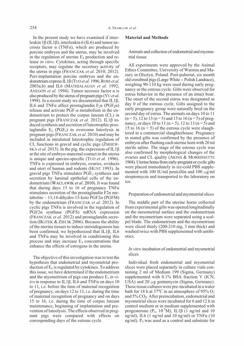
IL1â, IL6 and TNFá increased E1 release in vitro
(P<0.05) in endometrial explants harvested ondays 10 to 11 from gravid pigs and incubated in vi-
tro for 12 h as well as in endometrial explants har-vested on days 12 to 13 of pregnancy andincubated for 6 h (Fig. 1). After 12 h in vitro incu-bation on days 12 to 13 of pregnancy only IL6 (1ng/ml and 10 ng/ml) increased E1 release (P<0.05),while an effect of IL1â or TNFá was not observed(P>0.05). During days 15 to 16 of pregnancy theenhanced release of E1 was observed only in thepresence of IL6 after 12 h of in vitro incubation
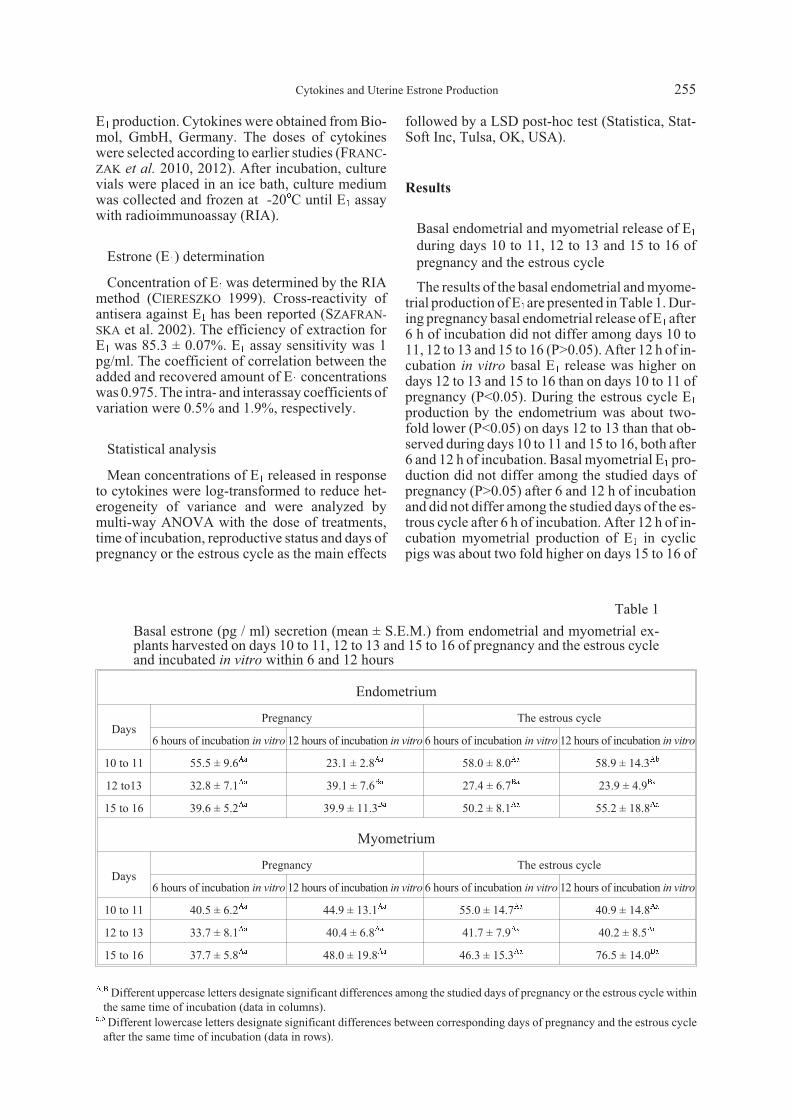
(Fig. 1). Neither IL1â, nor TNFá affected endome-trial E1 release during days 15 to 16 of pregnancy(P>0.05). In non-gravid pigs the stimulatory effectof IL1â, IL6 and TNFá on E1 release was observedonly on days 12 to 13 of the estrous cycle, both af-ter 6 and 12 h incubation (Fig. 2).
IL1â, IL6 and TNFá-induced myometrial E1 re-lease in vitro during early pregnancy and the es-trous cycle
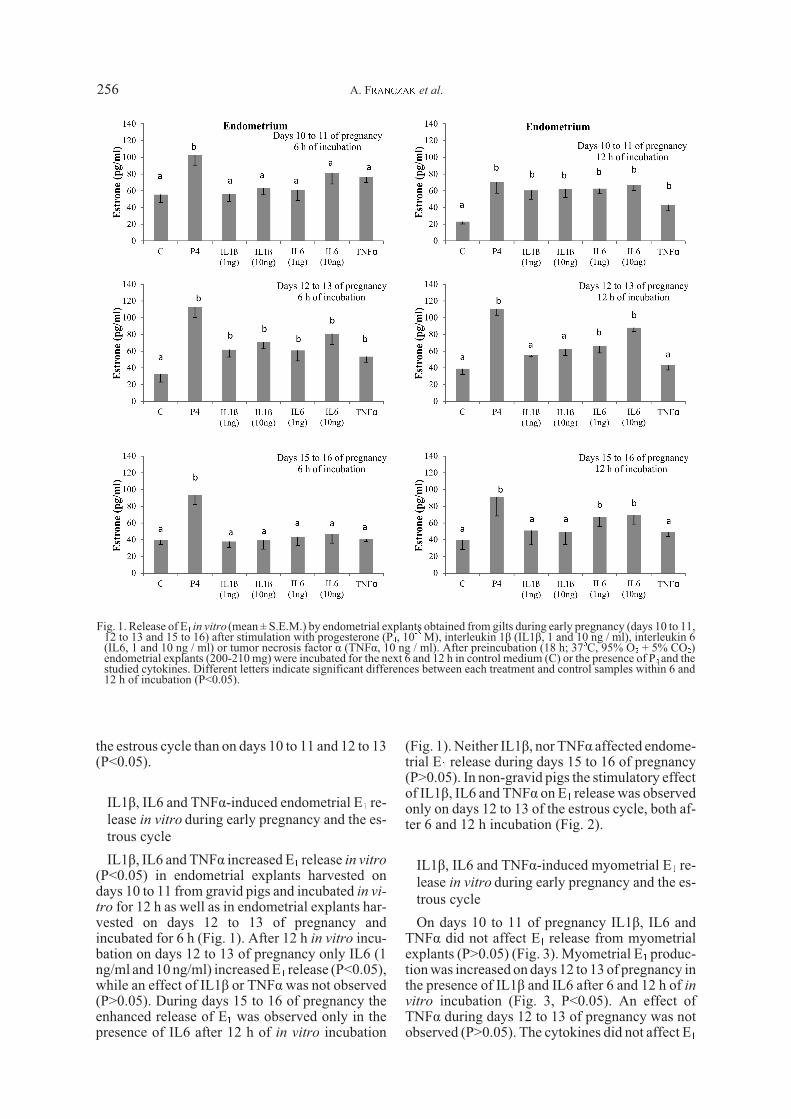
On days 10 to 11 of pregnancy IL1â, IL6 andTNFá did not affect E1 release from myometrialexplants (P>0.05) (Fig. 3). Myometrial E1 produc-tion was increased on days 12 to 13 of pregnancy inthe presence of IL1â and IL6 after 6 and 12 h of in
vitro incubation (Fig. 3, P<0.05). An effect ofTNFá during days 12 to 13 of pregnancy was notobserved (P>0.05). The cytokines did not affect E1
Fig. 1. Release of E1 in vitro (mean ± S.E.M.) by endometrial explants obtained from gilts during early pregnancy (days 10 to 11,12 to 13 and 15 to 16) after stimulation with progesterone (P4, 10-5 M), interleukin 1â (IL1â, 1 and 10 ng / ml), interleukin 6(IL6, 1 and 10 ng / ml) or tumor necrosis factor á (TNFá, 10 ng / ml). After preincubation (18 h; 37oC, 95% O2 + 5% CO2)endometrial explants (200-210 mg) were incubated for the next 6 and 12 h in control medium (C) or the presence of P4 and thestudied cytokines. Different letters indicate significant differences between each treatment and control samples within 6 and12 h of incubation (P<0.05).
A. FRANCZAK et al.256
12
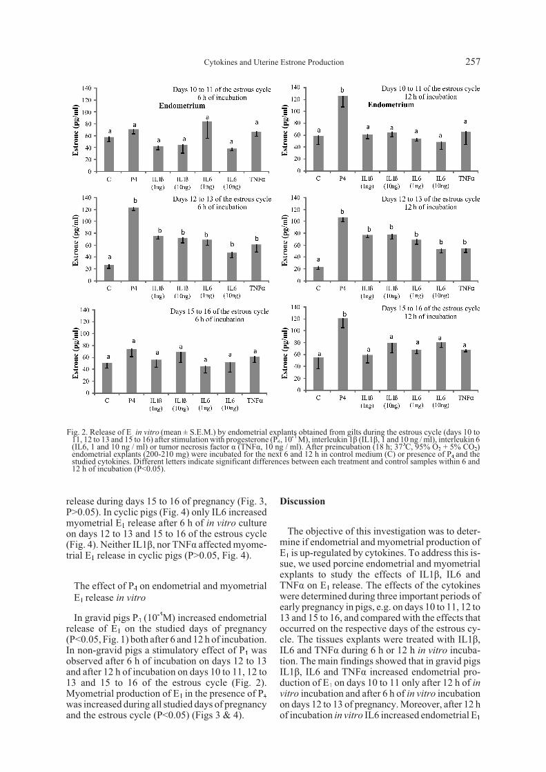
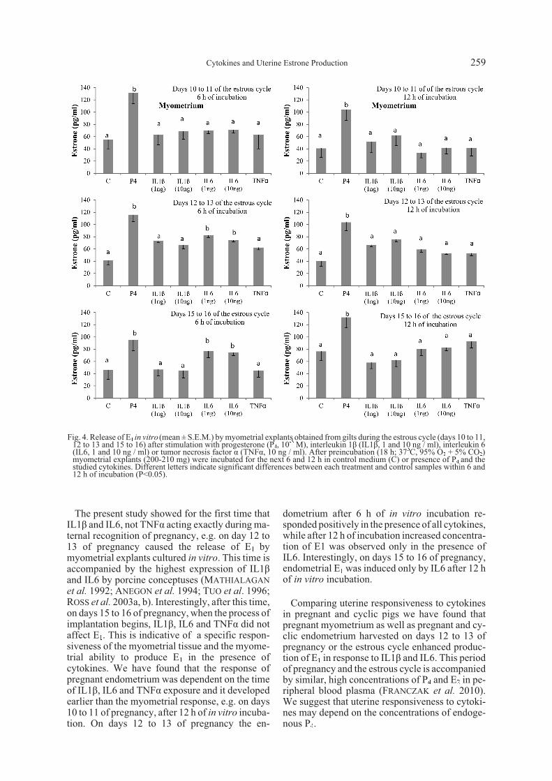
release during days 15 to 16 of pregnancy (Fig. 3,P>0.05). In cyclic pigs (Fig. 4) only IL6 increasedmyometrial E1 release after 6 h of in vitro cultureon days 12 to 13 and 15 to 16 of the estrous cycle(Fig. 4). Neither IL1â, nor TNFá affected myome-trial E1 release in cyclic pigs (P>0.05, Fig. 4).
The effect of P4 on endometrial and myometrialE1 release in vitro
In gravid pigs P4 (10-5M) increased endometrialrelease of E1 on the studied days of pregnancy(P<0.05, Fig. 1) both after 6 and 12 h of incubation.In non-gravid pigs a stimulatory effect of P4 wasobserved after 6 h of incubation on days 12 to 13and after 12 h of incubation on days 10 to 11, 12 to13 and 15 to 16 of the estrous cycle (Fig. 2).Myometrial production of E1 in the presence of P4was increased during all studied days of pregnancyand the estrous cycle (P<0.05) (Figs 3 & 4).
Discussion
The objective of this investigation was to deter-mine if endometrial and myometrial production ofE1 is up-regulated by cytokines. To address this is-sue, we used porcine endometrial and myometrialexplants to study the effects of IL1â, IL6 andTNFá on E1 release. The effects of the cytokineswere determined during three important periods ofearly pregnancy in pigs, e.g. on days 10 to 11, 12 to13 and 15 to 16, and compared with the effects thatoccurred on the respective days of the estrous cy-cle. The tissues explants were treated with IL1â,IL6 and TNFá during 6 h or 12 h in vitro incuba-tion. The main findings showed that in gravid pigsIL1â, IL6 and TNFá increased endometrial pro-duction of E1 on days 10 to 11 only after 12 h of in
vitro incubation and after 6 h of in vitro incubationon days 12 to 13 of pregnancy. Moreover, after 12 hof incubation in vitro IL6 increased endometrial E1
Fig. 2. Release of E1 in vitro (mean ± S.E.M.) by endometrial explants obtained from gilts during the estrous cycle (days 10 to11, 12 to 13 and 15 to 16) after stimulation with progesterone (P4, 10-5 M), interleukin 1â (IL1â, 1 and 10 ng / ml), interleukin 6(IL6, 1 and 10 ng / ml) or tumor necrosis factor á (TNFá, 10 ng / ml). After preincubation (18 h; 37oC, 95% O2 + 5% CO2)endometrial explants (200-210 mg) were incubated for the next 6 and 12 h in control medium (C) or presence of P4 and thestudied cytokines. Different letters indicate significant differences between each treatment and control samples within 6 and12 h of incubation (P<0.05).
Cytokines and Uterine Estrone Production 257
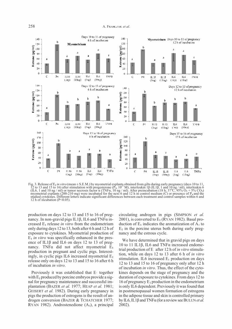
production on days 12 to 13 and 15 to 16 of preg-nancy. In non-gravid pigs IL1â, IL6 and TNFá in-creased E1 release in vitro from the endometriumonly during days 12 to 13, both after 6 h and 12 h ofexposure to cytokines. Myometrial production ofE1 in vitro was specifically enhanced in the pres-ence of IL1â and IL6 on days 12 to 13 of preg-nancy. TNFá did not affect myometrial E1production in pregnant and cyclic pigs. Interest-ingly, in cyclic pigs IL6 increased myometrial E1release only on days 12 to 13 and 15 to 16 after 6 hof incubation in vitro.
Previously it was established that E1 togetherwith E2 produced by porcine embryos provide a sig-nal for pregnancy maintenance and successful im-plantation (BAZER et al. 1977; HEAP et al. 1981;GEISERT et al. 1982). During early pregnancy inpigs the production of estrogens is the result of an-drogen conversion (BAZER & TCHATCHER 1977;RYAN 1982). Androstenedione (A4), a principal
circulating androgen in pigs (SIMPSON et al.
2001), is converted to E1 (RYAN 1982). Basal pro-duction of E1 indicates the aromatization of A4 toE1 in the porcine uterus both during early preg-nancy and the estrous cycle.
We have determined that in gravid pigs on days10 to 11 IL1â, IL6 and TNFá increased endome-trial production of E1 after 12 h of in vitro stimula-tion, while on days 12 to 13 after 6 h of in vitro
stimulation. IL6 increased E1 production on days12 to 13 and 15 to 16 of pregnancy only after 12 hof incubation in vitro. Thus, the effect of the cyto-kines depends on the stage of pregnancy and theduration of exposure to cytokines. From days 12 to16 of pregnancy E1 production in the endometriumis only IL6 dependent. Previously it was found thatin postmenopausal women formation of estrogensin the adipose tissue and skin is controlled primaryby IL6, IL1â and TNFá (for a review see BULUN et al.
2002).
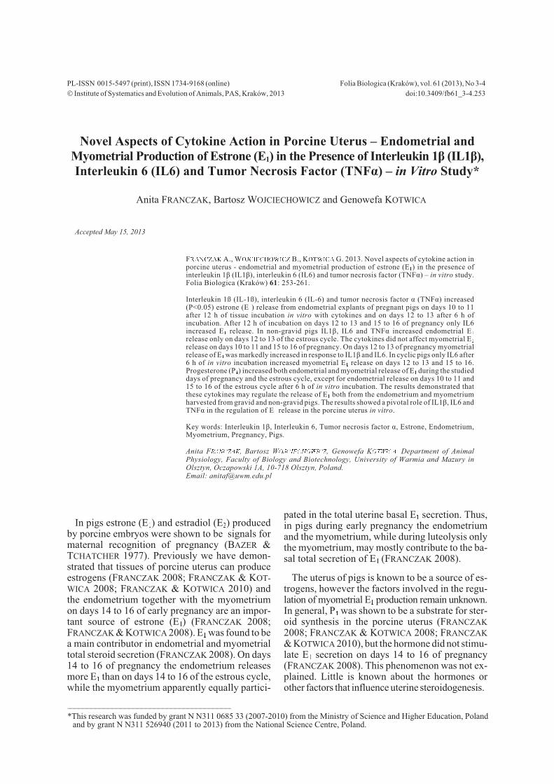
Fig. 3. Release of E1 in vitro (mean ± S.E.M.) by myometrial explants obtained from gilts during early pregnancy (days 10 to 11,12 to 13 and 15 to 16) after stimulation with progesterone (P4, 10-5 M), interleukin 1â (IL1â, 1 and 10 ng / ml), interleukin 6(IL6, 1 and 10 ng / ml) or tumor necrosis factor á (TNFá, 10 ng / ml). After preincubation (18 h; 37oC, 95% O2 + 5% CO2)myometrial explants (200-210 mg) were incubated for the next 6 and 12 h in control medium (C) or presence of P4 and thestudied cytokines. Different letters indicate significant differences between each treatment and control samples within 6 and12 h of incubation (P<0.05).
A. FRANCZAK et al.258
The present study showed for the first time thatIL1â and IL6, not TNFá acting exactly during ma-ternal recognition of pregnancy, e.g. on day 12 to13 of pregnancy caused the release of E1 bymyometrial explants cultured in vitro. This time isaccompanied by the highest expression of IL1âand IL6 by porcine conceptuses (MATHIALAGAN
et al. 1992; ANEGON et al. 1994; TUO et al. 1996;ROSS et al. 2003a, b). Interestingly, after this time,on days 15 to 16 of pregnancy, when the process ofimplantation begins, IL1â, IL6 and TNFá did notaffect E1. This is indicative of a specific respon-siveness of the myometrial tissue and the myome-trial ability to produce E1 in the presence ofcytokines. We have found that the response ofpregnant endometrium was dependent on the timeof IL1â, IL6 and TNFá exposure and it developedearlier than the myometrial response, e.g. on days10 to 11 of pregnancy, after 12 h of in vitro incuba-tion. On days 12 to 13 of pregnancy the en-
dometrium after 6 h of in vitro incubation re-sponded positively in the presence of all cytokines,while after 12 h of incubation increased concentra-tion of E1 was observed only in the presence ofIL6. Interestingly, on days 15 to 16 of pregnancy,endometrial E1 was induced only by IL6 after 12 hof in vitro incubation.
Comparing uterine responsiveness to cytokinesin pregnant and cyclic pigs we have found thatpregnant myometrium as well as pregnant and cy-clic endometrium harvested on days 12 to 13 ofpregnancy or the estrous cycle enhanced produc-tion of E1 in response to IL1â and IL6. This periodof pregnancy and the estrous cycle is accompaniedby similar, high concentrations of P4 and E2 in pe-ripheral blood plasma (FRANCZAK et al. 2010).We suggest that uterine responsiveness to cytoki-nes may depend on the concentrations of endoge-nous P4.
Fig. 4. Release of E1 in vitro (mean ± S.E.M.) by myometrial explants obtained from gilts during the estrous cycle (days 10 to 11,12 to 13 and 15 to 16) after stimulation with progesterone (P4, 10-5 M), interleukin 1â (IL1â, 1 and 10 ng / ml), interleukin 6(IL6, 1 and 10 ng / ml) or tumor necrosis factor á (TNFá, 10 ng / ml). After preincubation (18 h; 37oC, 95% O2 + 5% CO2)myometrial explants (200-210 mg) were incubated for the next 6 and 12 h in control medium (C) or presence of P4 and thestudied cytokines. Different letters indicate significant differences between each treatment and control samples within 6 and12 h of incubation (P<0.05).
Cytokines and Uterine Estrone Production 259
It is important to note that factors affecting uter-ine (endometrial and myometrial) secretion mayinfluence the efficiency of conceptus prolifera-tion, growth and development as well as uterineactivity and capacity (VALLET & CHRISTENSON
1996). Despite the fact that E1 is a less active estro-gen than estradiol (VALLET & CHRISTENSON 1996),E1 can be converted to more potent and active es-tradiol. Moreover E1 can be converted to catecho-lestrogens (CHAKRABORTY et al. 1989) whichmay have high biological activity (ROSENKRANS
et al. 1990). Thus, the level of E1 production by theuterus in general may be very important for modu-lation of endometrial protein secretion (TROUT et al.
1992) and for proper communication between em-bryos and the endometrium (ROBERTS & BAZER
1988). It was found that treatment of pregnant giltswith E1 resulted in increased endometrial secretionof nondialyzable macromolecules in culture, re-flecting total protein synthesis in the tissue (VAL-
LET & CHRISTENSON 1996). It is important to notethat the proper level of uterine estrogen synthesisand metabolism during early pregnancy may bea prerequisite for successful implantation. Previ-ously, we have found in pregnant endometrium in-creased expression of genes encoding sulfotransfe-rase 1E1 and sulfotransferase 2A1 (FRANCZAK et al.
2013). Sulfotransferases convert estrogens intonon-active estrogen sulphates. Thus, the mecha-nism of E1 metabolism may allow quick adjust-ment of steroid concentration in the uterus.
In conclusion: 1) endometrial and myometrialexplants of pigs release E1 in vitro in response tocytokines; 2) in pregnant pigs IL1â, IL6 and TNFáregulate endometrial production of E1 on days 10to 11 and on days 12 to 13 in a time of culture de-pendent manner; 3) on days 15 to 16 of pregnancyIL6 caused E1 release from the endometrium; 4) incyclic pigs IL1â, IL6 and TNFá increase E1 releasein vitro from the endometrium only during days 12to 13 of the estrous cycle; 5) in the presence ofIL1â and IL6 in gravid pigs myometrial produc-tion of E1 in vitro was enhanced specifically ondays 12 to 13 of pregnancy.
References
AKINS E.L., MORRISSETTE M.C. 1968. Gross ovarianchanges during oestrous cycle of swine. Am. J. Vet. Res. 29:1953-1957.
ANEGON I., CUTURI M.C., GODARD A., MOREAU M., TER-QUI M., MARTINAT-BOTTÉ F., SOULILLOU J.P. 1994. Pres-ence of leukaemia inhibitory factor and interleukin 6 inporcine uterine secretions prior to conceptus attachment.Cytokine 6: 493-499.
BAZER F.W., THATCHER W.W. 1977. Theory of maternal rec-ognition of pregnancy in swine based on estrogen controlledendocrine versus exocrine secretion of prostaglandin F2á byuterine endometrium. Prostaglandins 14: 397-401.
BLITEK A., ZIECIK A.J. 2006. Role of tumor necrosis factor al-pha in stimulation of prostaglandins F(2alpha) and E(2) re-lease by cultured porcine endometrial cells. Reprod. Dom.Anim. 41: 562-567.
BULUN S.E., YANG S., FANG Z., GURATES B., TAMURA M.,SEBASTIAN S. 2002. Estrogen production and metabolism inendometriosis. Ann. NY Acad. Sci. 955: 75-85.
CHAKRABORTY C., DEY S.K., DAVIS D.L. 1989. Pattern andtissue distribution of catechol estrogen forming activity bypig conceptuses during the peri-implantation period. J.Anim. Sci. 67: 991-998.
CIERESZKO R. 1999. Radioimmunoassay of steroid hormonesin biological fluids. In: PRZALA J. (ed.) Animal Physiology.Demonstrations and Methods UWM Press, Olsztyn;157-163. (in Polish).
FRANCZAK A. 2008. Endometrial and myometrial secretionof androgens and estrone during early pregnancy and lute-olysis in pigs. Reprod. Biol. 8: 213-228.
FRANCZAK A., KOTWICA G. 2008. Secretion of estradiol-17âby porcine endometrium and myometrium during earlypregnancy and luteolysis. Theriogenology 69: 283-289.
FRANCZAK A., KOTWICA G. 2010. Androgens and estradiol-17beta production by porcine uterine cells: In vitro study.Theriogenology 73: 232-241.
FRANCZAK A., WOJCIECHOWICZ B., KOTWICA G. 2013.Transcriptomic analysis of endometrium in pigs during earlypregnancy and luteolysis. Reprod. Biol. in press.
FRANCZAK A., ZMIJEWSKA A., KUROWICKA B., WOJCIE-CHOWICZ B., KOTWICA G. 2010. Interleukin 1ß-inducedsynthesis and secretion of PGE2 in the porcine uterus duringdays 10 to 11, 12 to 13 and 15 to16 of pregnancy and the es-trous cycle. J. Physiol. Pharmacol. 61: 733-742.
FRANCZAK A., ZMIJEWSKA A., KUROWICKA B., WOJCIE-CHOWICZ B., PETROFF B.K., KOTWICA G. 2012. The effectof tumor necrosis factor á (TNFá), interleukin 1â (IL1â) andinterleukin 6 (IL6) on endometrial PGF2á synthesis, me-tabolism and release in early-pregnant pigs. Theriogenology77: 155-165.
GEISERT R.D., RENEGAR R.H., THATCHER W.W., ROBERTSR.M., BAZER F.W. 1982. Establishment of pregnancy in thepig: I. Interrelationships between preimplantation develop-ment of the pig blastocyst and uterine endometrial secre-tions. Biol. Reprod. 27: 925-939.
HEAP R.B., FLINT A.P.F., HARTMAN P.E., GASBY J.E., STA-PLES L.E., ACKLAND N., HAMON M. 1981. Oestrogen pro-duction in early pregnancy. J. Endocrinol. 89: 77-94.
HUNT J.S. 1993. Expression and regulation of the tumor ne-crosis factor-alpha gene in the female reproductive tract. Re-prod. Fertil. Dev. 5: 141-153.
MATHIALAGAN N., BIXBY J.A., ROBERTS R.M. 1992. Ex-pression of interleukin-6 in porcine, ovine, and bovine pre-implantation conceptuses. Mol. Reprod. Dev. 32: 324-330.
ROBERTS R.M., BAZER F.W. 1988. The functions of uterinesecretions. J. Reprod. Fertil. 82: 875-892.
ROSENKRANS C.F., PARIA B.C. Jr., DAVIS D.L., MILLIKENG. 1990. In vitro synthesis of prostaglandin E and F2á by pigendometrium in the presence of estradiol, catechol estrogenand ascorbic acid. J. Anim. Sci. 68: 435-443.
ROSS J.W., ASHWORTH M.D., HURST A.G., MALAYER J.R.,GEISERT R.D. 2003a. Analysis and characterization of dif-ferential gene expression during rapid trophoblastic elonga-tion in the pig using suppression subtractive hybridization.Reprod. Biol. Endocrinol. 1: 23-35.
ROSS J.W., MALAYER J.R., RITCHEY J.W., GEISERT R.D.2003b. Characterization of the interleukin-1beta system dur-ing porcine trophoblastic elongation and early placental at-tachment. Biol. Reprod. 69: 1251-1259.
A. FRANCZAK et al.260
RYAN K.J. 1982. Biochemistry of aromatase: significance tofemale reproductive physiology. Cancer Res. Suppl. 42:3342-3344.
SIMPSON E.R., CLYNE C., SPEED C., RUBIN G., BULUN S.2001. Tissue-specific estrogen biosynthesis and metabo-lism. Ann. N Y Acad. Sci. 949: 58-66.
SZAFRANSKA B., ZIECIK A., OKRASA S. 2002. Primary antis-era against selected steroids or proteins and secondary an-tisera against-ã-globulins – an available tool for studies ofreproductive processes. Reprod. Biol. 2: 187-203.
TROUT W.E., HALL J.A., STALLINGS-MANN M.L., GALVINJ.M., ANTHONY R.V., ROBERTS R.M. 1992. Steroid regula-tion of the synthesis and secretion of retinol-binding proteinby the uterus of the pig. Endocrinology 130: 2557-2564.
TUO W., HARNEY J.P., BAZER F.W. 1996. Developmentallyregulated expression of interleukin-1 beta by peri-implantationconceptuses in swine. J. Reprod. Immunol. 31: 185-198.
VALLET J.L., CHRISTENSON R.K. 1996. The effect of estroneand estradiol treatment on endometrial total protein, utero-ferrin, and retinol-binding protein secretion during midpreg-nancy or midpseudopregnancy in swine. J. Anim. Sci. 74:2765-2772.
WACLAWIK A., BLITEK A., ZIECIK A.J. 2010. Oxytocin andtumor necrosis factor alpha stimulate expression of prosta-glandin E2 synthase and secretion of prostaglandin E2 by lu-minal epithelial cells of the porcine endometrium duringearly pregnancy. Reproduction 140: 613-622.
YU Z., GORDON J.R., KENDALL J., THACKER P.A. 1998. Ele-vations of tumor necrosis factor-alpha (TNF-alpha) messen-ger RNA levels in the uterus of pregnant gilts after oestrogentreatment. Anim. Reprod. Sci. 50: 57-67.
ZMIJEWSKA A., FRANCZAK A., KOTWICA G. 2013. Interleuk-in1â system in the corpora lutea of pigs during early preg-nancy and the estrous cycle. J. Reprod. Immunol. 98: 61-68.
Cytokines and Uterine Estrone Production 261