NOTES: UNIT 5- The Circulatory System part 2 Blood Vessels...
4
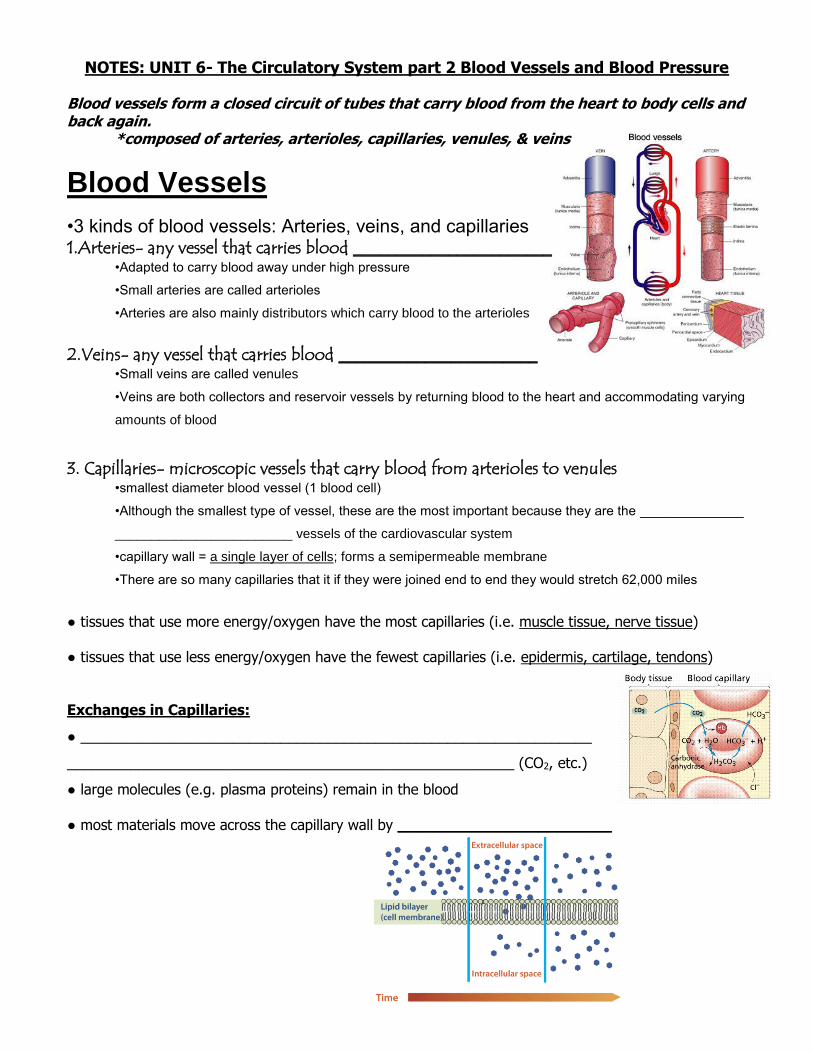
NOTES: UNIT 6- The Circulatory System part 2 Blood Vessels and Blood Pressure Blood vessels form a closed circuit of tubes that carry blood from the heart to body cells and back again. *composed of arteries, arterioles, capillaries, venules, & veins Blood Vessels •3 kinds of blood vessels: Arteries, veins, and capillaries 1.Arteries- any vessel that carries blood _______________________ •Adapted to carry blood away under high pressure •Small arteries are called arterioles •Arteries are also mainly distributors which carry blood to the arterioles 2.Veins- any vessel that carries blood _______________________ •Small veins are called venules •Veins are both collectors and reservoir vessels by returning blood to the heart and accommodating varying amounts of blood 3. Capillaries- microscopic vessels that carry blood from arterioles to venules •smallest diameter blood vessel (1 blood cell) •Although the smallest type of vessel, these are the most important because they are the ______________ ________________________ vessels of the cardiovascular system •capillary wall = a single layer of cells; forms a semipermeable membrane •There are so many capillaries that it if they were joined end to end they would stretch 62,000 miles ● tissues that use more energy/oxygen have the most capillaries (i.e. muscle tissue, nerve tissue) ● tissues that use less energy/oxygen have the fewest capillaries (i.e. epidermis, cartilage, tendons) Exchanges in Capillaries: ● ________________________________________________________________ ________________________________________________________ (CO2, etc.) ● large molecules (e.g. plasma proteins) remain in the blood ● most materials move across the capillary wall by _______________________
Transcript of NOTES: UNIT 5- The Circulatory System part 2 Blood Vessels...
NOTES: UNIT 6- The Circulatory System part 2 Blood Vessels and Blood Pressure Blood vessels form a closed circuit of tubes that carry blood from the heart to body cells and back again. *composed of arteries, arterioles, capillaries, venules, & veins
Blood Vessels
•3 kinds of blood vessels: Arteries, veins, and capillaries 1.Arteries- any vessel that carries blood _______________________
•Adapted to carry blood away under high pressure
•Small arteries are called arterioles
•Arteries are also mainly distributors which carry blood to the arterioles
2.Veins- any vessel that carries blood _______________________ •Small veins are called venules
•Veins are both collectors and reservoir vessels by returning blood to the heart and accommodating varying
amounts of blood
3. Capillaries- microscopic vessels that carry blood from arterioles to venules
•smallest diameter blood vessel (1 blood cell)
•Although the smallest type of vessel, these are the most important because they are the ______________
________________________ vessels of the cardiovascular system
•capillary wall = a single layer of cells; forms a semipermeable membrane
•There are so many capillaries that it if they were joined end to end they would stretch 62,000 miles
● tissues that use more energy/oxygen have the most capillaries (i.e. muscle tissue, nerve tissue) ● tissues that use less energy/oxygen have the fewest capillaries (i.e. epidermis, cartilage, tendons) Exchanges in Capillaries:
● ________________________________________________________________
________________________________________________________ (CO2, etc.)
● large molecules (e.g. plasma proteins) remain in the blood ● most materials move across the capillary wall by _______________________
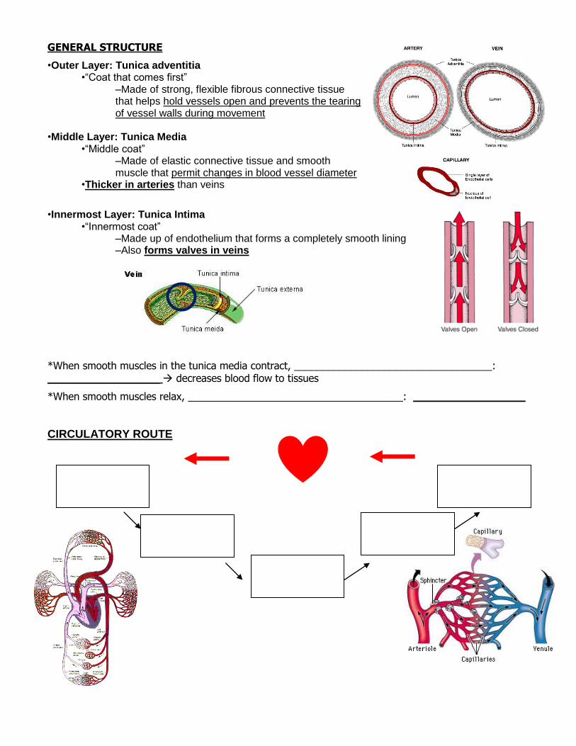
GENERAL STRUCTURE
•Outer Layer: Tunica adventitia •“Coat that comes first”
–Made of strong, flexible fibrous connective tissue that helps hold vessels open and prevents the tearing of vessel walls during movement
•Middle Layer: Tunica Media
•“Middle coat” –Made of elastic connective tissue and smooth muscle that permit changes in blood vessel diameter
•Thicker in arteries than veins
•Innermost Layer: Tunica Intima •“Innermost coat”
–Made up of endothelium that forms a completely smooth lining –Also forms valves in veins
*When smooth muscles in the tunica media contract, ___________________________________: _________________ decreases blood flow to tissues
*When smooth muscles relax, ______________________________________: _________________
CIRCULATORY ROUTE
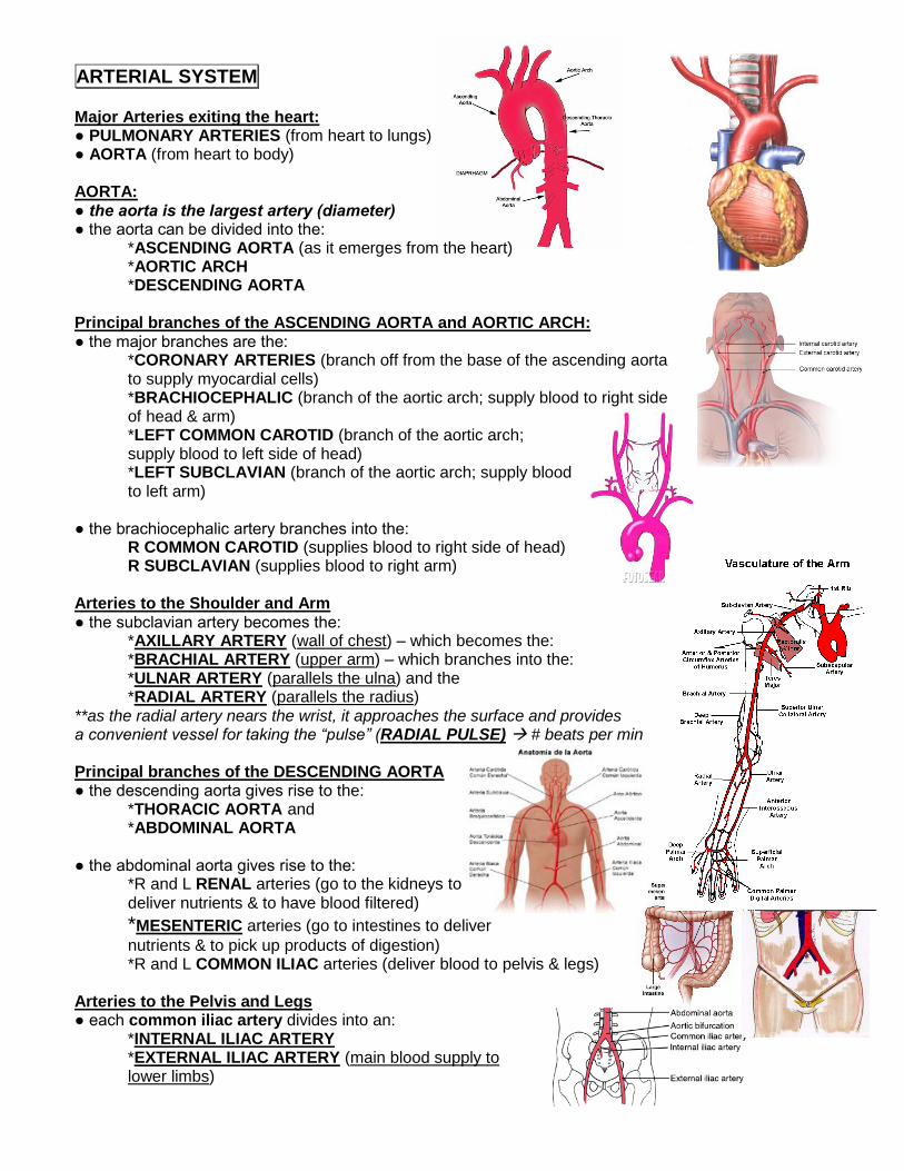
ARTERIAL SYSTEM Major Arteries exiting the heart: ● PULMONARY ARTERIES (from heart to lungs) ● AORTA (from heart to body) AORTA: ● the aorta is the largest artery (diameter) ● the aorta can be divided into the: *ASCENDING AORTA (as it emerges from the heart) *AORTIC ARCH
*DESCENDING AORTA Principal branches of the ASCENDING AORTA and AORTIC ARCH: ● the major branches are the:
*CORONARY ARTERIES (branch off from the base of the ascending aorta to supply myocardial cells) *BRACHIOCEPHALIC (branch of the aortic arch; supply blood to right side of head & arm) *LEFT COMMON CAROTID (branch of the aortic arch; supply blood to left side of head) *LEFT SUBCLAVIAN (branch of the aortic arch; supply blood to left arm)
● the brachiocephalic artery branches into the: R COMMON CAROTID (supplies blood to right side of head)
R SUBCLAVIAN (supplies blood to right arm) Arteries to the Shoulder and Arm ● the subclavian artery becomes the: *AXILLARY ARTERY (wall of chest) – which becomes the: *BRACHIAL ARTERY (upper arm) – which branches into the: *ULNAR ARTERY (parallels the ulna) and the *RADIAL ARTERY (parallels the radius) **as the radial artery nears the wrist, it approaches the surface and provides a convenient vessel for taking the “pulse” (RADIAL PULSE) # beats per min Principal branches of the DESCENDING AORTA ● the descending aorta gives rise to the: *THORACIC AORTA and *ABDOMINAL AORTA ● the abdominal aorta gives rise to the:
*R and L RENAL arteries (go to the kidneys to deliver nutrients & to have blood filtered)
*MESENTERIC arteries (go to intestines to deliver
nutrients & to pick up products of digestion) *R and L COMMON ILIAC arteries (deliver blood to pelvis & legs) Arteries to the Pelvis and Legs ● each common iliac artery divides into an: *INTERNAL ILIAC ARTERY
*EXTERNAL ILIAC ARTERY (main blood supply to lower limbs)
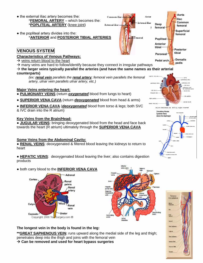
● the external iliac artery becomes the: *FEMORAL ARTERY – which becomes the: *POPLITEAL ARTERY (knee joint) ● the popliteal artery divides into the: *ANTERIOR and POSTERIOR TIBIAL ARTERIES
VENOUS SYSTEM
Characteristics of Venous Pathways: veins return blood to the heart many veins are hard to follow/identify because they connect in irregular pathways the larger veins typically parallel the arteries (and have the same names as their arterial counterparts)
(ex: renal vein parallels the renal artery; femoral vein parallels the femoral artery, ulnar vein parallels ulnar artery, etc.)
Major Veins entering the heart: ● PULMONARY VEINS (return oxygenated blood from lungs to heart)
● SUPERIOR VENA CAVA (return deoxygenated blood from head & arms)
● INFERIOR VENA CAVA (deoxygenated blood from torso & legs; both SVC & IVC drain into the R atrium) Key Veins from the Brain/Head: ● JUGULAR VEINS: bringing deoxygenated blood from the head and face back towards the heart (R atrium) ultimately through the SUPERIOR VENA CAVA
Some Veins from the Abdominal Cavity: ● RENAL VEINS: deoxygenated & filtered blood leaving the kidneys to return to heart ● HEPATIC VEINS: deoxygenated blood leaving the liver; also contains digestion products ● both carry blood to the INFERIOR VENA CAVA The longest vein in the body is found in the leg:
**GREAT SAPHENOUS VEIN: runs upward along the medial side of the leg and thigh; penetrates deep into the thigh and joins with the femoral vein Can be removed and used for heart bypass surgeries