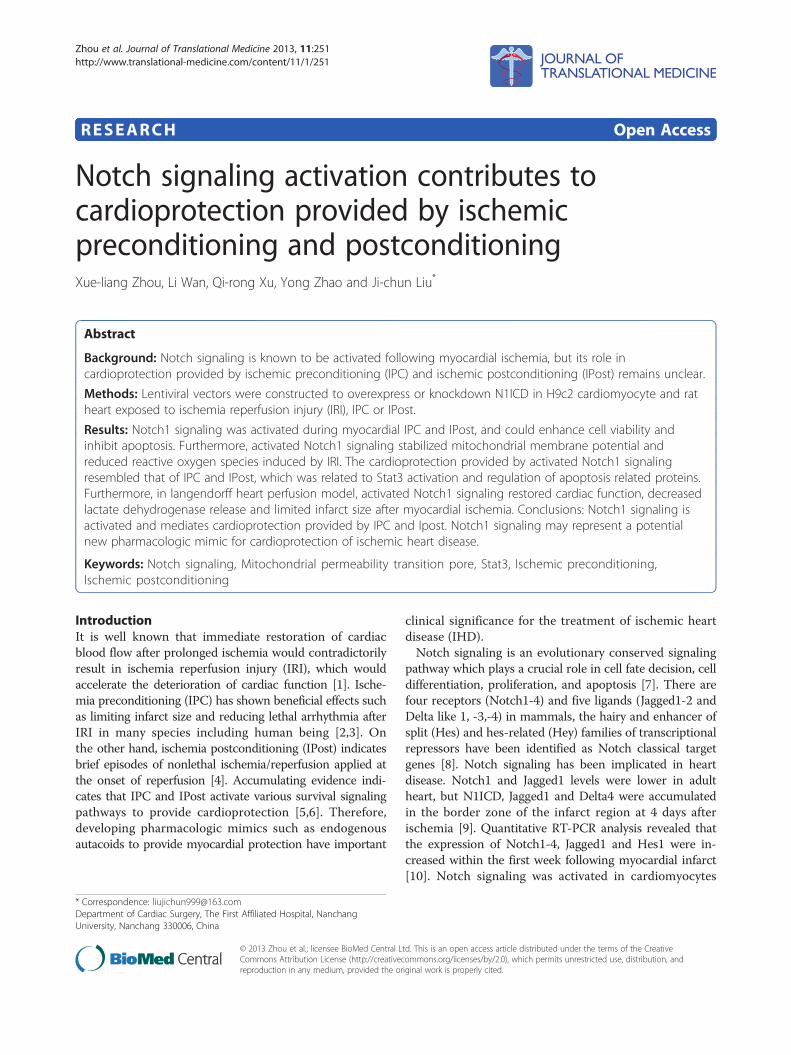
Secretome of apoptotic cells causes cardioprotection and ...
Zhou et al. Journal of Translational Medicine 2013, 11:251http://www.translational-medicine.com/content/11/1/251
RESEARCH Open Access
Notch signaling activation contributes tocardioprotection provided by ischemicpreconditioning and postconditioningXue-liang Zhou, Li Wan, Qi-rong Xu, Yong Zhao and Ji-chun Liu*
Abstract
Background: Notch signaling is known to be activated following myocardial ischemia, but its role incardioprotection provided by ischemic preconditioning (IPC) and ischemic postconditioning (IPost) remains unclear.
Methods: Lentiviral vectors were constructed to overexpress or knockdown N1ICD in H9c2 cardiomyocyte and ratheart exposed to ischemia reperfusion injury (IRI), IPC or IPost.
Results: Notch1 signaling was activated during myocardial IPC and IPost, and could enhance cell viability andinhibit apoptosis. Furthermore, activated Notch1 signaling stabilized mitochondrial membrane potential andreduced reactive oxygen species induced by IRI. The cardioprotection provided by activated Notch1 signalingresembled that of IPC and IPost, which was related to Stat3 activation and regulation of apoptosis related proteins.Furthermore, in langendorff heart perfusion model, activated Notch1 signaling restored cardiac function, decreasedlactate dehydrogenase release and limited infarct size after myocardial ischemia. Conclusions: Notch1 signaling isactivated and mediates cardioprotection provided by IPC and Ipost. Notch1 signaling may represent a potentialnew pharmacologic mimic for cardioprotection of ischemic heart disease.
Keywords: Notch signaling, Mitochondrial permeability transition pore, Stat3, Ischemic preconditioning,Ischemic postconditioning
IntroductionIt is well known that immediate restoration of cardiacblood flow after prolonged ischemia would contradictorilyresult in ischemia reperfusion injury (IRI), which wouldaccelerate the deterioration of cardiac function [1]. Ische-mia preconditioning (IPC) has shown beneficial effects suchas limiting infarct size and reducing lethal arrhythmia afterIRI in many species including human being [2,3]. Onthe other hand, ischemia postconditioning (IPost) indicatesbrief episodes of nonlethal ischemia/reperfusion applied atthe onset of reperfusion [4]. Accumulating evidence indi-cates that IPC and IPost activate various survival signalingpathways to provide cardioprotection [5,6]. Therefore,developing pharmacologic mimics such as endogenousautacoids to provide myocardial protection have important
* Correspondence: [email protected] of Cardiac Surgery, The First Affiliated Hospital, NanchangUniversity, Nanchang 330006, China
© 2013 Zhou et al.; licensee BioMed Central LCommons Attribution License (http://creativecreproduction in any medium, provided the or
clinical significance for the treatment of ischemic heartdisease (IHD).Notch signaling is an evolutionary conserved signaling
pathway which plays a crucial role in cell fate decision, celldifferentiation, proliferation, and apoptosis [7]. There arefour receptors (Notch1-4) and five ligands (Jagged1-2 andDelta like 1, -3,-4) in mammals, the hairy and enhancer ofsplit (Hes) and hes-related (Hey) families of transcriptionalrepressors have been identified as Notch classical targetgenes [8]. Notch signaling has been implicated in heartdisease. Notch1 and Jagged1 levels were lower in adultheart, but N1ICD, Jagged1 and Delta4 were accumulatedin the border zone of the infarct region at 4 days afterischemia [9]. Quantitative RT-PCR analysis revealed thatthe expression of Notch1-4, Jagged1 and Hes1 were in-creased within the first week following myocardial infarct[10]. Notch signaling was activated in cardiomyocytes
td. This is an open access article distributed under the terms of the Creativeommons.org/licenses/by/2.0), which permits unrestricted use, distribution, andiginal work is properly cited.
Zhou et al. Journal of Translational Medicine 2013, 11:251 Page 2 of 10http://www.translational-medicine.com/content/11/1/251
during post-infarction remodeling, and the expression ofNotch1, Jagged1 and Hes1 were upregulated in dilated orhypertrophic hearts [11]. In addition, Notch signaling canstimulate immature cardiomyocyte proliferation and pro-mote quiescent cardiomyocytes to reenter the cell cycle[12,13]. However, whether activated Notch signaling fol-lowing myocardial ischemia plays cardioprotective effectsduring IPC and IPost remains unclear.Therefore, in this study we investigated the role of
Notch1 signaling in cardioprotection provided by IPC andIPost, using both cardiomyocyte and rat heart model. Ourresults showed that the inhibition of Notch1 signaling viathe knockdown of N1ICD abrogated the cardioprotectionprovided by IPC and IPost.
Materials and methodsReagents and antibodiesDulbecco’s modified Eagle’s medium (DMEM) and trypsinwere from Gibco BRL (USA). Fetal bovine serum (FBS)was from NQBB International Biological Corporation(Australia). Cell counting kit-8 (CCK-8) and Reactive oxy-gen species assay kits were from Beyotime institute ofbiotechnology (China). Annexin V-FITC apoptosis de-tection kit was obtained from KeyGEN Biotech(China).Mitochondria staining kit (JC-1) was purchased fromMultiSciences Biotech (USA). Mitochondria isolation kitwas purchased from Applygen Technologies (China). LDHkits were purchased from Nanjing jiancheng bioengineeringinstitute, 2,3,5-triphenyltetrazolium chloride (TTC) wasfrom SolarbioTechnologies (China). Enhanced chemilumin-escence kit was purchased from Thermo Scientific (USA).Notch1 and Hes1 antibodies were purchased from Abcam
(USA), Flag, Bcl-xL and Bax antibodies were purchasedfrom Cell Signaling Technology (USA), Stat3, p-Stat3and β-Actin antibodies were purchased from Anbo Bio-technology Company (USA).
Cell cultureRat cardiac H9c2 cells (ATCC Rockville, USA) were cul-tured in DMEM supplemented with 10% FBS at 37°C ina humidified incubator with 5% CO2. The cells were fedevery 3 days and subcultured once reaching 90% conflu-ence. Cells were plated at an appropriate density accordingto each experimental design.
Virus vector construction and infectionRat N1ICD cDNA (Notch1:5456–7819) was generated byPCR using forward primer: 5′-GAGGATCCCCGGGTACCGGTCGCCACCATGGTGCTGCTGTCCCGCAAG-3′and reverse primer: 5′-TCATCCTTGTAGTCGCTAGCCTTAAATGCCTCTGGAATG-3′. The amplified fragmentwas subcloned into pGC-FU-EGFP-3Flag to get pGC-FU-N1ICD-3Flag vector. Rat N1ICD-shRNAs were designedaccording to Ambion with the following sequences:
GCACAGTGCTGAGTACCAA (N1ICD-shRNA-1), GCCTCTCCACCAATACCTT (N1ICD-shRNA-2), CCCACATTCCAGAGGCATT (N1ICD-shRNA-3), TCTGGATGCCCGAATGCAT (N1ICD-shRNA-4) with CTCGAG as theloop sequence. The oligonucleotide-annealed productswere subcloned into pGVC112 to get pGVC112-N1ICD-shRNA vectors. An irrelevant TTCTCCGAACGTGTCACGT was used as negative control.To generate lentiviral vectors, either 20 μg pGC-FU-
N1ICD-3Flag or pGVC112-N1ICD-shRNA was transfectedalong with 15 μg pHelper 1.0 and 10 μg pHelper 2.0 into293T cells in 10 cm plate at 80% confluence for 8 h. Themedium was replaced with fully supplemented DMEMand the supernatants were harvested after 72 h for virustitration, which were then stored at −80°C.To infect H9c2 cells, multiplicity infection (MOI) of
20 lentiviral particles with 5 μg/ml polybrene were addedinto the medium, 24 h later the medium was changedand H9c2 cells were cultured for an additional 72 h.To infect Sprague–Dawley rat, 100 μl pseudo viral
particles (1×10⁸/ml) were injected into the tail vein, 4weeks after infection, Sprague–Dawley rats were collectedfor analysis.
In vitro protocols of hypoxia/reoxygenation (H/R), IPC, IPostFor H/R, H9c2 cells were cultured in hypoxic solution ina hypoxic incubator (95% N2-5% CO2) for 3 h, subse-quently hypoxic solution was replaced with reoxygenationsolution, and the cells were cultured in a high oxygenincubator (95% O2-5% CO2) for 3 h.For IPC, H9c2 cells were cultured in hypoxic solution
in a hypoxic incubator (95% N2-5% CO2) for 10 min,subsequently the hypoxic solution was replaced withreoxygenation solution, and the cells were cultured in ahigh oxygen incubator (95% O2-5% CO2) for 10 min.The hypoxia-reoxygenation cycle was repeated 3 timesand followed by H/R treatment.For IPost, at the end of the 3 h hypoxia, the hypoxic
solution was replaced with reoxygenation solution andH9c2 cells were cultured in a high oxygen incubator for10 min. Next the medium was changed to hypoxic solu-tion and the cells were cultured in a hypoxic incubatorfor another 10 min. The reoxygenation-hypoxia cycle wasrepeated 3 times and followed by continuous culture inreoxygenation solution in a high oxygen incubator for 3 h.
Cell viability assayH9c2 cells were seeded into a 96-well plate at 10,000cells/well and cultured in 100 μL DMEM supplementedwith 10% FBS for 24 h, then treated as indicated. Finally,the cell viability was detected using CCK-8 Kit (Dojindo,Japan) according to the manufacturer’s instructions. Theabsorbance was measured at 450 nm using a Microplate
Zhou et al. Journal of Translational Medicine 2013, 11:251 Page 3 of 10http://www.translational-medicine.com/content/11/1/251
reader (Thermo, USA). The percentage of viable cellswas calculated with the control cells set as 100%.
Apoptosis assayH9c2 cells were harvested and 4×10⁵ cells were resuspendedin 500 μl binding buffer, then incubated with 5 μl AnnexinV-FITC and 5 μl propidium iodide (PI) at room temperaturein dark for 15 min. The samples were analyzed by flowcytometry with a BD FACSCalibur. Apoptosis ratio wasdefined as the ratio between Annexin V positive/PInegative cells and the total living cells.
Detection of intracellular reactive oxygen species (ROS)H9c2 cells were harvested and 105 cells were incubatedwith 10 μmol/L DCFH-DA at 37°C for 20 min. The DCFfluorescence was detected by BD FACSCalibur excitedat 488 nm and the emitted fluorescence was collectedat 525 nm.
Determination of mitochondrial membrane potential (ΔΨm)H9c2 cells were harvested and 105 cells were loaded withJC-1 (2 μM) at 37°C, 5% CO2 for 20 min, then analyzed byBD FACSCalibur excited at 488 nm and the emitted fluor-escence was collected at 530 nm and 590 nm. ΔΨm wasexpressed as the emitted fluorescence ratio (590/530 nm)in percentage to the initial level, whereas 0% level wasrecorded after complete mitochondrial depolarization withcarbonyl cyanide-p-trifluoromethoxyphenylhydra-zone.
Langendorff heart perfusionMale Sprague–Dawley rats (weight 220 to 250 g) wereobtained from Jiangxi University of Traditional ChineseMedicine (China). All animals were treated in accord-ance with the Guide for the Care and Use of LaboratoryAnimals, and approved by the Ethics Committee of thefirst affiliated hospital of Nanchang University (permitNo: NDYFY-2012-YYLSD-016). The rats were anesthe-tized with pentobarbital sodium (100 mg/kg intraperi-toneally). Hearts were excised and retrograde perfusedvia the aorta with modified Krebs-Henseleit buffer(NaCl 118 nM, KCl 4.7 nM, MgSO4 1.2 nM, CaCl2 2.5 nM,NaHCO3 2 nM, KH2PO4 1.2 nM, Glucose 11 nM, pH7.35-7.45) equilibrated with 95% O2-5% CO2. Leftventriculardeveloped pressure (LVDP), heart rate (HR)and intraventricular pressure (±dP/dt) were measuredby Powerlab pressure transducer with a balloon insertedinto the left ventricle. In the Control, Mock1 and Mock2groups, hearts were buffer perfused without ischemia.In ischemia/reperfusion (I/R) group, hearts were firstequilibrated for 20 min followed by 30 min of stop-flowischemia and 120 min of reperfusion. In IPC group,after 3 cycles of 5 min of ischemia and 5 min of reperfu-sion, hearts were subjected to 30 min of no-flow ische-mia, followed by 120 min of reperfusion. In IPost group,
hearts were subjected to 30 min of stop-flow ischemia,before 120 min of reperfusion, 3 cycles of 10 s of ische-mia and 10 s of reperfusion were applied.
Lactate dehydrogenase (LDH) assayCoronary effluent was collected at 1 min before ischemia,1 min after ischemia and 1 min after reperfusion to deter-mine LDH activity by using LDH kits according to themanufacturer’s instructions. LDH activity was expressedas international units per liter (IU/L).
Assessment of infarct sizeThe heart was frozen at −20°C for 30 min, then sectionedfrom apex to base into five to six 1 mm sections. The sec-tions were incubated in 1%TTC at 37°C for 30 min andfixed in 10% formaldehyde for 24 h. The normal myocar-dium was stained brick red with TTC while the infarctedportion was not stained and appeared pale white. Theinfarct area-IA (white) and the area at risk-AAR (redand white) from each section were measured using ImagePro 6.0 Plus software. For each section, infarct size wasexpressed as the percentage of IA/AAR.
Western blot analysisAt different time points of the experiemtns, cells or thehearts were harvested immediately and lysed in cell lysisbuffer (Beyotime Institute of Biotechnology, China) at 4°C.Protein samples were separated by 8-10% SDS-PAGE,then transferred to nitrocellulose membranes (Millipore)and blocked in 10% nonfat milk in TBST (150 mM NaCl,50 mM Tris pH 7.5, 0.1% Tween-20). Membranes wereincubated with primary antibodies overnight at 4°C, fol-lowed by incubation with secondary antibodies at roomtemperature for 1 h. The fluorescent signals were de-tected using enhanced chemiluminescence by ImageQuantLAS4000 (GE, USA). Relative protein levels were nor-malized to that of β-actin.
Statistical analysisData were analyzed using the SPSS version 12 statisticalanalysis package (SPSS Inc., Chicago, IL, USA). Exam-ined data were assessed using ANOVA. In each test, thedata were expressed as the mean±SD, and P<0.05 wasaccepted as statistically significant.
ResultsIdentification of LV-N1ICD and LV-N1ICD-shRNA vectorsIn 293T cells transfected with pGC-FU-N1ICD-3Flagvector, we observed ectopic expression of N1ICD(Figure 1A). Furthermore, in H9c2 cells and rats infectedwith lentiviral vector LV-N1ICD, we observed ectopicexpression of N1ICD in the cells or hearts (Figure 1B-C).In contrast, in 293T cells cotransfected with pGC-FU-N1ICD-3Flag vector and N1ICD-shRNA-1 to 4, we
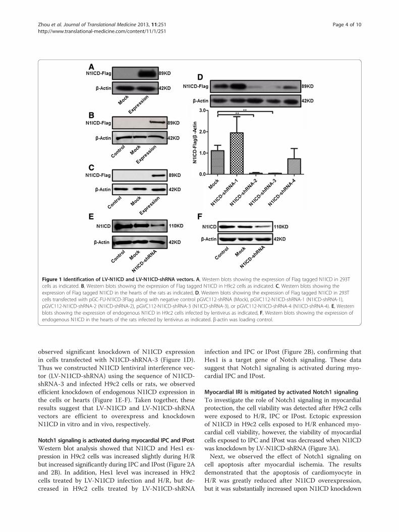
Figure 1 Identification of LV-N1ICD and LV-N1ICD-shRNA vectors. A, Western blots showing the expression of Flag tagged N1ICD in 293Tcells as indicated. B, Western blots showing the expression of Flag tagged N1ICD in H9c2 cells as indicated. C, Western blots showing theexpression of Flag tagged N1ICD in the hearts of the rats as indicated, D, Western blots showing the expression of Flag tagged N1ICD in 293Tcells transfected with pGC-FU-N1ICD-3Flag along with negative control pGVC112-shRNA (Mock), pGVC112-N1ICD-shRNA-1 (N1ICD-shRNA-1),pGVC112-N1ICD-shRNA-2 (N1ICD-shRNA-2), pGVC112-N1ICD-shRNA-3 (N1ICD-shRNA-3), or pGVC112-N1ICD-shRNA-4 (N1ICD-shRNA-4). E, Westernblots showing the expression of endogenous N1ICD in H9c2 cells infected by lentivirus as indicated, F, Western blots showing the expression ofendogenous N1ICD in the hearts of the rats infected by lentivirus as indicated. β-actin was loading control.
Zhou et al. Journal of Translational Medicine 2013, 11:251 Page 4 of 10http://www.translational-medicine.com/content/11/1/251
observed significant knockdown of N1ICD expressionin cells transfected with N1ICD-shRNA-3 (Figure 1D).Thus we constructed N1ICD lentiviral interference vec-tor (LV-N1ICD-shRNA) using the sequence of N1ICD-shRNA-3 and infected H9c2 cells or rats, we observedefficient knockdown of endogenous N1ICD expression inthe cells or hearts (Figure 1E-F). Taken together, theseresults suggest that LV-N1ICD and LV-N1ICD-shRNAvectors are efficient to overexpress and knockdownN1ICD in vitro and in vivo, respectively.
Notch1 signaling is activated during myocardial IPC and IPostWestern blot analysis showed that N1ICD and Hes1 ex-pression in H9c2 cells was increased slightly during H/Rbut increased significantly during IPC and IPost (Figure 2Aand 2B). In addition, Hes1 level was increased in H9c2cells treated by LV-N1ICD infection and H/R, but de-creased in H9c2 cells treated by LV-N1ICD-shRNA
infection and IPC or IPost (Figure 2B), confirming thatHes1 is a target gene of Notch signaling. These datasuggest that Notch1 signaling is activated during myo-cardial IPC and IPost.
Myocardial IRI is mitigated by activated Notch1 signalingTo investigate the role of Notch1 signaling in myocardialprotection, the cell viability was detected after H9c2 cellswere exposed to H/R, IPC or IPost. Ectopic expressionof N1ICD in H9c2 cells exposed to H/R enhanced myo-cardial cell viability, however, the viability of myocardialcells exposed to IPC and IPost was decreased when N1ICDwas knockdown by LV-N1ICD-shRNA (Figure 3A).Next, we observed the effect of Notch1 signaling on
cell apoptosis after myocardial ischemia. The resultsdemonstrated that the apoptosis of cardiomyocyte inH/R was greatly reduced after N1ICD overexpression,but it was substantially increased upon N1ICD knockdown
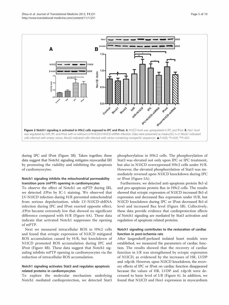
Figure 2 Notch1 signaling is activated in H9c2 cells exposed to IPC and IPost. A, N1ICD level was upregulated in IPC and IPost. B, Hes1 levelwas regulated by H/R, IPC and IPost with or without LV-N1ICD/LV-N1ICD-shRNA infection. Data were presented as mean±SD, n=3. Mock1 indicatedcells infected with empty vector, Mock2 indicated cells infected with vector containing nonspecfic sequences. ▲P>0.05, *P<0.05, **P<0.01.
Zhou et al. Journal of Translational Medicine 2013, 11:251 Page 5 of 10http://www.translational-medicine.com/content/11/1/251
during IPC and IPost (Figure 3B). Taken together, thesedata suggest that Notch1 signaling mitigates myocardial IRIby promoting the viability and inhibiting the apoptosisof cardiomyocytes.
Notch1 signaling inhibits the mitochondrial permeabilitytransition pore (mPTP) opening in cardiomyocytesTo observe the effect of Notch1 on mPTP during IRI,we detected ΔΨm by JC-1 staining. We observed thatLV-N1ICD infection during H/R prevented mitochondrialfrom serious depolarization, while LV-N1ICD-shRNAinfection during IPC and IPost exerted opposite effect,ΔΨm became extremely low that showed no significantdifference compared with H/R (Figure 4A). These dataindicate that activated Notch1 suppresses the openingof mPTP.Next we measured intracellular ROS in H9c2 cells
and found that ectopic expression of N1ICD mitigatedROS accumulation caused by H/R, but knockdown ofN1ICD promoted ROS accumulation during IPC andIPost (Figure 4B). These data suggest that Notch1 sig-naling inhibits mPTP opening in cardiomyocytes via thereduction of intracellular ROS accumulation.
Notch1 signaling activates Stat3 and regulates apoptosisrelated proteins in cardiomyocytesTo explore the molecular mechanism underlyingNotch1 mediated cardioprotection, we detected Stat3
phosphorylation in H9c2 cells. The phosphorylation ofStat3 was elevated not only upon IPC or IPC treatment,but also in N1ICD overexpressed H9c2 cells under H/R.However, the elevated phosphorylation of Stat3 was im-mediately reversed upon N1ICD knockdown during IPCor IPost (Figure 5A).Furthermore, we detected anti-apoptosis protein Bcl-xl
and pro-apoptosis protein Bax in H9c2 cells. The resultsshowed that ectopic expression of N1ICD increased Bcl-xlexpression and decreased Bax expression under H/R, butN1ICD knockdown during IPC or IPost decreased Bcl-xllevel and increased Bax level (Figure 5B). Collectively,these data provide evidence that cardioprotection effectsof Notch1 signaling are mediated by Stat3 activation andregulation of apoptosis related proteins.
Notch1 signaling contributes to the restoration of cardiacfunction in post-ischemia ratsAfter langendorff-perfused isolated heart models wereestablished, we measured the parameters of cardiac func-tion. The results showed that the recovery of cardiacfunction in I/R was strengthened by ectopic expressionof N1ICD, as evidenced by the increases of HR, LVDPand ±dp/dt. However, upon N1ICD knockdown, the recov-ery effects of IPC or IPost on cardiac function disappearedbecause the values of HR, LVDP and ±dp/dt were de-creased to basic level of I/R (Figure 6). In addition, wefound that N1ICD and Hes1 expression in myocardium
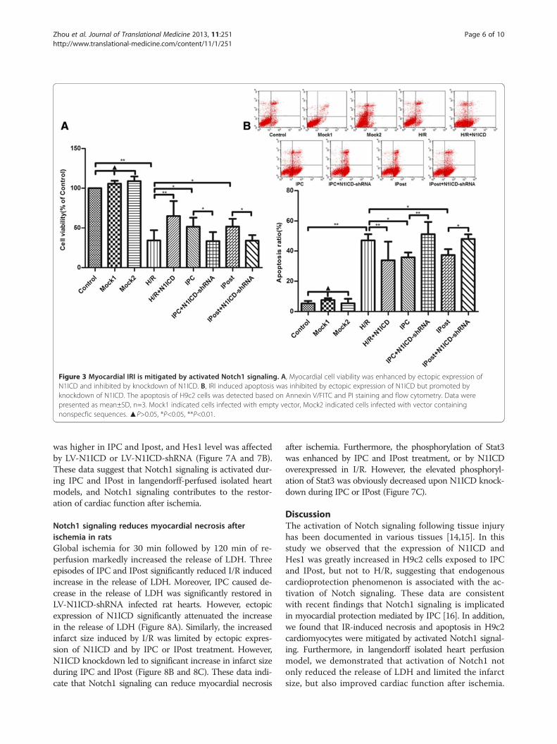
Figure 3 Myocardial IRI is mitigated by activated Notch1 signaling. A, Myocardial cell viability was enhanced by ectopic expression ofN1ICD and inhibited by knockdown of N1ICD. B, IRI induced apoptosis was inhibited by ectopic expression of N1ICD but promoted byknockdown of N1ICD. The apoptosis of H9c2 cells was detected based on Annexin V/FITC and PI staining and flow cytometry. Data werepresented as mean±SD, n=3. Mock1 indicated cells infected with empty vector, Mock2 indicated cells infected with vector containingnonspecfic sequences. ▲P>0.05, *P<0.05, **P<0.01.
Zhou et al. Journal of Translational Medicine 2013, 11:251 Page 6 of 10http://www.translational-medicine.com/content/11/1/251
was higher in IPC and Ipost, and Hes1 level was affectedby LV-N1ICD or LV-N1ICD-shRNA (Figure 7A and 7B).These data suggest that Notch1 signaling is activated dur-ing IPC and IPost in langendorff-perfused isolated heartmodels, and Notch1 signaling contributes to the restor-ation of cardiac function after ischemia.
Notch1 signaling reduces myocardial necrosis afterischemia in ratsGlobal ischemia for 30 min followed by 120 min of re-perfusion markedly increased the release of LDH. Threeepisodes of IPC and IPost significantly reduced I/R inducedincrease in the release of LDH. Moreover, IPC caused de-crease in the release of LDH was significantly restored inLV-N1ICD-shRNA infected rat hearts. However, ectopicexpression of N1ICD significantly attenuated the increasein the release of LDH (Figure 8A). Similarly, the increasedinfarct size induced by I/R was limited by ectopic expres-sion of N1ICD and by IPC or IPost treatment. However,N1ICD knockdown led to significant increase in infarct sizeduring IPC and IPost (Figure 8B and 8C). These data indi-cate that Notch1 signaling can reduce myocardial necrosis
after ischemia. Furthermore, the phosphorylation of Stat3was enhanced by IPC and IPost treatment, or by N1ICDoverexpressed in I/R. However, the elevated phosphoryl-ation of Stat3 was obviously decreased upon N1ICD knock-down during IPC or IPost (Figure 7C).
DiscussionThe activation of Notch signaling following tissue injuryhas been documented in various tissues [14,15]. In thisstudy we observed that the expression of N1ICD andHes1 was greatly increased in H9c2 cells exposed to IPCand IPost, but not to H/R, suggesting that endogenouscardioprotection phenomenon is associated with the ac-tivation of Notch signaling. These data are consistentwith recent findings that Notch1 signaling is implicatedin myocardial protection mediated by IPC [16]. In addition,we found that IR-induced necrosis and apoptosis in H9c2cardiomyocytes were mitigated by activated Notch1 signal-ing. Furthermore, in langendorff isolated heart perfusionmodel, we demonstrated that activation of Notch1 notonly reduced the release of LDH and limited the infarctsize, but also improved cardiac function after ischemia.
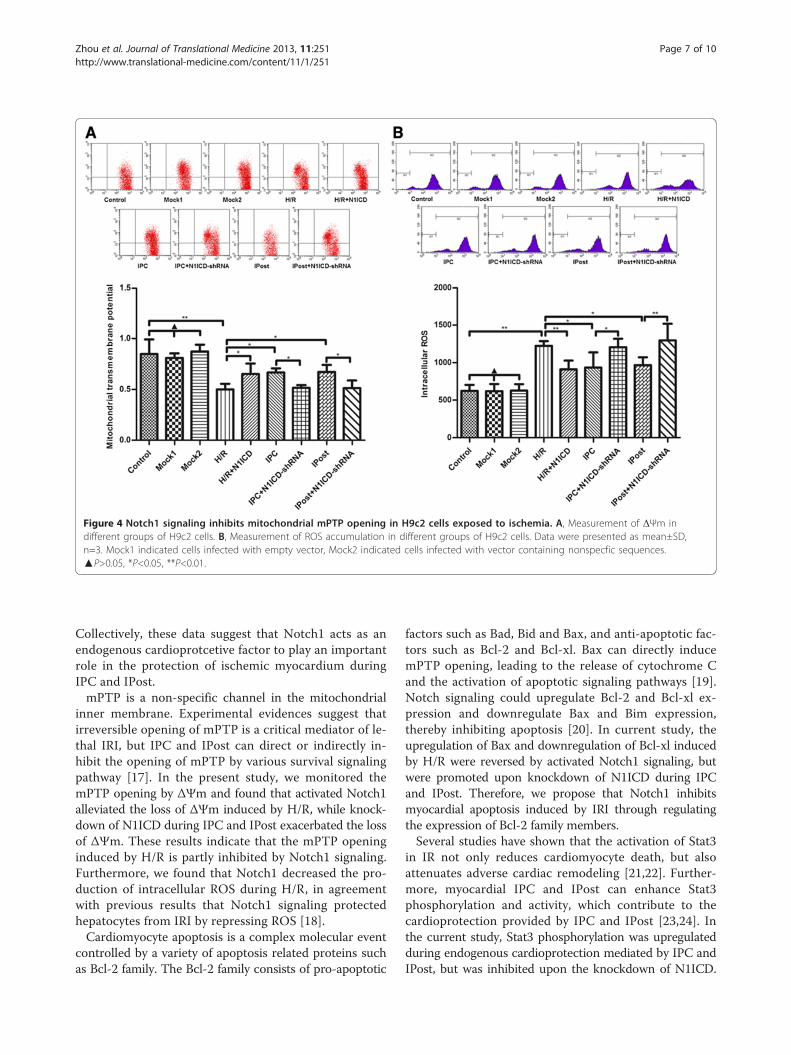
Figure 4 Notch1 signaling inhibits mitochondrial mPTP opening in H9c2 cells exposed to ischemia. A, Measurement of ΔΨm indifferent groups of H9c2 cells. B, Measurement of ROS accumulation in different groups of H9c2 cells. Data were presented as mean±SD,n=3. Mock1 indicated cells infected with empty vector, Mock2 indicated cells infected with vector containing nonspecfic sequences.▲P>0.05, *P<0.05, **P<0.01.
Zhou et al. Journal of Translational Medicine 2013, 11:251 Page 7 of 10http://www.translational-medicine.com/content/11/1/251
Collectively, these data suggest that Notch1 acts as anendogenous cardioprotcetive factor to play an importantrole in the protection of ischemic myocardium duringIPC and IPost.mPTP is a non-specific channel in the mitochondrial
inner membrane. Experimental evidences suggest thatirreversible opening of mPTP is a critical mediator of le-thal IRI, but IPC and IPost can direct or indirectly in-hibit the opening of mPTP by various survival signalingpathway [17]. In the present study, we monitored themPTP opening by ΔΨm and found that activated Notch1alleviated the loss of ΔΨm induced by H/R, while knock-down of N1ICD during IPC and IPost exacerbated the lossof ΔΨm. These results indicate that the mPTP openinginduced by H/R is partly inhibited by Notch1 signaling.Furthermore, we found that Notch1 decreased the pro-duction of intracellular ROS during H/R, in agreementwith previous results that Notch1 signaling protectedhepatocytes from IRI by repressing ROS [18].Cardiomyocyte apoptosis is a complex molecular event
controlled by a variety of apoptosis related proteins suchas Bcl-2 family. The Bcl-2 family consists of pro-apoptotic
factors such as Bad, Bid and Bax, and anti-apoptotic fac-tors such as Bcl-2 and Bcl-xl. Bax can directly inducemPTP opening, leading to the release of cytochrome Cand the activation of apoptotic signaling pathways [19].Notch signaling could upregulate Bcl-2 and Bcl-xl ex-pression and downregulate Bax and Bim expression,thereby inhibiting apoptosis [20]. In current study, theupregulation of Bax and downregulation of Bcl-xl inducedby H/R were reversed by activated Notch1 signaling, butwere promoted upon knockdown of N1ICD during IPCand IPost. Therefore, we propose that Notch1 inhibitsmyocardial apoptosis induced by IRI through regulatingthe expression of Bcl-2 family members.Several studies have shown that the activation of Stat3
in IR not only reduces cardiomyocyte death, but alsoattenuates adverse cardiac remodeling [21,22]. Further-more, myocardial IPC and IPost can enhance Stat3phosphorylation and activity, which contribute to thecardioprotection provided by IPC and IPost [23,24]. Inthe current study, Stat3 phosphorylation was upregulatedduring endogenous cardioprotection mediated by IPC andIPost, but was inhibited upon the knockdown of N1ICD.
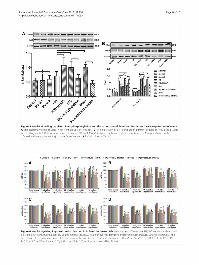
Figure 5 Notch1 signaling regulates Stat3 phosphorylation and the expression of Bcl-xl and Bax in H9c2 cells exposed to ischemia.A, The phosphorylation of Stat3 in different groups of H9c2 cells. B, The expression of Bcl-xl and Bax in different groups of H9c2 cells. β-actinwas loading control. Data were presented as mean±SD, n=3. Mock1 indicated cells infected with empty vector, Mock2 indicated cellsinfected with vector containing nonspecfic sequences. ▲P>0.05, *P<0.05, **P<0.01.
Figure 6 Notch1 signaling improves cardiac function in isolated rat hearts. A-D, Measurement of heart rate (HR), left ventricular developedpressure (LVDP) and maximal (dP/dtmax) and minimal (dP/dtmin) value of the first derivative of left ventricular pressure. Data were shown as thepercentage of the values recorded at 1 min before ischemia. Data were presented as mean±SD, n=6. a: I/R+N1ICD vs I/R; P<0.05; b: IPC vs I/R,P<0.05; c: IPC vs IPC+shRNA, P<0.05; d: IPost vs I/R, P<0.05; e: IPost vs IPost+shRNA, P<0.05.
Zhou et al. Journal of Translational Medicine 2013, 11:251 Page 8 of 10http://www.translational-medicine.com/content/11/1/251
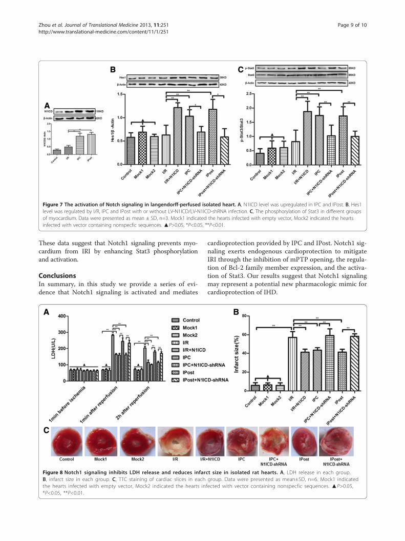
Figure 7 The activation of Notch signaling in langendorff-perfused isolated heart. A, N1ICD level was upregulated in IPC and IPost. B, Hes1level was regulated by I/R, IPC and IPost with or without LV-N1ICD/LV-N1ICD-shRNA infection. C, The phosphorylation of Stat3 in different groupsof myocardium. Data were presented as mean ± SD, n=3. Mock1 indicated the hearts infected with empty vector, Mock2 indicated the heartsinfected with vector containing nonspecfic sequences. ▲P>0.05, *P<0.05, **P<0.01.
Zhou et al. Journal of Translational Medicine 2013, 11:251 Page 9 of 10http://www.translational-medicine.com/content/11/1/251
These data suggest that Notch1 signaling prevents myo-cardium from IRI by enhancing Stat3 phosphorylationand activation.
ConclusionsIn summary, in this study we provide a series of evi-dence that Notch1 signaling is activated and mediates
Figure 8 Notch1 signaling inhibits LDH release and reduces infarB, infarct size in each group. C, TTC staining of cardiac slices in eachthe hearts infected with empty vector, Mock2 indicated the hearts in*P<0.05, **P<0.01.
cardioprotection provided by IPC and IPost. Notch1 sig-naling exerts endogenous cardioprotection to mitigateIRI through the inhibition of mPTP opening, the regula-tion of Bcl-2 family member expression, and the activa-tion of Stat3. Our results suggest that Notch1 signalingmay represent a potential new pharmacologic mimic forcardioprotection of IHD.
ct size in isolated rat hearts. A, LDH release in each group.group. Data were presented as mean±SD, n=6. Mock1 indicatedfected with vector containing nonspecfic sequences. ▲P>0.05,
Zhou et al. Journal of Translational Medicine 2013, 11:251 Page 10 of 10http://www.translational-medicine.com/content/11/1/251
Competing interestsThe authors declare that they have no competing interests.
Authors’ contributionsConceived and designed the experiments: J-cL. Performed the experiments:X-lZ, LW, Q-rX. Analyzed the data: YZ. Wrote the paper: X-lZ and J-cL. Allauthors read and approved the final manuscript.
AcknowledgmentsThis work was supported in part by grants from the National Natural ScienceFoundation of China (No: 81260024), the Natural Science Foundation ofJiangxi Province (No: 20122BAB205026, No: 20132BAB205033).
Received: 19 August 2013 Accepted: 30 September 2013Published: 8 October 2013
References1. Minamino T: Cardioprotection from ischemia/reperfusion injury: basic
and translational research. Circ J 2012, 76:1074–1082.2. Gerczuk PZ, Kloner RA: An update on cardioprotection: a review of the
latest adjunctive therapies to limit myocardial infarction size in clinicaltrials. J Am Coll Cardiol 2012, 59:969–978.
3. Apostolakis E, Baikoussis NG, Papakonstantinou NA: The role of myocardialischaemic preconditioning during beating heart surgery: biological aspectand clinical outcome. Interact Cardiovasc Thorac Surg 2012, 14:68–71.
4. Zhao ZQ, Corvera JS, Halkos ME, Kerendi F, Wang NP, Guyton RA,Vinten-Johansen J: Inhibition of myocardial injury by ischemicpostconditioning during reperfusion: comparison with ischemicpreconditioning. Am J Physiol Heart Circ Physiol 2003, 285:H579–588.
5. Huang C, Yitzhaki S, Perry CN, Liu W, Giricz Z, Mentzer RM Jr, Gottlieb RA:Autophagy induced by ischemic preconditioning is essential forcardioprotection. J Cardiovasc Transl Res 2010, 3:365–373.
6. Babiker FA, van Golde J, Vanagt WY, Prinzen FW: Pacing postconditioning:impact of pacing algorithm, gender, and diabetes on its myocardialprotective effects. J Cardiovasc Transl Res 2012, 5:727–734.
7. Schwanbeck R, Martini S, Bernoth K, Just U: The notch signaling pathway:molecular basis of cell context dependency. Eur J Cell Biol 2011, 90:572–581.
8. Kopan R: Notch signaling. Cold Spring Harb Perspect Biol 2012, 4:10.doi:10.1101/cshperspect.a011213.
9. Gude NA, Emmanuel G, Wu W, Cottage CT, Fischer K, Quijada P, Muraski JA,Alvarez R, Rubio M, Schaefer E, Sussman MA: Activation of notch-mediatedprotective signaling in the myocardium. Circ Res 2008, 102:1025–1035.
10. Kratsios P, Catela C, Salimova E, Huth M, Berno V, Rosenthal N, Mourkioti F:Distinct roles for cell-autonomous notch signaling in cardiomyocytes ofthe embryonic and adult heart. Circ Res 2010, 106:559–572.
11. Croquelois A, Domenighetti AA, Nemir M, Lepore M, Rosenblatt-Velin N,Radtke F, Pedrazzini T: Control of the adaptive response of the heart tostress via the notch1 receptor pathway. J Exp Med 2008, 205:3173–3185.
12. Collesi C, Zentilin L, Sinagra G, Giacca M: Notch1 signaling stimulatesproliferation of immature cardiomyocytes. J Cell Biol 2008, 183:117–128.
13. Campa VM, Gutierrez-Lanza R, Cerignoli F, Diaz-Trelles R, Nelson B, Tsuji T,Barcova M, Jiang W, Mercola M: Notch activates cell cycle reentry andprogression in quiescent cardiomyocytes. J Cell Biol 2008, 183:129–141.
14. Lovschall H, Tummers M, Thesleff I, Fuchtbauer EM, Poulsen K: Activation ofthe notch signaling pathway in response to pulp capping of rat molars.Eur J Oral Sci 2005, 113:312–317.
15. Givogri MI, de Planell M, Galbiati F, Superchi D, Gritti A, Vescovi A, de Vellis J,Bongarzone ER: Notch signaling in astrocytes and neuroblasts of the adultsubventricular zone in health and after cortical injury. Dev Neurosci 2006,28:81–91.
16. Yang Y, Duan W, Jin Z, Bi S, Yan J, Jin Y, Lu J, Yang J, Yi D: New role ofnotch-mediated signaling pathway in myocardial ischemicpreconditioning. Med Hypotheses 2011, 76:427–428.
17. Hausenloy DJ, Ong SB, Yellon DM: The mitochondrial permeabilitytransition pore as a target for preconditioning and postconditioning.Basic Res Cardiol 2009, 104:189–202.
18. Yu HC, Qin HY, He F, Wang L, Fu W, Liu D, Guo FC, Liang L, Dou KF, Han H:Canonical notch pathway protects hepatocytes from ischemia/reperfusioninjury in mice by repressing reactive oxygen species production throughjak2/stat3 signaling. Hepatology 2011, 54:979–98826.
19. Kinnally KW, Antonsson B: A tale of two mitochondrial channels, mac andptp, in apoptosis. Apoptosis 2007, 12:857–868.
20. Gao F, Yao M, Shi Y, Hao J, Ren Y, Liu Q, Wang X, Duan H: Notch pathwayis involved in high glucose-induced apoptosis in podocytes via bcl-2and p53 pathways. J Cell Biochem 2013, 114:1029–1038.
21. Boengler K, Buechert A, Heinen Y, Roeskes C, Hilfiker-Kleiner D, Heusch G,Schulz R: Cardioprotection by ischemic postconditioning is lost in agedand stat3-deficient mice. Circ Res 2008, 102:131–135.
22. Obana M, Maeda M, Takeda K, Hayama A, Mohri T, Yamashita T, Nakaoka Y,Komuro I, Matsumiya G, Azuma J, Fujio Y: Therapeutic activation of signaltransducer and activator of transcription 3 by interleukin-11 amelioratescardiac fibrosis after myocardial infarction. Circulation 2010, 121:684–691.
23. You L, Li L, Xu Q, Ren J, Zhang F: Postconditioning reduces infarct sizeand cardiac myocyte apoptosis via the opioid receptor and jak-statsignaling pathway. Mol Biol Rep 2011, 38:437–443.
24. Hausenloy DJ, Lecour S, Yellon DM: Reperfusion injury salvage kinase andsurvivor activating factor enhancement prosurvival signaling pathwaysin ischemic postconditioning: Two sides of the same coin. Antioxid RedoxSignal 2011, 14:893–907.
doi:10.1186/1479-5876-11-251Cite this article as: Zhou et al.: Notch signaling activation contributes tocardioprotection provided by ischemic preconditioning andpostconditioning. Journal of Translational Medicine 2013 11:251.
Submit your next manuscript to BioMed Centraland take full advantage of:
• Convenient online submission
• Thorough peer review
• No space constraints or color figure charges
• Immediate publication on acceptance
• Inclusion in PubMed, CAS, Scopus and Google Scholar
• Research which is freely available for redistribution
Submit your manuscript at www.biomedcentral.com/submit