Notable Articles of 2017 - The New England Journal of...
44
Notable Articles of 2017 A collection of articles selected by NEJM editors
Transcript of Notable Articles of 2017 - The New England Journal of...
Notable Articles of 2017
A collection of articles selected by NEJM editors
B Notable Articles of 2017
800.843.6356 | f: 781.891.1995 | [email protected] winter street, waltham, ma 02451-1413
nejmgroup.org
December 2017
Dear Reader,
2017 was the year of big data. One study took data from 61 million Americans and looked at the association between air pollution and mortality. The trial found that for every increase of 10 μg per cubic meter in fine particulate matter (PM2.5), there was an associated 7.3% increase in all-cause mortality. These findings stress the need for tighter regulation of air-pollutant levels, and make the point that we still have time to make a difference.
Another study analyzed data from 68.5 million people from 195 countries to find the trends in the prevalence of overweight and obesity among children and adults between 1980 and 2015. This study found that the global obesity epidemic is worsening in most parts of the world, but — as with the air pollution study — our future is not immutable.
As the medical information published in NEJM is regularly used in daily practice, we ensure each paper published meets exacting standards for editorial quality, clinical relevance, and impact on patient outcomes. Among all papers published in 2017, this “most notable” collection was selected by the editors as being the most meaningful in improving medical practice and patient care. We hope that you will take valuable insights from these articles.
Sincerely,
Jeffrey M. Drazen, M.D.Editor-In-Chief, The New England Journal of Medicine
ORIGINAL ARTICLE
Radiation with or without Antiandrogen Therapy in Recurrent Prostate Cancer. . . . . . 1
EDITORIAL: Improved Therapy for PSA Recurrence after Prostatectomy . . . . . . . . . . . 2
ORIGINAL ARTICLE
Survival and Neurodevelopmental Outcomes among Periviable Infants . . . . . . . . . . . 4
EDITORIAL: Neonatal Intensive Care — The Only Constant Is Change . . . . . . . . . . . . 5
ORIGINAL ARTICLE
Osimertinib or Platinum–Pemetrexed in EGFR T790M–Positive Lung Cancer . . . . . . . 8
ORIGINAL ARTICLE
Long-Term Outcomes of Imatinib Treatment for Chronic Myeloid Leukemia . . . . . . . 9
EDITORIAL: Imatinib Changed Everything . . . . . . . . . . . . . . . . . . . . . . . . . . . . . . . . . . 10
ORIGINAL ARTICLE
Mepolizumab or Placebo for Eosinophilic Granulomatosis with Polyangiitis . . . . . . 12
EDITORIAL: Targeting Eosinophils in Eosinophilic Granulomatosis with Polyangiitis . . 13
ORIGINAL ARTICLE
Air Pollution and Mortality in the Medicare Population . . . . . . . . . . . . . . . . . . . . . . . 15
EDITORIAL: Air Pollution Still Kills . . . . . . . . . . . . . . . . . . . . . . . . . . . . . . . . . . . . . . . . . 16
ORIGINAL ARTICLE
Health Effects of Overweight and Obesity in 195 Countries over 25 Years . . . . . . . . . 18
EDITORIAL: Global Health Effects of Overweight and Obesity . . . . . . . . . . . . . . . . . . 19
ORIGINAL ARTICLE
Trial of Tocilizumab in Giant-Cell Arteritis . . . . . . . . . . . . . . . . . . . . . . . . . . . . . . . . . . 21
EDITORIAL: Giant-Cell Arteritis — More Ecstasy, Less Agony . . . . . . . . . . . . . . . . . . . 22
ORIGINAL ARTICLE
Analysis of Plasma Epstein–Barr Virus DNA to Screen for Nasopharyngeal Cancer . . . 24
EDITORIAL: Plasma Epstein–Barr Virus DNA for Screening . . . . . . . . . . . . . . . . . . . . 25
ORIGINAL ARTICLE
Patent Foramen Ovale Closure or Anticoagulation vs. Antiplatelets after Stroke . . . . 27
EDITORIAL: Tipping Point for Patent Foramen Ovale Closure . . . . . . . . . . . . . . . . . . . 28
PERSPECTIVE
Letter to a Young Female Physician . . . . . . . . . . . . . . . . . . . . . . . . . . . . . . . . . . . . . . . 31
TABLE OF CONTENTS
The New England Journal of Medicine is a publication of NEJM Group, a division of the Massachusetts Medical Society. ©2017 Massachusetts Medical Society, All rights reserved.
(continued on next page)

PERSPECTIVE
Tragedy, Perseverance, and Chance — The Story of CAR-T Therapy . . . . . . . . . . . . . . 34
PERSPECTIVE
Stretching the Scope — Becoming Frontline Addiction-Medicine Providers . . . . . . . 37
(continued from previous page)
TABLE OF CONTENTS
Back to Table of Contents
1 Notable Articles of 2017 nejm.org
ORIGINAL ARTICLE
Read Full Article at NEJM.org
The new england journal of medicine
n engl j med 376;5 nejm.org February 2, 2017 417
established in 1812 February 2, 2017 vol. 376 no. 5
The authors’ full names, academic de-grees, and affiliations are listed in the Appendix. Address reprint requests to Dr. Shipley at the Department of Radiation Oncology, Massachusetts General Hospi-tal, 55 Fruit St., Cox 3, Boston, MA 02114, or at wshipley@ partners . org.
* A complete list of the investigators inthe NRG Oncology Radiation TherapyOncology Group (RTOG) is provided in the Supplementary Appendix, availableat NEJM.org.
N Engl J Med 2017;376:417-28.DOI: 10.1056/NEJMoa1607529Copyright © 2017 Massachusetts Medical Society.
BACKGROUNDSalvage radiation therapy is often necessary in men who have undergone radical pros-tatectomy and have evidence of prostate-cancer recurrence signaled by a persistently or recurrently elevated prostate-specific antigen (PSA) level. Whether antiandrogen therapy with radiation therapy will further improve cancer control and prolong overall survival is unknown.
METHODSIn a double-blind, placebo-controlled trial conducted from 1998 through 2003, we as-signed 760 eligible patients who had undergone prostatectomy with a lymphadenectomy and had disease, as assessed on pathological testing, with a tumor stage of T2 (confined to the prostate but with a positive surgical margin) or T3 (with histologic extension beyond the prostatic capsule), no nodal involvement, and a detectable PSA level of 0.2 to 4.0 ng per milliliter to undergo radiation therapy and receive either antiandrogen therapy (24 months of bicalutamide at a dose of 150 mg daily) or daily placebo tablets during and after radiation therapy. The primary end point was the rate of overall survival.
RESULTSThe median follow-up among the surviving patients was 13 years. The actuarial rate of overall survival at 12 years was 76.3% in the bicalutamide group, as compared with 71.3% in the placebo group (hazard ratio for death, 0.77; 95% confidence interval, 0.59 to 0.99; P = 0.04). The 12-year incidence of death from prostate cancer, as assessed by means of central review, was 5.8% in the bicalutamide group, as compared with 13.4% in the pla-cebo group (P<0.001). The cumulative incidence of metastatic prostate cancer at 12 years was 14.5% in the bicalutamide group, as compared with 23.0% in the placebo group (P = 0.005). The incidence of late adverse events associated with radiation therapy was similar in the two groups. Gynecomastia was recorded in 69.7% of the patients in the bicalutamide group, as compared with 10.9% of those in the placebo group (P<0.001).
CONCLUSIONSThe addition of 24 months of antiandrogen therapy with daily bicalutamide to salvage radiation therapy resulted in significantly higher rates of long-term overall survival and lower incidences of metastatic prostate cancer and death from prostate cancer than radiation therapy plus placebo. (Funded by the National Cancer Institute and AstraZeneca; RTOG 9601 ClinicalTrials.gov number, NCT00002874.)
a bs tr ac t
Radiation with or without Antiandrogen Therapy in Recurrent Prostate Cancer
W.U. Shipley, W. Seiferheld, H.R. Lukka, P.P. Major, N.M. Heney, D.J. Grignon, O. Sartor, M.P. Patel, J.-P. Bahary, A.L. Zietman, T.M. Pisansky, K.L. Zeitzer, C.A.F. Lawton, F.Y. Feng, R.D. Lovett, A.G. Balogh, L. Souhami,
S.A. Rosenthal, K.J. Kerlin, J.J. Dignam, S.L. Pugh, and H.M. Sandler, for the NRG Oncology RTOG*
Back to Table of Contents
2 Notable Articles of 2017 nejm.org
e d i t o r i a l
T h e n e w e ngl a nd j o u r na l o f m e dic i n e
n engl j med 376;5 nejm.org February 2, 2017484
Improved Therapy for PSA Recurrence after ProstatectomyIan M. Thompson, Jr., M.D.
In developed countries around the world, prostate cancer is the most common solid neoplastic dis-ease. In the United States, approximately half the men with localized prostate cancer undergo radi-cal prostatectomy.1 Prostate-specific antigen (PSA) levels should be undetectable after surgery; a de-tectable PSA level usually indicates disease recur-rence. Almost uniformly, patients who have pro-gression to metastatic disease and subsequently die from prostate cancer present with a detectable PSA level as the first evidence of cancer recurrence.
In many patients, residual or recurrent disease is limited to the prostatic bed or pelvis, a phenom-enon that has been confirmed by observations that adjuvant radiation therapy (in patients with high-risk cancer) or salvage radiation therapy (at the time of PSA recurrence) can result in long-term survival without evidence of disease. The progno-sis for patients with PSA recurrence is related to the initial tumor characteristics — grade, volume, and local stage. Despite randomized trials show-ing benefits of radiation after prostatectomy, the disease may recur and patients may ultimately die from prostate cancer.
Shipley and colleagues report in this issue of the Journal the long-term outcomes of a random-ized trial comparing pelvic radiation therapy plus 2 years of antiandrogen therapy with pelvic radia-tion therapy plus placebo in high-risk patients who have PSA recurrence after surgery.2 Using a mod-erate dose of radiation and high-dose (150 mg) bicalutamide as the androgen-deprivation therapy, the investigators found a 23% higher rate of over-all survival and a 51% lower rate of death from prostate cancer in the bicalutamide group than in the placebo group. The number needed to treat with the nonsteroidal antiandrogen drug bicalu-tamide to prevent one death from prostate cancer
was 20. By comparison, the number needed to treat (surgery or radiation therapy) to prevent one death from prostate cancer has been estimated to be 27, which shows the magnitude of the benefit of antiandrogen therapy.3 As expected, the primary side effect of bicalutamide was gynecomastia, which was seen in 70% of the men treated. This side effect can be distressing but can be mitigated by prophylactic radiation of the breast or by the administration of tamoxifen.4
Androgen-deprivation therapy with high-dose bicalutamide may be used in place of luteinizing hormone–releasing hormone–based (LHRH) ther-apy to reduce sexual and bone side effects. More commonly, contemporary trials of adjuvant ther-apy have used LHRH-based agents; most data suggest that LHRH-based agents have a similar effect as bicalutamide.5 Higher contemporaneous doses of radiation, delivered with intensity-modu-lated radiation therapy or other techniques, may have greater efficacy than the dose prescribed in the trial conducted by Shipley et al. Given the mag-nitude of benefit that was seen with bicalutamide in this trial, it is unlikely that higher doses of radiation would reduce this benefit, but the treat-ment outcomes would probably be even better.
Despite evidence supporting and guidelines calling for the use of salvage radiation therapy at the time that PSA becomes detectable, in clinical practice, radiation therapy is often not adminis-tered or treatment may be delayed until the PSA level continues to rise.6 The benefit of the con-clusions of this trial can be accrued only if sal-vage radiation therapy is administered appropri-ately; this is clearly an opportunity for national quality-improvement initiatives.
Androgen-deprivation therapy in combination with radiation therapy is worth serious consider-

Back to Table of Contents
3 Notable Articles of 2017 nejm.org Editorials
n engl j med 376;5 nejm.org February 2, 2017 485
ation in high-risk patients with PSA recurrence. Nonetheless, in the era of precision medicine, it is our ultimate goal to administer this therapy to patients who are likely to benefit. The RADICALS (Radiation Therapy and Androgen Deprivation Therapy in Treating Patients Who Have Under-gone Surgery for Prostate Cancer) trial (ongoing in Europe and Canada; ClinicalTrials.gov number, NCT00541047) will help to address this issue by investigating two critical questions. With evidence that adjuvant radiation therapy significantly re-duces the risk of disease recurrence and, in the National Cancer Institute clinical trial,7 reduced the risk of metastases and prolonged survival, the RADICALS trial is comparing adjuvant radiation therapy (in high-risk patients after surgery) with salvage radiation therapy (at the time of PSA re-currence). The other question that is addressed is the duration of androgen-deprivation therapy: none, 6 months, or 24 months. A recent secondary analysis of a randomized trial of androgen-depri-vation therapy showed that the benefit of 6 months of androgen-deprivation therapy added to radiation therapy was seen only in men with no or minimal coexisting conditions.8 Given the range of side effects of androgen deprivation in older men with coexisting conditions, especially in those with early cognitive decline or with the metabolic syn-drome, the use of antiandrogen therapy in lieu of LHRH-based therapies or the omission of hor-monal therapy entirely may be considered.9
This remarkable contribution by the National Clinical Trials Network of the National Cancer In-stitute shows the importance of randomized clini-cal trials with very long follow-up. Studies that incorporate interventions without proprietary in-tellectual property (e.g., surgery or radiation thera-
py) or pharmaceutical agents whose patents of-ten expire before the study is completed can be achieved only with the use of this invaluable national resource.
Disclosure forms provided by the author are available with the full text of this editorial at NEJM.org.
From the Christus Santa Rosa Health System and Christus On-cology Research Council, San Antonio, TX.
1. Gray PJ, Lin CC, Cooperberg MR, Jemal A, Efstathiou JA.Temporal trends and the impact of race, insurance, and socio-economic status in the management of localized prostate can-cer. Eur Urol 2016 September 2 (Epub ahead of print).2. Shipley WU, Seiferheld W, Lukka HR, et al. Radiation withor without antiandrogen therapy in recurrent prostate cancer. N Engl J Med 2017; 376: 417-28.3. Schröder FH, Hugosson J, Roobol MJ, et al. Screening andprostate cancer mortality: results of the European Randomised Study of Screening for Prostate Cancer (ERSPC) at 13 years of follow-up. Lancet 2014; 384: 2027-35.4. Tunio MA, Al-Asiri M, Al-Amro A, Bayoumi Y, Fareed M.Optimal prophylactic and definitive therapy for bicalutamide-induced gynecomastia: results of a meta-analysis. Curr Oncol 2012; 19(4): e280-e288.5. Iversen P, Tyrrell CJ, Kaisary AV, et al. Bicalutamide mono-therapy compared with castration in patients with nonmeta-static locally advanced prostate cancer: 6.3 years of followup. J Urol 2000; 164: 1579-82.6. Morgan TM, Hawken SR, Ghani KR, et al. Variation in theuse of postoperative radiotherapy among high-risk patients fol-lowing radical prostatectomy. Prostate Cancer Prostatic Dis 2016; 19: 216-21.7. Thompson IM, Tangen CM, Paradelo J, et al. Adjuvant radio-therapy for pathological T3N0M0 prostate cancer significantly reduces risk of metastases and improves survival: long-term fol-lowup of a randomized clinical trial. J Urol 2009; 181: 956-62.8. Giacalone NJ, Wu J, Chen MH, et al. Prostate-specific anti-gen failure and risk of death within comorbidity subgroups among men with unfavorable-risk prostate cancer treated in a randomized trial. J Clin Oncol 2016 September 6 (Epub ahead of print).9. Rhee H, Gunter JH, Heathcote P, et al. Adverse effects ofandrogen-deprivation therapy in prostate cancer and their man-agement. BJU Int 2015; 115: Suppl 5: 3-13.
DOI: 10.1056/NEJMe1614133Copyright © 2017 Massachusetts Medical Society.

Back to Table of Contents
4 Notable Articles of 2017 nejm.org
ORIGINAL ARTICLE
n engl j med 376;7 nejm.org February 16, 2017 617
established in 1812 February 16, 2017 vol. 376 no. 7
The new england journal of medicine
n engl j med 376;7 nejm.org February 16, 2017 617
a bs tr ac t
From the Department of Pediatrics, Duke University, Durham (N.Y., R.F.G., P.B.S., R.N.G., C.M.C.), and the Statistics and Epidemiology Unit, RTI International, Re-search Triangle Park (C.M.B., A.D.) — both in North Carolina; the Department of Pediatrics, Stanford University School of Medicine and Lucile Packard Chil-dren’s Hospital, Palo Alto, CA (S.R.H.); the Department of Pediatrics, Emory University School of Medicine and Chil-dren’s Healthcare of Atlanta, Atlanta (R.M.P.); the Department of Pediatrics, University of Iowa, Iowa City (E.F.B., M.A.R.); the Department of Pediatrics, University of Wisconsin, Madison (M.A.R.); the Department of Pediatrics, University of Texas Medical School at Houston, Houston (A.F.D.); the Depart-ment of Pediatrics, Women and Infants’ Hospital, Brown University, Providence, RI (B.R.V.); and the Eunice Kennedy Shriver National Institute of Child Health and Human Development, National In-stitutes of Health, Bethesda, MD (R.D.H.). Address reprint requests to Dr. Younge at the Department of Pediatrics, Duke University, DUMC Box 2739, Dur-ham, NC 27710, or at noelle . younge@ duke . edu.
* A complete list of investigators and participating sites in the Eunice Kenne-dy Shriver National Institute of Child Health and Human Development Neo-natal Research Network is provided in the Supplementary Appendix, available at NEJM.org.
N Engl J Med 2017;376:617-28.DOI: 10.1056/NEJMoa1605566Copyright © 2017 Massachusetts Medical Society.
BACKGROUNDData reported during the past 5 years indicate that rates of survival have increased among infants born at the borderline of viability, but less is known about how increased rates of survival among these infants relate to early childhood neurodevelopmental outcomes.METHODSWe compared survival and neurodevelopmental outcomes among infants born at 22 to 24 weeks of gestation, as assessed at 18 to 22 months of corrected age, across three consecutive birth-year epochs (2000–2003 [epoch 1], 2004–2007 [epoch 2], and 2008–2011 [epoch 3]). The infants were born at 11 centers that participated in the National Institute of Child Health and Human Development Neonatal Research Network. The primary outcome measure was a three-level outcome — survival without neurodevel-opmental impairment, survival with neurodevelopmental impairment, or death. After accounting for differences in infant characteristics, including birth center, we used multinomial generalized logit models to compare the relative risk of survival without neurodevelopmental impairment, survival with neurodevelopmental impairment, and death.RESULTSData on the primary outcome were available for 4274 of 4458 infants (96%) born at the 11 centers. The percentage of infants who survived increased from 30% (424 of 1391 infants) in epoch 1 to 36% (487 of 1348 infants) in epoch 3 (P<0.001). The per-centage of infants who survived without neurodevelopmental impairment increased from 16% (217 of 1391) in epoch 1 to 20% (276 of 1348) in epoch 3 (P = 0.001), where-as the percentage of infants who survived with neurodevelopmental impairment did not change significantly (15% [207 of 1391] in epoch 1 and 16% [211 of 1348] in epoch 3, P = 0.29). After adjustment for changes in the baseline characteristics of the infants over time, both the rate of survival with neurodevelopmental impairment (as compared with death) and the rate of survival without neurodevelopmental im-pairment (as compared with death) increased over time (adjusted relative risks, 1.27 [95% confidence interval {CI}, 1.01 to 1.59] and 1.59 [95% CI, 1.28 to 1.99], respectively).CONCLUSIONSThe rate of survival without neurodevelopmental impairment increased between 2000 and 2011 in this large cohort of periviable infants. (Funded by the National Institutes of Health and others; ClinicalTrials.gov numbers, NCT00063063 and NCT00009633.)
Survival and Neurodevelopmental Outcomes among Periviable Infants
Noelle Younge, M.D., M.H.S., Ricki F. Goldstein, M.D., Carla M. Bann, Ph.D., Susan R. Hintz, M.D., Ravi M. Patel, M.D., P. Brian Smith, M.D., M.P.H., M.H.S., Edward F. Bell, M.D., Matthew A. Rysavy, M.D., Ph.D.,
Andrea F. Duncan, M.D., Betty R. Vohr, M.D., Abhik Das, Ph.D., Ronald N. Goldberg, M.D., Rosemary D. Higgins, M.D., and C. Michael Cotten, M.D., M.H.S., for the Eunice Kennedy Shriver National Institute of Child Health
and Human Development Neonatal Research Network*
Read Full Article at NEJM.org

Back to Table of Contents
5 Notable Articles of 2017 nejm.org
E d i t o r i a l
T h e n e w e ngl a nd j o u r na l o f m e dic i n e
n engl j med 376;7 nejm.org February 16, 2017694
Neonatal Intensive Care — The Only Constant Is Change
Prakesh S. Shah, M.D.
The rate of survival of infants born extremely early — previously considered to be periviable (≤24 weeks) — has increased with advances in perinatal–neonatal care. However, concerns re-garding higher rates of neurodevelopmental impairment among survivors have been raised. A precise interpretation of outcomes in periviable neonates requires an understanding of compet-ing outcomes bias, differences in outcome re-porting, denominators used in the calculation of rates, and health care philosophies at the per-sonal, institutional, regional, and national level that influence care provision.
In this issue of the Journal, Younge et al.1 re-port data on 4227 neonates born at 22 to 24 weeks of gestation from 11 neonatal centers in the United States. The data were compared across three consecutive birth-year epochs (2000–2003 [epoch 1], 2004–2007 [epoch 2], and 2008–2011 [epoch 3]). Survival free of neurodevelopmental impairment increased between epoch 1 and epoch 3 (adjusted relative risk, 1.59; 95% confi-dence interval, 1.28 to 1.99). When the data are analyzed according to gestational age, improve-ments in survival are still not seen for infants born at 22 weeks; epoch 2 was the turning point for infants born at 23 weeks, and random varia-tions in outcomes characterized infants born at 24 weeks of gestation. There was a 4 percentage point increase in the rate of survival without clinically significant neurodevelopmental impair-ment from epoch 1 to epoch 3 (P = 0.001) and a 1 percentage point increase in the rate of survival with clinically significant neurodevelopmental impairment from epoch 1 to epoch 3 (P = 0.29). The study attempts to signal a progressive change in neonatal intensive care by reporting on the
largest cohort of periviable neonates from the United States and, more importantly, shows vari-ability across centers. However, limitations in-clude the exclusion of neonates not born in the 11 centers, evaluation of outcomes by arbitrary segmental epochs rather than by process control charts, and lack of generalizability — the study population represents only 4 to 5% of periviable neonates born in the United States.
In addition to the study by Younge et al., other multicenter studies have reported on perivi-able neonates (Table 1). Reported rates of death or clinically significant neurodevelopmental im-pairment were greater than 94% for infants born at 22 weeks, between 80% and 90% for infants born at 23 weeks, and between 51% and 72% for infants born at 24 weeks of gestation, with the exception of Japan and Sweden (Table 1). There is wide variation in these rates, but the key to a correct interpretation lies in the denominator used, as well as the differing definitions of neuro-developmental impairment in these reports. For example, in the study from Japan,7 data were from selected neonatal units, whereas in the studies from Sweden,4 France,3 and the United Kingdom,2 data included all births within a de-fined period. The classification of motor, cogni-tive, and sensory impairments that composed clinically significant neurodevelopmental impair-ment differed among studies. Therefore, it is dif-ficult to counsel families on the basis of these different population bases and different out-comes. Studies from the United Kingdom and France have attempted to overcome such limita-tions and provided results using several denom-inators — all alive fetuses, all births, live births, neonates in whom active intervention was at-
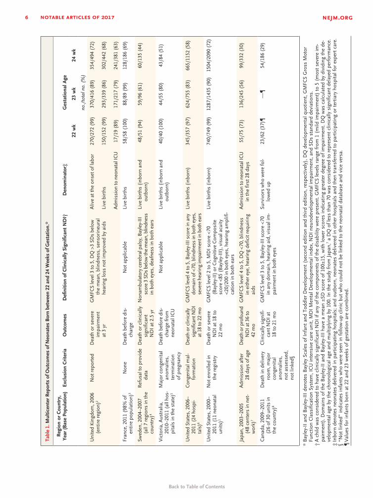
Back to Table of Contents
6 Notable Articles of 2017 nejm.org Editorial
n engl j med 376;7 nejm.org February 16, 2017 695
Tabl
e 1.
Mul
ticen
ter
Rep
orts
of O
utco
mes
of N
eona
tes
Bor
n be
twee
n 22
and
24
Wee
ks o
f Ges
tatio
n.*
Reg
ion
or C
ount
ry,
Year
(Bas
e Po
pula
tion)
Excl
usio
n C
rite
ria
Out
com
esD
efin
ition
of C
linic
ally
Sig
nific
ant
ND
I†D
enom
inat
or‡
Ges
tatio
nal A
ge
22 w
k23
wk
24 w
k
no./
tota
l no.
(%
)
Uni
ted
Kin
gdom
, 200
6 (e
ntir
e re
gion
)2N
ot r
epor
ted
Dea
th o
r se
vere
im
pair
men
t at
3 y
r
GM
FCS
leve
l 3 to
5, D
Q >
3 SD
s be
low
th
e m
ean,
blin
dnes
s, s
enso
rine
ural
he
arin
g lo
ss n
ot im
prov
ed b
y ai
ds
Aliv
e at
the
onse
t of l
abor
Live
bir
ths
Adm
issi
on to
neo
nata
l IC
U
270/
272
(99)
150/
152
(99)
17/1
9 (8
9)
370/
416
(89)
293/
339
(86)
171/
217
(79)
354/
494
(72)
302/
442
(68)
241/
381
(63)
Fran
ce, 2
011
(98%
of
entir
e po
pula
tion)
3N
one
Dea
th b
efor
e di
s-ch
arge
Not
app
licab
leLi
ve b
irth
s58
/58
(100
)88
/89
(99)
128/
186
(69)
Swed
en, 2
004–
2007
(a
ll 7
regi
ons
in th
e co
untr
y)4
Ref
usal
to p
rovi
de
data
Dea
th o
r cl
inic
ally
si
gnifi
cant
N
DI a
t 2.5
yr
Non
ambu
lato
ry c
ereb
ral p
alsy
, Bay
ley-
III
scor
e 3
SDs
belo
w th
e m
ean,
blin
dnes
s in
bot
h ey
es, d
eafn
ess
in b
oth
ears
Live
bir
ths
(inb
orn
and
outb
orn)
48/5
1 (9
4)59
/96
(61)
60/1
35 (
44)
Vic
tori
a, A
ustr
alia
, 20
10–2
011
(all
hos-
pita
ls in
the
stat
e)5
Maj
or c
onge
nita
l an
omal
ies,
te
rmin
atio
n
of p
regn
ancy
Dea
th b
efor
e di
s-ch
arge
from
ne
onat
al IC
U
Not
app
licab
leLi
ve b
irth
s (i
nbor
n an
d ou
tbor
n)40
/40
(100
)44
/55
(80)
43/8
4 (5
1)
Uni
ted
Stat
es, 2
006–
2011
(24
hos
pi-
tals
)6
Con
geni
tal m
al-
form
atio
nD
eath
or c
linic
ally
si
gnifi
cant
ND
I at
18
to 2
2 m
o
GM
FCS
leve
l 4 to
5, B
ayle
y-II
I sco
re in
any
do
mai
n of
<70
, blin
dnes
s in
bot
h ey
es,
seve
re h
earin
g im
pairm
ent i
n bo
th e
ars
Live
bir
ths
(inb
orn)
345/
357
(97)
624/
755
(83)
665/
1152
(58
)
Uni
ted
Stat
es, 2
000–
2011
(11
neo
nata
l un
its)1
Not
enr
olle
d in
th
e re
gist
ryD
eath
or
seve
re
ND
I at 1
8 to
22
mo
GM
FCS
leve
l 2 to
5, M
DI s
core
<70
(B
ayle
y-II
) or
Cog
nitiv
e C
ompo
site
sc
ore
<85
(Bay
ley-
III)
, vis
ual a
cuity
<2
0/20
0 in
bot
h ey
es, h
eari
ng a
mpl
ifi-
catio
n in
bot
h ea
rs
Live
bir
ths
(inb
orn)
740/
749
(99)
1287
/143
5 (9
0)15
04/2
090
(72)
Japa
n, 2
003–
2005
(4
8 ce
nter
s in
net
-w
ork)
7
Adm
issi
on a
fter
28
day
s of
age
Dea
th o
r pro
foun
d N
DI a
t 36
to
42 m
o
GM
FCS
leve
l 4 to
5, D
Q <
70, b
lindn
ess
in
eith
er e
ye, h
eari
ng d
efic
it re
quir
ing
aids
Adm
issi
on to
neo
nata
l IC
U
in th
e fir
st 2
8 da
ys55
/75
(73)
136/
245
(56)
99/3
32 (
30)
Can
ada,
200
9–20
11
(26
of 3
0 un
its in
th
e co
untr
y)8
Dea
th in
del
iver
y ro
om, m
ajor
co
ngen
ital
anom
alie
s,
not a
sses
sed,
no
t lin
ked§
Clin
ical
ly s
igni
fi-ca
nt N
DI a
t 18
to 2
1 m
o
GM
FCS
leve
l 3 to
5, B
ayle
y-II
I sco
re <
70
in a
ny d
omai
n, h
eari
ng a
id, v
isua
l im
-pa
irm
ent i
n bo
th e
yes
Surv
ivor
s w
ho w
ere
fol-
low
ed u
p23
/62
(37)
¶—
¶54
/186
(29
)
* B
ayle
y-II
and
Bay
ley-
III
deno
tes
Bay
ley
Scal
es o
f Inf
ant
and
Todd
ler
Dev
elop
men
t (s
econ
d ed
ition
and
thi
rd e
ditio
n, r
espe
ctiv
ely)
, DQ
dev
elop
men
tal q
uotie
nt, G
MFC
S G
ross
Mot
orFu
nctio
n C
lass
ifica
tion
Syst
em, I
CU
inte
nsiv
e ca
re u
nit,
MD
I M
enta
l Dev
elop
men
tal i
ndex
, ND
I ne
urod
evel
opm
enta
l im
pair
men
t, an
d SD
s st
anda
rd d
evia
tions
.†
A c
hild
was
con
side
red
to h
ave
clin
ical
ly s
igni
fican
t N
DI
if an
y of
the
com
pone
nts
of t
he d
isab
ility
wer
e pr
esen
t. G
MFC
S le
vels
ran
ge fr
om 1
(m
ild im
pair
men
t) t
o 5
(mos
t se
vere
im-
pair
men
t). D
omai
ns o
f the
Bay
ley-
II a
nd B
ayle
y-II
I ha
ve a
mea
n ±S
D s
core
of 1
00±1
5, w
ith lo
wer
sco
res
indi
catin
g gr
eate
r de
gree
of i
mpa
irm
ent.
DQ
was
cal
cula
ted
by d
ivid
ing
the
de-
velo
pmen
tal a
ge b
y th
e ch
rono
logi
cal a
ge a
nd m
ultip
lyin
g by
100
. In
the
stud
y fr
om J
apan
,7 a
DQ
of l
ess
than
70
was
con
side
red
to r
epre
sent
clin
ical
ly s
igni
fican
t de
laye
d pe
rfor
man
ce.
‡ In
born
den
otes
neo
nate
s de
liver
ed in
par
ticip
atin
g ho
spita
ls; a
nd o
utbo
rn, n
eona
tes
deliv
ered
in o
ther
hos
pita
ls a
nd t
hen
tran
sfer
red
to p
artic
ipat
ing
or t
ertia
ry h
ospi
tal f
or e
xper
t ca
re.
§ “N
ot li
nked
” in
dica
tes
infa
nts
who
wer
e se
en in
follo
w-u
p cl
inic
but
who
cou
ld n
ot b
e lin
ked
to t
he n
eona
tal d
atab
ase
and
vice
ver
sa.
¶ V
alue
s fo
r in
fant
s bo
rn a
t 22
and
23
wee
ks o
f ges
tatio
n ar
e co
mbi
ned.
Back to Table of Contents
7 Notable Articles of 2017 nejm.org Editorial
n engl j med 376;7 nejm.org February 16, 2017696
tempted, and neonates who were admitted to neonatal units. However, caution is needed when counting all births; the termination of pregnan-cies at 22 to 24 weeks of gestation because of major congenital malformations may artificially increase the calculated mortality rate.
As neonatal care advances, new reports of survival and outcomes at periviable gestational ages will emerge. A consistent reporting frame-work is needed to permit comparisons of results and to use them to create benchmarks and pur-sue quality improvement. Death and neurodevel-opmental impairment are competing outcomes, and reports need to delineate them in combina-tion and in isolation. Determination of what constitutes neurodevelopmental impairment and whose perspectives are considered (health care workers, parents, or children) remains debatable. Undoubtedly, our obligations to society will be unfulfilled if survivors are not followed longitu-dinally to better guide neonatal, postneonatal, infantile, and childhood care and to improve the quality of life for patients and families.
No discussion on neonatal outcomes is com-plete without consideration of the philosophy of care provision at these gestational ages. Of the national guidelines reviewed previously,9 none have suggested active care for neonates born at 22 to 23 weeks of gestation. Differences in the initiation of resuscitation for such neonates have explained a significant proportion of variation in outcomes between centers.6 “Gentler” approaches to provision of care have initiated a mini-revo-lution in neonatology. In providing care for periviable neonates, opportunities for testing sev-eral organ-protective strategies (e.g., less invasive respiratory support, tolerance in permitting levels of physiological measures that are outside the normal range, melatonin therapy, and adminis-tration of erythropoietin) in a rigorous manner should be a priority. Nonetheless, one must not
forget the implementation of proven interven-tions, because outcomes can be greatly improved if we act on existing knowledge.10 Reports of out-comes in periviable neonates, such as the study by Younge et al., remind us that the only con-stant thing in neonatal intensive care is change.
Disclosure forms provided by the author are available with the full text of this article at NEJM.org.
From the Department of Pediatrics and Institute of Health Pol-icy, Management, and Evaluation, Mount Sinai Hospital, and the University of Toronto — both in Toronto.
This article was updated on February 16, 2017, at NEJM.org.
1. Younge N, Goldstein RF, Bann CM, et al. Survival and neuro-developmental outcomes among periviable infants. N Engl J Med 2017; 376: 617-28.2. Moore T, Hennessy EM, Myles J, et al. Neurological and devel-opmental outcome in extremely preterm children born in England in 1995 and 2006: the EPICure studies. BMJ 2012; 345: e7961.3. Ancel PY, Goffinet F, EPIPAGE-2 Writing Group. Survival and morbidity of preterm children born at 22 through 34 weeks’ gestation in France in 2011: results of the EPIPAGE-2 cohort study. JAMA Pediatr 2015; 169: 230-8.4. Serenius F, Källén K, Blennow M, et al. Neurodevelopmental outcome in extremely preterm infants at 2.5 years after active perinatal care in Sweden. JAMA 2013; 309: 1810-20.5. Boland RA, Davis PG, Dawson JA, Doyle LW. Outcomes of infants born at 22-27 weeks’ gestation in Victoria according to outborn/inborn birth status. Arch Dis Child Fetal Neonatal Ed 2016 August 16 (Epub ahead of print).6. Rysavy MA, Li L, Bell EF, et al. Between-hospital variation in treatment and outcomes in extremely preterm infants. N Engl J Med 2015; 372: 1801-11.7. Ishii N, Kono Y, Yonemoto N, Kusuda S, Fujimura M. Out-comes of infants born at 22 and 23 weeks’ gestation. Pediatrics 2013; 132: 62-71.8. Synnes A, Luu TM, Moddemann D, et al. Determinants of developmental outcomes in a very preterm Canadian cohort. Arch Dis Child Fetal Neonatal Ed 2016 October 6 (Epub ahead of print).9. Guillén Ú, Weiss EM, Munson D, et al. Guidelines for the management of extremely premature deliveries: a systematic re-view. Pediatrics 2015; 136: 343-50.10. Zeitlin J, Manktelow BN, Piedvache A, et al. Use of evidence based practices to improve survival without severe morbidity for very preterm infants: results from the EPICE population based cohort. BMJ 2016; 354: i2976.
DOI: 10.1056/NEJMe1616539Copyright © 2017 Massachusetts Medical Society.
apply for jobs at the nejm careercenter
Physicians registered at the NEJM CareerCenter can apply for jobs electronically. A personal account created when you register allows you to apply for positions,
using your own cover letter and CV, and keep track of your job-application history. Visit NEJMjobs.org for more information.
Back to Table of Contents
8 Notable Articles of 2017 nejm.org T h e n e w e ngl a nd j o u r na l o f m e dic i n e
n engl j med 376;7 nejm.org February 16, 2017 629
The authors’ full names, academic de-grees, and affiliations are listed in the Ap-pendix. Address reprint requests to Dr. Mok at the Department of Clinical Oncol-ogy, Chinese University of Hong Kong, Prince of Wales Hospital, Hong Kong, China, or at tony@ clo . cuhk . edu . hk.
* A complete list of the AURA3 Investiga-tors is provided in the SupplementaryAppendix, available at NEJM.org.
Drs. Mok and Wu contributed equally to this article.
This article was published on December 6, 2016, at NEJM.org.
N Engl J Med 2017;376:629-40.DOI: 10.1056/NEJMoa1612674Copyright © 2016 Massachusetts Medical Society.
BACKGROUNDOsimertinib is an epidermal growth factor receptor tyrosine kinase inhibitor (EGFR-TKI) that is selective for both EGFR-TKI sensitizing and T790M resistance mutations in patients with non–small-cell lung cancer. The efficacy of osimertinib as compared with platinum-based therapy plus pemetrexed in such patients is unknown.
METHODSIn this randomized, international, open-label, phase 3 trial, we assigned 419 pa-tients with T790M-positive advanced non–small-cell lung cancer, who had disease progression after first-line EGFR-TKI therapy, in a 2:1 ratio to receive either oral osimertinib (at a dose of 80 mg once daily) or intravenous pemetrexed (500 mg per square meter of body-surface area) plus either carboplatin (target area under the curve, 5 [AUC5]) or cisplatin (75 mg per square meter) every 3 weeks for up to six cycles; maintenance pemetrexed was allowed. In all the patients, disease had progressed during receipt of first-line EGFR-TKI therapy. The primary end point was investigator-assessed progression-free survival.
RESULTSThe median duration of progression-free survival was significantly longer with osimer-tinib than with platinum therapy plus pemetrexed (10.1 months vs. 4.4 months; haz-ard ratio; 0.30; 95% confidence interval [CI], 0.23 to 0.41; P<0.001). The objective response rate was significantly better with osimertinib (71%; 95% CI, 65 to 76) than with platinum therapy plus pemetrexed (31%; 95% CI, 24 to 40) (odds ratio for objec-tive response, 5.39; 95% CI, 3.47 to 8.48; P<0.001). Among 144 patients with metas-tases to the central nervous system (CNS), the median duration of progression-free survival was longer among patients receiving osimertinib than among those receiv-ing platinum therapy plus pemetrexed (8.5 months vs. 4.2 months; hazard ratio, 0.32; 95% CI, 0.21 to 0.49). The proportion of patients with adverse events of grade 3 or higher was lower with osimertinib (23%) than with platinum therapy plus pemetrexed (47%).
CONCLUSIONSOsimertinib had significantly greater efficacy than platinum therapy plus peme-trexed in patients with T790M-positive advanced non–small-cell lung cancer (includ-ing those with CNS metastases) in whom disease had progressed during first-line EGFR-TKI therapy. (Funded by AstraZeneca; AURA3 ClinicalTrials.gov number, NCT02151981.)
A BS TR AC T
Osimertinib or Platinum–Pemetrexed in EGFR T790M–Positive Lung Cancer
T.S. Mok, Y.-L. Wu, M.-J. Ahn, M.C. Garassino, H.R. Kim, S.S. Ramalingam, F.A. Shepherd, Y. He, H. Akamatsu, W.S.M.E. Theelen, C.K. Lee, M. Sebastian, A. Templeton, H. Mann, M. Marotti, S. Ghiorghiu,
and V.A. Papadimitrakopoulou, for the AURA3 Investigators*
Original Article
Read Full Article at NEJM.org
Back to Table of Contents
9 Notable Articles of 2017 nejm.org The new england journal of medicine
n engl j med 376;10 nejm.org March 9, 2017 917
established in 1812 March 9, 2017 vol. 376 no. 10
The authors’ affiliations are listed in the Appendix. Address reprint requests to Dr. Hochhaus at Klinik für Innere Med-izin II, Abteilung Hämatologie–Onkolo-gie, Universitätsklinikum Jena, Am Klini-kum 1, 07740 Jena, Germany, or at andreas . hochhaus@ med . uni-jena . de.
* A complete list of participating investi-gators in the International RandomizedStudy of Interferon and STI571 (IRIS) is provided in the Supplementary Appen-dix, available at NEJM.org.
N Engl J Med 2017;376:917-27.DOI: 10.1056/NEJMoa1609324Copyright © 2017 Massachusetts Medical Society.
BACKGROUNDImatinib, a selective BCR-ABL1 kinase inhibitor, improved the prognosis for pa-tients with chronic myeloid leukemia (CML). We conducted efficacy and safety analyses on the basis of more than 10 years of follow-up in patients with CML who were treated with imatinib as initial therapy.
METHODSIn this open-label, multicenter trial with crossover design, we randomly assigned patients with newly diagnosed CML in the chronic phase to receive either imatinib or interferon alfa plus cytarabine. Long-term analyses included overall survival, response to treatment, and serious adverse events.
RESULTSThe median follow-up was 10.9 years. Given the high rate of crossover among patients who had been randomly assigned to receive interferon alfa plus cytarabine (65.6%) and the short duration of therapy before crossover in these patients (median, 0.8 years), the current analyses focused on patients who had been ran-domly assigned to receive imatinib. Among the patients in the imatinib group, the estimated overall survival rate at 10 years was 83.3%. Approximately half the pa-tients (48.3%) who had been randomly assigned to imatinib completed study treat-ment with imatinib, and 82.8% had a complete cytogenetic response. Serious ad-verse events that were considered by the investigators to be related to imatinib were uncommon and most frequently occurred during the first year of treatment.
CONCLUSIONSAlmost 11 years of follow-up showed that the efficacy of imatinib persisted over time and that long-term administration of imatinib was not associated with unac-ceptable cumulative or late toxic effects. (Funded by Novartis Pharmaceuticals; IRIS ClinicalTrials.gov numbers, NCT00006343 and NCT00333840.)
a bs tr ac t
Long-Term Outcomes of Imatinib Treatment for Chronic Myeloid Leukemia
Andreas Hochhaus, M.D., Richard A. Larson, M.D., François Guilhot, M.D., Jerald P. Radich, M.D., Susan Branford, Ph.D., Timothy P. Hughes, M.D., Michele Baccarani, M.D., Michael W. Deininger, M.D., Ph.D.,
Francisco Cervantes, M.D., Satoko Fujihara, Ph.D., Christine-Elke Ortmann, M.Sc., Hans D. Menssen, M.D., Hagop Kantarjian, M.D., Stephen G. O’Brien, M.D., Ph.D., and Brian J. Druker, M.D., for the IRIS Investigators*
Read Full Article at NEJM.org
original Article

Back to Table of Contents
10 Notable Articles of 2017 nejm.org
e d i t o r i a l
T h e n e w e ngl a nd j o u r na l o f m e dic i n e
n engl j med 376;10 nejm.org March 9, 2017982
Imatinib Changed Everything
Dan L. Longo, M.D.
The initial successes of combination chemotherapy were stunning. Childhood acute leukemia, several forms of lymphoma, and testicular cancer all became largely curable malignant conditions. Adjuvant chemotherapy led to dramatically longer survival among persons with breast cancer. The fundamental principle of chemotherapy was to target dividing cells, because it was assumed that more rapid proliferation than “normal” was an underlying common defect in cancer. The effective agents generally attacked DNA or the mitotic spindle. Some tumors, such as breast cancer and prostate cancer, were susceptible to hormonal manipulation because of growth regulation of their normaltissue counterparts.
Gradually data emerged that, except in the case of Burkitt’s lymphoma, the fraction of tumor cells that were actively progressing through the cell cycle at any given time was quite small and generally no greater than in normal bone marrow, skin, or intestinal mucosa. Yet efforts to use chemotherapy in patients with other types of cancer continued with rare success and with wellknown toxic effects.
Then along came Brian Druker. His focus on chronic myeloid leukemia (CML) was both clever and lucky. What we know now, which we didn’t know then, is that CML is less complex genetically than most cancers. The introduction of the bcr-abl gene product into murine hematopoietic stem cells was sufficient for the reproduction of humanlike disease in these animals,13 an unusual finding. These results led to the idea that interference with the function of this chimeric gene product might exert antitumor effects. This was a new idea.
Novartis (then called Ciba Geigy) had made a number of compounds that were capable of inhibiting protein tyrosine kinases, and one from the 2phenylaminopyrimidine class (called CGP 57148 in some places and STI 571 in others and known today as imatinib) interfered with the viral oncogene vabl and plateletderived growth factor receptor activity in vitro and in mice.4 It also inhibited the growth of BCRABL–positive cells.5 Five years later, Druker and colleagues reported data from 54 patients with CML who had been treated with imatinib in a phase 1 study; complete hematologic responses were seen in 53 of the 54 patients, and in 7 patients, the Philadelphia chromosome was no longer detectable.6 No other drug had achieved such results.
In this issue of the Journal, Hochhaus, Druker, and colleagues7 report data from more than 10 years of followup in a randomized trial comparing interferon alfa and cytarabine with imatinib as initial therapy in patients with CML. The estimated survival rate at 10 years among patients in the imatinib group was 83%; in the course of the trial, 83% of the patients in the imatinib group had a complete cytogenetic response. Patients were mainly treated continuously, and no unexpected late toxic effects emerged. Approximately one patient in five had a stable deep molecular response for 1 year or longer, and some had the therapy discontinued, although this was not done on a consistent basis. Approximately 40% of the patients who stopped therapy remained in remission for 3 years or longer, with the rest having a relapse. Thus, imatinib is highly successful at controlling the disease in the long term, but few, if any, patients would be consid
Back to Table of Contents
11 Notable Articles of 2017 nejm.org Editorials
n engl j med 376;10 nejm.org March 9, 2017 983
ered to be “cured” (in a stable remission and not taking any therapy). Yet the imatinib story suggested that the understanding of the pathogenesis of this tumor led to a less toxic and more effective treatment approach. Subsequent studies revealed other cancers in which imatinib was active through a different target,8 identified the molecular mechanisms of imatinib resistance (and showed that they are often shared by other tyrosine kinase inhibitors),9 and led to the design of new second and thirdgeneration agents.10
The development of imatinib fundamentally altered the field of oncology. Priorities shifted from agents that were active on dividing cells to understanding the biology of individual types of cancer. Once genetic analysis of tumors began, nearly all the cancer types had more complex genetic abnormalities than did CML, but the complexity gave rise to a revolution in cancer nosology. We now recognize that the grouping of tumors on the basis of the appearance of a hematoxylin and eosin–stained tissue fragment examined under a light microscope lumps together entities that are distinct both genetically and clinically. Lung cancer is now considered to be at least eight or nine entities, and the number of variants is continuing to expand. That is the good news. The bad news is that the inherent genetic instability of many cancers also facilitates the development of resistance to these interventions. In some instances, new genetic abnormalities create vulnerabilities that can be attacked by new agents.
The future of oncology is more hopeful now. The analysis of cancer at the genetic level and the development of (mainly) oral agents that can inhibit driver mutations constitute a conceptual departure from the medical oncology of the 1990s. An increasing number of agents hit an increasing number of targets. Tumor heterogeneity and mechanisms of resistance still limit our therapies, but the analysis of plasma for tumor DNA may help us to detect and anticipate the adaptation of the tumor to therapy and to select secondary treatments that target those
changes. On top of the genetic guidance of therapeutic decisions is the remarkable recent progress in activating the immune system as a weapon in the battle. In this arena, too, much more needs to be learned.
We need to resist the temptation to be selfcongratulatory. The prognosis for patients with common cancers is improving somewhat, but none of the new tools appears to cure a majority of patients. We still need to learn how to combine therapies that have different targets, to identify patients who are likely to have a response, and to define mechanisms of resistance. Although the journey to cancer cure has just begun, the use of imatinib to treat CML has pointed oncology in a new direction.
Disclosure forms provided by the author are available with the full text of this editorial at NEJM.org.
1. Daley GQ, Van Etten RA, Baltimore D. Induction of chronicmyelogenous leukemia in mice by the P210bcr/abl gene of the Philadelphia chromosome. Science 1990; 247: 82430.2. Heisterkamp N, Jenster G, ten Hoeve J, Zovich D, PattengalePK, Groffen J. Acute leukaemia in bcr/abl transgenic mice. Nature 1990; 344: 2513.3. Kelliher MA, McLaughlin J, Witte ON, Rosenberg N. Induction of a chronic myelogenous leukemialike syndrome in mice with vabl and BCR/ABL. Proc Natl Acad Sci U S A 1990; 87: 664953.4. Buchdunger E, Zimmermann J, Mett H, et al. Inhibition ofthe Abl proteintyrosine kinase in vitro and in vivo by a 2phenylaminopyrimidine derivative. Cancer Res 1996; 56: 1004.5. Druker BJ, Tamura S, Buchdunger E, et al. Effects of a selective inhibitor of the Abl tyrosine kinase on the growth of BcrAbl positive cells. Nat Med 1996; 2: 5616.6. Druker BJ, Talpaz M, Resta DJ, et al. Efficacy and safety of aspecific inhibitor of the BCRABL tyrosine kinase in chronic myeloid leukemia. N Engl J Med 2001; 344: 10317.7. Hochhaus A, Larson RA, Guilhot F, et al. Longterm outcomes of imatinib treatment for chronic myeloid leukemia. N Engl J Med 2017; 376:91727.8. Joensuu H, Roberts PJ, SarlomoRikala M, et al. Effect ofthe tyrosine kinase inhibitor STI571 in a patient with a metastatic gastrointestinal stromal tumor. N Engl J Med 2001; 344: 10526.9. Gorre ME, Mohammed M, Ellwood K, et al. Clinical resistance to STI571 cancer therapy caused by BCRABL gene mutation or amplification. Science 2001; 293: 87680.10. Weisberg E, Manley PW, Breitenstein W, et al. Characterization of AMN107, a selective inhibitor of native and mutant BcrAbl. Cancer Cell 2005; 7: 12941.
DOI: 10.1056/NEJMe1700833
Copyright © 2017 Massachusetts Medical Society.

Back to Table of Contents
12 Notable Articles of 2017 nejm.org T h e n e w e ngl a nd j o u r na l o f m e dic i n e
n engl j med 376;20 nejm.org May 18, 2017 1921
The authors’ full names, academic de-grees, and affiliations are listed in the Ap-pendix. Address reprint requests to Dr. Wechsler at National Jewish Health, 1400 Jackson St., Denver, CO 80230, or at mikewechsler@ gmail . com.
*A complete list of the members of theEGPA Mepolizumab Study Team is pro-vided in the Supplementary Appendix,available at NEJM.org.
Drs. Weller and Gleich contributed equally to this article.
N Engl J Med 2017;376:1921-32.DOI: 10.1056/NEJMoa1702079Copyright © 2017 Massachusetts Medical Society.
BACKGROUNDEosinophilic granulomatosis with polyangiitis is an eosinophilic vasculitis. Mepo-lizumab, an anti–interleukin-5 monoclonal antibody, reduces blood eosinophil counts and may have value in the treatment of eosinophilic granulomatosis with polyangiitis.
METHODSIn this multicenter, double-blind, parallel-group, phase 3 trial, we randomly as-signed participants with relapsing or refractory eosinophilic granulomatosis with polyangiitis who had received treatment for at least 4 weeks and were taking a stable prednisolone or prednisone dose to receive 300 mg of mepolizumab or placebo, administered subcutaneously every 4 weeks, plus standard care, for 52 weeks. The two primary end points were the accrued weeks of remission over a 52-week period, according to categorical quantification, and the proportion of participants in remission at both week 36 and week 48. Secondary end points included the time to first relapse and the average daily glucocorticoid dose (during weeks 48 through 52). The annualized relapse rate and safety were assessed.
RESULTSA total of 136 participants underwent randomization, with 68 participants assigned to receive mepolizumab and 68 to receive placebo. Mepolizumab treatment led to significantly more accrued weeks of remission than placebo (28% vs. 3% of the par-ticipants had ≥24 weeks of accrued remission; odds ratio, 5.91; 95% confidence inter-val [CI], 2.68 to 13.03; P<0.001) and a higher percentage of participants in remission at both week 36 and week 48 (32% vs. 3%; odds ratio, 16.74; 95% CI, 3.61 to 77.56; P<0.001). Remission did not occur in 47% of the participants in the mepolizumab group versus 81% of those in the placebo group. The annualized relapse rate was 1.14 in the mepolizumab group, as compared with 2.27 in the placebo group (rate ratio, 0.50; 95% CI, 0.36 to 0.70; P<0.001). A total of 44% of the participants in the mepolizumab group, as compared with 7% of those in the placebo group, had an average daily dose of prednisolone or prednisone of 4.0 mg or less per day dur-ing weeks 48 through 52 (odds ratio, 0.20; 95% CI, 0.09 to 0.41; P<0.001). The safety profile of mepolizumab was similar to that observed in previous studies.
CONCLUSIONSIn participants with eosinophilic granulomatosis with polyangiitis, mepolizumab resulted in significantly more weeks in remission and a higher proportion of par-ticipants in remission than did placebo, thus allowing for reduced glucocorticoid use. Even so, only approximately half the participants treated with mepolizumab had protocol-defined remission. (Funded by GlaxoSmithKline and the National Institute of Allergy and Infectious Diseases; ClinicalTrials.gov number, NCT02020889.)
A BS TR AC T
Mepolizumab or Placebo for Eosinophilic Granulomatosis with Polyangiitis
M.E. Wechsler, P. Akuthota, D. Jayne, P. Khoury, A. Klion, C.A. Langford, P.A. Merkel, F. Moosig, U. Specks, M.C. Cid, R. Luqmani, J. Brown, S. Mallett,
R. Philipson, S.W. Yancey, J. Steinfeld, P.F. Weller, and G.J. Gleich, for the EGPA Mepolizumab Study Team*
Original Article
Read Full Article at NEJM.org
Back to Table of Contents
13 Notable Articles of 2017 nejm.org
Editorials
n engl j med 376;20 nejm.org May 18, 2017 1985
Targeting Eosinophils in Eosinophilic Granulomatosis with Polyangiitis
Ratko Djukanovic, M.D., and Paul M. O’Byrne, M.B.
Eosinophilic granulomatosis with polyangiitis, first described in the early 1950s by Dr. Jacob Churg and Dr. Lotte Strauss (hence the original name, the Churg–Strauss syndrome), is a rare condition that can affect many organ systems, most commonly the lung, with the majority of patients presenting with asthma symptoms, oc-casionally complicated by the fleeting presence of pulmonary infiltrates.1 Many different treat-ments have been tried for eosinophilic granu-lomatosis with polyangiitis, with limited or no success, and systemic glucocorticoids are the standard treatment for both eosinophilic granu-lomatosis with polyangiitis1 and severe refractory asthma.2
The predominance of eosinophils in the pe-ripheral blood and tissues in patients with eosino-philic granulomatosis with polyangiitis has sug-gested a central role for eosinophils in the pathogenesis of this disease. Several small, open-label studies3-5 have shown evidence that the blocking of interleukin-5, a cytokine known to be involved in the maturation, tissue accumulation, activation, and survival of eosinophils, with the monoclonal antibody mepolizumab provides clin-ical benefit in patients with eosinophilic granu-lomatosis with polyangiitis. Mepolizumab has already been shown to reduce the incidence of severe exacerbations among patients with severe refractory eosinophilic asthma.6,7 In this issue of the Journal, Wechsler and colleagues8 report the results of a placebo-controlled, double-blind trial of mepolizumab in participants who had relaps-ing eosinophilic granulomatosis with polyangiitis or who had eosinophilic granulomatosis with polyangiitis that was refractory to treatment with oral glucocorticoids, but the trial excluded partici-pants with organ-threatening or life-threatening disease.
The trial showed a benefit of mepolizumab with regard to the two primary end points: the accrued weeks of disease remission and the pro-portion of participants who were in remission at weeks 36 and 48 of the trial. The annualized
relapse rate was approximately 50% lower in the mepolizumab group than in the placebo group, a finding that is similar to that observed with mepolizumab with regard to exacerbation rates among patients with severe eosinophilic asthma.7 Although the trial was powered to evaluate re-lapses on the basis of a worsening condition in any of three categories (asthma, vasculitis, and sinonasal disease alone or in combination), the benefit of treatment was slightly greater with regard to relapses defined according to exacerbat-ing asthma-based or sinonasal-based symptoms. This finding suggests that the vasculitic compo-nent of eosinophilic granulomatosis with poly-angiitis may respond less well to mepolizumab than do other components of the disease.
After remission, some participants in this trial had a relapse during treatment with mepolizu-mab. This situation raises questions about the mechanism of relapses of eosinophilic granulo-matosis with polyangiitis (in particular, the role of eosinophils) and the dose of mepolizumab that was used in the trial. Eosinophils have been the focus of research for decades but gained mechanistic and biomarker prominence in the study of severe asthma after studies showed that the use of sputum or blood eosinophil counts as biomarkers to adjust the dose of inhaled gluco-corticoids9 or to enrich the population of patients with severe eosinophilic asthma for treatment with mepolizumab effectively reduced exacer-bation frequency.6,7 As has been the case with asthma, in the current trial, the blood eosino-phil count was a risk factor for relapse, in that participants with a blood eosinophil count of 150 or more per cubic millimeter at enrollment benefited markedly from mepolizumab versus placebo (odds ratio, 26.10), whereas those with a blood eosinophil count of less than 150 per cu-bic millimeter did not (odds ratio, 0.95).8 Also, the dose of mepolizumab that was used (300 mg monthly) was higher than the dose approved for severe eosinophilic asthma (100 mg monthly) but lower than that used in an initial study involving
editorial

Back to Table of Contents
14 Notable Articles of 2017 nejm.org T h e n e w e ngl a nd j o u r na l o f m e dic i n e
n engl j med 376;20 nejm.org May 18, 20171986
patients with severe asthma (750 mg monthly).6 It is uncertain whether a dose of 300 mg monthly is the most effective dose for treating patients with eosinophilic granulomatosis with polyangi-itis, in whom blood eosinophil counts are often much higher than in patients with severe eosino-philic asthma. Unfortunately, blood eosinophil counts were not recorded during exacerbations, which is a missed opportunity to explore the pathobiologic features of exacerbations.
All the participants in this trial had relapsing or refractory eosinophilic granulomatosis with polyangiitis, despite taking at least 7.5 mg of prednisone per day. The reduction in the predni-sone dose that occurred with mepolizumab shows an important benefit from treatment, with 18% of the participants being able to discontinue pred-nisone completely. The trial design allowed the reduction of the oral glucocorticoid dose as early as 4 weeks after trial onset, which leaves an open question as to whether the effect of mepo-lizumab on the prevention of relapse may have been better if the dose of glucocorticoids had remained constant throughout the trial.
After many years of research into eosinophilic diseases and failure of treatment with anti–inter-leukin-5 antibodies in all patients with severe asthma,10 drug developers are increasingly using biomarkers to select patients who are most likely to have a response to a given treatment. After this proof-of-concept study, additional research is needed to identify biomarkers that inform suc-cess and failure of mepolizumab in patients with eosinophilic granulomatosis with polyangiitis and to elucidate the fate of tissue eosinophils, espe-cially in vasculitic lesions. Further studies may discover previously unknown pro-eosinophilic mechanisms or identify eosinophil-independent mechanisms in eosinophilic granulomatosis with polyangiitis. Future trials will also need not only to establish the appropriate dosing of mepoliz-
umab but also to include participants with life-threatening eosinophilic granulomatosis with polyangiitis who were not included in this trial and possibly to evaluate synergy with immuno-suppressants such as azathioprine and cyclo-phosphamide.
Disclosure forms provided by the authors are available with the full text of this article at NEJM.org.
From Clinical and Experimental Sciences, Faculty of Medicine, University of Southampton, and National Institute for Health Research Southampton Biomedical Research Centre, Southamp-ton, United Kingdom (R.D.); and Firestone Institute for Respira-tory Health, St. Joseph’s Healthcare, and the Department of Medicine, Michael G. DeGroote School of Medicine, McMaster University, Hamilton, ON, Canada (P.M.O.).
1. Groh M, Pagnoux C, Baldini C, et al. Eosinophilic granulo-matosis with polyangiitis (Churg-Strauss) (EGPA) Consensus Task Force recommendations for evaluation and management. Eur J Intern Med 2015; 26: 545-53.2. Reddel HK, Bateman ED, Becker A, et al. A summary of thenew GINA strategy: a roadmap to asthma control. Eur Respir J 2015; 46: 622-39.3. Kahn JE, Grandpeix-Guyodo C, Marroun I, et al. Sustainedresponse to mepolizumab in refractory Churg-Strauss syndrome. J Allergy Clin Immunol 2010; 125: 267-70.4. Kim S, Marigowda G, Oren E, Israel E, Wechsler ME. Mepo-lizumab as a steroid-sparing treatment option in patients with Churg-Strauss syndrome. J Allergy Clin Immunol 2010; 125: 1336-43.5. Moosig F, Gross WL, Herrmann K, Bremer JP, Hellmich B.Targeting interleukin-5 in refractory and relapsing Churg-Strauss syndrome. Ann Intern Med 2011; 155: 341-3.6. Nair P, Pizzichini MM, Kjarsgaard M, et al. Mepolizumab forprednisone-dependent asthma with sputum eosinophilia. N Engl J Med 2009; 360: 985-93.7. Ortega HG, Liu MC, Pavord ID, et al. Mepolizumab treat-ment in patients with severe eosinophilic asthma. N Engl J Med 2014; 371: 1198-207.8. Wechsler ME, Akuthota P, Jayne D, et al. Mepolizumab or pla-cebo for eosinophilic granulomatosis with polyangiitis. N Engl J Med 2017; 376: 1921-32.9. Green RH, Brightling CE, McKenna S, et al. Asthma exacer-bations and sputum eosinophil counts: a randomised controlled trial. Lancet 2002; 360: 1715-21.10. Flood-Page P, Swenson C, Faiferman I, et al. A study to eval-uate safety and efficacy of mepolizumab in patients with moder-ate persistent asthma. Am J Respir Crit Care Med 2007; 176: 1062-71.
DOI: 10.1056/NEJMe1704402Copyright © 2017 Massachusetts Medical Society.

Back to Table of Contents
15 Notable Articles of 2017 nejm.org
Read Full Article at NEJM.org
The new england journal of medicine
n engl j med 376;26 nejm.org June 29, 2017 2513
established in 1812 June 29, 2017 vol. 376 no. 26
From the Departments of Environmental Health (Q.D., Yan Wang, A.Z., P.K., J.D.S.) and Biostatistics (Yun Wang, C.C., F.D.), Harvard T.H. Chan School of Public Health, Boston. Address reprint requests to Dr. Dominici at Harvard T.H. Chan School of Public Health, Biostatistics Department, Bldg. 2, 4th Flr., 655 Huntington Ave., Boston, MA 02115, or at fdominic@ hsph . harvard . edu.
N Engl J Med 2017;376:2513-22.DOI: 10.1056/NEJMoa1702747Copyright © 2017 Massachusetts Medical Society.
BACKGROUNDStudies have shown that long-term exposure to air pollution increases mortality. However, evidence is limited for air-pollution levels below the most recent Na-tional Ambient Air Quality Standards. Previous studies involved predominantly urban populations and did not have the statistical power to estimate the health effects in underrepresented groups.
METHODSWe constructed an open cohort of all Medicare beneficiaries (60,925,443 persons) in the continental United States from the years 2000 through 2012, with 460,310,521 person-years of follow-up. Annual averages of fine particulate matter (particles with a mass median aerodynamic diameter of less than 2.5 μm [PM2.5]) and ozone were estimated according to the ZIP Code of residence for each en-rollee with the use of previously validated prediction models. We estimated the risk of death associated with exposure to increases of 10 μg per cubic meter for PM2.5 and 10 parts per billion (ppb) for ozone using a two-pollutant Cox proportional-hazards model that controlled for demographic characteristics, Medicaid eligibil-ity, and area-level covariates.
RESULTSIncreases of 10 μg per cubic meter in PM2.5 and of 10 ppb in ozone were associ-ated with increases in all-cause mortality of 7.3% (95% confidence interval [CI], 7.1 to 7.5) and 1.1% (95% CI, 1.0 to 1.2), respectively. When the analysis was re-stricted to person-years with exposure to PM2.5 of less than 12 μg per cubic meter and ozone of less than 50 ppb, the same increases in PM2.5 and ozone were as-sociated with increases in the risk of death of 13.6% (95% CI, 13.1 to 14.1) and 1.0% (95% CI, 0.9 to 1.1), respectively. For PM2.5, the risk of death among men, blacks, and people with Medicaid eligibility was higher than that in the rest of the population.
CONCLUSIONSIn the entire Medicare population, there was significant evidence of adverse effects related to exposure to PM2.5 and ozone at concentrations below current national standards. This effect was most pronounced among self-identified racial minori-ties and people with low income. (Supported by the Health Effects Institute and others.)
a bs tr ac t
Air Pollution and Mortality in the Medicare PopulationQian Di, M.S., Yan Wang, M.S., Antonella Zanobetti, Ph.D., Yun Wang, Ph.D., Petros Koutrakis, Ph.D.,
Christine Choirat, Ph.D., Francesca Dominici, Ph.D., and Joel D. Schwartz, Ph.D.
original Article

Back to Table of Contents
16 Notable Articles of 2017 nejm.org
e d i t o r i a l
T h e n e w e ngl a nd j o u r na l o f m e dic i n e
n engl j med 376;26 nejm.org June 29, 2017 2591
Air Pollution Still Kills
Rebecca E. Berger, M.D., Ramya Ramaswami, M.B., B.S., M.P.H., Caren G. Solomon, M.D., M.P.H., and Jeffrey M. Drazen, M.D.
In late October 1948, a dense smog descended over the town of Donora, Pennsylvania. The town was home to a zinc plant and a steel mill, both run by the United States Steel Corporation. Susan Gnora, a 62-year-old resident of Donora, started to gasp and cough as the smog descended.1 She died the next day. Dr. William Rongaus, a physi-cian and a member of the board of health, went door to door, treating patients for their respira-tory symptoms and encouraging them to leave town if they could. Many thousands were ill, and at least 20 people died in one of the worst air-pollution disasters in U.S. history. The Donora tragedy transformed our perception of smog from a nuisance to a potential killer.
We started to improve air quality with the Clean Air Act of 1963. In 1970, Richard Nixon established the Environmental Protection Agency (EPA) by executive order, and the Clean Air Act was amended to institute National Ambient Air Quality Standards (NAAQS), which set exposure limits for six major air pollutants.2 Among the pollutants regulated by the EPA is fine particu-late matter — inhalable particles with an aero-dynamic diameter of less than 2.5 μm (PM2.5). Major contributors to PM2.5 in the United States include various types of transportation and the coal-fired generation of electricity.3,4 Since the 1970s, hundreds of articles have been written establishing an association between PM2.5 and poor health outcomes, including asthma, ische-mic heart disease, and all-cause mortality in ur-ban populations.5,6 In response to these findings, regulators have lowered NAAQS for the allow-able amount of PM2.5 in the air.7 Current NAAQS,
last updated in 2012, set an annual mean PM2.5 level of 12 μg per cubic meter. This standard, which is to be reviewed every 5 years, aims to protect the population, especially those who are particularly sensitive to the adverse effects of air pollution, including children, elderly persons, and persons with cardiopulmonary disease.2 As com-munities meet these stricter standards, fewer people will become sick and die as a result of air pollution. A 2011 report from the EPA projected that by 2020, amendments to the Clean Air Act would prevent more than 230,000 premature deaths, largely as a result of reductions in PM2.5 levels.8 But are current standards sufficient to protect public health?
Di et al. now report in the Journal the results of a large study, including more than 60 million Medicare beneficiaries from the years 2000 through 2012, that addresses the association be-tween annual average levels of PM2.5 and ozone,9 as measured at the ZIP Code level, and mortality. For every increase of 10 μg per cubic meter in PM2.5, there was an associated 7.3% increase in all-cause mortality (95% confidence interval [CI], 7.1 to 7.5), after adjustment for demographic characteristics, Medicaid eligibility, and area-level covariates. Below the current NAAQS for PM2.5 of 12 μg per cubic meter, the data showed that each increase in PM2.5 of 10 μg per cubic meter was associated with an even greater increase (13.6%) in mortality (95% CI, 13.1 to 14.1). There was no appreciable level below which the risk of death tapered off — and thus no “safe” level of PM2.5. Owing to the large size of the co-hort, Di et al. were able to perform robust sub-

Back to Table of Contents
17 Notable Articles of 2017 nejm.org T h e n e w e ngl a nd j o u r na l o f m e dic i n e
n engl j med 376;26 nejm.org June 29, 20172592
group analyses and identified greater risks of death associated with air pollutants among blacks and Medicaid-eligible populations; moreover, these groups were more likely to be exposed to higher pollutant levels.
The findings of Di et al. stress the need for tighter regulation of air-pollutant levels, includ-ing the imposition of stricter limits on levels of PM2.5. Despite compelling data, the Trump ad-ministration is moving headlong in the opposite direction. In March, Trump signed an executive order that lifted a moratorium on new leases for coal mined on public and tribal lands and began a process to dismantle guidelines intended to re-duce emissions from coal-fired electricity plants.10 Earlier this month, he announced his intention to withdraw the United States from the Paris climate agreement. Although these actions were primarily intended to undo efforts made by the Obama administration to address climate change, the potentially dire consequences also include increasing people’s exposure to particulate matter. In addition, EPA Administrator Scott Pruitt has not ruled out the possibility of revoking a waiver included in the 1970 Clean Air Act that allows California to set limits on automotive tailpipe emissions that are more stringent than national standards11; 15 states have adopted California’s standards. Revoking this waiver could have the effect of exposing more than 100 million Amer-icans to higher levels of automobile emissions. Trump’s proposed budget includes crippling cuts to the EPA, including cuts in funding for both federal and state enforcement of regulations. The increased air pollution that would result from loosening current restrictions would have devas-tating effects on public health.
In explaining his withdrawal from the Paris climate agreement, Trump stated, “I was elected to represent the citizens of Pittsburgh, not Paris.” Ironically, Pittsburgh is less than 30 miles from the Donora Smog Museum, where a sign reads,
“Clean Air Started Here.” With the report by Di et al. adding to the large body of evidence indi-cating the risks of air pollution, even at current standards, we must redouble our commitment to clean air. If such protections lapse, Americans will suffer and we are doomed to repeat history. Do we really want to breathe air that kills us?
Disclosure forms provided by the authors are available with the full text of this editorial at NEJM.org.
1. Templeton D. Cleaner air is legacy left by Donora’s killer1948 smog. Pittsburgh Post-Gazette. October 29, 1998 (http://old .post-gazette .com/ magazine/ 19981029smog1 .asp).2. National Ambient Air Quality Standards — criteria air pollut-ants: NAAQS table. Washington, DC: Environmental Protection Agency (https:/ / www .epa .gov/ criteria-air-pollutants/ naaqs-table).3. Laden F, Neas LM, Dockery DW, Schwartz J. Association offine particulate matter from different sources with daily mortal-ity in six U.S. cities. Environ Health Perspect 2000; 108: 941-7.4. Thurston GD, Burnett RT, Turner MC, et al. Ischemic heartdisease mortality and long-term exposure to source-related components of U.S. fine particle air pollution. Environ Health Perspect 2016; 124: 785-94.5. Atkinson RW, Kang S, Anderson HR, Mills IC, Walton HA.Epidemiological time series studies of PM2.5 and daily mortality and hospital admissions: a systematic review and meta-analysis. Thorax 2014; 69: 660-5.6. Hoek G, Krishnan RM, Beelen R, et al. Long-term air pollu-tion exposure and cardio-respiratory mortality: a review. Environ Health 2013; 12: 43.7. National Ambient Air Quality Standards — table of historical particulate matter (PM). Washington, DC: Environmental Protec-tion Agency (https:/ / www .epa .gov/ pm-pollution/ table-historical -particulate-matter-pm-national-ambient-air-quality-standards -naaqs).8. Benefits and costs of the Clean Air Act 1990-2020, the sec-ond prospective study. Washington, DC: Environmental Protec-tion Agency Office of Air and Radiation, 2011.9. Di Q, Wang Y, Zanobetti A, et al. Air pollution and mortalityin the Medicare population. N Engl J Med 2017; 376: 2513-22.10. Presidential Executive Order on promoting energy indepen-dence and economic growth. Press release of The White House, Washington, DC: March 28, 2017.11. Hearing on nomination of Attorney General Scott Pruitt tobe Administrator of the U.S. Environmental Protection Agency — questions for the record for the Honorable E. Scott Pruitt: hearing before the Senate Environment and Public Works Com-mittee, January 18, 2017 (https:/ / www .epw .senate .gov/ public/ _cache/ files/ 1291a5e0-b3aa-403d-8ce3-64cb2ef86851/ spw-011817 .pdf).
DOI: 10.1056/NEJMe1706865
Copyright © 2017 Massachusetts Medical Society.
Back to Table of Contents
18 Notable Articles of 2017 nejm.org The new england journal of medicine
n engl j med 377;1 nejm.org July 6, 2017 13
established in 1812 July 6, 2017 vol. 377 no. 1
*The names, academic degrees, and af-filiations of the authors, who are mem-bers of the Global Burden of Disease(GBD) 2015 Obesity Collaborators, arelisted in the Appendix. The authors as-sume responsibility for the content andintegrity of this article. Address reprintrequests to Dr. Murray at the Institutefor Health Metrics and Evaluation, Uni-versity of Washington, 2301 5th Ave.,Suite 600, Seattle, WA 98121, or at cjlm@ uw . edu.
This article was published on June 12, 2017, at NEJM.org.
This is the New England Journal of Medi-cine version of record, which includes all Journal editing and enhancements. The Author Final Manuscript, which is the au-thor’s version after external peer review and before publication in the Journal, is available under a CC BY license at PMC5477817.
N Engl J Med 2017;377:13-27.DOI: 10.1056/NEJMoa1614362Copyright © 2017 Massachusetts Medical Society.
BACKGROUNDAlthough the rising pandemic of obesity has received major attention in many countries, the effects of this attention on trends and the disease burden of obe-sity remain uncertain.
METHODSWe analyzed data from 68.5 million persons to assess the trends in the prevalence of overweight and obesity among children and adults between 1980 and 2015. Using the Global Burden of Disease study data and methods, we also quantified the burden of disease related to high body-mass index (BMI), according to age, sex, cause, and BMI in 195 countries between 1990 and 2015.
RESULTSIn 2015, a total of 107.7 million children and 603.7 million adults were obese. Since 1980, the prevalence of obesity has doubled in more than 70 countries and has continuously increased in most other countries. Although the prevalence of obesity among children has been lower than that among adults, the rate of in-crease in childhood obesity in many countries has been greater than the rate of increase in adult obesity. High BMI accounted for 4.0 million deaths globally, nearly 40% of which occurred in persons who were not obese. More than two thirds of deaths related to high BMI were due to cardiovascular disease. The dis-ease burden related to high BMI has increased since 1990; however, the rate of this increase has been attenuated owing to decreases in underlying rates of death from cardiovascular disease.
CONCLUSIONSThe rapid increase in the prevalence and disease burden of elevated BMI highlights the need for continued focus on surveillance of BMI and identification, implemen-tation, and evaluation of evidence-based interventions to address this problem. (Funded by the Bill and Melinda Gates Foundation.)
a bs tr ac t
Health Effects of Overweight and Obesity in 195 Countries over 25 Years
The GBD 2015 Obesity Collaborators*
original article
Read Full Article at NEJM.org
Back to Table of Contents
19 Notable Articles of 2017 nejm.org
e d i t o r i a l
T h e n e w e ngl a nd j o u r na l o f m e dic i n e
n engl j med 377;1 nejm.org July 6, 201780
Global Health Effects of Overweight and Obesity
Edward W. Gregg, Ph.D., and Jonathan E. Shaw, M.D.
The Global Burden of Disease (GBD) study that is now reported in the Journal offers a discourag-ing reminder that the global obesity epidemic is worsening in most parts of the world and that its implications regarding both physical health and economic health remain ominous.1 The study, in which researchers assembled data from 195 coun-tries to model trends in overweight and obesity and related morbidity and mortality, showed that the prevalence of obesity has more than doubled since 1980 and is now 5% in children and 12% in adults — findings that mirror similar global trends in type 2 diabetes. Apart from a possible recent plateau in the prevalence of obesity in high-income countries, the prevalence has increased in all other sociodemographic strata.
On the encouraging side, despite this increase in prevalence, the effect of high body-mass index (BMI) on population-level age-adjusted rates of death and disability has not grown, which sug-gests that obese persons are healthier and live longer now than in previous decades because of better care and risk-factor management. Unfor-tunately, even this success brings a new burden, since the mix of increased prevalence and de-creased mortality leads to more years spent with obesity and more time for the damaging coexist-ing illnesses, such as type 2 diabetes and chronic kidney disease, to develop.
The most worrisome finding is the approxi-mate tripling of obesity seen in youth and young adults of developing, middle-income countries such as China, Brazil, and Indonesia. An early onset of obesity is likely to translate into a high cumulative incidence of type 2 diabetes, hyper-tension, and chronic kidney disease. These find-ings come on the heels of reports from the
United States that the incidence of type 2 diabetes in youth has increased substantially in minority populations, and when type 2 diabetes occurs in youth, it brings a much higher prevalence of complications than does type 1 diabetes.2,3 Since reductions in diabetes complications have been dominated by improvements among older adults, an increased incidence of diabetes among chil-dren may shift a proportionately greater load of morbidity into middle age4 and spread the burden of chronic disease more fully across the entire age distribution, even as populations continue to age.
The findings of the GBD investigators are an impressive and essential effort to provide policy-makers with both global and country-specific estimates that most countries alone lack. How-ever, some of the modeling assumptions in the current report might obscure important varia-tion in both the threats and the successes under-lying the obesity epidemic. First, the assumption that the risk of outcomes at any given level of obesity is uniform across populations could skew morbidity estimates. For example, at any given level of BMI, Asians have been shown to have a higher absolute risk of diabetes and hyperten-sion and African Americans to have a lower risk of cardiovascular disease than other groups.5 Once chronic conditions such as diabetes and cardiovascular disease develop, the associated relative risk of death may vary according to loca-tion — as was recently seen in Mexico, where the relative risk of death associated with diabetes far exceeds that in the United States and Europe.6 Second, there may be important, missed varia-tion in the high end of the BMI distribution, which disproportionately drives the development of type 2 diabetes and other coexisting illnesses.7
Back to Table of Contents
20 Notable Articles of 2017 nejm.org Editorials
n engl j med 377;1 nejm.org July 6, 2017 81
In some regions, the high prevalence of severe obesity may persist even when levels of overweight and obesity appear to plateau. Finally, global find-ings only hint at some of the actual successes in prevention that may finally be under way. In the United States, the past decade has brought an apparent peak and plateau in the prevalence of obesity and diagnosed diabetes, decreases in the intake of overall calories and of sugared bever-ages, and increasing levels of physical activity.8-10 Similarly, more communities in the United States now report reductions in the incidence of child-hood obesity and adult type 2 diabetes.
Gaps in available data have forced the GBD researchers to make the best of a checkerboard of periodic and suboptimal data to provide a global picture. However, the magnitude of obesity-related morbidity and the demands for effective public health decision making point to the need for improvements in at least three types of data: efficient, continuous surveillance systems to as-sess risk factors, prevalence, care, and outcomes of chronic diseases; cohorts in more diverse populations to capture variation in progression to outcomes; and platforms for natural experimen-tal studies to determine which of the interventions are working locally and why. Although obesity and diabetes have become a shared global bur-den requiring a strong response from govern-ments, their determinants and effects — and particularly their solutions — also depend on the specific environment in which people live. Better data systems would permit policymakers in the hardest hit areas of the world to respond more quickly and to shorten the long learning period that is typically required to overcome chronic diseases.
The views expressed in this editorial are those of the authors and do not necessarily represent the official position of the Cen-ters for Disease Control and Prevention.
Disclosure forms provided by the authors are available with the full text of this editorial at NEJM.org.
From the Division of Diabetes Translation, National Center for Chronic Disease Prevention and Health Promotion, Centers for Disease Control and Prevention, Atlanta (E.W.G.); and the Divi-sion of Clinical and Population Health, Baker Heart and Diabe-tes Institute, Melbourne, VIC, Australia ( J.E.S.).
This editorial was published on June 12, 2017, at NEJM.org.
1. GBD 2015 Obesity Collaborators. Health effects of overweight and obesity in 195 countries over 25 years. N Engl J Med 2017; 377: 13-27.2. Mayer-Davis EJ, Lawrence JM, Dabelea D, et al. Incidencetrends of type 1 and type 2 diabetes among youths, 2002–2012. N Engl J Med 2017; 376: 1419-29.3. Dabelea D, Stafford JM, Mayer-Davis EJ, et al. Association oftype 1 diabetes vs type 2 diabetes diagnosed during childhood and adolescence with complications during teenage years and young adulthood. JAMA 2017; 317: 825-35.4. Gregg EW, Sattar N, Ali MK. The changing face of diabetescomplications. Lancet Diabetes Endocrinol 2016; 4: 537-47.5. Karter AJ, Schillinger D, Adams AS, et al. Elevated rates ofdiabetes in Pacific Islanders and Asian subgroups: the Diabetes Study of Northern California (DISTANCE). Diabetes Care 2013; 36: 574-9.6. Alegre-Díaz J, Herrington W, López-Cervantes M, et al. Dia-betes and cause-specific mortality in Mexico City. N Engl J Med 2016; 375: 1961-71.7. Kivimaki M, Kuosma E, Ferrie JE, et al. Overweight, obesity,and risk of cardiometabolic multimorbidity: pooled analysis of individual-level data for 120 813 adults from 16 cohort studies from the USA and Europe. Lancet Public Health 2017 May 19 (Epub ahead of print).8. Ogden CL, Carroll MD, Lawman HG, et al. Trends in obesityprevalence among children and adolescents in the United States, 1988-1994 through 2013-2014. JAMA 2016; 315: 2292-9.9. Prevalence of childhood obesity in the United States, 2011-2014. Atlanta: Centers for Disease Control and Prevention (https:/ / www .cdc .gov/ obesity/ data/ childhood .html).10. Geiss LS, Kirtland K, Lin J, et al. Changes in diagnosed dia-betes, obesity, and physical inactivity prevalence in US counties, 2004-2012. PLoS One 2017; 12(3): e0173428.DOI: 10.1056/NEJMe1706095Copyright © 2017 Massachusetts Medical Society.

Back to Table of Contents
21 Notable Articles of 2017 nejm.org The new england journal of medicine
n engl j med 377;4 nejm.org July 27, 2017 317
established in 1812 July 27, 2017 vol. 377 no. 4
The authors’ full names, academic de‑grees, and affiliations are listed in the Ap‑pendix. Address reprint requests to Dr. Stone at Massachusetts General Hospi‑tal, 55 Fruit St., Boston, MA 02114, or at jhstone@ mgh . harvard . edu.
N Engl J Med 2017;377:317-28.DOI: 10.1056/NEJMoa1613849Copyright © 2017 Massachusetts Medical Society.
BACKGROUNDGiant-cell arteritis commonly relapses when glucocorticoids are tapered, and the prolonged use of glucocorticoids is associated with side effects. The effect of the in-terleukin-6 receptor alpha inhibitor tocilizumab on the rates of relapse during gluco-corticoid tapering was studied in patients with giant-cell arteritis.
METHODSIn this 1-year trial, we randomly assigned 251 patients, in a 2:1:1:1 ratio, to receive subcutaneous tocilizumab (at a dose of 162 mg) weekly or every other week, combined with a 26-week prednisone taper, or placebo combined with a prednisone taper over a period of either 26 weeks or 52 weeks. The primary outcome was the rate of sus-tained glucocorticoid-free remission at week 52 in each tocilizumab group as com-pared with the rate in the placebo group that underwent the 26-week prednisone taper. The key secondary outcome was the rate of remission in each tocilizumab group as compared with the placebo group that underwent the 52-week prednisone taper. Dosing of prednisone and safety were also assessed.
RESULTSSustained remission at week 52 occurred in 56% of the patients treated with tocili-zumab weekly and in 53% of those treated with tocilizumab every other week, as compared with 14% of those in the placebo group that underwent the 26-week prednisone taper and 18% of those in the placebo group that underwent the 52-week prednisone taper (P<0.001 for the comparisons of either active treatment with pla-cebo). The cumulative median prednisone dose over the 52-week period was 1862 mg in each tocilizumab group, as compared with 3296 mg in the placebo group that underwent the 26-week taper (P<0.001 for both comparisons) and 3818 mg in the placebo group that underwent the 52-week taper (P<0.001 for both comparisons). Serious adverse events occurred in 15% of the patients in the group that received to-cilizumab weekly, 14% of those in the group that received tocilizumab every other week, 22% of those in the placebo group that underwent the 26-week taper, and 25% of those in the placebo group that underwent the 52-week taper. Anterior ischemic optic neuropathy developed in one patient in the group that received tocilizumab every other week.
CONCLUSIONSTocilizumab, received weekly or every other week, combined with a 26-week pred-nisone taper was superior to either 26-week or 52-week prednisone tapering plus placebo with regard to sustained glucocorticoid-free remission in patients with gi-ant-cell arteritis. Longer follow-up is necessary to determine the durability of remis-sion and safety of tocilizumab. (Funded by F. Hoffmann–La Roche; ClinicalTrials.gov number, NCT01791153.)
a bs tr ac t
Trial of Tocilizumab in Giant-Cell ArteritisJ.H. Stone, K. Tuckwell, S. Dimonaco, M. Klearman, M. Aringer, D. Blockmans, E. Brouwer, M.C. Cid, B. Dasgupta,
J. Rech, C. Salvarani, G. Schett, H. Schulze‑Koops, R. Spiera, S.H. Unizony, and N. Collinson
Read Full Article at NEJM.org
original Article

Back to Table of Contents
22 Notable Articles of 2017 nejm.org
e d i t o r i a l
T h e n e w e ngl a nd j o u r na l o f m e dic i n e
n engl j med 377;4 nejm.org July 27, 2017 385
Giant-Cell Arteritis — More Ecstasy, Less Agony
David B. Hellmann, M.D.
If Irving Stone had written a book about giant-cell arteritis rather than about Michelangelo, he might have chosen The Ecstasy and the Agony as the more appropriate title. As many physicians know, di-agnosing giant-cell arteritis and witnessing the patient’s dramatic initial response to treatment are much more fulfilling than managing a disease that lasts for months or years and that leads to the use of glucocorticoids in doses that result in a lit-any of side effects, including weight gain, hyper-tension, diabetes, and osteoporosis. This vexing challenge of treating giant-cell arteritis explains why doctors and patients will welcome the results of the trial conducted by Stone et al., now pub-lished in the Journal,1 that suggest that there is an effective glucocorticoid-sparing treatment for giant-cell arteritis.
Giant-cell arteritis, the most common systemic vasculitis among North Americans, causes gran-ulomatous inflammation chiefly of the aortic arch and the extracranial portion of the carotid artery.2,3 The disease manifests after 50 years of age, most frequently with headache, polymyalgia rheumati-ca, jaw claudication, and visual loss. The laboratory hallmark of active disease is a markedly elevated erythrocyte sedimentation rate (or C-reactive pro-tein [CRP] level). The most feared complication is blindness, which is usually irreversible. Early diagnosis and treatment with prednisone at a daily dose of 40 to 60 mg per day prevents blind-ness and dramatically ameliorates symptoms. The tapering of prednisone usually begins after the first 2 to 4 weeks of treatment. Although a minor-ity of patients can taper off prednisone over a pe-riod of 3 to 6 months, the majority have repeated cycles of flares and remissions that result in a yo-yoing of prednisone therapy over a period of
many months or years. Unfortunately, alternatives to glucocorticoids have not been reliably effective in randomized, controlled trials.
Although the cause of giant-cell arteritis is un-known, activated dendritic cells, T cells, macro-phages, and the cytokines they secrete (including interferon-γ, interleukin-17, interleukin-1, and in-terleukin-6) play central roles in the pathogenesis of this disorder.4,5 Might blocking one of these pathways treat giant-cell arteritis?6,7 To answer this question, some investigators have tested tocili-zumab, an inhibitor of interleukin-6 receptor alpha that has been previously approved for the treatment of rheumatoid arthritis. In 2016, a phase 2 trial showed that tocilizumab allowed predni-sone tapering without a return of symptoms.8 Stone et al. now report the results of a randomized, double-blind, placebo-controlled, phase 3 trial that convincingly shows that tocilizumab can result in sustained prednisone-free remissions in giant-cell arteritis over a period of 52 weeks.
In this trial, patients with active giant-cell arte-ritis were randomly assigned to one of two groups that received different doses of tocilizumab ad-ministered subcutaneously and combined with a 26-week prednisone taper or to one of two groups that received placebo and prednisone with the prednisone tapered over a period of either 26 weeks or 52 weeks. Sustained remission at week 52, the primary outcome, occurred in 53% and 56% of the patients treated with the two different doses of tocilizumab, as compared with 14% and 18% of those in the two placebo groups. Over the 52-week trial period, patients treated with tocili-zumab also had higher quality-of-life scores and received approximately half the cumulative dose of prednisone as those in the placebo groups.

Back to Table of Contents
23 Notable Articles of 2017 nejm.org T h e n e w e ngl a nd j o u r na l o f m e dic i n e
n engl j med 377;4 nejm.org July 27, 2017386
Adverse events occurred with similar frequency in the tocilizumab groups and the placebo groups, but serious adverse events developed less often in patients who received tocilizumab than in those who received placebo.
Despite these impressive results and the recent approval by the Food and Drug Administration for the use of subcutaneously administered tocili-zumab in patients with giant-cell arteritis, addi-tional studies are needed before tocilizumab can be recommended for all patients with active giant-cell arteritis. As Stone et al. emphasize, the long-term effect of tocilizumab cannot be determined in 1 year. Although tocilizumab was much more effective than prednisone alone, only approxi-mately 50% of the patients treated with tocili-zumab had prednisone-free remission. Perhaps blocking other cytokines or cells (such as T cells) or interrupting multiple pathways will be more effective.9 The previous report of a patient with giant-cell arteritis who appeared to have remis-sion with tocilizumab but had a myocardial in-farction and was found to have active arteritis suggests that tocilizumab may suppress manifes-tations of giant-cell arteritis (especially the elevat-ed CRP level) without eliminating the arteritis.6
Moreover, since other studies have shown that interleukin-6, in addition to causing inflamma-tion, might also promote angiogenesis and pro-tect against blindness in patients with giant-cell arteritis, it will be important to determine the long-term risk of visual loss or stroke.10 Larger studies will be useful in determining whether ischemic events will be a risk with the long-term use of the drug. Although the current trial showed fewer serious side effects with tocilizumab than with placebo, the drug carries black-box warnings about the risk of opportunistic infection. Although
I believe that the report by Stone et al. is likely to herald the coming of more ecstasy and less agony for patients with giant-cell arteritis, pend-ing longer-term evaluations, I will reserve tocili-zumab for patients who are at high risk for seri-ous side effects from prednisone and for patients who have repeated flares that are not manage-able with low doses of prednisone.
Disclosure forms provided by the author are available with the full text of this editorial at NEJM.org.
From the Johns Hopkins University School of Medicine, Balti-more.
1. Stone JH, Tuckwell K, Dimonaco S, et al. Trial of tocilizu-mab in giant-cell arteritis. N Engl J Med 2017; 377: 317-28.2. Hoffman GS. Giant cell arteritis. Ann Intern Med 2016; 165(9): ITC65-ITC80.3. Salvarani C, Pipitone N, Versari A, Hunder GG. Clinical fea-tures of polymyalgia rheumatica and giant cell arteritis. Nat Rev Rheumatol 2012; 8: 509-21.4. Samson M, Corbera-Bellalta M, Audia S, et al. Recent ad-vances in our understanding of giant cell arteritis pathogenesis. Autoimmun Rev 2017 May 28 (Epub ahead of print).5. Zhang H, Watanabe R, Berry GJ, et al. Immunoinhibitory checkpoint deficiency in medium and large vessel vasculitis. Proc Natl Acad Sci U S A 2017; 114(6): E970-E9796. Unizony S, Arias-Urdaneta L, Miloslavsky E, et al. Tocili-zumab for the treatment of large-vessel vasculitis (giant cell ar-teritis, Takayasu arteritis) and polymyalgia rheumatica. Arthritis Care Res (Hoboken) 2012; 64: 1720-9.7. Unizony S, Stone JH, Stone JR. New treatment strategies in large-vessel vasculitis. Curr Opin Rheumatol 2013; 25: 3-9.8. Villiger PM, Adler S, Kuchen S, et al. Tocilizumab for induc-tion and maintenance of remission in giant cell arteritis: a phase 2, randomised, double-blind, placebo-controlled trial. Lancet 2016; 387: 1921-7.9. Langford CA, Cuthbertson D, Ytterberg SR, et al. A random-ized, double-blind trial of abatacept (CTLA-4Ig) for the treat-ment of giant cell arteritis. Arthritis Rheumatol 2017; 69: 837-45.10. Hernández-Rodríguez J, Segarra M, Vilardell C, et al. Elevat-ed production of interleukin-6 is associated with a lower inci-dence of disease-related ischemic events in patients with giant-cell arteritis: angiogenic activity of interleukin-6 as a potential protective mechanism. Circulation 2003; 107: 2428-34.
DOI: 10.1056/NEJMe1706439Copyright © 2017 Massachusetts Medical Society.
Back to Table of Contents
24 Notable Articles of 2017 nejm.org The new england
journal of medicine
n engl j med 377;6 nejm.org August 10, 2017 513
established in 1812 August 10, 2017 vol. 377 no. 6
From the Li Ka Shing Institute of Health Sciences (K.C.A.C., W.K.J.L., S.W.I.C., C.M., I.O.L.T., S.Y.M.L., G.C., R.W.K.C., Y.M.D.L.), the Department of Chemical Pathology (K.C.A.C., W.K.J.L., S.W.I.C., C.M., I.O.L.T., S.Y.M.L., G.C., R.W.K.C., Y.M.D.L.), State Key Laboratory of Oncology in South China, Sir Y.K. Pao Centre for Cancer (K.C.A.C., W.K.J.L., S.L.C., S.W.I.C., C.M., I.O.L.T., S.Y.M.L., G.C., E.P.H., B.B.Y.M., R.W.K.C., S.-F.L., A.T.C.C., Y.M.D.L.), De-partment of Otorhinolaryngology, Head and Neck Surgery ( J.K.S.W., W.K.J.L., A.C.H.), Department of Imaging and In-terventional Radiology (A.K.), Jockey Club School of Public Health and Primary Care (B.C.Y.Z.), and Department of Clin-ical Oncology (S.L.C., E.P.H., B.B.Y.M., S.-F.L., A.T.C.C.), Chinese University of Hong Kong, Prince of Wales Hospital — all in Hong Kong. Address reprint re-quests to Dr. K.C. Allen Chan or Dr. Lo at the Department of Chemical Pathology, Chinese University of Hong Kong, Prince of Wales Hospital, 30–32 Ngan Shing St., Shatin, Hong Kong, or at allen@ cuhk . edu . hk or loym@ cuhk . edu . hk.
*Deceased.
N Engl J Med 2017;377:513-22.DOI: 10.1056/NEJMoa1701717Copyright © 2017 Massachusetts Medical Society.
BACKGROUNDCirculating cell-free Epstein–Barr virus (EBV) DNA is a biomarker for nasopharyngeal carcinoma. We conducted a prospective study to investigate whether EBV DNA in plasma samples would be useful to screen for early nasopharyngeal carcinoma in asymptomatic persons.METHODSWe analyzed EBV DNA in plasma specimens to screen participants who did not have symptoms of nasopharyngeal carcinoma. Participants with initially positive results were retested approximately 4 weeks later, and those with persistently positive EBV DNA in plasma underwent nasal endoscopic examination and magnetic resonance imaging (MRI).RESULTSA total of 20,174 participants underwent screening. EBV DNA was detectable in plasma samples obtained from 1112 participants (5.5%), and 309 (1.5% of all participants and 27.8% of those who initially tested positive) had persistently positive results on the re-peated sample. Among these 309 participants, 300 underwent endoscopic examination, and 275 underwent both endoscopic examination and MRI; of these participants, 34 had nasopharyngeal carcinoma. A significantly higher proportion of participants with naso-pharyngeal carcinoma that was identified by screening had stage I or II disease than in a historical cohort (71% vs. 20%, P<0.001 by the chi-square test) and had superior 3-year progression-free survival (97% vs. 70%; hazard ratio, 0.10; 95% confidence interval, 0.05 to 0.18). Nine participants declined to undergo further testing, and 1 of them presented with advanced nasopharyngeal carcinoma 32 months after enrollment. Nasopharyngeal carcinoma developed in only 1 participant with negative EBV DNA in plasma samples within 1 year after testing. The sensitivity and specificity of EBV DNA in plasma samples in screening for nasopharyngeal carcinoma were 97.1% and 98.6%, respectively.CONCLUSIONSAnalysis of EBV DNA in plasma samples was useful in screening for early asymptomatic nasopharyngeal carcinoma. Nasopharyngeal carcinoma was detected significantly earlier and outcomes were better in participants who were identified by screening than in those in a historical cohort. (Funded by the Kadoorie Charitable Foundation and the Research Grants Council of the Hong Kong government; ClinicalTrials.gov number, NCT02063399.)
a bs tr ac t
Analysis of Plasma Epstein–Barr Virus DNA to Screen for Nasopharyngeal Cancer
K.C. Allen Chan, F.R.C.P.A., John K.S. Woo, F.R.C.S., Ann King, F.R.C.R., Benny C.Y. Zee, Ph.D., W.K. Jacky Lam, F.R.C.S., Stephen L. Chan, F.R.C.P., Sam W.I. Chu, B.Sc., Constance Mak, B.S.N.,
Irene O.L. Tse, B.N., Samantha Y.M. Leung, B.N., Gloria Chan, R.N., Edwin P. Hui, F.R.C.P., Brigette B.Y. Ma, M.D., Rossa W.K. Chiu, F.R.C.P.A., Sing-Fai Leung, F.R.C.R.,* Andrew C. van Hasselt, F.R.C.S.,
Anthony T.C. Chan, F.R.C.P., and Y.M. Dennis Lo, F.R.S.
original Article
Read Full Article at NEJM.org
Back to Table of Contents
25 Notable Articles of 2017 nejm.org
E d i t o r i a l
T h e n e w e ngl a nd j o u r na l o f m e dic i n e
n engl j med 377;6 nejm.org August 10, 2017584
Plasma Epstein–Barr Virus DNA for Screening
Richard F. Ambinder, M.D., Ph.D.
In this issue of the Journal, Chan et al.1 report on the monitoring of Epstein–Barr virus (EBV) DNA in plasma samples to screen a population that was at high risk for nasopharyngeal carcinoma. Although interest in monitoring circulating cell-free tumor DNA has surged, the application of this method to early detection of tumors remains challenging.2,3 Among the challenges are assay sensitivity and specificity. In healthy persons, circulating cell-free DNA is composed of DNA originating from normal cells. In early-stage cancer, only a small fraction of cell-free DNA is derived from tumor. Sensitive screening for cancer-associated mutations in turn yields false positive results, because with increasing age, people in whom cancer will never develop dur-ing their lifetimes nonetheless acquire cancer-associated mutations.
The targeted EBV DNA sequence described in the article by Chan and colleagues allowed high sensitivity. Each nasopharyngeal carcinoma cell carries approximately 50 copies of the EBV ge-nome. The particular sequence target chosen for amplification is repeated approximately 10 times in tandem in the EBV genome. Thus, each tumor cell harbors approximately 500 target sequences for amplification, some of which are released into the circulation and can be detected by means of a polymerase-chain-reaction assay.
Specificity has been another problem. Although EBV is consistently associated with nasopharyn-geal carcinoma (i.e., viral DNA and RNA pro-teins can be detected in tumor cells), the infec-tion is ubiquitous, and one might justly be skeptical about screening for early nasopharyn-geal cancers by testing to detect viral DNA. In
the vast majority of adults, latent EBV is harbored in a fraction of resting memory B cells. EBV shedding in saliva can be intermittently detected in adults indefinitely after primary infection.
In devising a screening procedure, Chan et al. chose not to select a cutoff value for the detec-tion of EBV in plasma so as not to compromise the sensitivity. Any amplification signal was con-sidered to be a positive result. A previous study by some of the same researchers showed that patients with nasopharyngeal carcinoma tended to have persistently positive results, whereas those without nasopharyngeal carcinoma did not.4 Thus, the investigators minimized false positives by conducting a second evaluation ap-proximately 4 weeks after the first. Patients with persistently positive results were defined as “screen-positive.” The positive predictive value of the screening protocol was 11%. Because early-stage nasopharyngeal carcinoma is usually cured with local radiation therapy and because almost half the participants with nasopharyngeal carci-noma in this study had stage I disease, the find-ings are clinically important and the data pre-sented suggest that lives have been saved because of this screening.
Viral-load measurements have proved useful in the management of hepatitis B, hepatitis C, human immunodeficiency virus, and cytomegalo-virus infections. As measurements of EBV DNA in plasma become more widely used, it will be important that measurement of the EBV DNA viral copy number not be misinterpreted as a measurement of virions. The target of measure-ment in plasma samples obtained from patients with nasopharyngeal carcinoma is predominantly

Back to Table of Contents
26 Notable Articles of 2017 nejm.org Editorial
n engl j med 377;6 nejm.org August 10, 2017 585
viral sequences released from tumor cells into cell-free blood — a very important distinction.5 Virion DNA is tightly packed inside a protein capsid, which is in turn inside a lipid envelope. Production of virion DNA involves a viral DNA polymerase that can be inhibited by nucleoside analogues such as acyclovir and ganciclovir. In contrast, viral DNA in latently infected cells per-sists as a nuclear plasmid or episome that is replicated by host-cell DNA polymerases in syn-chrony with the cell cycle such that after mitosis, daughter cells also carry viral DNA. In tumors, virus is predominantly latent and cellular prolif-eration results in the perpetuation of nuclear plasmids. Thus, the addition of antiviral agents that inhibit the EBV DNA polymerase and block the production of virions would not be expected to alter either measurements of EBV DNA in plasma or the natural history of an established tumor.
A further caution is that in a retrospective survey of unselected patients in whom EBV DNA was detected in plasma samples at Johns Hopkins Hospital, approximately 1% had nasopharyngeal carcinoma.6 Thus, as Chan et al. warn, the posi-tive predictive value of EBV DNA in plasma to screen for nasopharyngeal carcinoma is much lower outside of endemic areas, high-risk popu-lations, or high-risk persons than it is in these
areas and populations. However, the study by Chan et al. shows that in the right context, population screening of plasma DNA is a very promising approach to detect early-stage cancer. Like cervical cytologic testing or testing for hu-man papillomavirus in the cervix for early detec-tion of cervical cancer, plasma EBV DNA screen-ing may profoundly change the natural history of nasopharyngeal carcinoma.
Disclosure forms provided by the author are available with the full text of this editorial at NEJM.org.
From Johns Hopkins School of Medicine, Baltimore.
1. Chan KCA, Woo JKS, King A, et al. Analysis of plasma Epstein–Barr virus DNA to screen for nasopharyngeal cancer. N Engl J Med 2017; 377:513-22.2. Diaz LA Jr, Bardelli A. Liquid biopsies: genotyping circulat-ing tumor DNA. J Clin Oncol 2014; 32: 579-86.3. Bardelli A, Pantel K. Liquid biopsies, what we do not know(yet). Cancer Cell 2017; 31: 172-9.4. Chan KC, Hung EC, Woo JK, et al. Early detection of naso-pharyngeal carcinoma by plasma Epstein-Barr virus DNA analy-sis in a surveillance program. Cancer 2013; 119: 1838-44.5. Chan KC, Zhang J, Chan AT, et al. Molecular characteriza-tion of circulating EBV DNA in the plasma of nasopharyngeal carcinoma and lymphoma patients. Cancer Res 2003; 63: 2028-32.6. Kanakry JA, Hegde AM, Durand CM, et al. The clinical sig-nificance of EBV DNA in the plasma and peripheral blood mono-nuclear cells of patients with or without EBV diseases. Blood 2016; 127: 2007-17.
DOI: 10.1056/NEJMe1706815
Copyright © 2017 Massachusetts Medical Society.
apply for jobs at the nejm careercenter
Physicians registered at the NEJM CareerCenter can apply for jobs electronically. A personal account created when you register allows you to apply for positions,
using your own cover letter and CV, and keep track of your job-application history. Visit NEJMjobs.org for more information.

Back to Table of Contents
27 Notable Articles of 2017 nejm.org The new england journal of medicine
n engl j med 377;11 nejm.org September 14, 2017 1011
established in 1812 September 14, 2017 vol. 377 no. 11
The authors’ full names, academic degrees, and affiliations are listed in the Appen-dix. Address reprint requests to Dr. Mas at the Department of Neurology, Hôpital Sainte-Anne, 1 rue Cabanis, 75014 Paris, France, or at jl . mas@ ch-sainte-anne . fr.
* A complete list of the Patent Foramen Ovale Closure or Anticoagulants versus Antiplatelet Therapy to Prevent Stroke Recurrence (CLOSE) investigators is pro-vided in the Supplementary Appendix, available at NEJM.org.
This article was updated on September 14, 2017, at NEJM.org.
N Engl J Med 2017;377:1011-21.DOI: 10.1056/NEJMoa1705915Copyright © 2017 Massachusetts Medical Society.
BACKGROUNDTrials of patent foramen ovale (PFO) closure to prevent recurrent stroke have been inconclusive. We investigated whether patients with cryptogenic stroke and echocardiographic features representing risk of stroke would benefit from PFO closure or anticoagulation, as compared with antiplatelet therapy.METHODSIn a multicenter, randomized, open-label trial, we assigned, in a 1:1:1 ratio, patients 16 to 60 years of age who had had a recent stroke attributed to PFO, with an associated atrial septal aneurysm or large interatrial shunt, to transcatheter PFO closure plus long-term antiplatelet therapy (PFO closure group), antiplatelet therapy alone (antiplatelet-only group), or oral antico-agulation (anticoagulation group) (randomization group 1). Patients with contraindications to anticoagulants or to PFO closure were randomly assigned to the alternative noncontraindi-cated treatment or to antiplatelet therapy (randomization groups 2 and 3). The primary out-come was occurrence of stroke. The comparison of PFO closure plus antiplatelet therapy with antiplatelet therapy alone was performed with combined data from randomization groups 1 and 2, and the comparison of oral anticoagulation with antiplatelet therapy alone was per-formed with combined data from randomization groups 1 and 3.RESULTSA total of 663 patients underwent randomization and were followed for a mean (±SD) of 5.3±2.0 years. In the analysis of randomization groups 1 and 2, no stroke occurred among the 238 patients in the PFO closure group, whereas stroke occurred in 14 of the 235 patients in the an-tiplatelet-only group (hazard ratio, 0.03; 95% confidence interval, 0 to 0.26; P<0.001). Proce-dural complications from PFO closure occurred in 14 patients (5.9%). The rate of atrial fibrilla-tion was higher in the PFO closure group than in the antiplatelet-only group (4.6% vs. 0.9%, P = 0.02). The number of serious adverse events did not differ significantly between the treatment groups (P = 0.56). In the analysis of randomization groups 1 and 3, stroke occurred in 3 of 187 pa-tients assigned to oral anticoagulants and in 7 of 174 patients assigned to antiplatelet therapy alone.CONCLUSIONSAmong patients who had had a recent cryptogenic stroke attributed to PFO with an associated atrial septal aneurysm or large interatrial shunt, the rate of stroke recurrence was lower among those assigned to PFO closure combined with antiplatelet therapy than among those assigned to antiplatelet therapy alone. PFO closure was associated with an increased risk of atrial fibrillation. (Funded by the French Ministry of Health; CLOSE ClinicalTrials.gov number, NCT00562289.)
a bs tr ac t
Patent Foramen Ovale Closure or Anticoagulation vs. Antiplatelets after Stroke
J.-L. Mas, G. Derumeaux, B. Guillon, E. Massardier, H. Hosseini, L. Mechtouff, C. Arquizan, Y. Béjot, F. Vuillier, O. Detante, C. Guidoux, S. Canaple, C. Vaduva, N. Dequatre-Ponchelle, I. Sibon, P. Garnier, A. Ferrier, S. Timsit, E. Robinet-Borgomano, D. Sablot, J.-C. Lacour, M. Zuber, P. Favrole, J.-F. Pinel, M. Apoil, P. Reiner, C. Lefebvre,
P. Guérin, C. Piot, R. Rossi, J.-L. Dubois-Randé, J.-C. Eicher, N. Meneveau, J.-R. Lusson, B. Bertrand, J.-M. Schleich, F. Godart, J.-B. Thambo, L. Leborgne, P. Michel, L. Pierard, G. Turc, M. Barthelet, A. Charles-Nelson, C. Weimar,
T. Moulin, J.-M. Juliard, and G. Chatellier, for the CLOSE Investigators*
Read Full Article at NEJM.org
original Article
Back to Table of Contents
28 Notable Articles of 2017 nejm.org
E d i t o r i a l
T h e n e w e ngl a nd j o u r na l o f m e dic i n e
n engl j med 377;11 nejm.org September 14, 2017 1093
Tipping Point for Patent Foramen Ovale Closure
Allan H. Ropper, M.D.
On the basis of what I had read previously in the Journal, I recently explained to my 44-year-old patient that closing his patent foramen ovale (PFO) after his stroke was not advisable. How can we now have three trials showing that clo-sure prevents recurrent stroke, given that in the past 5 years, the Journal published articles from three other trials that showed the opposite? It would be simple if the conversion from a negative to a positive outlook with respect to PFO closure could be explained by studying the various anti-platelet and anticoagulant treatments, or the various durations of follow-up among the trials, or the tyranny of a P value of 0.05, as discussed previously by other editorialists,1 but I found it futile to discover the answer in these details.
I will review the history; a tabular summary is also provided as a scorecard to assist in follow-ing the trail of trials and their names (Table 1). It begins with the CLOSURE I trial in 20122; the findings were flatly negative but, as pointed out by an editorialist,8 the trial had entry criteria that allowed inclusion of patients who had had strokes such as lacunes that would not benefit from PFO closure. The extended follow-up of the RESPECT trial,7 reported in this issue of the Journal, is the most provocative of the trials with positive results because it serves as its own con-trol, in that the entry criteria and treatments were the same as those of the original trial; the main difference was that the median duration of follow-up was 2.1 years in the original trial4 and 5.9 years in the extended follow-up. During that interval of follow-up, the number of patients who had a stroke increased from 9 to 18 in the PFO closure group and from 16 to 28 in the medical-
therapy group (P = 0.046 for the difference be-tween the treatment groups at the extended follow-up time point); note that there was a higher percentage of patients in the medical-therapy group than in the PFO closure group who withdrew from the trial before completion of the extended follow-up period. However, the longer duration of follow-up alone is probably not the reason for a change from negative to positive results, as evidenced by the PC trial,3 in which findings were again emphatically negative despite a mean duration of follow-up of 4 years.
A hint to explaining the discrepancies in re-sults among the trials may be the stringent entry criteria in the CLOSE trial,6 the results of which are also reported in this issue of the Journal, which required that patients have a large inter-atrial shunt at rest (more than 30 microbubbles in the left atrium within three cardiac cycles after opacification of the right atrium) or an atrial septal aneurysm (a septum primum excur-sion greater than 10 mm). Although the rates of stroke in the PFO closure groups of all six PFO trials were low (generally less than 5%), in the CLOSE trial, no patient in the PFO closure group had a stroke, whereas stroke occurred in 6% of the patients in the antiplatelet-only group. The Gore REDUCE trial,5 a trial with positive results that are also reported in this issue of the Journal, represented a middle ground by including pa-tients with a moderate-to-large interatrial shunt but not requiring that patients have an atrial septal aneurysm (approximately 20% of the pa-tients in the PFO closure group were found to have one at the time of the procedure). There-fore, in patients who have had a stroke, are
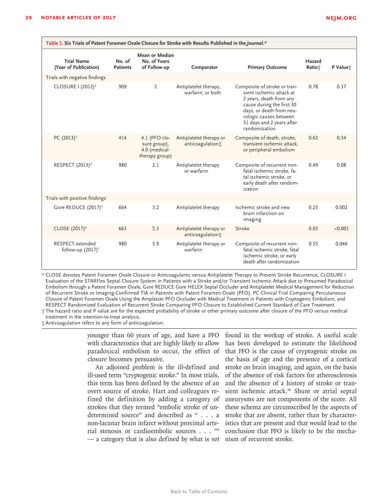
Back to Table of Contents
29 Notable Articles of 2017 nejm.org T h e n e w e ngl a nd j o u r na l o f m e dic i n e
n engl j med 377;11 nejm.org September 14, 20171094
younger than 60 years of age, and have a PFO with characteristics that are highly likely to allow paradoxical embolism to occur, the effect of closure becomes persuasive.
An adjoined problem is the ill-defined and ill-used term “cryptogenic stroke.” In most trials, this term has been defined by the absence of an overt source of stroke. Hart and colleagues re-fined the definition by adding a category of strokes that they termed “embolic stroke of un-determined source” and described as “ . . . a non-lacunar brain infarct without proximal arte-rial stenosis or cardioembolic sources . . . ”9 — a category that is also defined by what is not
found in the workup of stroke. A useful scale has been developed to estimate the likelihood that PFO is the cause of cryptogenic stroke on the basis of age and the presence of a cortical stroke on brain imaging, and again, on the basis of the absence of risk factors for atherosclerosis and the absence of a history of stroke or tran-sient ischemic attack.10 Shunt or atrial septal aneurysms are not components of the score. All these schema are circumscribed by the aspects of stroke that are absent, rather than by character-istics that are present and that would lead to the conclusion that PFO is likely to be the mecha-nism of recurrent stroke.
Trial Name (Year of Publication)
No. of Patients
Mean or Median No. of Years of Follow-up Comparator Primary Outcome
Hazard Ratio† P Value†
Trials with negative findings
CLOSURE I (2012)2 909 2 Antiplatelet therapy, war farin, or both
Composite of stroke or tran-sient ischemic attack at 2 years, death from any cause during the first 30 days, or death from neu-rologic causes between 31 days and 2 years after randomization
0.78 0.37
PC (2013)3 414 4.1 (PFO clo-sure group), 4.0 (medical-
therapy group)
Antiplatelet therapy or anticoagulation‡
Composite of death, stroke, transient ischemic attack, or peripheral embolism
0.63 0.34
RESPECT (2013)4 980 2.1 Antiplatelet therapy or warfarin
Composite of recurrent non-fatal ischemic stroke, fa-tal ischemic stroke, or early death after random-ization
0.49 0.08
Trials with positive findings
Gore REDUCE (2017)5 664 3.2 Antiplatelet therapy Ischemic stroke and new brain infarction on imaging
0.23 0.002
CLOSE (2017)6 663 5.3 Antiplatelet therapy or anticoagulation‡
Stroke 0.03 <0.001
RESPECT extended follow-up (2017)7
980 5.9 Antiplatelet therapy or warfarin
Composite of recurrent non-fatal ischemic stroke, fatal ischemic stroke, or early death after randomization
0.55 0.046
* CLOSE denotes Patent Foramen Ovale Closure or Anticoagulants versus Antiplatelet Therapy to Prevent Stroke Recurrence, CLOSURE IEvaluation of the STARFlex Septal Closure System in Patients with a Stroke and/or Transient Ischemic Attack due to Presumed ParadoxicalEmbolism through a Patent Foramen Ovale, Gore REDUCE Gore HELEX Septal Occluder and Antiplatelet Medical Management for Reductionof Recurrent Stroke or Imaging-Confirmed TIA in Patients with Patent Foramen Ovale (PFO), PC Clinical Trial Comparing PercutaneousClosure of Patent Foramen Ovale Using the Amplatzer PFO Occluder with Medical Treatment in Patients with Cryptogenic Embolism, andRESPECT Randomized Evaluation of Recurrent Stroke Comparing PFO Closure to Established Current Standard of Care Treatment.
† The hazard ratio and P value are for the expected probability of stroke or other primary outcome after closure of the PFO versus medical treatment in the intention-to-treat analysis.
‡ Anticoagulation refers to any form of anticoagulation.
Table 1. Six Trials of Patent Foramen Ovale Closure for Stroke with Results Published in the Journal.*
Back to Table of Contents
30 Notable Articles of 2017 nejm.org Editorial
n engl j med 377;11 nejm.org September 14, 2017 1095
The evidence for causation of embolic stroke in any given person is, of course, circumstantial (e.g., atrial fibrillation or carotid stenosis), and it seems reasonable that the presence of a PFO and a sizable interatrial shunt should similarly no longer result in the categorization of a stroke as cryptogenic. One conclusion from the six trials described above is that the potential benefit from closure is determined on the basis of the positive characteristics of the PFO rather than on the basis of exclusionary factors that make a stroke cryptogenic. Restricting PFO closure en-tirely to patients with high-risk characteristics of the PFO may perhaps be too conservative, but the boundaries of the features that support the pro-cedure are becoming clearer.
Disclosure forms provided by the author are available with the full text of this editorial at NEJM.org.
This editorial was updated on September 20, 2017, at NEJM.org.
1. Messé SR, Kent DM. Still no closure on the question of PFOclosure. N Engl J Med 2013; 368: 1152-3.2. Furlan AJ, Reisman M, Massaro J, et al. Closure or medical
therapy for cryptogenic stroke with patent foramen ovale. N Engl J Med 2012; 366: 991-9.3. Meier B, Kalesan B, Mattle HP, et al. Percutaneous closure ofpatent foramen ovale in cryptogenic embolism. N Engl J Med 2013; 368: 1083-91.4. Carroll JD, Saver JL, Thaler DE, et al. Closure of patent fora-men ovale versus medical therapy after cryptogenic stroke. N Engl J Med 2013; 368: 1092-100.5. Søndergaard L, Kasner SE, Rhodes JF, et al. Patent foramenovale closure or antiplatelet therapy for cryptogenic stroke. N Engl J Med 2017; 377: 1033-42.6. Mas J-L, Derumeaux G, Guillon B, et al. Patent foramenovale closure or anticoagulation vs. antiplatelets after stroke. N Engl J Med 2017; 377: 1011-21.7. Saver JL, Carroll JD, Thaler DE, et al. Long-term outcomes ofpatent foramen ovale closure or medical therapy after stroke. N Engl J Med 2017; 377: 1022-32.8. Johnston SC. Patent foramen ovale closure — closing thedoor except for trials. N Engl J Med 2012; 366: 1048-50.9. Hart RG, Diener HC, Coutts SB, et al. Embolic strokes ofundetermined source: the case for a new clinical construct. Lan-cet Neurol 2014; 13: 429-38.10. Kent DM, Ruthazer R, Weimar C, et al. An index to identifystroke-related vs incidental patent foramen ovale in cryptogenic stroke. Neurology 2013; 81: 619-25.
DOI: 10.1056/NEJMe1709637
Copyright © 2017 Massachusetts Medical Society.
apply for jobs at the nejm careercenter
Physicians registered at the NEJM CareerCenter can apply for jobs electronically. A personal account created when you register allows you to apply for positions,
using your own cover letter and CV, and keep track of your job-application history. Visit nejmjobs.org for more information.
Back to Table of Contents
31 Notable Articles of 2017 nejm.org
Perspective
The NEW ENGLAND JOURNAL of MEDICINE
May 18, 2017
n engl j med 376;20 nejm.org May 18, 2017 1901
This past June, I participated in an orientation session dur-
ing which new interns were asked to write self-addressed letters ex-pressing their hopes and anxiet-ies. The sealed envelopes were col-lected and then returned 6 months later, when I’m sure the interns felt encouraged to see how far they’d come.
This exercise, in which the in-tern serves as both letter writer and recipient, both novice and vet-eran, offers a new twist on an old tradition. In 1855, James Jackson published Letters to a Young Physi-cian Just Entering Upon Practice. More recent additions to this epistolary canon include Richard Selzer’s Letters to a Young Doctor, which ap-peared in 1982, and Treatment Kind and Fair: Letters to a Young Doctor, which Perri Klass published in 2007 on the occasion of her son’s entry into medical school.
When I started my internship 30 years ago, I wasn’t invited to share my hopes and anxieties in a letter — or anywhere else, for that matter. In fact, I recall no
orientation at all, other than lin-ing up to receive a stack of ill-fitting white uniforms, a tuber-culin skin test, and a hasty and not particularly reassuring re-view of CPR.
Perhaps the memory of my own abrupt initiation explains my re-sponse as I sat at the conference table watching the new interns hunched earnestly over their let-ters: I was filled with longing. I wanted so much to tell them, particularly the women — more than half the group, I was pleased to note — what I wished I’d known. Even more, I yearned to tell my younger self what I wished I’d known. As the in-terns wrote, I composed a letter of my own.
Dear Young Female Physician:I know you are excited and
also apprehensive. These feelings are not unwarranted. The hours you will work, the body of knowl-edge you must master, and the responsibility you will bear for people’s lives and well-being are daunting. I’d be worried if you
weren’t at least a little worried.As a woman, you face an ad-
ditional set of challenges, but you know that already. On your urol-ogy rotation in medical school, you were informed that your pres-ence was pointless since “no self-respecting man would go to a lady urologist.”
There will be more sexism, some infuriating, some merely an-noying. As a pregnant resident, I inquired about my hospital’s ma-ternity-leave policy for house of-ficers and was told that it was a great idea and I should draft one. Decades into practice, when I call in a prescription, some pharma-cists still ask for the name of the doctor I’m calling for.
And there will be more serious and damaging discrimination as well. It pains me to tell you that in 2017, as I’m nearing the end of my career, female physicians earn on average $20,000 less than our male counterparts (even allowing for factors such as numbers of publications and hours worked)1; are still underrepresented in lead-
Letter to a Young Female PhysicianSuzanne Koven, M.D.
Physicians, Firearms, and Free Speech
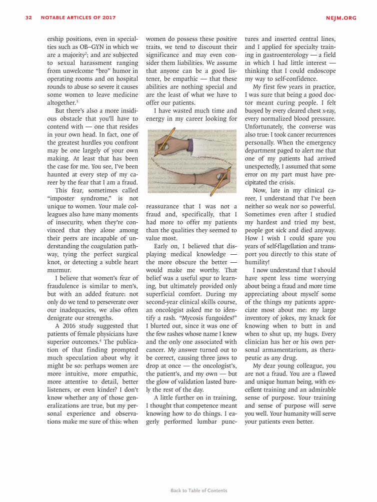
Back to Table of Contents
32 Notable Articles of 2017 nejm.org PERSPECTIVE
1902
Physicians, Firearms, and Free Speech
n engl j med 376;20 nejm.org May 18, 2017
ership positions, even in special-ties such as OB–GYN in which we are a majority2; and are subjected to sexual harassment ranging from unwelcome “bro” humor in operating rooms and on hospital rounds to abuse so severe it causes some women to leave medicine altogether.3
But there’s also a more insidi-ous obstacle that you’ll have to contend with — one that resides in your own head. In fact, one of the greatest hurdles you confront may be one largely of your own making. At least that has been the case for me. You see, I’ve been haunted at every step of my ca-reer by the fear that I am a fraud.
This fear, sometimes called “imposter syndrome,” is not unique to women. Your male col-leagues also have many moments of insecurity, when they’re con-vinced that they alone among their peers are incapable of un-derstanding the coagulation path-way, tying the perfect surgical knot, or detecting a subtle heart murmur.
I believe that women’s fear of fraudulence is similar to men’s, but with an added feature: not only do we tend to perseverate over our inadequacies, we also often denigrate our strengths.
A 2016 study suggested that patients of female physicians have superior outcomes.4 The publica-tion of that finding prompted much speculation about why it might be so: perhaps women are more intuitive, more empathic, more attentive to detail, better listeners, or even kinder? I don’t know whether any of those gen-eralizations are true, but my per-sonal experience and observa-tions make me sure of this: when
women do possess these positive traits, we tend to discount their significance and may even con-sider them liabilities. We assume that anyone can be a good lis-tener, be empathic — that these abilities are nothing special and are the least of what we have to offer our patients.
I have wasted much time and energy in my career looking for
reassurance that I was not a fraud and, specifically, that I had more to offer my patients than the qualities they seemed to value most.
Early on, I believed that dis-playing medical knowledge — the more obscure the better — would make me worthy. That belief was a useful spur to learn-ing, but ultimately provided only superficial comfort. During my second-year clinical skills course, an oncologist asked me to iden-tify a rash. “Mycosis fungoides!” I blurted out, since it was one of the few rashes whose name I knew and the only one associated with cancer. My answer turned out to be correct, causing three jaws to drop at once — the oncologist’s, the patient’s, and my own — but the glow of validation lasted bare-ly the rest of the day.
A little further on in training, I thought that competence meant knowing how to do things. I ea-gerly performed lumbar punc-
tures and inserted central lines, and I applied for specialty train-ing in gastroenterology — a field in which I had little interest — thinking that I could endoscope my way to self-confidence.
My first few years in practice, I was sure that being a good doc-tor meant curing people. I felt buoyed by every cleared chest x-ray, every normalized blood pressure. Unfortunately, the converse was also true: I took cancer recurrences personally. When the emergency department paged to alert me that one of my patients had arrived unexpectedly, I assumed that some error on my part must have pre-cipitated the crisis.
Now, late in my clinical ca-reer, I understand that I’ve been neither so weak nor so powerful. Sometimes even after I studied my hardest and tried my best, people got sick and died anyway. How I wish I could spare you years of self-flagellation and trans-port you directly to this state of humility!
I now understand that I should have spent less time worrying about being a fraud and more time appreciating about myself some of the things my patients appre-ciate most about me: my large inventory of jokes, my knack for knowing when to butt in and when to shut up, my hugs. Every clinician has her or his own per-sonal armamentarium, as thera-peutic as any drug.
My dear young colleague, you are not a fraud. You are a flawed and unique human being, with ex-cellent training and an admirable sense of purpose. Your training and sense of purpose will serve you well. Your humanity will serve your patients even better.
Back to Table of Contents
33 Notable Articles of 2017 nejm.org PERSPECTIVE
1903
Physicians, Firearms, and Free Speech
n engl j med 376;20 nejm.org May 18, 2017
Sincerely,Suzanne Koven, M.D.Harvard Medical SchoolMassachusetts General HospitalBoston, MA
Disclosure forms provided by the author are available at NEJM.org.
From Harvard Medical School and Massa-chusetts General Hospital, Boston.
1. Jena AB, Olenski AR, Blumenthal DM. Sex differences in physician salaries in U.S. public medical schools. JAMA Intern Med 2016; 176: 1294-304.2. Jena AB, Khullar D, Ho O, Olenski AR, Blumenthal DM. Sex differences in academ-ic rank in US medical schools in 2014. JAMA 2015; 314: 1149-58.3. Lowes R. Most female physicians report sexual harassment at job. Medscape (http://www .medscape .com/ viewarticle/ 866853).4. Tsugawa Y, Jena AB, Figueroa JF, Orav
EJ, Blumenthal DM, Jha AK. Comparison of hospital mortality and readmission rates for Medicare patients treated by male vs female physicians. JAMA Intern Med 2017; 177: 206-13.
DOI: 10.1056/NEJMp1702010Copyright © 2017 Massachusetts Medical Society.
Back to Table of Contents
34 Notable Articles of 2017 nejm.org
Perspective
The NEW ENGLAND JOURNAL of MEDICINE
October 5, 2017
n engl j med 377;14 nejm.org October 5, 2017 1309
I n 2010, 5-year-old Emily White-head was diagnosed with acute
lymphoblastic leukemia (ALL). Though her parents were told that if you had to have a kid with cancer, ALL was the best one to have, Emily’s course was hardly typical. After two rounds of chemotherapy, necrotizing fas-ciitis developed in both legs and she barely avoided amputation. Sixteen months later, she had a relapse. Bone marrow transplan-tation was recommended, but the Whiteheads, concerned about toxic effects, sought a second opinion at Children’s Hospital of Pennsylvania. There they learned about a new therapy, developed by University of Pennsylvania inves-tigators and known as CART-19, which involved genetically engi-neering the patient’s own T cells to kill tumor cells.
Unfortunately, a clinical trial had not yet been cleared by the Food and Drug Administration (FDA), and Emily’s leukemic cells were doubling daily. So Emily returned to her local hospital and received another round of intensive chemotherapy, which bought her 3 weeks but no re-mission. Out of options, one on-cologist recommended hospice. But “That just didn’t make sense to us,” says Tom Whitehead, Emily’s father. When the White-heads said they wanted to re-turn to Children’s Hospital, the oncologist told them that hos-pice was preferable to enter-ing Emily into an experimental study that wouldn’t help her get better.
But her parents opted to en-roll her in a study, and she be-came the first child to receive
CART-19. As a result, not only is she now a thriving 12-year-old, but her survival helped reener-gize a line of research that was nearing failure. In August 2017, the FDA approved the first chi-meric antigen receptor T-cell (CAR-T) therapy, Novartis’s tisa-genlecleucel, which uses the Penn-developed technology, for patients up to 25 years of age with re-lapsed or refractory ALL. Though the indication is narrow, the re-sults are striking in a patient population with otherwise limit-ed options: 83% of the 63 evalu-able children who received tisa-genlecleucel in Novartis’s phase 2 trial had complete elimination of malignant cells at 3 months.1
The approval is probably the first of many for CAR-T prod-ucts. Gilead recently announced its $11.9 billion acquisition of
Tragedy, Perseverance, and Chance — The Story of CAR-T TherapyLisa Rosenbaum, M.D.
Making Patients and Doctors Happier

Back to Table of Contents
35 Notable Articles of 2017 nejm.org PERSPECTIVE
1310
Making Patients and Doctors Happier
n engl j med 377;14 nejm.org October 5, 2017
Kite Pharma, whose CAR-T tech-nology, initially developed at the National Institutes of Health (NIH), has shown efficacy in patients with chemorefractory, aggressive B-cell non-Hodgkin’s lymphoma.2 And some 40 other companies, many in partnership with academic institutions, are racing to develop CAR-T tech-nologies for myriad indications. Though early data are most prom-ising for other hematologic can-cers, such as relapsed chronic lymphocytic leukemia (CLL),3 sim-
ilar therapies may eventually prove effective for solid tumors as well.
The emergence of CAR-T therapy, like most scientific ad-vances, ref lects the incremental insights of hundreds of scien-tists over decades — from sur-geon William Coley’s recogni-tion of the immune system’s potential for treating cancer when, in 1893, he injected strep-tococcus into inoperable osteo-sarcoma and observed the tumor shrink, to the making of the first CAR-T cells by the Israeli immunologist Zelig Eshhar in 1993.4 Indeed, the story of CAR-T therapy says as much about the methodical nature of scientific progress as it does about the passions that sustain it.
As Carl June, the immunolo-gist who led the development of Penn’s CAR-T technology, re-called, “So many times, I almost had to quit.” June spent his ear-ly career developing a technique to boost immune function in patients with HIV by modifying their T cells and inducing pro-liferation ex vivo. Though he and his colleague Bruce Levine would later build on this technique to engineer patients’ T cells to at-tack leukemia, June might have continued focusing solely on the
basic science. But in 1996, his 41-year-old wife was diagnosed with ovarian cancer. June tried unsuccessfully to get a pharma-ceutical company to provide the tools he needed to attempt im-munotherapy. When his wife died in 2001, June resolved to apply emerging immunologic insights to the development of cancer therapies, even though that meant creating a biotech infrastructure within academia.
The translational hurdles re-mained formidable. The field had been dogged by skepticism and setbacks, and the NIH wouldn’t fund a clinical trial. Once again, tragedy propelled the research forward. In 2001, Barbara and Edward Netter, having watched
their daughter-in-law die of breast cancer, started the Alliance for Cancer Gene Therapy (ACGT), hoping to develop alternative ap-proaches. In 2008, ACGT granted June and his coinvestigator David Porter $1 million, enough to treat their first three patients with re-lapsed CLL with CART-19. Two of the three patients achieved com-plete remission, but the investiga-tors ran out of funding. Knowing they couldn’t prove efficacy statis-tically, they published their find-ings as case reports.3,5 Soon, the National Cancer Institute offered June a grant, and Novartis li-censed Penn’s CAR-T technology. But June acknowledges the tenu-ous nature of anecdote: “Were we lucky? Were they representative? Would it be durable?”
Indeed, anecdote can easily break a field rather than make it: the death of Jesse Gelsinger in a trial at Penn had set the field of gene therapy back at least a decade. And as both June and Stephan Grupp, the Children’s Hospital oncologist and princi-pal investigator of the CART-19 trial in children, emphasized, had Emily died, the CAR-T field would probably have died with her. But though unexpected tox-ic effects in phase 1 studies can fell any new therapy, the unfor-tunate reality is that it often takes time, and human lives, to distinguish fatal toxic effects from those that can be managed. As Grupp explained, “There was no way to predict a great deal of what we learned. The toxicity is-sues can only be learned from human beings.”
Emily Whitehead was a case in point. After receiving her third dose of CART-19, she developed high fevers, respiratory failure, and shock necessitating the use of three pressors. Though Emily
The emergence of CAR-T therapy, like most scientific advances, reflects the incremental
insights of hundreds of scientists over decades. Indeed, the story of CAR-T therapy says as much about the methodical nature of scientific progress as it does about
the passions that sustain it.

Back to Table of Contents
36 Notable Articles of 2017 nejm.org PERSPECTIVE
1311
Tragedy, Perseverance, and Chance
n engl j med 377;14 nejm.org October 5, 2017
was experiencing what’s now un-derstood to be cytokine-release syndrome, which occurred in 78% of patients in Novartis’s phase 2 trial, it wasn’t clear at the time what was driving this response, much less how to treat it.
That she survived gives new meaning to the adage “Chance favors the prepared mind.” Per protocol, participants’ blood was sent for cytokine analysis, with about a 2-week turnaround time. But as Emily rapidly deteriorated, Grupp called the lab and begged them to run Emily’s blood more quickly. Two hours later, in time for his 3 p.m. lab meeting, Grupp learned that Emily’s level of in-terleukin-6 was elevated 1000-fold. He recalls the meeting, as everyone pored over the results. “No one thought we should be thinking about this thing, IL6,” Grupp explained. “It isn’t even made by T cells.” That fact, how-ever, made interleukin-6 accept-able for Emily’s doctors to tar-get, since any interference with T-cell function could interfere with the antileukemic activity, without which she would die. But how to quash interleukin-6? As Grupp and his lab members started Googling, June, giving a talk in Seattle, received the re-sults and had an idea. His daugh-ter, who has juvenile rheumatoid arthritis, had recently started tak-ing tocilizumab, a monoclonal antibody that targets interleu-kin-6. As the investigators con-verged on a similar conclusion, one hurdle remained: how to get the drug in time for Emily?
Once again, they got lucky. Tocilizumab was on the hospi-tal’s formulary for rheumatolog-ic indications, which meant that rather than having to wait for up to 2 days, by 8 p.m. that evening, Emily received a dose. Within
hours, she began to improve, so dramatically that her doctors could barely wean the pressors fast enough. On her seventh birthday, Emily woke up. Eight days later, on the basis of a bone marrow biopsy, Grupp reported that the treatment had worked.
Though the remissions achieved with CAR-T therapy are impres-sive, much remains unknown. CAR-T products vary in ways that will have implications for both efficacy and toxicity. Some variation arises from the chime-ric antigen receptors (CARs) them-selves, which are programmed to recognize various antigens and contain various types and num-bers of costimulatory domains to induce proliferation. In the case of CART-19, for instance, the engineered T cells bind to lym-phocytes displaying the CD19 antigen, a hallmark of leukemic B cells and thus an attractive target because humans can tol-erate B-cell aplasia. Identifying antigen targets in solid tumors while preventing destruction of healthy tissue remains challeng-ing, however, especially since the tumor microenvironment can be immunologically hostile to intro-duction of a CAR. Moreover, tox-icities remain formidable. Though tocilizumab is now often used to manage the cytokine-release syndrome, other toxic effects, such as cerebral edema, remain poorly understood and difficult to manage.
Meanwhile, the CAR-T dis-cussion has become dominated by cost concerns. Critics argue that tisagenlecleucel’s $475,000 price tag is unaffordable and unjustifiable given the taxpayer-supported basic research under-pinning its development, while manufacturers point to the tre-mendous investment required to
produce the drug and fund tri-als. With many patients unable to afford their medications and ongoing instances of unconscio-nable drug-company profiteering, these discussions are both es-sential and complex. Regardless of the finances, we all hope that these remissions are prolonged or, even better, turn out to be cures. There is no way to know whether they will without pro-longed observation, but while we carefully observe each patient, it is important to remember that therapeutic advances are motivat-ed by more than money — that it’s the hope, vision, and perse-verance of both patients and in-vestigators that have made this critical conversation possible.
Disclosure forms provided by the author are available at NEJM.org.
Dr. Rosenbaum is a national correspondent for the Journal.
This article was published on September 13, 2017, at NEJM.org.
1. Buechner J, Grupp SA, Maude SL, et al. Global registration trial of efficacy and safety of CTL019 in pediatric and young adult patients with relapsed/refractory (R/R) acute lymphoblastic leukemia (ALL): update to the interim analysis. Clin Lymphoma Myeloma Leuk 2017; Suppl 2: S263-S264. ab-stract.2. Kochenderfer JN, Dudley ME, Kassim SH, et al. Chemotherapy-refractory diffuse large B-cell lymphoma and indolent B-cell malignancies can be effectively treated with autologous T cells expressing an anti-CD19 chimeric antigen receptor. J Clin On-col 2015; 33: 540-9.3. Porter DL, Levine BL, Kalos M, Bagg A, June CH. Chimeric antigen receptor–modi-fied T cells in chronic lymphoid leukemia. N Engl J Med 2011; 365: 725-33.4. Yang Y. Cancer immunotherapy: har-nessing the immune system to battle can-cer. J Clin Invest 2015; 125: 3335-7.5. Kalos M, Levine BL, Porter DL, et al. T cells with chimeric antigen receptors have potent antitumor effects and can es-tablish memory in patients with advanced leukemia. Sci Transl Med 2011; 3: 95ra73.
DOI: 10.1056/NEJMp1711886
Copyright © 2017 Massachusetts Medical Society.Tragedy, Perseverance, and Chance
Back to Table of Contents
37 Notable Articles of 2017 nejm.org
Perspective
The NEW ENGLAND JOURNAL of MEDICINE
August 24, 2017
n engl j med 377;8 nejm.org August 24, 2017 705
On our infectious diseases (ID) consult ser-vice, we recently cared for Mr. C., a young man with Staphylococcus aureus tricuspid valve
endocarditis, septic arthritis, and empyema that
were consequences of his opioid use disorder (OUD). Several years earlier, he had started taking oxy-codone at parties, and eventually, when the cost of pills became prohibitive, he’d progressed to injecting heroin. His days were consumed by the logistics of ob-taining heroin to stave off the exhausting cycle of opioid with-drawal. Despite his deep desire to stop using, he was initially am-bivalent when we offered to start treatment with buprenorphine, which is commonly coformulated with naloxone as Suboxone (Reck-itt Benckiser). “Doc,” he said, “you gotta understand that as an ad-dict, the scariest thing right now is the idea of putting another opioid in my body, even if it’s go-ing to help me.”
Although Mr. C. had done well on buprenorphine in the past, accumulating several months of recovery, he felt overwhelmed by the prospect of starting the pro-cess again. In the days after his clinical status stabilized and the ID service defined his antibiotic course, we kept visiting Mr. C. on the ward. We confronted the dual imperatives to treat his infection and his OUD to reduce his near-term chance of dying from an overdose or relapsed infection. During our visits, we discussed his resolving empyema, but also his cravings, withdrawal symp-toms, and readiness to start bu-prenorphine treatment. On the day before his discharge, as he faced impending relapse, Mr. C. decided he was ready. That afternoon, we
completed an observed buprenor-phine induction and made an ap-pointment to see him the follow-ing week in the ID clinic for ongoing buprenorphine and anti-biotic treatment.
As the opioid use and over-dose epidemic ravages the United States, bearing witness to the physical and psychosocial conse-quences of addiction has become part of many physicians’ daily work. Despite our position on the epidemic’s front lines, the remark-able reality is that we remain systematically undertrained and underengaged in addiction-treat-ment efforts.1 Though we have taken steps toward recognizing our profession’s complicity in the epidemic’s roots, most physicians feel paralyzed when it comes to effecting change for individual patients.
The history of medicine is, in part, the history of physicians stretching the scope of their prac-tice to answer the pressing needs
Stretching the Scope — Becoming Frontline Addiction-Medicine ProvidersAlison B. Rapoport, M.D., and Christopher F. Rowley, M.D.
Stretching the Scope

Back to Table of Contents
38 Notable Articles of 2017 nejm.org PERSPECTIVE
706
Stretching the Scope
n engl j med 377;8 nejm.org August 24, 2017
of their times. In the face of OUD, a treatable illness with a striking capacity to rapidly and definitive-ly alter the lives of our patients, their families, and the communi-ties we serve, we have been late and ineffective in our response. In recent years, the number of hospitalizations for the medical consequences of OUD has esca-lated, and in 2015 alone, more than 33,000 people died in the United States from opioid-related overdose.2 Yet rates of active physician engagement in addiction treatment remain embarrassing-ly low.
At some point, it became cul-turally acceptable to treat all conditions in a patient except ad-diction. It’s a diagnosis still fre-quently and falsely regarded as untreatable — a convenient as-sumption driven by the stigma against people with this disease. ID specialists have historically been ardent advocates for social justice and public health, cham-pioning patients on the margins of society who have stigmatizing illnesses. In the age of the opioid epidemic, treatment of life-threat-ening infections arising from injection drug use accounts for an increasing proportion of our practice. Far too often, however, infections that we treat resolve while underlying substance use disorders are left to fester.
Under the federal Drug Addic-tion Treatment Act of 2000, phy-sicians who register with the Drug Enforcement Administra-tion, regardless of their subspe-cialty, can receive a waiver to pre-scribe buprenorphine for OUD treatment after undergoing 8 hours of training. According to the Sub-stance Abuse and Mental Health Services Administration, the fed-eral body that oversees the bu-prenorphine waiver program, there
are currently 37,448 physicians with such waivers,3 representing only approximately 4% of all pro-fessionally active U.S. physicians.4 Nationally, the distribution of phy-sicians with waivers is grossly uneven, and many suffering com-munities are left with little to no capacity for buprenorphine treat-ment. Obtaining a waiver is one concrete action that all physi-cians can take to help stem the tide of this epidemic. Physicians practicing in clinical contexts in which long-term prescribing is not possible can prescribe a short course of buprenorphine therapy as a bridge to long-term treat-ment managed by one of a grow-ing number of primary care phy-sicians and psychiatrists.
As a small group of ID fellows and faculty practicing at Beth Israel Deaconess Medical Center, a large tertiary care hospital in Boston, we have pursued this strategy. We offer buprenorphine in conjunction with antibiotics to patients who are hospitalized with infectious complications of injec-tion drug use. We ask patients about injection practices, counsel them about harm reduction, and prescribe intranasal naloxone for overdose reversal, recognizing that OUD is marked by both recovery and relapse. We partner with col-leagues in social work to build viable treatment plans to facili-tate recovery and eventually trans-fer addiction care to long-term programs. As we have waited for institutional capacity to increase, we have also started to offer in-patient buprenorphine induction for patients without concurrent infection.
We anticipated some resistance on both the institutional and the provider levels, but in practice, we have largely encountered ap-preciation, and our work has
served as one impetus for a larger hospital initiative to address the opioid crisis. This pilot program was born in our ID division, but we believe it is replicable by any physician group — for example, surgical teams discharging pa-tients admitted with OUD-related complications or psychiatry teams discharging patients with both substance use disorder and men-tal illness. For all physicians, it is vital to recognize that medication treatment for OUD is a corner-stone of recovery for most patients, and when it’s omitted, high rates of relapse are consistently ob-served.
We are wading into the turbu-lent waters of our patients’ lives to see them through to a time when they are clear of their in-fection and on the continuum of recovery. Though our efforts are still relatively new, we have been changed by the experience. Some of our patients have had relapses or haven’t returned for care. But we’ve also seen remarkable suc-cesses — patients who presented in the depths of addiction and illness who have subsequently re-connected with their families, have started to work again, and now use opioids less or not at all. By providing the bridge to long-term addiction treatment, we have ob-served patients remain in care at higher rates and start to mend their badly damaged sense of trust in a medical system that has long treated them with judg-ment and neglect.
We are providing this care out-side the realm of traditional ID consultation because the crisis demands it. Today in the United States, another 91 people will die from an opioid overdose.5 Under the watchful eyes of physicians, many people survive their acute illnesses only to die in public rest-
Back to Table of Contents
39 Notable Articles of 2017 nejm.org PERSPECTIVE
707
Stretching the Scope
n engl j med 377;8 nejm.org August 24, 2017
rooms, in private homes, or on the street. There are many inspiring examples of physicians and health care communities that have simi-larly stretched the scope of their practice, and lives have been saved as a result. We believe it’s time for more of us to join the movement.
Two months after being dis-charged, Mr. C. continues to re-ceive buprenorphine treatment. He gets his prescriptions through a program close to his home, where
he attends weekly group meetings and individual counsel-ing sessions. He
wholly understands the gravity of his infection; his heart valve has been left severely damaged, and
he still feels weak. But he has re-connected with friends and fam-ily and is making plans to return to work. He is in early recovery from his OUD and from the cha-os, social isolation, and depres-sion that come with it. As we see it, the medical community is also in early recovery — moving past implicit biases, stigma, and fear to connect with our patients and respond to a defining crisis of our time.
Disclosure forms provided by the authors are available at NEJM.org.
From the Division of Infectious Diseases, Beth Israel Deaconess Medical Center, and Harvard Medical School — both in Boston.
1. Office of the Surgeon General. Facing addiction in America: the Surgeon General’s
report on alcohol, drugs, and health. Wash-ington, DC: Department of Health and Hu-man Services, November 2016.2. Rudd RA, Seth P, David F, Scholl L. In-creases in drug and opioid-involved overdose deaths — United States, 2010–2015. MMWR Morb Mortal Wkly Rep 2016; 65: 1445-52.3. Physician and program data: Substance Abuse and Mental Health Services Adminis-tration (https:/ / www .samhsa .gov/ programs -campaigns/ medication-assisted-treatment/ physician-program-data).4. Henry J. Kaiser Family Foundation. State health facts: total professionally active physi-cians (http://www .kff .org/ other/ state-indicator/ total-active-physicians/ ?currentTimeframe =0&sortModel=%7B%22colId%22:%22 Location%22,%22sort%22:%22asc%22%7D).5. Opioid overdose: understanding the epi-demic. Atlanta: Centers for Disease Control and Prevention, 2016 (https:/ / www .cdc .gov/ drugoverdose/ epidemic).
DOI: 10.1056/NEJMp1706492Copyright © 2017 Massachusetts Medical Society.Stretching the Scope
Threats to Information Security
An audio interview with Dr. Rapoport
is available at NEJM.org

Recent achievements in medicine have resulted in progress beyond what many could have imagined just decades ago. New science and technology have empowered physicians to make better, faster treatment decisions. Our understanding of the human genome and targeted drug research is producing major improvements in treatments for cancer, heart disease, and many other chronic illnesses.
Connecting medicine’s thought leaders and practitioners to advances in science and practice is the unifying goal of the New England Journal of Medicine, NEJM Journal Watch, NEJM Knowledge+, and NEJM Catalyst, the premier products of NEJM Group.
NEJM Group brings together the people and expertise behind these products to drive innovation and ensure rigorous quality in our ongoing mission to further advance knowledge, learning, and practice.
NEJM Group. Working together to advance research and improve care. NEJMGroup.org
Inspiring Insights in Clinical Care