Normal Endometrium and Infertility Evaluation
27
biopsy or curettage is part of a comprehensive workup of the patient in the operating room that includes laparoscopy, hysteroscopy, or hysterosalpingography to assess the presence or absence of uterine or tubal lesions that con- tribute to infertility. In these cases, the endome- trial sampling may not be timed as precisely for the mid- to late luteal phase. Nonetheless, his- tologic evaluation provides the gynecologist with information regarding the response of the endometrium to hormonal stimulation, includ- ing indirect evidence of ovulatory function. The secretory phase is constant in the normal cycle, lasting 14 days from the time of ovula- tion to the onset of menstruation. 1 Variations in cycle length occur because the proliferative phase of the cycle varies, both between cycles and between women. Accordingly, the gynecol- ogist correlates the cycle date by histology with the woman’s cycle date based on the time of onset of the upcoming menstrual period, not the last menstrual period. Ovulation with se- cretory endometrial changes ceases in most women by age 53, although rarely ovulation with secretory endometrium and a confirmed corpus luteum of the ovary has been seen up to a least age 56 ( personal observation). The biopsy findings help confirm that ovula- tion occurred, and indicate whether there was sufficient secretory effect, mediated by proges- terone, during the luteal phase. To utilize fully the morphologic interpretation, the gynecolo- gist compares the histologic date to the clinical data, including the date of the rise in the basal body temperature, the time of the serum 7 2 Normal Endometrium and Infertility Evaluation The histologic features of what constitutes “normal” endometrium change with a woman’s age, through the premenarchal, reproductive, perimenopausal, and postmenopausal years. 1–3 During the reproductive years, the cyclical hor- monal changes of the menstrual cycle provide a continuously changing morphologic pheno- type that is “normal.” In biopsy specimens, the combination of these cyclical changes along with artifacts and limited sampling can make normal patterns difficult to interpret. During the reproductive years, deviations from normal, either in histologic pattern or in temporal rela- tionship to ovulation, often indicate underlying abnormalities that may cause female infertility. The endometrial biopsy is an important part of the evaluation of the woman with infertil- ity. 4;5 Biopsies for the evaluation of infertility often are performed in the office using a small curette or a Pipelle aspirator and therefore the specimens tend to be small. 6 Occasionally, General Considerations in Histologic Evaluation . . . . . . . . . . . . . . . . . . . . . . . . . 8 Histologic Dating of the Normal, Cycling Endometrium . . . . . . . . . . . . . . . . 10 Proliferative Phase Endometrium . . . . . 11 Secretory Phase Endometrium . . . . . . . 12 Menstrual Endometrium . . . . . . . . . . . . 19 Pitfalls in Dating . . . . . . . . . . . . . . . . . . . . 21 Artifacts and Contaminants . . . . . . . . . . . 23 Luteal Phase Defect and Abnormal Secretory Phase Patterns . . . . . . . . . . . . . . 26 Clinical Queries and Reporting . . . . . . . . 29
Transcript of Normal Endometrium and Infertility Evaluation

biopsy or curettage is part of a comprehensiveworkup of the patient in the operating roomthat includes laparoscopy, hysteroscopy, or hysterosalpingography to assess the presenceor absence of uterine or tubal lesions that con-tribute to infertility. In these cases, the endome-trial sampling may not be timed as precisely forthe mid- to late luteal phase. Nonetheless, his-tologic evaluation provides the gynecologistwith information regarding the response of theendometrium to hormonal stimulation, includ-ing indirect evidence of ovulatory function.
The secretory phase is constant in the normalcycle, lasting 14 days from the time of ovula-tion to the onset of menstruation.1 Variations incycle length occur because the proliferativephase of the cycle varies, both between cyclesand between women. Accordingly, the gynecol-ogist correlates the cycle date by histology withthe woman’s cycle date based on the time ofonset of the upcoming menstrual period, notthe last menstrual period. Ovulation with se-cretory endometrial changes ceases in mostwomen by age 53, although rarely ovulationwith secretory endometrium and a confirmedcorpus luteum of the ovary has been seen up toa least age 56 (personal observation).
The biopsy findings help confirm that ovula-tion occurred, and indicate whether there wassufficient secretory effect, mediated by proges-terone, during the luteal phase. To utilize fullythe morphologic interpretation, the gynecolo-gist compares the histologic date to the clinicaldata, including the date of the rise in the basalbody temperature, the time of the serum
7
2Normal Endometrium and Infertility Evaluation
The histologic features of what constitutes“normal” endometrium change with a woman’sage, through the premenarchal, reproductive,perimenopausal, and postmenopausal years.1–3
During the reproductive years, the cyclical hor-monal changes of the menstrual cycle providea continuously changing morphologic pheno-type that is “normal.” In biopsy specimens, thecombination of these cyclical changes alongwith artifacts and limited sampling can makenormal patterns difficult to interpret. Duringthe reproductive years, deviations from normal,either in histologic pattern or in temporal rela-tionship to ovulation, often indicate underlyingabnormalities that may cause female infertility.
The endometrial biopsy is an important partof the evaluation of the woman with infertil-ity.4;5 Biopsies for the evaluation of infertilityoften are performed in the office using a smallcurette or a Pipelle aspirator and therefore the specimens tend to be small.6 Occasionally,
General Considerations in HistologicEvaluation . . . . . . . . . . . . . . . . . . . . . . . . . 8Histologic Dating of the Normal,Cycling Endometrium . . . . . . . . . . . . . . . . 10
Proliferative Phase Endometrium . . . . . 11Secretory Phase Endometrium . . . . . . . 12Menstrual Endometrium . . . . . . . . . . . . 19
Pitfalls in Dating . . . . . . . . . . . . . . . . . . . . 21Artifacts and Contaminants . . . . . . . . . . . 23Luteal Phase Defect and AbnormalSecretory Phase Patterns . . . . . . . . . . . . . . 26Clinical Queries and Reporting . . . . . . . . 29
luteinizing hormone (LH) surge, transvaginalultrasound evaluation of follicular or corpusluteum development, serum progesteronelevel, or subtraction of 14 days from the onsetof menses.4;7–9 Consequently, the biopsy typi-cally is timed to coincide with the luteal (secre-tory) phase of the cycle. In addition to definingthe precise histologic date, an endometrialbiopsy is part of the infertility workup toexclude other organic uterine abnormalities.
This chapter reviews the morphologic varia-tions caused by ovarian hormonal stimulationthat provide a background for the interpreta-tion of endometrial biopsies in infertilitypatients. These patterns include changes result-ing from normal hormonal fluctuations duringthe menstrual cycle and variations in normaldevelopment that are caused by abnormalitiesin the endogenous ovarian hormonal levelsduring the reproductive years. The latter repre-sent the so-called dysfunctional abnormali-ties that are, for the most part, due to abnor-malities in ovarian follicular development or inhormone production by the corpus luteum.Ovarian dysfunction also can result in abnormalbleeding, and Chapter 5 reviews dysfunctionaluterine bleeding caused by ovulatory abnor-malities. During gestation the endometriumundergoes other “normal,” that is, physiologic,alterations as discussed in Chapter 3.
General Considerations inHistologic Evaluation
Histologic evaluation begins with identificationof surface epithelium, a prerequisite for orient-ing the underlying glands and stroma. The sur-face epithelium is less responsive to sex steroidhormones than the underlying glands, but itoften shows alteration in pathologic conditions,especially when the abnormalities are subtle orfocal. For example, during the proliferativephase, estrogenic stimulation induces develop-ment of ciliated cells along the surface.10 In con-trast, ciliated surface epithelial cells are farmore frequent in pathologic conditions, par-ticularly those associated with unopposedestrogen stimulation, such as hyperplasia andmetaplasia.2;3;11–13
The subsurface endometrium is divided intotwo regions, the functionalis (stratum spongio-sum) and the basalis (stratum basale) (Fig. 2.1).The functionalis, situated between the surfaceepithelium and the basalis, is important to evaluate because it shows the greatest degreeof hormonal responsiveness. The size and distribution of glands as well as the cytologicfeatures of the glandular epithelial cells are important features in the histologic evalua-tion. Under normal conditions, the glandsshould be regularly spaced and have a per-pendicular arrangement from the basalis to the surface epithelium. In the secretory phase,the endometrium also shows a stratum com-pactum, a thin region beneath the surfaceepithelium. In the stratum compactum thestroma is dense and the glands are straight and narrow, even when the glands in the functionalis are tortuous. The basalis adjoinsthe myometrium, serving to regenerate thefunctionalis and surface epithelium followingshedding during menses. The endometrium ofthe basalis is less responsive to steroid hor-mones, and typically shows irregularly shaped,inactive appearing glands, dense stroma, andaggregates of spiral arteries. The spiral arteriesof the basalis (basal arteries) have thicker mus-cular walls than those in the functionalis. Inbiopsies, tissue fragments that contain basalisoften do not have surface epithelium. Theglands and stroma of the basalis cannot bedated, as they are unresponsive to steroid hormones. A specimen consisting solely ofendometrium from the basalis is thereforeinadequate for dating.
Tissue from the lower uterine segment oristhmus is another region of the endometriumthat is less responsive to steroid hormones. Inthe lower uterine segment the endometriumhas shorter, poorly developed, inactive glandsdispersed in a distinctive stroma (Fig. 2.2). Thecolumnar cells lining the glands resemble thoseof the corpus. Some glands near the junctionwith the endocervix show a transition to muci-nous endocervical-type epithelium.The stromalcells in the lower uterine segment are elongateand resemble fibroblasts with more abundanteosinophilic cytoplasm, in contrast to the ovalto rounded stromal cells with minimum cyto-plasm seen in the corpus.
8 2. Normal Endometrium and Infertility Evaluation

The tangential orientation of the functionalisin biopsies and the tortuosity of the glands, par-ticularly in the late proliferative phase, oftenlead to irregular cross sections of the tissue. In
this instance, gland development can be diffi-cult to assess. Furthermore, not all fragments oftissue in a biopsy or curettage include surfaceepithelium, which helps to orient the glands.
General Considerations in Histologic Evaluation 9
Figure 2.1. Normal secretory phase endometrium.Surface epithelium orients the tissue.The midportionof the tissue consists of functionalis where glands,stroma, and blood vessels demonstrate the typicalpatterns of maturation through the menstrual cycle.
The basalis in the lower portion of the illustrationconsists of irregular, closely spaced glands, densestroma, and aggregates of arteries. The stratum com-pactum is composed of the surface epithelium and asubjacent thin layer of dense stroma.

Nonetheless, at least focally, portions of better-oriented glands usually can be traced throughthe functionalis to the surface epithelium, andthese foci are critical for assessing appropriateglandular and stromal development.
Histologic Dating of the Normal,Cycling Endometrium
In the ovulatory patient, normal endometriumhas two phases.The first is the proliferative (fol-licular or preovulatory) phase characterized bygrowth of glands, stroma, and vessels that isinfluenced by estradiol produced mainly bygranulosa cells in the ovarian follicles. Fol-lowing ovulation, the secretory (luteal or postovulatory) phase reflects the effect of the combined production of progesterone andestradiol by luteinized granulosa and theca cells
of the corpus luteum.4 The regular sequence ofmorphologic changes determined by the fluctu-ating levels of ovarian steroid hormones formsthe basis for histologic dating.
Dating uses an arbitrarily defined “normal”cycle of 28 days, with day 1 the first day of men-strual bleeding.1 Histologic dating is mostprecise in the postovulatory secretory phase,as the follicular phase can be highly variable in length. Furthermore, proliferative phasechanges are not as discrete as those in the secre-tory phase. The date of the secretory phase isexpressed either as the specific day of the 28-day menstrual cycle, assuming ovulation occurson day 14, or is stated as the postovulatory day(e.g., secretory day 21 or postovulatory [P.O.]day 7). Local custom often determines the pre-ferred method of stating the histologic date.
There are nine histologic features of theglands and stroma that determine the phase of
10 2. Normal Endometrium and Infertility Evaluation
Figure 2.2. Lower uterine segment. Small, poorly developed glands are seen in nonreactive stroma that iscomposed of widely spaced spindle cells. Tissue from the lower uterine segment cannot be dated.
the cycle and the histologic date (Table 2.1).1
Five of these features affect glands: (1) tortu-osity, (2) gland mitoses, (3) orientation of nuclei(pseudostratified versus basal), (4) basal sub-nuclear cytoplasmic vacuoles, and (5) luminalsecretions with secretory exhaustion. Four features relate to the stromal: (6) edema, (7)mitoses, (8) predecidual change, and (9) infil-tration of granular lymphocytes. Practically, themost important glandular features are orienta-tion of nuclei, subnuclear cytoplasmic vacuoles,and luminal secretions with secretory exhaus-tion (3, 4, and 5), and the most importantstromal features are edema, predecidualchange, and granular lymphocytic infiltration(6, 8, and 9). These salient features are usuallyreadily apparent when present, allowing thepathologist to assign a histologic date.
Proliferative Phase Endometrium
During the proliferative phase, the endo-metrium grows from about 0.5mm up to 4.0 to5.0mm in thickness, so by the late proliferativephase, a biopsy obtains a moderate amount oftissue. Proliferative endometrium has threestages: early, mid, and late (Table 2.2).2 Thesedivisions are seldom used in dating biopsies,however. Usually the diagnosis of proliferativephase alone is sufficient, indicating that theendometrium is growing, shows a normal glan-dular distribution, and evidence of ovulation isnot present.
Growth of endometrium is the main charac-teristic of the proliferative phase (Figs. 2.3 and
2.4). Glands and stroma show brisk mitoticactivity. In early proliferative phase endo-metrium, the functionalis contains small,tubular glands. The glands progressively elon-gate and become tortuous from the mid- to the late proliferative phase because the glandgrowth is disproportionate to the stromalgrowth. Despite the tortuosity, the glands main-tain a relatively regular spacing between eachother. Throughout the proliferative phase, theepithelium lining the glands has pseudostrati-fied, oval nuclei with small nucleoli and densebasophilic cytoplasm. The pseudostratifiednuclei remain oriented to the basement mem-brane, but some nuclei are raised above thebasement membrane, giving a two-dimensionallayering of the nuclei. The pseudostratificationof the nuclei and the presence of mitotic activ-ity in the glands and stroma are two constantfeatures of the proliferative phase.
In the proliferative phase, the stromal cellsare widely separated in the functionalis (Fig.2.4).They are small and oval, with dense nuclei,scant wisps of cytoplasm, and ill-defined cellborders. Some stromal edema is normal at mid-proliferative phase. A few lymphocytes also arescattered throughout the stroma, being mostprominent around the vessels. Small spiralarteries and thin-walled venules are present.
The orientation and outline of proliferativephase glands and their relationship to intactstroma are important features for recognizing
Histologic Dating of the Normal, Cycling Endometrium 11
Table 2.1. Morphologic features used in endome-trial dating.
Glandular changes1. Tortuosity2. Mitoses3. Orientation of nuclei (pseudostratified or basal)a
4. Subnuclear cytoplasmic vacuolesa
5. Secretory exhaustion (luminal secretions)a
Stromal changes1. Edemaa
2. Mitoses3. Predeciduaa
4. Granular lymphocyte infiltratea
a Salient features used in dating the secretory phase.
Table 2.2. Proliferative phase changes.a
Early (4–7 days)Thin regenerating epitheliumShort narrow glands with epithelial mitosesStroma compact with mitoses (cells stellate or spindle
shaped)
Mid (8–10 days)Long, curving glandsColumnar surface epitheliumStroma variably edematous, mitoses frequent
Late (11–14 days)Tortuous glandsPseudostratified nucleiModerately dense, actively growing stroma
a These changes are subtle. They are rarely used for actualdating.
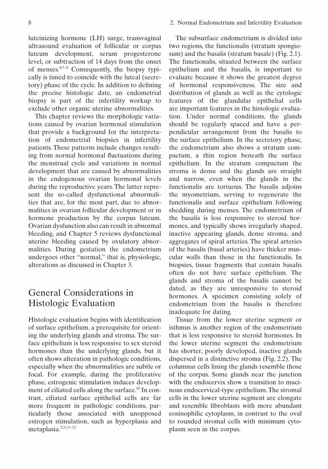
Figure 2.3. Proliferative endometrium. Glands aretubular and regularly spaced in abundant stroma.The stroma contains small vessels with thin walls.Both the glandular and the stromal cells show
this normal pattern, as hyperplastic glands orglands in a polyp can have cytologic featuresidentical to those of glands in the proliferativephase. The regular spacing and uniform shapeof the glands are characteristics of normal proliferative endometrium. Assessing gland orientation can be complicated, however, bybiopsy-induced fragmentation, an especiallycommon artifact in early to mid-proliferativephase biopsies when the mucosa is still thin.Detached and disrupted glands may appearabnormally crowded or irregular. To separatefragmentation artifact from true abnormalities,it is important to assess the integrity of thestroma as well as the glands and to use surfaceepithelium to help orient the tissue fragments.Detached and poorly oriented glands that showpseudostratified nuclei and mitotic activityusually represent proliferative endometriumunless better-oriented tissue suggests another
diagnosis. Also, proliferative phase glands fre-quently show the telescoping artifact (seelater).
Secretory Phase Endometrium
In the secretory phase, the glands and stromadevelop in an orderly sequence and display spe-cific histologic features of secretory activityfrom histologic day 16 through day 28. Theendometrium attains a thickness of up to 7.0 to8.0mm. Unlike in the proliferative phase, thechanges in the glands and stroma are relativelydiscrete, varying sharply from one day to thenext, thus permitting accurate dating. Dating of the first half of the secretory phase is basedprimarily on glandular changes whereas datingof the second half is based mainly on stromalalterations (Table 2.3).
12 2. Normal Endometrium and Infertility Evaluation
mitotic activity. Focal hemorrhage beneath thesurface epithelium is a result of the biopsy and doesnot represent a pathologic change.
Histologic Dating of the Normal, Cycling Endometrium 13
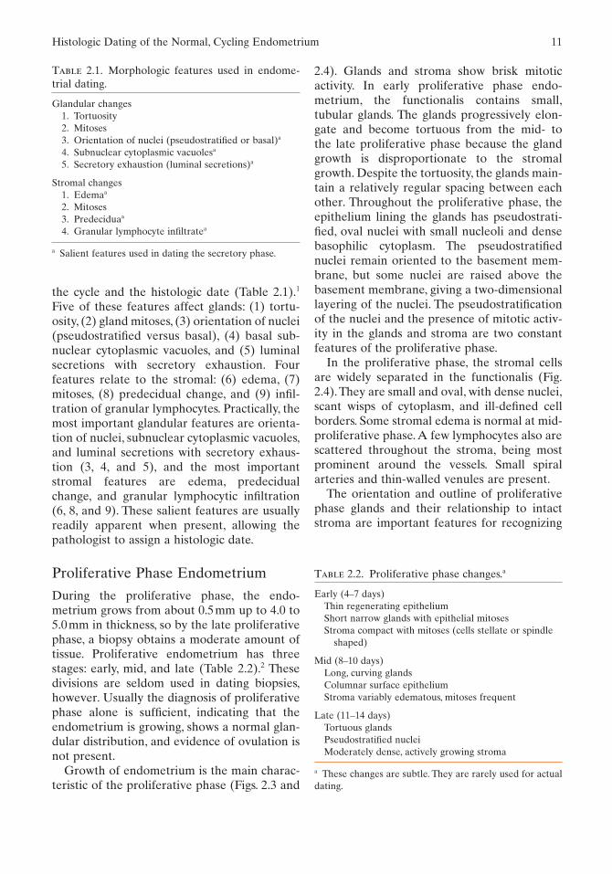
Figure 2.4. Proliferative endometrium. The prolif-erative phase gland shows pseudostratified nucleiwith mitotic activity. The stromal cells have oval
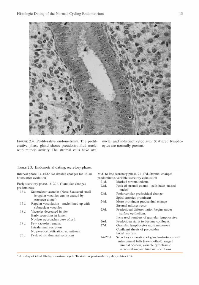
Table 2.3. Endometrial dating, secretory phase.
Interval phase, 14–15d.a No datable changes for 36–48hours after ovulation
Early secretory phase, 16–20d. Glandular changespredominate
16d. Subnuclear vacuoles (Note: Scattered smallirregular vacuoles can be caused byestrogen alone.)
17d. Regular vacuolation—nuclei lined up withsubnuclear vacuoles
18d. Vacuoles decreased in sizeEarly secretions in lumenNucleus approaches base of cell.
19d. Few vacuoles remain.Intraluminal secretionNo pseudostratification, no mitoses
20d. Peak of intraluminal secretions
Mid- to late secretory phase, 21–27d. Stromal changespredominate, variable secretory exhaustion
21d. Marked stromal edema22d. Peak of stromal edema—cells have “naked
nuclei”23d. Periarteriolar predecidual change
Spiral arteries prominent24d. More prominent predecidual change
Stromal mitoses recur.25d. Predecidual differentiation begins under
surface epithelium.Increased numbers of granular lymphocytes
26d. Predecidua starts to become confluent.27d. Granular lymphocytes more numerous
Confluent sheets of predeciduaFocal necrosis
24–27d. Secretory exhaustion of glands—tortuous withintraluminal tufts (saw-toothed), raggedluminal borders, variable cytoplasmicvacuolization, and lumenal secretions
nuclei and indistinct cytoplasm. Scattered lympho-cytes are normally present.
a d. = day of ideal 28-day menstrual cycle. To state as postovulatory day, subtract 14

The morphologic changes of the secretoryphase begin 36 to 48 hours after ovulation.There is an interval phase of 36 to 48 hoursbetween ovulation and the first recognizablehistologic changes of the endometrium attrib-utable to ovulation. During the interval phase,the glands become more tortuous and begin toshow subnuclear vacuoles (Fig. 2.5). The firstdiagnostic evidence of ovulation, however, isthe presence of abundant subnuclear glycogenvacuoles in the undulating, tortuous glands(Fig. 2.6). At this time the stroma is indistin-guishable from that of the late proliferativephase. Because focal subnuclear vacuolizationmay occur in the proliferative phase, at least50% of the glands should contain vacuoles toconfirm ovulation. In addition, at least 50% ofthe cells in a gland should contain vacuoles. Ifthe 50% rule is not fully met, but the clinicalhistory and morphology suggest recent ovula-tion, the endometrium may be in the interval
phase. Special stains for glycogen add little toroutine histologic evaluation for establishingthe presence of secretory changes.
Subnuclear vacuoles are abundant by day 17,and by day 18 the vacuoles begin to move fromthe basal to the supranuclear cytoplasm (Fig.2.7). Concurrently, the nuclei become basallyoriented and line up in a single layer perpen-dicular to the basement membrane. The cyto-plasmic contents then form mucin that isexpelled into the gland lumen. Luminal secre-tions peak at day 20 (Fig. 2.8).
After day 20, the stromal changes are moreimportant for dating than the glandularchanges. Nonetheless, the glands continue toshow increasing tortuosity, and variableamounts of luminal secretions persist until justbefore menses. From days 20 to 22 the glandsin the functionalis begin to show secretoryexhaustion, a change that becomes more promi-nent by days 24 to 25 (Fig. 2.9). Secretory
14 2. Normal Endometrium and Infertility Evaluation
Figure 2.5. Interval endometrium. The glands main-tain proliferative phase characteristics and showscattered subnuclear vacuoles. The extent of cyto-
plasmic vacuolization is not sufficient to be certainovulation has occurred.

Figure 2.6. Early secretory endometrium, days16–17. Postovulatory changes are clearly presentwith a regular distribution of subnuclear vacuoles inthe serpiginous glands.The stroma shows no changes
Figure 2.7. Early secretory endometrium, days 17–18. Glandular cell vacuoles remain prominent but beginto migrate to the supranuclear cytoplasm. A portion of the stroma shows mild edema.
compared to the late proliferative phase. Inset: Everygland cell contains a vacuole, resulting in a uniformalignment of nuclei away from the basement mem-brane.

Figure 2.8. Mid-secretory endometrium, days 20–21. Glands are distended with secretions.The stroma showsedema and there is no predecidual change.
Figure 2.9. Late secretory endometrium, days 23–24. Predecidual stromal change is evident around spiralarteries with intervening zones showing edema. The glands are tortuous and show secretory exhaustion.

Histologic Dating of the Normal, Cycling Endometrium 17
exhaustion is characterized by the presence ofa single layer of cells that lie in disarray withloss of orientation. The cytoplasmic borderalong the luminal surface becomes ragged, andluminal secretions are usually, although notinvariably, present. By days 24 to 25 the glandsoften develop a serrated, “saw-toothed”luminal border (Fig. 2.10). The glandular cellsmay continue to show a variable degree of vac-uolization throughout the remainder of thesecretory phase. Cytoplasmic vacuolization is aphysiologic change as long as the glands other-wise have appropriate tortuosity; the cytoplas-mic changes from vacuolization to completesecretory exhaustion with no vacuoles repre-sent a continuum of normal development. Byday 27, cellular necrosis (apoptosis) becomesevident with accumulation of nuclear debris inthe basal cytoplasm of the glandular epithelialcells. Throughout the secretory phase, the
glands in the stratum compactum immediatelybeneath the surface epithelium remain smalland tubular despite their increasing tortuosityin the functionalis.
As the glandular cells develop cytoplasmicvacuoles and produce luminal secretions,edema, the first stromal change, begins andpeaks quickly at days 21 to 22 (Table 2.3). Oncestromal changes begin, the glandular changesare less important for dating. Because of theedema, the stromal cells take on the so-callednaked nucleus appearance at days 21 to 22.With this change the stromal cells are widelydispersed and have small nuclei with scant,imperceptible cytoplasm (Fig. 2.8). This phaseof pure stromal edema is brief, and the subse-quent predecidual transformation of the stromabecomes the main feature in dating the latesecretory phase. Although stromal edema ismaximal at days 21 to 22, edema begins in a
Figure 2.10. Late secretory endometrium. Stromal cells around spiral arteries show predecidual change withincreased cytoplasm. The gland shows secretory exhaustion with patchy cytoplasmic vacuolization.

patchy distribution in the early secretory phaseat day 17 to 18. Therefore, some edema in theearlier portion of the secretory phase does notrepresent an irregularity of maturation.
Predecidual change characterizes the latesecretory phase (days 23 to 28). With theappearance of predecidua (not “pseudode-cidua”), the cells gain identifiable cytoplasm(Fig. 2.9). These cells become oval to polygonalshaped in the functionalis and show a moder-ate amount of eosinophilic to amphophiliccytoplasm (Figs. 2.10 and 2.11). Just below thesurface epithelium they can be spindle shaped.Cell borders of predecidual cells often areindistinct in formalin-fixed specimens. Prede-cidual transformation begins on day 23 aroundspiral arteries, making the walls appear thicker
and leading to prominence of the vessels (Fig.2.9). In the predecidua there is a resurgence ofstromal mitotic activity at day 24, while theglandular epithelium lacks mitoses. Predecidualchange expands, extending to the subsurfacestroma on day 25. The predecidual changearound vessels and beneath the surface epithe-lium becomes confluent, forming larger sheetsby day 26 (Fig. 2.12). Predecidua is easy to rec-ognize when advanced, but this change can besubtle when it is early and not confluent. Inter-vening stroma often shows some edema, anddating remains based on the most advancedchanges. By day 27, predecidual change isextensive.
With predecidual transformation, the stromashows a gradually increasing number of smaller
18 2. Normal Endometrium and Infertility Evaluation
Figure 2.11. Predecidua and granular lymphocytesin late secretory endometrium. Predecidualizedstromal cells in the late secretory phase appear ovalto polygonal with a moderate amount of pale cyto-
plasm. At this time in the secretory phase, mitoticactivity recurs in the stromal cells. Stromal granularlymphocytes are scattered throughout the stroma.These cells have dark, often lobulated nuclei.
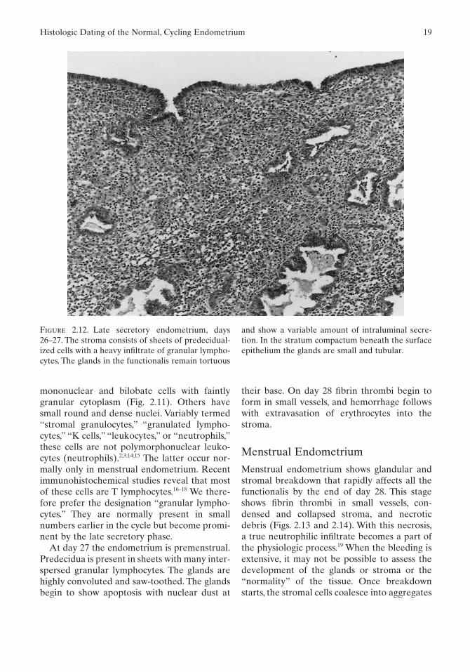
mononuclear and bilobate cells with faintlygranular cytoplasm (Fig. 2.11). Others havesmall round and dense nuclei. Variably termed“stromal granulocytes,” “granulated lympho-cytes,” “K cells,” “leukocytes,” or “neutrophils,”these cells are not polymorphonuclear leuko-cytes (neutrophils).2;3;14;15 The latter occur nor-mally only in menstrual endometrium. Recentimmunohistochemical studies reveal that mostof these cells are T lymphocytes.16–18 We there-fore prefer the designation “granular lympho-cytes.” They are normally present in smallnumbers earlier in the cycle but become promi-nent by the late secretory phase.
At day 27 the endometrium is premenstrual.Predecidua is present in sheets with many inter-spersed granular lymphocytes. The glands arehighly convoluted and saw-toothed. The glandsbegin to show apoptosis with nuclear dust at
their base. On day 28 fibrin thrombi begin toform in small vessels, and hemorrhage followswith extravasation of erythrocytes into thestroma.
Menstrual Endometrium
Menstrual endometrium shows glandular andstromal breakdown that rapidly affects all thefunctionalis by the end of day 28. This stageshows fibrin thrombi in small vessels, con-densed and collapsed stroma, and necroticdebris (Figs. 2.13 and 2.14). With this necrosis,a true neutrophilic infiltrate becomes a part ofthe physiologic process.19 When the bleeding isextensive, it may not be possible to assess thedevelopment of the glands or stroma or the“normality” of the tissue. Once breakdownstarts, the stromal cells coalesce into aggregates
Histologic Dating of the Normal, Cycling Endometrium 19
Figure 2.12. Late secretory endometrium, days26–27. The stroma consists of sheets of predecidual-ized cells with a heavy infiltrate of granular lympho-cytes. The glands in the functionalis remain tortuous
and show a variable amount of intraluminal secre-tion. In the stratum compactum beneath the surfaceepithelium the glands are small and tubular.

Figure 2.13. Menstrual endometrium. Hemorrhage into the stroma forms lakes of erythrocytes. The hemorrhage disrupts the glands and stroma, although the tortuosity of the glands persists.
Figure 2.14. Menstrual endometrium.With stromal hemorrhage, the predecidual cells collapse and they losetheir abundant cytoplasm.

and clusters that often show little cytoplasm.With extensive stromal collapse during men-struation, the predecidual change in the stromalcells becomes indistinct (Figs. 2.14 and 2.15).The extensive breakdown also can result instriking morphologic alterations with artifac-tual glandular crowding. As a result, menstrualendometrium can be confused with hyperplasiaor even carcinoma if the background bleedingpattern is not recognized. Conversely, hyper-plasia and carcinoma are proliferative pro-cesses that rarely show extensive breakdown of the type displayed by menstrual endo-metrium. Because of the artifacts induced by the breakdown and bleeding of the men-strual phase, this tissue is not suitable for eval-uation of glandular and stromal development.Some advocate biopsy at the onset of bleedingto be certain that the procedure does not inter-rupt an early pregnancy, but this tissue is not optimal unless obtained very early in the
menstrual phase before breakdown becomesextensive.4
Pitfalls in Dating
The preceding description summarizes thebasic histologic changes of endometrial devel-opment. In addition to understanding thenormal morphology in ideal situations, oneneeds to consider a number of practical pointswhen interpreting the endometrial biopsy.There are several caveats and potential pitfalls,knowledge of which assists in accurate diagno-sis of normal endometrium and helps avoiderrors in dating. The following, in our opinion,are especially important aspects to consider inevaluating this biopsy material:
1. Endometrium with surface epithelium isbest for interpretation. Absence of surfaceepithelium compromises the interpretation.
Pitfalls in Dating 21
Figure 2.15. Menstrual endometrium. Glands andstroma near the basalis undergo collapse as thesuperficial tissue sloughs. The glands retain tortuous
shapes but show nuclear dust accumulating in thesubnuclear cytoplasm (arrowheads). Predecidualchange in the stroma has become indistinct.
2. Tissue from the lower uterine segment or basalis is not satisfactory for dating.Endometrium from these regions does notrespond fully to hormones.
3. Straight, tubular glands beneath the sur-face are normal and not a sign of irregularity inmaturation in the late secretory phase.
4. Scattered subnuclear vacuoles in glandsare not sufficient evidence of ovulation. To becertain that ovulation has occurred, more than50% of the glands must show subnuclear vacuoles.
5. The presence of secretions in the glandu-lar lumen does not indicate secretory endome-trium. Proliferative, hyperplastic, and neoplasticglands can contain luminal secretions. It is theglandular cytoplasm and nuclear changes thatare most important for determining the pres-ence or absence of secretory changes.
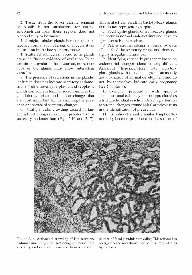
6. Focal glandular crowding caused by tan-gential sectioning can occur in proliferative orsecretory endometrium (Figs. 2.16 and 2.17).
This artifact can result in back-to-back glandsthat do not represent hyperplasia.
7. Focal cystic glands or nonreactive glandscan occur in normal endometrium and have nosignificance by themselves.
8. Patchy stromal edema is normal by days17 to 18 of the secretory phase and does notsignify irregular maturation.
9. Identifying very early pregnancy based onendometrial changes alone is very difficult.Apparent “hypersecretory” late secretoryphase glands with vacuolated cytoplasm usuallyare a variation of normal development and donot, by themselves, indicate early pregnancy(see Chapter 3).
10. Compact predecidua with spindle-shaped stromal cells may not be appreciated asa true predecidual reaction. Directing attentionto stromal changes around spiral arteries assistsin the identification of predecidua.
11. Lymphocytes and granular lymphocytesnormally become prominent in the stroma of
22 2. Normal Endometrium and Infertility Evaluation
Figure 2.16. Artifactual crowding of late secretoryendometrium. Tangential sectioning of normal latesecretory endometrium near the basalis yields a
pattern of focal glandular crowding. This artifact hasno significance and should not be misinterpreted ashyperplasia.

the late secretory phase.These do not representinflammation.
12. If the tissue is difficult to date because ofapparent discordance in features, the possibil-ity of chronic endometritis or a polyp should beconsidered.
13. The endometrium cannot be dated accurately when polyps, inflammation, or otherabnormalities are present.
Artifacts and Contaminants
Besides variations in the normal anatomy, suchas the basalis and lower uterine segment,several artifacts of the biopsy often complicatethe histologic patterns. One frequent artifact istissue fragmentation caused by mechanical dis-ruption of the tissue. As a result, glands are
detached from the surrounding stroma, andfragmented glands become randomly oriented,often appearing closely spaced (Fig. 2.18). Thisartifact should not be mistaken for real crowd-ing that occurs in hyperplasia or carcinoma.Fragmentation and close apposition of dis-parate tissues such as cervical epithelium andfunctionalis also lead to confusing patterns.Artifactually crowded glands lack a continuousinvestment of tissue and are not connected byintervening stroma. These latter features helpin recognition of the artifact. Fragmentationalso is a common feature of atrophy (seeChapter 5).
Another frequent change is so-called tele-scoping of glands.20 Telescoping may occur in either proliferative or secretory phaseendometrium (Fig. 2.19), but it also complicatesmany nonphysiologic conditions. Telescoping
Artifacts and Contaminants 23
Figure 2.17. Artifactual crowding of late secretoryendometrium. This is another example of late se-cretory phase glands that appear crowded due to tangential sectioning. This pattern is neither
hyperplastic nor “hypersecretory,” however. Identi-fying surface epithelium elsewhere in the sectionsoften helps to avoid misinterpretation of thesenormal glands.

Figure 2.18. Artifactual fragmentation. Normal pro-liferative phase endometrium is fragmented as aresult of the procedure. The glands have a haphaz-
Figure 2.19. Telescoping artifact. Normal secretoryendometrium shows telescoping artifact with agland-in-gland appearance. This common alteration
ard arrangement that should not be confused with asignificant abnormality. Focal “telescoping” artifactalso is present (arrow).
is an apparent result of the biopsy procedure and hasno significance.

results in a pattern of an apparent gland withinthe lumen of another gland and can mimichyperplasia or neoplasia (Fig. 2.20). This arti-fact seems to be a result of mechanical disrup-tion and “snap back” of the gland duringcurettage, resulting in intussusception whichrarely occurs in hysterectomy specimens. Tan-gential sectioning of tortuous glands also con-tributes to this phenomenon. Fortunately,telescoping rarely presents difficulty in inter-pretation once the observer understands thephenomenon. In questionable cases, the cytol-ogy of the glandular cells and comparison withsurrounding tissue establishes this change as anartifact.
Endometrial biopsies also often contain con-taminants from the cervix. Most of these conta-minants are obvious. Strips of bland squamousor mucinous epithelium and irregular pools of
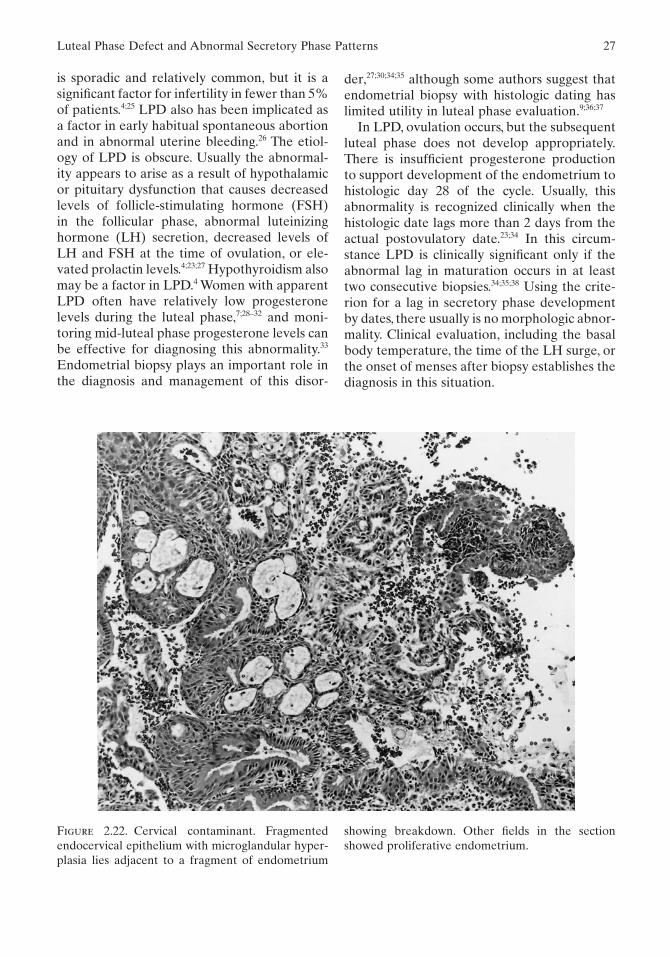
extracellular mucin are common (Fig. 2.21).Theextracellular mucin may contain neutrophils,cell debris, macrophages, or giant cells that arenormal components with no pathologic signifi-cance in the absence of inflammation in theendometrial stroma. Occasionally, benign cervi-cal contaminants become more complex andtroublesome in biopsies. Endocervical glandswith squamous metaplasia or microglandularhyperplasia yield complex patterns, but theseelements are cytologically bland and usuallyblend into more typical cervical epithelium (Fig.2.22). In questionable cases, continuity withendometrial surface epithelium may help toestablish origin in the corpus. It is also helpful tolook at the surrounding stroma and see if it is ofendometrial or endocervical type. Rarely, anendometrial biopsy also may reveal an admix-ture of fragments of tissue from cervical dyspla-
Artifacts and Contaminants 25
Figure 2.20. Telescoping with focal artifactual glandcrowding. Focus of disrupted secretory glands showsapparent gland crowding caused by telescoping and
fragmentation. This is a common artifact seen innormal proliferative and secretory endometriumthat should not be mistaken for hyperplasia.

Figure 2.21. Cervical contaminants. Left: Fragmen-tation has resulted in a strip of mucinous glandularepithelium adjacent to detached proliferative phase
sia, squamous carcinoma, or adenocarcinoma.Chapter 10 addresses the differential diagnosisof endocervical versus endometrial carcinoma.
Occasionally, curettage yields sheets of histi-ocytes with no associated mucin or other tissue(Fig. 2.23). These histiocytes apparently residein the endometrial cavity, and show the typicalhistiocyte cytology with a lobulated nucleus andamphophilic cytoplasm. We have seen them inassociation with hydrometra and with benignbleeding patterns. They apparently represent aresponse to intracavitary debris and have beenreferred to as “nodular histiocytic hyperplasia”because they can represent a nodular accumu-lation of histiocytes in sections.21 They arebenign but can mimic endometrial stromal cellsor stromal cell lesions. Immunohistochemicalstains for histiocyte markers, such as lysozymeor KP 1, can facilitate their recognition. Stromalfoam cells, in contrast, represent stromal cells
and macrophages that are filled with lipid fromerythrocytes in areas of chronic nonphysiologicbleeding.22 Separate fragments of adipose tissuewith clearly identifiable fat cells in an endome-trial biopsy almost always represent omentumor extrauterine pelvic soft tissue and indicateperforation of the uterus. We have also occa-sionally seen colonic mucosa in endometrialbiopsies. In these circumstances the clinicianshould be notified immediately.
Luteal Phase Defect andAbnormal Secretory Phase Patterns
Luteal phase defect (LPD), or inadequateluteal phase, is a recognized cause of infertility,so-called ovulatory infertility.4;23–25 This disorder
glands. Right: Amorphous endocervical mucus witha few macrophages may be admixed with endome-trial tissues. This finding has no clinical significance.
26 2. Normal Endometrium and Infertility Evaluation
is sporadic and relatively common, but it is asignificant factor for infertility in fewer than 5%of patients.4;25 LPD also has been implicated asa factor in early habitual spontaneous abortionand in abnormal uterine bleeding.26 The etiol-ogy of LPD is obscure. Usually the abnormal-ity appears to arise as a result of hypothalamicor pituitary dysfunction that causes decreasedlevels of follicle-stimulating hormone (FSH) in the follicular phase, abnormal luteinizinghormone (LH) secretion, decreased levels ofLH and FSH at the time of ovulation, or ele-vated prolactin levels.4;23;27 Hypothyroidism alsomay be a factor in LPD.4 Women with apparentLPD often have relatively low progesteronelevels during the luteal phase,7;28–32 and moni-toring mid-luteal phase progesterone levels canbe effective for diagnosing this abnormality.33
Endometrial biopsy plays an important role inthe diagnosis and management of this disor-
der,27;30;34;35 although some authors suggest thatendometrial biopsy with histologic dating haslimited utility in luteal phase evaluation.9;36;37
In LPD, ovulation occurs, but the subsequentluteal phase does not develop appropriately.There is insufficient progesterone productionto support development of the endometrium tohistologic day 28 of the cycle. Usually, thisabnormality is recognized clinically when thehistologic date lags more than 2 days from theactual postovulatory date.23;34 In this circum-stance LPD is clinically significant only if theabnormal lag in maturation occurs in at leasttwo consecutive biopsies.34;35;38 Using the crite-rion for a lag in secretory phase developmentby dates, there usually is no morphologic abnor-mality. Clinical evaluation, including the basalbody temperature, the time of the LH surge, orthe onset of menses after biopsy establishes thediagnosis in this situation.
Luteal Phase Defect and Abnormal Secretory Phase Patterns 27
Figure 2.22. Cervical contaminant. Fragmentedendocervical epithelium with microglandular hyper-plasia lies adjacent to a fragment of endometrium
showing breakdown. Other fields in the sectionshowed proliferative endometrium.

Because of inadequate progesterone produc-tion, LPD may also cause abnormalities in thedevelopment of secretory endometrium.35;39 Infact, experimental evidence shows that varia-tions in the relative amounts of the sex steroidhormones, estradiol and progesterone, affectendometrial development.40 In these experi-mental conditions, relatively low doses of estro-gen and progesterone result in glandular andstromal hypoplasia. Higher doses of estrogenbut low doses of progesterone result in stromalinadequacy, while high levels of progesteroneand low doses of estrogen lead to glandularinadequacy. These data suggest that variationsin follicle and corpus luteum development withdecreases in hormone production alter thedevelopment of secretory endometrium follow-ing ovulation.
Morphologic features of LPD other than alag in the histologic date are poorly character-
ized. LPD may cause discordance in the devel-opment of the glands and the stroma.35;39;41–43
The resulting pattern is that of irregular matu-ration, with different areas showing a marked(greater than 4 days) variation in development.Although LPD can be reflected in endometrialmorphologic abnormalities such as irregularmaturation, there are no large-scale studies thathave clearly identified specific pathologic fea-tures of this condition.
On occasion, endometrial biopsies showabnormal secretory phase patterns.35;39;41;43–45 Insuch cases the endometrium typically showssecretory changes that cannot be assigned toany day of the normal cycle (Figs. 2.24 and2.25). The pattern may show true irregular maturation with a large variation in the patternof endometrial development from field to field.For example, some areas may show early secre-tory changes characteristic of about days 18 to
28 2. Normal Endometrium and Infertility Evaluation
Figure 2.23. Histiocytes. A detached aggregate ofhistiocytes superficially resembles endometrialstroma. Inset: The cells have cytologic features of his-
tiocytes with oval, folded nuclei and faintly vacuo-lated cytoplasm. A lack of intrinsic vasculature helpsto indicate that this is a contaminant.
Clinical Queries and Reporting 29
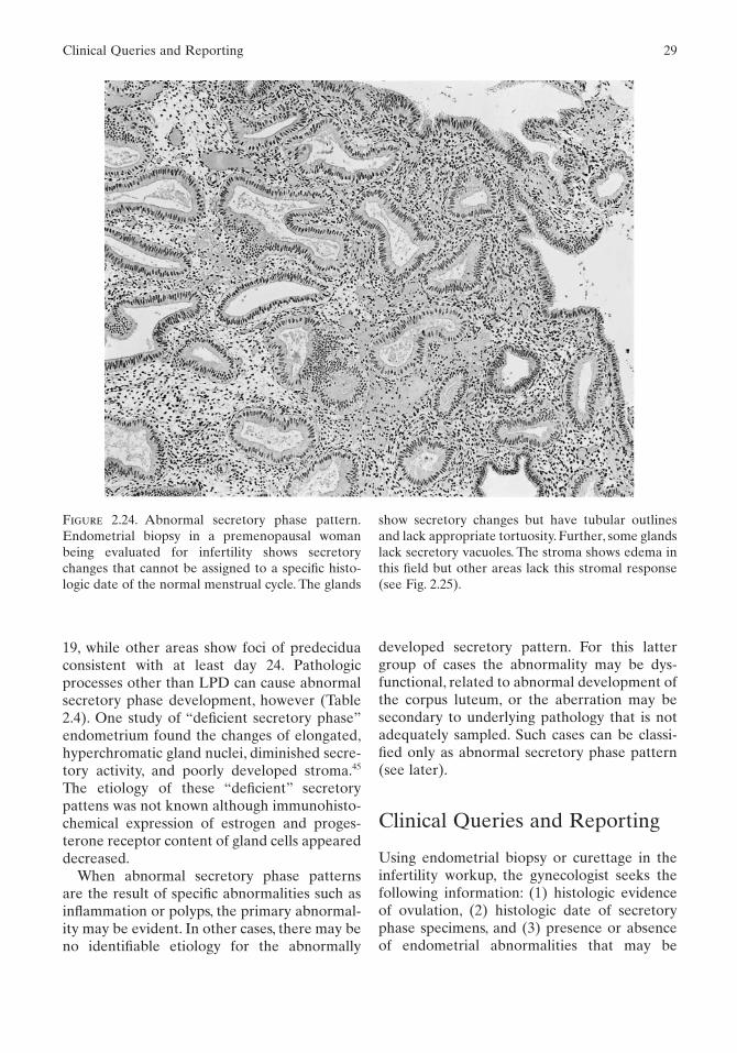
Figure 2.24. Abnormal secretory phase pattern.Endometrial biopsy in a premenopausal womanbeing evaluated for infertility shows secretorychanges that cannot be assigned to a specific histo-logic date of the normal menstrual cycle. The glands
19, while other areas show foci of predeciduaconsistent with at least day 24. Pathologicprocesses other than LPD can cause abnormalsecretory phase development, however (Table2.4). One study of “deficient secretory phase”endometrium found the changes of elongated,hyperchromatic gland nuclei, diminished secre-tory activity, and poorly developed stroma.45
The etiology of these “deficient” secretorypattens was not known although immunohisto-chemical expression of estrogen and proges-terone receptor content of gland cells appeareddecreased.
When abnormal secretory phase patterns are the result of specific abnormalities such asinflammation or polyps, the primary abnormal-ity may be evident. In other cases, there may beno identifiable etiology for the abnormally
show secretory changes but have tubular outlinesand lack appropriate tortuosity. Further, some glandslack secretory vacuoles. The stroma shows edema inthis field but other areas lack this stromal response(see Fig. 2.25).
developed secretory pattern. For this lattergroup of cases the abnormality may be dys-functional, related to abnormal development ofthe corpus luteum, or the aberration may besecondary to underlying pathology that is notadequately sampled. Such cases can be classi-fied only as abnormal secretory phase pattern(see later).
Clinical Queries and Reporting
Using endometrial biopsy or curettage in theinfertility workup, the gynecologist seeks thefollowing information: (1) histologic evidenceof ovulation, (2) histologic date of secretoryphase specimens, and (3) presence or absenceof endometrial abnormalities that may be

responsible for infertility. Secretory phasechanges indicate that ovulation has occurred.Dating the secretory phase gives a generalassessment of progesterone production by thecorpus luteum and the ability of the endo-metrium to respond to progesterone.
In practice, a span of up to 3 days in datesfrom field to field is acceptable as normal, espe-cially in mid- to late secretory endometriumwhere areas of edematous stroma alternatewith areas of predecidualized stroma. Also, theglandular changes in the late secretory phasecan be highly variable. The pathologist shoulddate the tissue on the basis of the mostadvanced changes using a 2-day span (e.g., days24 to 25). Dating the secretory phase is some-what subjective and neither completely exactnor reproducible, and therefore it is also impor-tant that everyone involved in the interpreta-
30 2. Normal Endometrium and Infertility Evaluation
Figure 2.25. Abnormal secretory phase pattern.Another area from the specimen shown in Fig. 2.24shows poorly developed secretory glands and dense
stroma that lacks edema. The cause of this type ofabnormality cannot be determined by morphologyalone.
Table 2.4. Causes of undatable endometrium.
Hormonal effectsAnovulationLuteal phase defectPersistent corpus luteumExogenous hormonesPregnancy
Organic lesionsPolypsLeiomyomasChronic inflammationHyperplasiaCarcinomaAtrophy
Sampling problemsFragmentationLack of surface epithelium

tion and clinical application of histologic datingunderstand the limitations of this morphologicassessment.46;47 Studies of interobserver varia-tion show that 60% to 80% or more of endome-trial biopsies for dating are within 2 days ofeach other when evaluated by experiencedpathologists.47 Furthermore, 80% of the timethe dates are within 2 days of the expected daycompared with basal body temperature andmenstrual dates.48 Some investigators find thatthe criteria for histologic dating do not havesufficient precision to determine the degree ofcorpus luteum function in the late secretoryphase.49
The field-to-field variations in normal latesecretory endometrium probably lead to someof the interobserver and intraobserver varia-tions found in dating.With experience, a pathol-ogist should be able to provide a reasonableassessment of the endometrial development inthe secretory phase.
Obviously for the infertility patient everyattempt should be made to provide an accuratedate, but there are cases in which precise histo-logic dating is not possible. When accuratedating cannot be done, it is important, if pos-sible, to indicate why (Table 2.4). Hormonaleffects, various organic lesions, and samplingproblems all can make dating difficult or impos-sible. Other organic factors such as inflamma-tion, adhesions, or polyps may interfere withpregnancy (see Chapter 7).These abnormalitiesaffect fertility by altering the development ofthe glands and stroma, thereby preventingnormal implantation or mechanically disrupt-ing the early implanting placenta. Therefore,when present these abnormalities should bereported. If factors preclude reporting a specificdate, an attempt should be made to decidewhether the endometrium is proliferative orsecretory, because secretory phase develop-ment generally indicates that ovulation hasoccurred.
On occasion the biopsy shows an abnormalsecretory pattern that cannot be histologicallydated. In such cases, the abnormal pattern maybe due to an LPD or to some other pathologicfactor that is not identifiable in the specimen.In practice, a descriptive diagnosis of the
changes (e.g., “secretory with irregular matura-tion” or “abnormal secretory phase pattern”)with a description of the abnormality is suffi-cient to indicate that the secretory develop-ment is not normal. The gynecologist can usethis information in combination with other clin-ical observations to determine its significanceand possible cause. Descriptive diagnosesshould be used carefully, however. The term“dyssynchronous endometrium” has been usedto describe apparent alterations in secretoryphase development. “Dyssynchronous” is not aword with a specific connotation and thereforeits use can be confusing unless there is clearcommunication between the pathologist andthe gynecologist regarding its meaning.
Recently, morphometric analysis has beenattempted to increase the accuracy of endome-trial histologic dating.50 It was found that fivemorphometric measurements, including mitoticrate in gland cells, amount of luminal secretion,volume fraction of gland occupied by gland cell,amount of pseudostratification of gland cells,and amount of predecidual reaction, added precision to histologic dating.50 Furthermore,immunohistochemical analysis for specificsecretory products of the endometrium andintegrins may help to identify LPDs.51–54 Othercell products such as cyclin E and p27 also maybe useful in assessing secretory phase develop-ment.55 These data indicate that further refine-ments in evaluation of normal endometriummay evolve that have clinical utility. At present,however, routine histologic evaluation remainsa cost-effective method of determining the relative degree of endometrial developmentthrough the menstrual cycle.
References(1) Noyes RW, Hertig AT, Rock J. Dating the
endometrial biopsy. Fertil Steril 1950; 1:3–25.(2) Dallenbach-Hellweg G. Histopathology of
the endometrium. 4th ed. New York: Springer-Verlag; 1987.
(3) Hendrickson MR, Kempson RL. Surgicalpathology of the uterine corpus (Major prob-lems in pathology series). Volume 12. Philadel-phia: W.B. Saunders; 1980.
References 31

(4) Speroff L, Glass RH, Kase NG. Clinical gyne-cologic endocrinology and infertility. 6th ed.Baltimore: Lippincott Williams & Wilkins;1999.
(5) Merrill JA. The interpretation of endometrialbiopsies. Clin Obstet Gynecol 1991; 34:211–221.
(6) Hill GA, Herbert CMI, Parker RA, Wentz AC.Comparison of late luteal phase endometrialbiopsies using the Novak curette or Pipelleendometrial suction curette. Obstet Gynecol1989; 73:443–446.
(7) Hecht BR, Bardawil WA, Khan-Dawood FS,Dawood MY. Luteal insufficiency: Correlationbetween endometrial dating and integratedprogesterone output in clomiphene citrate-induced cycles. Am J Obstet Gynecol 1990;163:1986–1991.
(8) Shoupe D, Mishell DRJ, Lacarra M, Lobo RA,Horenstein J, d’Ablaing G et al. Correlation ofendometrial maturation with four methods ofestimating day of ovulation. Obstet Gynecol1989; 73:88–92.
(9) Peters AJ, Lloyd RP, Coulam CB. Prevalence ofout-of-phase endometrial biopsy specimens.Am J Obstet Gynecol 1992; 166:1738–1746.
(10) Masterson R, Armstrong EM, More IAR. Thecyclical variation in the percentage of ciliatedcells in the normal human endometrium.J Reprod Fertil 1975; 42:537–540.
(11) Hendrickson MR, Kempson RL. Ciliated carci-noma-a variant of endometrial adenocarci-noma. A report of 10 cases. Int J GynecolPathol 1983; 2:1–12.
(12) Ronnett BM, Kurman RJ. Precursor lesions ofendometrial carcinoma. In: Kurman R, ed.Blaustein’s pathology of the female genitaltract. 5th ed. New York: Springer-Verlag; 2002:467–500.
(13) Silverberg SG, Kurman RJ. Tumors of theuterine corpus and gestational trophoblasticdisease. Atlas of tumor pathology, 3rd series,Fascicle 3. Washington, DC: Armed ForcesInstitute of Pathology; 1992.
(14) Buckley CH, Fox H. Biopsy pathology of theendometrium. 2nd ed. London: Arnold, 2002.
(15) Mutter GL, Ferenczy A. Anatomy and histol-ogy of the uterine corpus. In: Kurman RJ, ed.Blaustein’s pathology of the female genitaltract. 5th ed. New York: Springer-Verlag; 2002.
(16) Bulmer JN, Lunny DP, Hagin SV. Immunohis-tochemical characterization of stromal leuco-cytes in nonpregnant human endometrium.Am J Reprod Immmunol Microbiol 1988;17:83–90.
(17) King A, Wellings V, Gardner L, Loke YW.Immunocytochemical characterization of theunusual large granular lymphocytes in humanendometrium throughout the menstrual cycle.Hum Immunol 1989; 24:195–205.
(18) Bulmer JN, Hollings D, Ritson A. Immunocy-tochemical evidence that endometrial stromalgranulocytes are granulated lymphocytes.J Pathol 1987; 153:281–288.
(19) Poropatich C, Rojas M, Silverberg SG. Poly-morphonuclear leukocytes in the endometriumduring the normal menstrual cycle. Int JGynecol Pathol 1987; 6:230–234.
(20) Numers CV. On the traumatic effect of curet-tage on the endometrial biopsy, with special ref-erence to so-called invagination pictures andthe “crumbling endometrium.” Acta ObstetGynecol Scand 1942; 28:305–313.
(21) Kim KR, Lee YH, Ro JY. Nodular histiocytichyperplasia of the endometrium. Int J GynecolPathol 2002; 21:141–146.
(22) Silver SA, Sherman ME. Morphologic andimmunophenotypic characterization of foamcells in endometrial lesions. Int J GynecolPathol 1998; 17:140–145.
(23) Jones GS. Luteal phase insufficiency. ClinObstet Gynecol 1972; 16:255–273.
(24) Wentz AC. Diagnosing luteal phase inadequacy.Fertil Steril 1982; 37:334.
(25) Wentz AC, Kossoy LR, Parker RA. The impactof luteal phase indaequacy in an infertile population. Am J Obstet Gynecol 1990;162:937–945.
(26) Sherman ME, Mazur MT, Kurman RJ. Benigndiseases of the endometrium. In: Kurman RJ,ed. Blaustein’s pathology of the female genitaltract. 5th ed. New York: Springer-Verlag; 2002:421–466.
(27) Daly DC, Walters CA, Soto-Albors CE,Riddick DH. Endometrial biopsy during treat-ment of luteal phase defects is predictive oftherapeutic outcome. Fertil Steril 1983; 40:305–310.
(28) Rosenfeld DL, Garcia C. A comparison ofendometrial histology with simultaneousplasma progesterone determinations in infer-tile women. Fertil Steril 1976; 27:1256–1266.
(29) Annos T, Thompson IE, Taymor ML. Lutealphase deficiency and infertility: Difficultiesencountered in diagnosis and treatment. ObstetGynecol 1980; 55:705–710.
(30) Rosenfeld DL, Chudow S, Bronson RA. Diag-nosis of luteal phase inadequacy. ObstetGynecol 1980; 56:193–196.
32 2. Normal Endometrium and Infertility Evaluation

(31) Cooke ID, Morgan CA, Parry TE. Correlationof endometrial biopsy and plasma proges-terone levels in infertile women. J ObstetGynaecol Br Commonw 1972; 79:647–650.
(32) Cumming DC, Honore LH, Scott JZ, WilliamsKP. The late luteal phase in infertile women:Comparison of simultaneous endometrialbiopsy and progesterone levels. Fertil Steril1985; 43:715–719.
(33) Jordan J, Craig K, Clifton DK, Soules MR.Luteal phase defect: The sensitivity and speci-ficity of diagnostic methods in common clinicaluse. Fertil Steril 1994; 62:54–62.
(34) Wentz AC. Endometrial biopsy in the evalua-tion of infertility. Fertil Steril 1980; 33:121–124.
(35) Witten BI, Martin SA. The endometrial biopsyas a guide to the management of luteal phasedefect. Fertil Steril 1985; 44:460–465.
(36) Balasch J, Fabregues F, Creus M, Vanrell JA.The usefulness of endometrial biopsy for lutealphase evaluation in infertility. Hum Reprod1992; 7:973–977.
(37) Scott RT, Snyder RR, Strickland DM, TyburskiCC, Bagnall JA, Reed KR, et al. The effect ofinterobserver variation in dating endometrialhistology on the diagnosis of luteal phasedefects. Fertil Steril 1988; 50:888–892.
(38) Downs KA, Gibson M. Basal body temperaturegraph and the luteal phase defect. Fertil Steril1983; 40:466–468.
(39) Dallenbach-Hellweg G. The endometrium ofinfertility. A review. Pathol Res Pract 1984;178:527–537.
(40) Good RG, Moyer DL. Estrogen-progesteronerelationships in the development of secretoryendometrium. Fertil Steril 1968; 19:37–45.
(41) Gillam JS. Study of the inadequate secretionphase endometrium. Fertil Steril 1955; 6:18–36.
(42) Deligdisch L. Hormonal pathology of theendometrium. Mod Pathol 2000; 13:285–294.
(43) Dallenbach-Hellweg G. Changes in theendometrium caused by endogenous hormonaldysfunction. Verh Dtsch Ges Pathol 1997; 81:213–218.
(44) Noyes RW. The underdeveloped secretoryendometrium. Am J Obstet Gynecol 1959; 77:929–945.
(45) Thornburgh I, Anderson MC. The endometrialdeficient secretory phase. Histopathology 1997;30:11–15.
(46) Zaino R, Novotney D, Ireland D, Meyer W,Fritz M, Lessey B, et al. Dating the endometrialbiopsy: A re-evaluation. Mod Pathol 2003; 16:216A.
(47) Duggan MA, Brashert P, Ostor A, Scurry J,Billson V, Kneafsey P, et al. The accuracy andinterobserver reproducibility of endometrialdating. Pathology 2001; 33:292–297.
(48) Noyes RW, Haman JO. The accuracy ofendometrial dating. Fertil Steril 1953;4:504–517.
(49) Li T-C, Dockery P, Rogers AW, Cooke ID. Howprecise is histologic dating of endometriumusing the standard dating criteria? Fertil Steril1989; 51:759–763.
(50) Li TC, Rogers AW, Dockery P, Lenton EA,Cooke ID. A new method of histologic datingof human endometrium in the luteal phase.Fertil Steril 1988; 50:52–60.
(51) Graham RA, Seif MW, Aplin JD, Li TC, CookeID, Rogers AW, et al. An endometrial factor in unexplained infertility. Br Med J 1990;300:1428–1431.
(52) Seif MW, Aplin JD, Buckley CH. Luteal phasedefect: The possibility of an immunohistochem-ical diagnosis. Fertil Steril 1989; 51:273–279.
(53) Lessey BA, Castelbaum AJ, Wolf L, Greene W,Paulson M, Meyer WR, et al. Use of integrinsto date the endometrium. Fertil Steril 2000;73:779–787.
(54) Ordi J, Creus M, Ferrer B, Fabregues F,Carmona F, Casamitjana R, et al. Midlutealendometrial biopsy and alphavbeta3 integrinexpression in the evaluation of the endo-metrium in infertility: implications for fecun-dity. Int J Gynecol Pathol 2002; 21:231–238.
(55) Dubowy RL, Feinberg RF, Keefe DL, DoncelGF, Williams SC, McSweet JC, et al. Improvedendometrial assessment using cyclin E and p27.Fertil Steril 2003; 80:146–156.
References 33