NIH Public Access injury Expert Rev Respir Med · Pathophysiology, management and ... smoke...
27
Pathophysiology, management and treatment of smoke inhalation injury Sebastian Rehberg † , Department of Anesthesiology, The University of Texas Medical Branch, 301 University Boulevard, Galveston, TX 77555, USA, Tel.: +1 409 772 6405, Fax: +1 409 772 6409, [email protected] Marc O Maybauer, Department of Anesthesiology, The University of Texas Medical Branch, 301 University Boulevard, Galveston, TX 77555, USA Perenlei Enkhbaatar, Department of Anesthesiology, The University of Texas Medical Branch, 301 University Boulevard, Galveston, TX 77555, USA Dirk M Maybauer, Department of Anesthesiology, The University of Texas Medical Branch, 301 University Boulevard, Galveston, TX 77555, USA Yusuke Yamamoto, and Department of Anesthesiology, The University of Texas Medical Branch, 301 University Boulevard, Galveston, TX 77555, USA Daniel L Traber Department of Anesthesiology, The University of Texas Medical Branch, 301 University Boulevard, Galveston, TX 77555, USA Abstract Smoke inhalation injury continues to increase morbidity and mortality in burn patients in both the third world and industrialized countries. The lack of uniform criteria for the diagnosis and definition of smoke inhalation injury contributes to the fact that, despite extensive research, mortality rates have changed little in recent decades. The formation of reactive oxygen and nitrogen species, as well as the procoagulant and antifibrinolytic imbalance of alveolar homeostasis, all play a central role in the pathogenesis of smoke inhalation injury. Further hallmarks include massive airway obstruction owing to cast formation, bronchospasm, the increase in bronchial circulation and transvascular fluid flux. Therefore, anticoagulants, antioxidants and bronchodilators, especially when administered as an aerosol, represent the most promising treatment strategies. The purpose of this review article is to provide an overview of the pathophysiological changes, management and treatment options of smoke inhalation injury based on the current literature. Keywords acute lung injury; anticoagulants; antioxidants; β 2 -agonists; carbon monoxide; cyanide; nitric oxide; reactive oxygen species †Author for correspondence Department of Anesthesiology, The University of Texas Medical Branch, 301 University Boulevard, Galveston, TX 77555, USA, Tel.: +1 409 772 6405, Fax: +1 409 772 6409, [email protected]. For reprint orders, please contact [email protected] NIH Public Access Author Manuscript Expert Rev Respir Med. Author manuscript; available in PMC 2010 April 1. Published in final edited form as: Expert Rev Respir Med. 2009 June 1; 3(3): 283–297. doi:10.1586/ERS.09.21. NIH-PA Author Manuscript NIH-PA Author Manuscript NIH-PA Author Manuscript
-
Upload
truongdieu -
Category
Documents
-
view
215 -
download
1
Transcript of NIH Public Access injury Expert Rev Respir Med · Pathophysiology, management and ... smoke...

Pathophysiology, management and treatment of smoke inhalationinjury
Sebastian Rehberg†,Department of Anesthesiology, The University of Texas Medical Branch, 301 University Boulevard,Galveston, TX 77555, USA, Tel.: +1 409 772 6405, Fax: +1 409 772 6409, [email protected]
Marc O Maybauer,Department of Anesthesiology, The University of Texas Medical Branch, 301 University Boulevard,Galveston, TX 77555, USA
Perenlei Enkhbaatar,Department of Anesthesiology, The University of Texas Medical Branch, 301 University Boulevard,Galveston, TX 77555, USA
Dirk M Maybauer,Department of Anesthesiology, The University of Texas Medical Branch, 301 University Boulevard,Galveston, TX 77555, USA
Yusuke Yamamoto, andDepartment of Anesthesiology, The University of Texas Medical Branch, 301 University Boulevard,Galveston, TX 77555, USA
Daniel L TraberDepartment of Anesthesiology, The University of Texas Medical Branch, 301 University Boulevard,Galveston, TX 77555, USA
AbstractSmoke inhalation injury continues to increase morbidity and mortality in burn patients in both thethird world and industrialized countries. The lack of uniform criteria for the diagnosis and definitionof smoke inhalation injury contributes to the fact that, despite extensive research, mortality rates havechanged little in recent decades. The formation of reactive oxygen and nitrogen species, as well asthe procoagulant and antifibrinolytic imbalance of alveolar homeostasis, all play a central role in thepathogenesis of smoke inhalation injury. Further hallmarks include massive airway obstructionowing to cast formation, bronchospasm, the increase in bronchial circulation and transvascular fluidflux. Therefore, anticoagulants, antioxidants and bronchodilators, especially when administered asan aerosol, represent the most promising treatment strategies. The purpose of this review article isto provide an overview of the pathophysiological changes, management and treatment options ofsmoke inhalation injury based on the current literature.
Keywordsacute lung injury; anticoagulants; antioxidants; β2-agonists; carbon monoxide; cyanide; nitric oxide;reactive oxygen species
†Author for correspondence Department of Anesthesiology, The University of Texas Medical Branch, 301 University Boulevard,Galveston, TX 77555, USA, Tel.: +1 409 772 6405, Fax: +1 409 772 6409, [email protected] reprint orders, please contact [email protected]
NIH Public AccessAuthor ManuscriptExpert Rev Respir Med. Author manuscript; available in PMC 2010 April 1.
Published in final edited form as:Expert Rev Respir Med. 2009 June 1; 3(3): 283–297. doi:10.1586/ERS.09.21.
NIH
-PA Author Manuscript
NIH
-PA Author Manuscript
NIH
-PA Author Manuscript

Smoke inhalation injury is generally defined as the inhalation of thermal or chemical irritants.With more than 23,000 injuries and 5000–10,000 deaths per year in the USA alone, smokeinhalation injury represents a major cause of morbidity and mortality in burn patients [1]. Inaddition, disasters such as the Station Nightclub Fire in Rhode Island in 2005 [2], or thedevastating terrorism attacks on the World Trade Center in New York (NY, USA) [3,4] andthe Pentagon in Washington (DC, USA) [5] in 2001 are associated with a high incidence ofinhalation injuries. Among the 790 injured survivors from the World Trade Center attack, forexample, 49% suffered from inhalation injury caused by toxic compounds in the smoke anddust [3,4]. Compared with isolated burns, a combined injury with smoke inhalation isassociated with increases in fluid requirements [6], the incidence of pulmonary complications[7] and mortality [7–9]. It is important to remember however that smoke inhalation injury isnot ‘only’ an adjunct to burn trauma; it is an independent injury in and of itself.
According to the WHO, more than 1 billion people develop airway and pulmonaryinflammation as a result of inhaled smoke from indoor cooking fires, forest fires and burningof crops [10,11]. In addition, smoke toxicity is increasing because industrial products haveshifted from woods and natural materials towards lighter construction materials, synthetics andpetrochemicals, which ignite and burn two- to three-times hotter and faster. Thus, theprobability that fire victims will breath in smoke and toxic gases is increased, because theyhave less time to escape.
While there has been remarkable progress in the treatment of cutaneous burns in recent decades,mortality rates of patients with inhalation injury have changed little in the past 20 years [12].The heterogeneity of smoke and the resulting differences in clinical symptoms may be onereason why a definition and specific diagnostic criteria of smoke inhalation injury are stilllacking. The purpose of this review article is to provide an overview of the pathophysiologicalchanges, management and treatment options of smoke inhalation injury based on the currentliterature.
Pathophysiology & clinical symptomsSmoke is composed of a gas phase and a particle phase. Particle size and tidal volume determinetheir distribution in the lung. Physiologically, the nasopharynx clears the inspiratory air of themajority of particles with a diameter larger than 5 μm [13]. During a fire, however, victims(both conscious and unconscious) breathe through the mouth owing to the nasopharyngealirritation. As a result, particle deposition in the airway is much greater, causing progressivecellular injury and severe lung injury. The gas phase causes predominantly proximal airwayand local damage, although some long-acting oxidants are able to reach distal lung tissues.
Smoke inhalation injury can be divided into three different types of injury [12]:
• Thermal injury, which is mostly restricted to the upper airway (exception: blast injuryor steam inhalation)
• Chemical irritation of the respiratory tract
• Systemic toxicity owing to toxic gases
The location of the injury depends on the ignition source, the size of the particles in the smoke,the duration of exposure and the solubility of the gases. Based on the predominant localization,smoke inhalation injury is classified into injuries of [14]:
• The upper airway
• The tracheobronchial system or lower airway
• The lung parenchyma
Rehberg et al. Page 2
Expert Rev Respir Med. Author manuscript; available in PMC 2010 April 1.
NIH
-PA Author Manuscript
NIH
-PA Author Manuscript
NIH
-PA Author Manuscript

• Systemic toxicity
This classification will be used in the following sections to represent the different effects ofsmoke inhalation injury. Figure 1 summarizes the pathogenesis of lung injury following smokeinhalation.
Upper airwayOwing to highly efficient heat exchange in the oro- and nasopharynx [15], the leading injuryin the upper airway, above the vocal cords, is caused by thermal injury. In accordance to burnsof other body areas, heat destroys the epithelial layer, denatures proteins and activates thecomplement cascade leading to the release of histamine and the formation of xanthine oxidase[16]. This enzyme catalyses the breakdown of purins to uric acid and thereby releases reactiveoxygen species (ROS) such as superoxide [17]. Superoxide represents a highly reactivemolecule, which is physiologically stabilized by its formation to hydrogen peroxide catalyzedby superoxide dismutase [18]. At the same time, nitric oxide (NO) formation by endothelialcells is increased by histamine stimulation. In this case, the reaction with NO to form reactivenitrogen species (RNS), such as peroxynitrite, is faster [18]. Both ROS and RNS cause anincreased permeability of endothelium for proteins, resulting in edema formation. In addition,eicosanoids and IL-8 are released after injury, leading to the attraction of polymorphonuclearcells [19,20], which amplify the inflammatory process, for example by ROS production. Inparallel, activation of pulmonary C-fiber receptors by inflammation and irritants of smokecauses vasodilation by increasing NO production, further aggravating edema formation [21].
While the immediate injury results in erythema, ulceration and edema (Figure 2), clinicalsymptoms, such as stridor, dyspnea or increased work of breathing might not be obvious untilthe edema is sufficiently large enough to impair the airway diameter significantly. This timedifference can amount up to 18 h or even longer [22]. In the case of combined burn and smokeinhalation injury, however, the aggressive fluid administration necessary to treat the burn shockpromotes edema formation [23]. In addition, accompanying face and neck burns might causeanatomic distortion or external compression of the upper airway, further complicating properairway management [22]. In addition to the inflammation, damage of ciliary function impairsthe physiological cleaning process of the airway, resulting in an increased risk of bacterialinfections for several weeks. Furthermore, the increased production of viscous secretionscauses distal airway obstruction and atelectasis, thereby impairing pulmonary gas exchange[22,24].
Tracheobronchial systemWith rare exceptions, such as the inhalation of steam, injury to the tracheobronchial area isusually caused by chemicals in smoke. The airway is richly innervated by vasomotor andsensory nerve endings [25]. Smoke inhalation stimulates these nerves to release neuropeptids[26], which are potent bronchoconstrictors. Under physiological conditions, the mucosaproduces neutral endopeptidases, which neutralize these toxic agents. Owing to severe cellulardamage, this neutralization is lost [22]. Neuropeptidases also attract and activate neutrophils,resulting in the production of ROS [20]. Simultaneously, the activity of neuronal NO synthases(nNOS, NOS-1) is upregulated by neuropeptides [27]. NOS catalyze the formation of NO andL-citrulline from the amino acid L-arginine in a complex five-electron redox reaction. Threedifferent isoforms of NOS have been identified in mammals: nNOS and endothelial NOS(eNOS, NOS-3) are constitutive isoforms, while the inducible isoenzyme (iNOS, NOS-2) canbe upregulated by oxidative stress and systemic inflammation [28]. Notably, in case of substrate(L-arginine) or cofactor limitation, for example under pathophysiological conditions, NOS canalso produce superoxide [29]. NO reacts with ROS such as superoxide to form RNS, forexample peroxinitrite. Although this highly cytotoxic RNS represents the most cited example,
Rehberg et al. Page 3
Expert Rev Respir Med. Author manuscript; available in PMC 2010 April 1.
NIH
-PA Author Manuscript
NIH
-PA Author Manuscript
NIH
-PA Author Manuscript

most of the ROS and RNS are able to interact with proteins, DNA or lipids by oxidation,nitration or nitrosylation, causing the inactivation of key enzymes such as the glycolyticenzyme glyceraldehydes-3-phosphate dehydrogenase or produce a single-strand breakage inDNA [30]. DNA damage leads to the formation of poly(ADP ribose) polymerase (PARP), anuclear enzyme involved in DNA repair [31,32]. It has been shown that PARP activation canbe induced by NO and its toxic products such as RNS [33].
Although the physiological function of PARP is beneficial, PARP activation depletes cellularATP levels by forming ADP, possibly resulting in cellular dysfunction and apoptosis [32,34].In addition, PARP activates nuclear factor κB (NF-κB), independent of its action to form PAR[35,36]. NF-κB is known to upregulate IL-8 [37], resulting in the attraction and activation ofpolymorphonuclear cells and consecutively in the production of ROS. NF-κB also stimulatesiNOS formation [38], thereby accelerating the production of NO. NO and ROS react to formRNS, leading to DNA damage and PARP activation, and thereby a positive feedback loop isestablished [27].
Histologically, there is evidence of damage to the mucosal lining and peribronchialinflammation [14,22,39–42]. Loss of bronchial epithelium results in a profuse transudate witha high protein content. While these secretions form foam material during the early response,this transudate/exudate mixture solidifies during the pathogenesis, forming obstructivematerial, which might occlude lower airways. These airway ‘casts’ (Figure 3) are composedof mucus secretions, denuded airway epithelial cells, inflammatory cells and fibrin [24].Especially when volume-controlled ventilator settings are selected, obstruction can promote abarotrauma of still-ventilated areas. Besides massive airway obstruction, further hallmarks ofsmoke inhalation injury include bronchospasm, increases in bronchial circulation andtransvascular fluid flux [43]. A tenfold increase in bronchial blood flow within 20 min aftersmoke inhalation was reported in an experimental study. In addition, the same animalsdemonstrated a sixfold increase in pulmonary transvascular fluid flux [44]. Both of thesechanges promote the development of airway edema. Further clinical symptoms includepersistent coughing and wheezing, soot-containing airway secretions, increased work ofbreathing resulting in hypoventilation, erythema, hyperemia and increased pulmonary shunting[22]. The latter is caused by the loss of hypoxic pulmonary vasoconstriction owing to high NOproduction [14].
Lung parenchymaThe damage of lung parenchyma following smoke inhalation injury is delayed. The timedifference between the initial trauma and a decrease in arterial oxygen tension to inspiratoryoxygen fraction (PaO2:FiO2) is correlated with the severity of the lung injury, and representsa consequence of changes described for the upper airway and the tracheobronchial system[14]. Alveolar injury is characterized by alveolar collapse and atelectasis owing to increasedtransvascular fluid flux, a lack of surfactant and a loss of hypoxic pulmonary vasoconstriction,resulting in impaired oxygenation. In addition, a severe imbalance in alveolar hemostasis,increased procoagulatory and decreased antifibrinolytic activity, leads to massive fibrindeposition in the airway, which prevents regular ventilation and causes a ventilation/perfusionmismatch [43]. By inhibiting surfactant, fibrin itself leads to atelectasis [45] and can attractinflammatory cells [46].
Activated neutrophils play a central role in the pathogenesis of smoke inhalation injury. First,they initiate and continuously support the pathological pathway described previously byproducing ROS and releasing proteases, such as elastase [27]. Second, neutrophils activatedin the bronchial circulation are drained into the pulmonary microvasculature [14]. Sinceactivated neutrophils are stiff owing to F-actin activation, they are not able to traverse thepulmonary capillaries as usual by changing their shape [14]. Instead, they adhere to the alveolar
Rehberg et al. Page 4
Expert Rev Respir Med. Author manuscript; available in PMC 2010 April 1.
NIH
-PA Author Manuscript
NIH
-PA Author Manuscript
NIH
-PA Author Manuscript
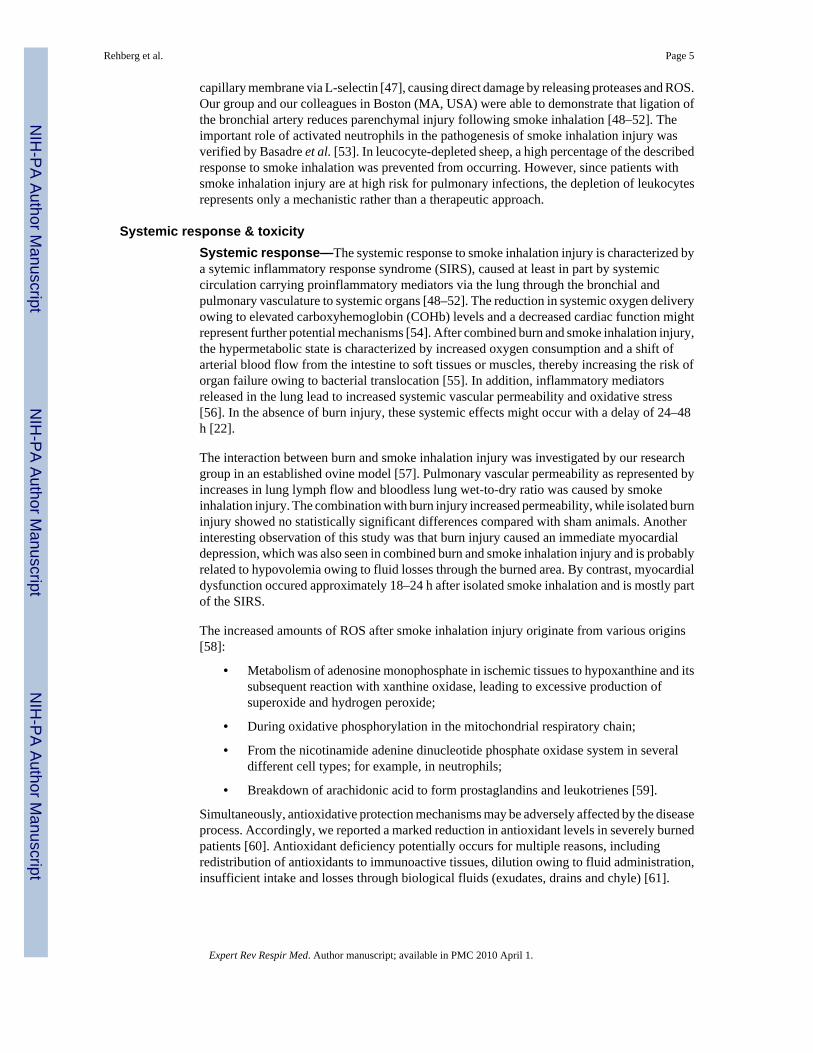
capillary membrane via L-selectin [47], causing direct damage by releasing proteases and ROS.Our group and our colleagues in Boston (MA, USA) were able to demonstrate that ligation ofthe bronchial artery reduces parenchymal injury following smoke inhalation [48–52]. Theimportant role of activated neutrophils in the pathogenesis of smoke inhalation injury wasverified by Basadre et al. [53]. In leucocyte-depleted sheep, a high percentage of the describedresponse to smoke inhalation was prevented from occurring. However, since patients withsmoke inhalation injury are at high risk for pulmonary infections, the depletion of leukocytesrepresents only a mechanistic rather than a therapeutic approach.
Systemic response & toxicitySystemic response—The systemic response to smoke inhalation injury is characterized bya sytemic inflammatory response syndrome (SIRS), caused at least in part by systemiccirculation carrying proinflammatory mediators via the lung through the bronchial andpulmonary vasculature to systemic organs [48–52]. The reduction in systemic oxygen deliveryowing to elevated carboxyhemoglobin (COHb) levels and a decreased cardiac function mightrepresent further potential mechanisms [54]. After combined burn and smoke inhalation injury,the hypermetabolic state is characterized by increased oxygen consumption and a shift ofarterial blood flow from the intestine to soft tissues or muscles, thereby increasing the risk oforgan failure owing to bacterial translocation [55]. In addition, inflammatory mediatorsreleased in the lung lead to increased systemic vascular permeability and oxidative stress[56]. In the absence of burn injury, these systemic effects might occur with a delay of 24–48h [22].
The interaction between burn and smoke inhalation injury was investigated by our researchgroup in an established ovine model [57]. Pulmonary vascular permeability as represented byincreases in lung lymph flow and bloodless lung wet-to-dry ratio was caused by smokeinhalation injury. The combination with burn injury increased permeability, while isolated burninjury showed no statistically significant differences compared with sham animals. Anotherinteresting observation of this study was that burn injury caused an immediate myocardialdepression, which was also seen in combined burn and smoke inhalation injury and is probablyrelated to hypovolemia owing to fluid losses through the burned area. By contrast, myocardialdysfunction occured approximately 18–24 h after isolated smoke inhalation and is mostly partof the SIRS.
The increased amounts of ROS after smoke inhalation injury originate from various origins[58]:
• Metabolism of adenosine monophosphate in ischemic tissues to hypoxanthine and itssubsequent reaction with xanthine oxidase, leading to excessive production ofsuperoxide and hydrogen peroxide;
• During oxidative phosphorylation in the mitochondrial respiratory chain;
• From the nicotinamide adenine dinucleotide phosphate oxidase system in severaldifferent cell types; for example, in neutrophils;
• Breakdown of arachidonic acid to form prostaglandins and leukotrienes [59].
Simultaneously, antioxidative protection mechanisms may be adversely affected by the diseaseprocess. Accordingly, we reported a marked reduction in antioxidant levels in severely burnedpatients [60]. Antioxidant deficiency potentially occurs for multiple reasons, includingredistribution of antioxidants to immunoactive tissues, dilution owing to fluid administration,insufficient intake and losses through biological fluids (exudates, drains and chyle) [61].
Rehberg et al. Page 5
Expert Rev Respir Med. Author manuscript; available in PMC 2010 April 1.
NIH
-PA Author Manuscript
NIH
-PA Author Manuscript
NIH
-PA Author Manuscript

Systemic toxicity—A direct systemic effect of smoke inhalation injury is caused byinhalation of toxic gases during the combustion of organic and inorganic substances. Withrespect to morbidity and mortality, the two most relevant gases are carbon monoxide (CO) andcyanide. Other toxic gases and their symptoms are listed in Table 1.
Carbon monoxide—Carbon monoxide is not only one of the most frequent immediatecauses of death following smoke inhalation injury, but also of poisoning deaths in the USA. Itaccounts for approximately 15,000 emergency room visits and 500 unintentional deaths eachyear [62]. CO is an odorless, colorless gas with an affinity for hemoglobin more than 200-timeshigher than that of oxygen [63]. Accordingly, inhalation of only 0.1% CO mixture can resultin a life-threatening COHb level of 50% [14]. Oxygen delivery to organs is further decreasedby a left shift of the oxygen–hemoglobin dissociation curve, impairing tissue oxygenavailability [64]. In addition, CO inhibits hepatic cytochromes, leading to mitochondrialoxidative stress and membrane damage owing to lipid peroxidation [63].
Clinical symptoms vary depending on the concentration of CO, the duration of exposure andpre-existing morbidity of the patient. Generally, a COHb level of more than 90% may lead toimmediate cardiac arrest [65]. Clinical symptoms mainly involve neurological andcardiovascular manifestations. Owing to a lack of alternatives, diagnosis of CO poisoning isstill based on the measurement of COHb levels. However, the strength of the correlationbetween COHb levels and the severity of poisoning, the prognosis or the choice of therapy isdiscussed controversially [66]. Cellular mechanisms, such as caspase-mediated apoptosis, mayplay an additional role in CO poisoning [67]. Importantly, it has to be taken into account thatthe usual pulse oximeter can not differentiate between COHb and oxyhemoglobin and thatvenous blood underestimates the arterial COHb content [68]. However, there are now devicesavailable (Masimo Set® Rad 57™) enabling physicians or paramedics to determine COHblevels noninvasively at the injury scene. In contrast to usual pulse oximeters, Masimo Set Rad57 measures seven or more instead of two wavelengths.
Cyanide—Hydrogen cyanide (HCN) represents the gaseous form of cyanide (CN), which isa colorless gas with the odor of bitter almonds; however, HCN is difficult to detect at the siteof a fire, as most inhalation injuries represent mixed intoxications. Notably, CN is a normalhuman metabolite as reflected by plasma levels of 0.3 mg/l in nonsmokers and 0.5 mg/l insmokers [69]. All cells, but mostly those in the liver, are able to convert CN into thiocyanide,which is excreted in the urine via the enzyme rhodanase. In case of large amounts of CN, thissystem can be overcome, especially in burned or traumatized patients who are hypovolemic[22]. Toxicity of CN is based on hypoxic states owing to the reversible inhibition of cytochromec oxidase, which is the terminal oxidase of the respiratory chain. The definition of fatal bloodlevels, however, varies from 1–3 to 5 mg/l [70]. The lack of a precise definition may be onereason for controversial discussions regarding the role of CN poisoning within smokeinhalation injury. Advocates cite studies reporting increased CN plasma levels in nonsurvivorsas compared with survivors of fire disasters [71,72]. By contrast, opponents argue that CN islikely to be rapidly consumed in fires. Davies and colleagues described CN concentrations inthe smoke of 250 ppm, which went down to below 10 ppm at 8 min [73]. Neither the short-term exposure limit (15 ppm), nor the short-term lethal concentration (350 ppm) were exceeded[70]. Nevertheless, CN originates from numerous compounds during combustion (Table 1),resulting in an increased probability of CN intoxication in a fire victim. At the same time,diagnosis of CN poisoning represents a challenge at the injury scene, as generally describedsymptoms (e.g., increased lactate levels and base deficit, or metabolic acidosis) may also becaused by asphyxiation, under-resuscitation, CO poisoning or associated traumatic injury.However, owing to the high probability of its presence at a fire scene (especially in case ofcombustion of certain materials such as plastics), in combination with the difficult diagnosis
Rehberg et al. Page 6
Expert Rev Respir Med. Author manuscript; available in PMC 2010 April 1.
NIH
-PA Author Manuscript
NIH
-PA Author Manuscript
NIH
-PA Author Manuscript

and its lethal potential, CN intoxication should be carefully considered in every patient withsmoke inhalation injury.
ManagementAt the injury scene
The first priority at the injury scene is rescue of the victim from the source of fire to stop theexposure time. This is usually the responsibility of firefighters. In order to reduce COHb levelsas soon as possible, a high flow of 100% oxygen should be administered via facemaskimmediately. The next step includes a short but careful body check to estimate the extent ofsmoke inhalation and to assess accompanying injuries such as burns and/or trauma. In addition,it is important to determine whether the victim has been exposed to an explosion, since thiscan cause barotrauma to the lung. If possible, information about comorbidities should beobtained. Indications of inhalation injury are summarized in Box 1. Usual cardiopulmonarymonitoring (electrocardiogram, pulse oxymeter and noninvasive blood pressure) should beestablished.
Box 1
Indications and symptoms of inhalation injury
• Facial and neck burns
• Burned lips and vibrissae
• Soot-containing airway secretions
• Pathological respiration patterns (coughing, stridor and hoarseness)
• Dyspnea
• Cyanosis
• Neurological symptoms (current or past unconsciousness, dizziness, nausea andvomiting)
After these basic measures, a decision needs to be made on how best to secure the airway ofthe patient. On the one hand, the risk of a rapidly developing airway edema has to be takeninto account even if no dyspnea is present. On the other hand, endotracheal intubation itself,especially at the injury scene, is associated with undeniable risks such as esophageal intubation,aspiration, barotrauma or laryngeal trauma. Therefore, in the authors’ opinion, prophylacticendotracheal intubation is not generally advised in every patient and, depending on thetechnical skills, for every physician or paramedic. Nevertheless, it is important to consider thatairway management will only become more difficult over time. Patients with heat and smokeinhalation injury combined with extensive face or neck burns definitely have to be intubated.In the case of oral burn without inhalation injury, an airway secured early represents the safestapproach. However, victims with smoke inhalation injury but no facial or neck burns can becarefully observed and later be intubated, if necessary [22].
If the patient is endotracheally intubated, the tube should be carefully secured. Accidentalremoval of the endotracheal tube is easy and may be lethal. In cases of vocal cord damage,tracheostomy may be necessary to prevent further damage. The patient’s head should beelevated to minimize facial and airway edema. As a matter of course, hemodynamic stabilityis a prerequisite. Aerolized epinephrine or corticosteroids may be beneficial to reduce upperairway edema [22]. However, there is no conclusive evidence for the efficacy of these treatmentstrategies. After initial stabilization of the patient, it might be very helpful for further treatment
Rehberg et al. Page 7
Expert Rev Respir Med. Author manuscript; available in PMC 2010 April 1.
NIH
-PA Author Manuscript
NIH
-PA Author Manuscript
NIH
-PA Author Manuscript

to obtain information about the fire source, combustion products and the estimated duration ofexposure. In case of specific intoxications, for example, HCN poisoning, recommendedtherapies should be started (please see the section entitled ‘Mechanical ventilation strategies’).
In-hospital treatmentAfter adequate airway management and arrival in the emergency room, the treatment ofaccompanying burn injuries or trauma usually represents the first, most immediate priority.Inhalation injury, by comparison, often develops with a latency of several hours. Nevertheless,airway management and oxygenation status of the patient, regardless if intubated or not, needto be reevaluated frequently to allow clinicians to react to the dynamic development of smokeinhalation injury [22]. After stabilization of cardiopulmonary hemodynamics and pulmonarygas exchange, the assumed diagnosis of smoke inhalation injury needs to be verified. However,as there are currently no uniform criteria available, diagnosis of smoke inhalation injury isusually a subjective decision based on a combination of history (please see the section entitled‘At the injury scene’) and physical examination, which are confirmed by diagnostics [12]. Thebronchoscopical examination of the airway represents the gold standard to detect apathognomonic mucosal hyperemia. Chest radiographs may show signs of diffuse atelectases,pulmonary edema or bronchopneumonia. However, during the initial period, the degree ofinjury is usually underestimated based on the chest x-ray, as the injury is mainly confined tothe airways [74].
Although many observational studies compared outcomes with various grading systems [75–77], there is still no uniform algorithm for assessing inhalation injury. As a result, no reliableindicators of progressive respiratory failure in patients with smoke inhalation injury have beenidentified so far [78,79]. This failure is largely explained by the extreme heterogeneity of theclinical presentation. In addition, the delay in the manifestation and development of acute lunginjury (ALI) as a consequence of SIRS, initiated by accompanying burns or trauma, complicatethe evaluation of the isolated effects of smoke inhalation. Useful tools to monitor smokeinhalation injury include frequent blood gas and sputum analyses [22].
Appropriate fluid resuscitation of patients with smoke inhalation injury is still subject tocontroversial debates. It has been demonstrated that smoke inhalation injury increases fluidrequirements in burned patients [6,80]. This, of course, does not inevitably indicate that isolatedsmoke inhalation injury is associated with increased fluid requirements. By contrast, over-resuscitation may increase pulmonary microvascular pressures and might thereby lead toincreased edema formation under the high permeability conditions in early lung injury [57,81]. In a retrospective study of more than 2346 trauma patients, Plurad et al. found an increasedincidence of late post-traumatic acute respiratory distress syndrome (ARDS) with risingvolumes of administered cristalloids and packed red blood cells [82]. Unfortunately, there iscurrently no evidence for the specific patient population of isolated smoke inhalation injury.Against the background of no proven benefit and the potential risk of detrimental effects,however, increased amount of fluids should be avoided in patients with isolated smokeinhalation injury. Instead, fluid resuscitation should be guided by urine output andhemodynamic parameters of the individual patient. Thereby, rather dynamic parameters, suchas changes in pulse pressure, rather than static parameters, such as central venous or pulmonaryartery occlusion pressures, might be helpful.
After acute therapy, treatment is mainly focused on three cornerstones: first, maintenance andrestoration of a sufficient gas exchange. Thus, respiratory ventilator settings have to be adjustedfrequently to guarantee the most efficient ventilatory support and to minimize ventilation-associated side effects, such as baro- or volutrauma (please see secion entitled ‘Nebulizedtreatments’). Second, vigorous bronchial toilet should be performed to clear the airway andavoid airway occlusion. Mucociliary action is extensively impaired owing to the extensive
Rehberg et al. Page 8
Expert Rev Respir Med. Author manuscript; available in PMC 2010 April 1.
NIH
-PA Author Manuscript
NIH
-PA Author Manuscript
NIH
-PA Author Manuscript

structural damage of heat and chemicals in the smoke [22]. In combination with an impairedcough reflex and increased tenacious secretions, this injury bears a high risk of occlusion ofsmaller airways, leading to atelectasis and infection [83]. Third, therefore, very carefulinfection surveillance is advised. Impairment of alveolar macrophage function and post-traumatic immunodeficiency also represent important risk factors, but a prophylacticadministration of antibiotics is not recommended to avoid the development of resistantorganisms [84].
TreatmentThe following section provides a brief overview of current therapeutic strategies in respect tosmoke inhalation injury. These recommendations are based on limited evidence from clinicaland experimental trials as well as expert opinions resulting from clinical experience.
Mechanical ventilation strategiesSeveral years ago, mechanical ventilation was performed by using high inspiratory oxygenconcentrations and tidal volumes of 10–15 ml/kg in a volume-controlled mode. However, “thisapproach may have violated the injunctive, first to do no harm (Primum non nocere)” [85].During the last two decades, mechanical ventilation has been investigated extensively. Lowtidal volume ventilation with associated permissive hypercapnia has been shown to effectivelyreduce ventilator-induced lung injury, and currently represents the standard of care [86]. TheAssessment of Low Tidal Volume and Elevated End Expiratory Pressure to Obviate AcuteLung Injury (ALVEOLI) trial revealed no effects of higher positive end-expiratory pressure(PEEP) levels in ARDS patients [87]. In addition, prone positioning has been shown to haveno beneficial effect on mortality, despite a transient improvement in oxygenation [88].
Although smoke inhalation predisposes patients to ALI and ARDS, especially in combinationwith burns, it remains to be determined if these results also apply to the specific patientpopulation after smoke inhalation. In the ARDS Network Study concerning low tidal volumeventilation, for example, patients with burns of more than 30% total body surface area (TBSA)have been excluded. A direct transfer of these approaches may be limited in clinical practiceowing to the unique clinical and pathophysiological features of smoke inhalation. Ventilationprotocols differ not only between different burn centers but also between individual physicians.Apart from conventional pressure-controlled low tidal volume ventilation, multiple strategiesfor mechanical ventilation are currently used for the treatment of smoke inhalation injury,isolated as well as in combination with burns.
Airway pressure release ventilation (APRV), first described in 1987 by Stock and colleagues[89], is a time-triggered, pressure-limited, time-cycled ventilation mode [90]. It provides twolevels of airway pressures during two time periods: a long, high-level and a short, low-levelperiod. In this way, lower peak but higher mean airway pressures are produced compared withconventional ventilation strategies, resulting in lower tidal volumes and more effective alveolarrecruitment. In addition, APRV allows spontaneous breathing during the whole inspiration–expiration cycle. Possible side effects include higher intrinsic PEEP, secondary to increasedairway resistance and short expiratory times, resulting in hyperinflation of the lungs [90]. Tworandomized, controlled trials in mechanically ventilated patients – not patients with smokeinhalation injury – revealed beneficial effects on oxygenation and lower end-inflation pressures[91,92]. However, there was no reduction in mortality. In conclusion, further research isnecessary to determine whether APRV represents a beneficial approach for the ventilation ofpatients suffering from smoke inhalation injury.
The volumetric diffusive ventilator (VDR; Percussionaire Corp., Sandpoint, ID, USA) is apneumatically powered, pressure-limited ventilator that stacks oscillatory breaths to a selected
Rehberg et al. Page 9
Expert Rev Respir Med. Author manuscript; available in PMC 2010 April 1.
NIH
-PA Author Manuscript
NIH
-PA Author Manuscript
NIH
-PA Author Manuscript

peak airway pressure by means of a sliding venturi called Phasitron®, resulting in low tidalvolumes. Exhalation is passive and a level of continuous positive airway pressure can beselected. In addition, VDR re-establishes physiologic diffusive gas exchange, while standardventilation modes induce a convective gas exchange [93]. In contrast to APRV, the effects ofVDR have been studied in patients with smoke inhalation injury. A prospective clinical analysisrevealed an improved gas exchange and a decrease in peak pressures [94]. In addition, aretrospective study in 330 patients with inhalation injury even reported a lower mortality rate[95]. While these studies compared the VDR to high-volume ventilatory strategies, dataregarding a comparison with modern low tidal volume ventilation are still lacking. This mayrepresent one reason why VDR is not universally accepted. Another factor might be that theVDR differs from other ventilators, and therefore requires special training. In addition, tidaland minute volumes cannot be monitored and humidified air, as well as nebulized saline, arenecessary to prevent airway desiccation [93].
High-frequency oscillatory ventilation (HFOV) uses extremely low tidal volumes of 1–2 ml/kg applied with high frequencies of 3–15 Hz and mean airway pressures of 30–40 cmH2O.This ventilation regime prevents alveolar trauma [96] and leads to improved lung recruitment,thereby improving oxygenation, allowing lower inspiratory oxygen concentrations andconsequently limiting oxygen toxicity [97]. However, its use after smoke inhalation injury maybe limited by copious secretions and small airway obstruction. In addition, gas trapping andhypercapnia will be hard to control using HFOV [97]. Accordingly, HFOV failed to improvePaO2:FiO2 ratios in patients with combined burn and smoke inhalation injury, while itimproved oxygenation in isolated burn patients within 8 h [97,98]. Furthermore, theadministration of nebulized adjunctive therapies is not possible with this ventilation strategy.
Taken together, all these ventilation modes, with the HFOV as an extreme, apply low tidalvolumes and high mean airway pressures or PEEP, respectively, to improve oxygenation andairway recruitment. Patients with inhalation injury suffer from increased mucous secretionsand even airway cast formation to a greater extent than ARDS patients in general. Therefore,clearance of secretions represents an essential factor within the therapeutic regime. However,airway clearance is best supported by high tidal volumes and low PEEP [81], therebycontradicting the ventilation strategies for improved oxygenation. As a consequence,physicians need to balance these competing objectives based on the individual situation of thepatient until future studies might provide more evidence.
External arterial venous CO2 removal represents a unique form of CO2 removal, which is drivenby endogenous arterial pressure [99]. Several studies in animal models of smoke inhalationinjury revealed a reduction in morbidity and mortality [100–102]. However, clinical evidenceis lacking.
In conclusion, mechanical ventilation of patients after smoke inhalation injury will furtherrepresent a tightrope walk between providing sufficient oxygenation and causing as littlecollateral harm as possible. While there are several specific ventilation devices and strategiesavailable, large, randomized, controlled multicenter studies are warranted to clarify theirpotential usefulness in patients with smoke inhalation injury.
Nebulized treatmentsAgainst the background that the lung is the primary injured organ following smoke inhalationinjury and that mechanical ventilation is frequently necessary, the administration of therapeuticcompounds via nebulization directly to the affected organ seems to be more than reasonable.Based on the complex pathogenesis of smoke inhalation injury, drugs with differentmechanisms of action, such as bronchodilatators, anticoagulants, antioxidants and
Rehberg et al. Page 10
Expert Rev Respir Med. Author manuscript; available in PMC 2010 April 1.
NIH
-PA Author Manuscript
NIH
-PA Author Manuscript
NIH
-PA Author Manuscript

corticosteroids have been investigated. In each case, however, the key to any successful aerosoltherapy is the consistent delivery into the lung and to the distal airways.
Bronchodilators, such as β2-agonists, improve respiratory mechanics by decreasing airflowresistance and peak airway pressures. This results in improved dynamic compliance [103]. Inaddition, β2-agonists provide anti-inflammatory properties, represented by a decrease ininflammatory mediators such as histamine, leukotrienes and TNF-α [104,105]. Finally, β2-agonists are associated with improved airspace fluid clearance and stimulation of epithelialrepair [106]. While clinical evidence for ALI/ARDS in general is present [107,108], theliterature discussing smoke inhalation is limited. Using an established ovine model ofcombined burn (40% cutaneous third-degree burn) and smoke inhalation injury (48 breath ofcotton smoke, average COHb of 75% injury), our research group was able to show a reductionof pulmonary vascular permeability, pulmonary edema and airway pressures, as well as animprovement of the PaO2:FiO2 ratio by continuous administration of nebulized albuterol at 20or 40 mg/h compared with placebo treatment [109]. Clinical evidence is restricted toretrospective data in children. Continuous nebulized albuterol (10–40 mg/h) in children withsmoke inhalation injury and a PaO2:FiO2 ratio less than 200 mmHg led to improvedoxygenation and lung compliance in the first 72 h after treatment. No serious adverse events(e.g., tachycardia or hypokalemia) have been reported [110].
Inhaled NO is thought to improve oxygenation and to reduce increased pulmonary vascularresistance by reversing the ventilation-perfusion mismatch in ARDS owing to selectivevasodilation in well-ventilated lung areas. Experimental studies in animals with smokeinhalation injury revealed a consistent reduction in pulmonary hypertension but variable resultsregarding pulmonary shunting [111,112]. Single-center case studies in patients with combinedburn and smoke inhalation injury demonstrated a significant increase in the PaO2:FiO2 ratio,with no additional advantage of doses exceeding 20 ppm [113,114]. Several randomized,controlled trials as well as recent meta-analyses in the general population of ARDS patientsrevealed only a positive effect on the patient’s oxygenation for 24 h (in some studies until 48h) [115–117], but no advantages in respect to mortality, ventilator-free days, duration ofventilation or pulmonary hypertension could be verified. On the contrary, there was even astrong trend towards increased mortality in patients receiving NO as compared with placebotreatment [115].
Against the evidence of beneficial effects of NO synthesis inhibition [27,29,118], inhaledadministration of NO for the treatment of smoke inhalation injury seems questionable. Whilea possibly increased production of RNS is only hypothetical at this time point, there are severalknown, unwanted effects of nebulized NO administration. Long-term treatment might lead tothe diffusion of NO into poorly ventilated areas, abolishing the selective pulmonaryvasoconstriction and consecutively increasing pulmonary shunt volume. In addition, surfactantinhibition, edematous changes and continuing fibrosis may over-ride any benefit of inhaledNO therapy. Although findings are controversial, renal dysfunction and methemoglobinemiahave also been described [115]. Based on the current literature, the standard treatment of smokeinhalation injury with inhaled NO cannot be recommended. However, short-term inhaledadministration of NO might be considered as a rescue treatment to improve oxygenation for ashort period in patients with acute, life-threatening hypoxemia [116].
Owing to increased procoagulatory activity following smoke inhalation injury [43], theaerolized administration of anticoagulants seems to be more than promising. In an ovine model,Brown et al. first described a reduction of mortality after smoke inhalation induced ARDS byaerolized heparin in 1988 [119]. Interestingly, Murakami and colleagues reported that heparinnebulization had only partial or no beneficial effects in ovine ARDS after combined burn andsmoke inhalation injury [120]. This might be explained by the lower levels of antithrombin in
Rehberg et al. Page 11
Expert Rev Respir Med. Author manuscript; available in PMC 2010 April 1.
NIH
-PA Author Manuscript
NIH
-PA Author Manuscript
NIH
-PA Author Manuscript

bronchoalveolar lavage fluid after combined burn and smoke inhalation injury, as the effectsof heparin depend on the binding to antithrombin, thereby enhancing the ability of antithrombinto inhibit fibrin and factor Xa by 2000–6000-fold [114]. In accordance, combined nebulizationof heparin and recombinant human antithrombin (rhAT) in the same model significantlyimproved pulmonary function by reducing airway obstruction [121]. However, heparin bindingto antithrombin inhibits the anti-inflammatory effects of antithrombin [43]. In addition,aerolized antithrombin administration did not affect the reduced systemic plasma levels ofantithrombin [121]. Recently, our research group was able to demonstrate that the combinationof intravenous rhAT and nebulized heparin effectively attenuated pulmonary injury followingcombined burn and smoke inhalation injury [122]. In children with combined burn and smokeinhalation injury, nebulization of heparin and N-acetylcysteine significantly decreased re-intubation rates, the incidence of atelectasis and mortality [123]. Notably, these children alsoreceived transfusions of fresh frozen plasma, a rich source of antithrombin. These promisingresults, however, need to be verified in randomized, controlled multicenter trials.
As mentioned earlier, following combined burn and smoke inhalation injury, the oxidative–antioxidative balance is disturbed by an increase in ROS and a parallel decrease in antioxidants[124]. Accordingly, the antioxidant Vitamin E or α-tocopherol is markedly reduced in patientswith major burns (>50% TBSA) and combined smoke inhalation injury [60]. Our study groupreported that prophylactic administration of α-tocopherol (5 mg/kg orally) 24 h prior to injuryattenuated the decrease in pulmonary function and the increase in vascular permeability thatoccurs after combined burn and smoke inhalation injury in sheep [125]. Of special interest aretwo studies in sheep after combined 40% TBSA burn and smoke inhalation injury, showingthe practicability and efficacy of local antioxidant administration to the lung by nebulization[126,127]. Both α- and γ-tocopherol attenuated the progressive decrease in pulmonary functionrepresented by higher PaO2:FiO2 ratios compared with control animals. In addition, nebulizedγ-tocopherol decreased lung lymph flow, lung wet-to-dry weight ratio, and markers ofoxidative and nitrosative tissue injury more effectively than α-tocopherol. Plasma levels of γ-tocopherol were not increased, suggesting a successful local administration potentiallyavoiding systemic side effects. The superiority of γ-tocopherol over α-tocopherol might bebased on the higher ability of γ-tocopherol to scavenge RNS as well as ROS [128].
Intravenous treatmentsWhile the efficacy of aerolized compounds always depends on consistent delivery into the lungand to the distal airways, the intravenous injection of a compound represents a relatively sureway of administration. Accordingly, the following section discusses some of the intravenous,therapeutic approaches of smoke inhalation injury.
The progressively developing upper airway edema induced by smoke inhalation injuryrepresents one of the major indications for endotracheal intubation. Aerolized epinephrine orcorticosteroids as therapeutic approaches have been proposed [22]; however, conclusiveevidence for these treatment strategies is still missing. One of the reasons might be the varyingefficacy of the aerolized drug delivery, especially in emergency situations. Therefore,intravenous administration of corticosteroids should be the treatment of choice.
Vitamin C has been shown to decrease oxidative damage in several experimental [129,130]and clinical [131,132] trials in different critical diseases. Unfortunately, to the best of ourknowledge, there are no studies investigating the effects of Vitamin C administration onisolated smoke inhalation injury. However, Tanaka and colleagues revealed very impressiveresults in a randomized, controlled trial by adding 66 mg/kg/h Vitamin C to the continuousinfusion in patients who had over 30% TBSA burns [132]. Fluid requirements, body weightgain, wound edema and duration of mechanical ventilation were significantly reduced.Notably, 73% of the included patients in this study were diagnosed with inhalation injury after
Rehberg et al. Page 12
Expert Rev Respir Med. Author manuscript; available in PMC 2010 April 1.
NIH
-PA Author Manuscript
NIH
-PA Author Manuscript
NIH
-PA Author Manuscript

admission to the hospital, with equal distribution between groups. A subgroup analysis revealedimproved oxygenation, represented by higher PaO2:FiO2 ratios, beginning at 18 h after injury.In addition, duration of mechanical ventilation was reduced in patients receiving Vitamin C(12 ± 9 days) as compared with the control group (21 ± 16 days). No differences were seen inPEEP level, inspiratory oxygen fraction or the incidence of pneumonia. Nevertheless, it mustbe explicitly stated that no direct data exist to confirm a beneficial effect of intravenous VitaminC treatment on patients with smoke inhalation injury.
As mentioned in the section ‘Nebulized treatments’, combined burn and smoke inhalationinjury causes an acquired antithrombin deficiency, which represents an independent predictorof length of hospital stay and mortality [133,134]. In addition to its anticoagulatory properties,antithrombin provides anti-inflammatory effects. Antithrombin inhibits the inflammatorysignal transduction in and promotes prostacyclin release from endothelial cells. Thereby, anti-thrombin directly attenuates lung inflammation and edema after combined burn and smokeinhalation injury [135]. Murakami et al. reported decreased airway obstruction by casts, higherurine outputs and mean arterial pressures after/or during the continuous administration of 100μg/kg rhAT over 24 h as compared with control animals in an ovine model of smoke inhalationinjury and pneumonia [136]. In a small clinical study of Kowal-Vern and colleagues, patientswith 20% or more TBSA burn and smoke inhalation injury who received rhAT had increasedPaO2:FiO2 ratios and significantly fewer episodes of pneumonia compared with standardtherapy alone [113]. In conclusion, restoring physiological antithrombin levels in patients withsmoke inhalation injury has the potential to improve survival and to decrease inflammation.Again, however, clinical evidence regarding isolated smoke inhalation injury is missing.
Specific intoxicationsCarbon monoxide—Current treatment recommendations of CO poisoning include cessationof exposure, administration of 100% oxygen and supportive care [67]. Hyperbaric oxygenationtherapy (HBO) is sometimes used to encourage rapid displacement of CO from hemoglobinand to reduce the duration of the hypoxic state. Administration of 100% oxygen at 3atmospheres reduces the CO half-life from 250 min at ambient pressure to 30 min [14]. In spiteof this efficiency, the use of HBO remains controversial [67] owing to the questionedcorrelation between COHb levels and outcomes [66] in combination with limited access to thepatient during HBO therapy, which has a severe impact on treatment quality of combined burninjuries.
Key issues
• Smoke inhalation injury continues to increase morbidity and mortality in burnpatients not only in the third world, but also in industrialized countries.
• Smoke inhalation injury can cause thermal and chemical injuries, as well assystemic toxicity.
• The ignition source, the size of the particles in the smoke, the duration of exposureand the solubility of the gases all determine the location and the severity of smokeinhalation injury.
• The formation of reactive oxygen and nitrogen species, as well as the procoagulantand antifibrinolytic imbalance of alveolar homeostasis, play a central role in thepathogenesis of smoke inhalation injury.
• Massive airway obstruction owing to cast formation, bronchospasm, the increasein bronchial circulation and transvascular fluid flux represent further hallmarks ofsmoke inhalation injury.
Rehberg et al. Page 13
Expert Rev Respir Med. Author manuscript; available in PMC 2010 April 1.
NIH
-PA Author Manuscript
NIH
-PA Author Manuscript
NIH
-PA Author Manuscript

• The development of clinical symptoms, such as erythema, dyspnea or massiveairway edema, may be delayed, but are ultimately fatal.
• Specific intoxications such as cyanide or carbon monoxide need to be consideredcritically and treated, if necessary.
• Clinical management consists preponderantly of supportive care.
• Clinical evidence for specific therapeutic approaches is lacking.
• Anticoagulants, antioxidants and bronchodilators currently represent the mostpromising treatment strategies.
• Aerolized administration of compounds seems to be feasible, safe and effective.
• The optimal ventilation protocol still needs to be defined.
• Establishing clear criteria for the diagnosis and grading of smoke inhalation injuryis essential for the design and success of randomized, controlled multicenter trials.
A second potential argument for HBO is that CO binds not only to hemoglobin, but also tocytochrome oxidase, and thus delayed neurological sequelae may be prevented by HBO. ACochrane database review of six randomized, controlled trials, however, did not reveal abeneficial effect of HBO compared with standard treatment with respect to neurologicalsequelae [137]. These results should be interpreted with care, however, because flaws indesign and analyses were evident in all the included trials. In summary, all patients withCO intoxication should be treated with 100% oxygen. HBO might be a useful therapeuticoption in patients with severe neurological symptoms and high COHb concentrations(>50%), but without major burns and severe pulmonary injury.
Cyanide—The adequate treatment of HCN poisoning following smoke inhalation injury iscurrently under discussion. Several antidotes are available: the ‘cyanide antidote kit’ includesamyl nitrite, thiosulfate and sodium nitrite [70], and is based on the classic treatment approachby Chen and Rose [138]. As these substances are methemoglobin generators, which mayadditionally impair oxygen transport, they should only be used in case of proven diagnosis(increased plasma levels of CN) and under continuous monitoring in an intensive care unit.Methemoglobin chelates CN to form cyanomethemoglobin, which, as it dissociates, allowsfree CN to be converted to thiocyanite by liver mitochondrial enzymes (rhodanase) usingthiosulfate as a substrate. Thiocyanate is then excreted into the urine [138]. n contrast to theseantidotes, hydroxycobalamin, a Vitamin B12 derivative, actively binds CN by formingcyanocobalamin, which will be directly excreted via the kidney [139,140]. In case ofintoxication with 1 mg CN, 50 mg/kg hydroxycobalamin is recommended [141]. Because itaverts methemoglobin production, hydroxycobalamin can even be used in the preclinicalsetting. Accordingly, hydroxycobalamin represents the active compound of the ‘cyanokit’,which is used in the prehospital management of smoke inhalation injury in Europe with areported improvement in mortality [140].
Aggressive restoration of cardiopulmonary function augments the hepatic clearance of CN viathe enzyme rhodanase [142], and has been reported to be successful in severe CN poisoning(blood levels 5.6–9 mg/l) as well as after ingestion [143–145] or smoke inhalation [146], evenwithout the use of antidotes. Therefore, the standard care of CN poisoning should combine theaggressive supportive therapy with the causal treatment using hydroxycobalamin.
Rehberg et al. Page 14
Expert Rev Respir Med. Author manuscript; available in PMC 2010 April 1.
NIH
-PA Author Manuscript
NIH
-PA Author Manuscript
NIH
-PA Author Manuscript

Expert commentaryAgainst the background of the current literature, there was a remarkable increase in ourknowledge regarding the pathogenesis of smoke inhalation injury during the last 10–15 years.There are several promising therapeutic approaches, such as the β2-agonists, antioxidants oranticoagulants, and nebulization of the use of different ventilation modes. In addition, the trendgoes to a combination of different compounds rather than a single ‘magic bullet’. Bearing inmind the complex pathophysiology and different clinical presentations of smoke inhalationinjury, this is more than justifiable.
At the same time, however, we cannot be satisfied with the clinical progress made. The factthat mortality rates of smoke inhalation injury did not change is a more than obvious indicatorfor the lacking success of our current efforts. The future challenge, then, will exist largely intransferring our expanding theoretical knowledge adequately into daily clinical practice.Owing to the relatively small patient population, however, no single-center study will be ableto recruit enough patients for a randomized, controlled trial with enough power to prove ordisprove the efficiency of a therapeutic approach in an adequate time period. To achieve thisgoal, the cooperation and communication between burn centers should be intensified.
Five-year viewDuring the recent Consensus Conference for Inhalation Injury (Couer d’Alene, ID, USA),invited experts in the field of smoke inhalation injury met to “define what basic researchelements are lacking and what additional information/definitions/technology is needed toadvance inhalation injury research” [147]. Definition of criteria for the diagnosis and gradingof inhalation injury received the highest ranked priority. These would represent the basis foruniform data acquisition, enabling the evaluation of short- and long-term efficiency of currentas well as new therapeutic approaches. Therefore, the authors fully agree and hope that thisgoal will be achieved within the next 5 years.
AcknowledgmentsFinancial & competing interests disclosure
Daniel L Traber has received NIH grant NIH-P012 GM066312 – Pathophysiology of lung injury by smoke inhalation.The authors have no other relevant affiliations or financial involvement with any organization or entity with a financialinterest in or financial conflict with the subject matter or materials discussed in the manuscript apart from thosedisclosed.
No writing assistance was utilized in the production of this manuscript.
ReferencesPapers of special note have been highlighted as:
• of interest
•• of considerable interest
1. Alcorta R. Smoke inhalation & acute cyanide poisoning. Hydrogen cyanide poisoning provesincreasingly common in smoke-inhalation victims. JEMS 2004;29(8):S6–S15.
2. Harrington DT, Biffl WL, Cioffi WG. The Station nightclub fire. J Burn Care Rehabil 2005;26(2):141–143. [PubMed: 15756115]
3. Yurt RW, Bessey PQ, Bauer GJ, et al. A regional burn center’s response to a disaster: September 11,2001, and the days beyond. J Burn Care Rehabil 2005;26(2):117–124. [PubMed: 15756112]
Rehberg et al. Page 15
Expert Rev Respir Med. Author manuscript; available in PMC 2010 April 1.
NIH
-PA Author Manuscript
NIH
-PA Author Manuscript
NIH
-PA Author Manuscript

4. CDC. Rapid assessment of injuries among survivors of the terrorist attack on the World Trade Center– New York City, September 2001. JAMA 2002;287(7):835–838. [PubMed: 11862961]
5. Jordan MH, Hollowed KA, Turner DG, Wang DS, Jeng JC. The Pentagon attack of September 11,2001: a burn center’s experience. J Burn Care Rehabil 2005;26(2):109–116. [PubMed: 15756111]
6. Dai NT, Chen TM, Cheng TY, et al. The comparison of early fluid therapy in extensive flame burnsbetween inhalation and noninhalation injuries. Burns 1998;24(7):671–675. [PubMed: 9882069]
7•. Shirani KZ, Pruitt BA Jr, Mason AD Jr. The influence of inhalation injury and pneumonia on burnmortality. Ann Surg 1987;205(1):82–87. Points out the important role of inhalation injury in patientssuffering from burn trauma. [PubMed: 3800465]
8. Suzuki M, Aikawa N, Kobayashi K, Higuchi R. Prognostic implications of inhalation injury in burnpatients in Tokyo. Burns 2005;31(3):331–336. [PubMed: 15774289]
9. Tredget EE, Shankowsky HA, Taerum TV, Moysa GL, Alton JD. The role of inhalation injury in burntrauma. A Canadian experience. Ann Surg 1990;212(6):720–727. [PubMed: 2256764]
10. Schwela DH. Public health and the Air Management Information System (AMIS). Epidemiology1999;10(5):647–655. [PubMed: 10468445]
11. Schwela D. Cooking smoke: a silent killer. People Planet 1997;6(3):24–25. [PubMed: 12321046]12. Woodson LC. Diagnosis and grading of inhalation injury. J Burn Care Res 2009;30(1):143–145.
[PubMed: 19060739]13. Gamsu G, Weintraub RM, Nadel JA. Clearance of tantalum from airways of different caliber in man
evaluated by a roentgenographic method. Am Rev Respir Dis 1973;107(2):214–224. [PubMed:4683579]
14••. Traber, DL.; Herndon, DN.; Enkhbaatar, P.; Maybauer, MO.; Maybauer, DM. The pathophysiologyof inhalation injury. In: Herndon, DN., editor. Total Burn Care. Saunders Elsevier; PA, USA: 2007.p. 248-261.The current pathophysiological knowledge about inhalation injury
15. Clark WR, Bonaventura M, Myers W. Smoke inhalation and airway management at a regional burnunit: 1974–1983. Part I: diagnosis and consequences of smoke inhalation. J Burn Care Rehabil1989;10(1):52–62. [PubMed: 2921259]
16. Friedl HP, Till GO, Trentz O, Ward PA. Roles of histamine, complement and xanthine oxidase inthermal injury of skin. Am J Pathol 1989;135(1):203–217. [PubMed: 2570531]
17. Granger DN. Role of xanthine oxidase and granulocytes in ischemia–reperfusion injury. Am J Physiol1988;255(6 Pt 2):H1269–H1275. [PubMed: 3059826]
18. Maybauer MO, Maybauer DM, Herndon DN, Traber DL. The role of superoxide dismutase insystemic inflammation. Shock 2006;25(2):206–207. [PubMed: 16525361]
19. Herndon DN, Abston S, Stein MD. Increased thromboxane B2 levels in the plasma of burned andseptic burned patients. Surg Gynecol Obstet 1984;159(3):210–213. [PubMed: 6433494]
20. Vindenes H, Ulvestad E, Bjerknes R. Increased levels of circulating interleukin-8 in patients withlarge burns: relation to burn size and sepsis. J Trauma 1995;39(4):635–640. [PubMed: 7473946]
21. Tari C, Baranink J. Upper airway neurological mechanisms. Curr Opin Allergy Clin Immunol2002;2:1149.
22. Demling RH. Smoke inhalation lung injury: an update. Eplasty 2008;8:e27. [PubMed: 18552974]23. Navar PD, Saffle JR, Warden GD. Effect of inhalation injury on fluid resuscitation requirements after
thermal injury. Am J Surg 1985;150(6):716–720. [PubMed: 4073365]24. Cox RA, Burke AS, Soejima K, et al. Airway obstruction in sheep with burn and smoke inhalation
injuries. Am J Respir Cell Mol Biol 2003;29(3 Pt 1):295–302. [PubMed: 12936906]25. Perez Fontan JJ. On lung nerves and neurogenic injury. Ann Med 2002;34(4):226–240. [PubMed:
12371707]26. Fontan JJ, Cortright DN, Krause JE, et al. Substance P and neurokinin-1 receptor expression by
intrinsic airway neurons in the rat. Am J Physiol Lung Cell Mol Physiol 2000;278(2):L344–L355.[PubMed: 10666119]
27. Rehberg S, Maybauer MO, Maybauer DM, et al. The role of nitric oxide and reactive nitrogen speciesin experimental ARDS. Front Biosci. 2009 (In press).
28. Steudel W, Kirmse M, Weimann J, et al. Exhaled nitric oxide production by nitric oxide synthase-deficient mice. Am J Respir Crit Care Med 2000;162(4 Pt 1):1262–1267. [PubMed: 11029328]
Rehberg et al. Page 16
Expert Rev Respir Med. Author manuscript; available in PMC 2010 April 1.
NIH
-PA Author Manuscript
NIH
-PA Author Manuscript
NIH
-PA Author Manuscript
29. Westphal M, Enkhbaatar P, Schmalstieg FC, et al. Neuronal nitric oxide synthase inhibition attenuatescardiopulmonary dysfunctions after combined burn and smoke inhalation injury in sheep. Crit CareMed 2008;36(4):1196–1204. [PubMed: 18379246]
30. Fink MP. Role of reactive oxygen and nitrogen species in acute respiratory distress syndrome. CurrOpin Crit Care 2002;8(1):6–11. [PubMed: 12205400]
31. Gero D, Szabo C. Poly(ADP-ribose) polymerase: a new therapeutic target? Curr Opin Anaesthesiol2008;21(2):111–121. [PubMed: 18443476]
32. Pacher P, Szabo C. Role of the peroxynitrite-poly(ADP-ribose) polymerase pathway in humandisease. Am J Pathol 2008;173(1):2–13. [PubMed: 18535182]
33. Szabo C. DNA strand breakage and activation of poly-ADP ribosyltransferase: a cytotoxic pathwaytriggered by peroxynitrite. Free Radic Biol Med 1996;21(6):855–869. [PubMed: 8902531]
34. Lobo SM, Orrico SR, Queiroz MM, et al. Pneumonia-induced sepsis and gut injury: effects of a poly-(ADP-ribose) polymerase inhibitor. J Surg Res 2005;129(2):292–297. [PubMed: 16139303]
35. Jagtap P, Szabo C. Poly(ADP-ribose) polymerase and the therapeutic effects of its inhibitors. NatRev Drug Discov 2005;4(5):421–440. [PubMed: 15864271]
36. Chiarugi A, Moskowitz MA. Poly(ADP-ribose) polymerase-1 activity promotes NF-κB-driventranscription and microglial activation: implication for neurodegenerative disorders. J Neurochem2003;85(2):306–317. [PubMed: 12675907]
37. Li LF, Ouyang B, Choukroun G, et al. Stretch-induced IL-8 depends on c-Jun NH2-terminal andnuclear factor-κB-inducing kinases. Am J Physiol Lung Cell Mol Physiol 2003;285(2):L464–L475.[PubMed: 12716652]
38. Yamaza T, Masuda KF, Tsukiyama Y, et al. NF-κB activation and iNOS expression in the synovialmembrane of rat temporomandibular joints after induced synovitis. J Dent Res 2003;82(3):183–188.[PubMed: 12598546]
39. Herndon DN, Traber DL, Niehaus GD, Linares HA, Traber LD. The pathophysiology of smokeinhalation injury in a sheep model. J Trauma 1984;24(12):1044–1051. [PubMed: 6512897]
40•. Traber, DL.; Enkhbaatar, P. Thermal lung injury and acute smoke inhalation. In: Fishman, DA.,editor. Fishman’s Pulmonary Diseases and Disorders. McGraw-Hill Medical Publishing Company;NY, USA: 2008. Comprehensive summary of the current knowledge regarding pathophysiology ofthermal lung injury and acute smoke inhalation
41. Traber DL, Hawkins HK, Enkhbaatar P, et al. The role of the bronchial circulation in the acute lunginjury resulting from burn and smoke inhalation. Pulm Pharmacol Ther 2007;20(2):163–166.[PubMed: 16798035]
42. Traber DL, Traber LD. Airway blood flow changes and airway obstruction following lung injury.Arch Physiol Biochem 2003;111(4):297–300. [PubMed: 15764058]
43. Enkhbaatar P, Herndon DN, Traber DL. Use of nebulized heparin in the treatment of smoke inhalationinjury. J Burn Care Res 2009;30(1):159–162. [PubMed: 19180699]
44. Abdi S, Herndon D, McGuire J, Traber L, Traber DL. Time course of alterations in lung lymph andbronchial blood flows after inhalation injury. J Burn Care Rehabil 1990;11(6):510–515. [PubMed:2286604]
45. Seeger W, Stohr G, Wolf HR, Neuhof H. Alteration of surfactant function due to protein leakage:special interaction with fibrin monomer. J Appl Physiol 1985;58(2):326–338. [PubMed: 3838543]
46. Ciano PS, Colvin RB, Dvorak AM, McDonagh J, Dvorak HF. Macrophage migration in fibrin gelmatrices. Lab Invest 1986;54(1):62–70. [PubMed: 3941542]
47. Sakurai H, Schmalstieg FC, Traber LD, Hawkins HK, Traber DL. Role of L-selectin in physiologicalmanifestations after burn and smoke inhalation injury in sheep. J Appl Physiol 1999;86(4):1151–1159. [PubMed: 10194196]
48. Abdi S, Herndon DN, Traber LD, et al. Lung edema formation following inhalation injury: role ofthe bronchial blood flow. J Appl Physiol 1991;71(2):727–734. [PubMed: 1938747]
49. Efimova O, Volokhov AB, Iliaifar S, Hales CA. Ligation of the bronchial artery in sheep attenuatesearly pulmonary changes following exposure to smoke. J Appl Physiol 2000;88(3):888–893.[PubMed: 10710383]
50. Hales CA, Barkin P, Jung W, et al. Bronchial artery ligation modifies pulmonary edema after exposureto smoke with acrolein. J Appl Physiol 1989;67(3):1001–1006. [PubMed: 2793693]
Rehberg et al. Page 17
Expert Rev Respir Med. Author manuscript; available in PMC 2010 April 1.
NIH
-PA Author Manuscript
NIH
-PA Author Manuscript
NIH
-PA Author Manuscript

51. Sakurai H, Johnigan R, Kikuchi Y, et al. Effect of reduced bronchial circulation on lung fluid fluxafter smoke inhalation in sheep. J Appl Physiol 1998;84(3):980–986. [PubMed: 9480960]
52. Sakurai H, Soejima K, Nozaki M, Traber LD, Traber DL. Effect of ablated airway blood flow onsystemic and pulmonary microvascular permeability after smoke inhalation in sheep. Burns 2007;33(7):885–891. [PubMed: 17493760]
53. Basadre JO, Sugi K, Traber DL, et al. The effect of leukocyte depletion on smoke inhalation injuryin sheep. Surgery 1988;104(2):208–215. [PubMed: 3400056]
54. Abdi S, Traber LD, Herndon DN, Rogers CS, Traber DL. Effects of ibuprofen on airway vascularresponse to cotton smoke injury. Eur J Pharmacol 1995;293(4):475–481. [PubMed: 8748701]
55. Sakurai H, Traber LD, Traber DL. Altered systemic organ blood flow after combined injury withburn and smoke inhalation. Shock 1998;9(5):369–374. [PubMed: 9617888]
56. Demling RH, Knox J, Youn YK, LaLonde C. Oxygen consumption early postburn becomes oxygendelivery dependent with the addition of smoke inhalation injury. J Trauma 1992;32(5):593–598.discussion 599. [PubMed: 1588648]
57. Soejima K, Schmalstieg FC, Sakurai H, Traber LD, Traber DL. Pathophysiological analysis ofcombined burn and smoke inhalation injuries in sheep. Am J Physiol Lung Cell Mol Physiol 2001;280(6):L1233–L1241. [PubMed: 11350803]
58. Rehberg S, Maybauer MO, Maybauer DM, et al. The antioxidants Vitamin E and Vitamin C fornutritional support in critical ill patients: beneficial or harmful? Adv Anaesthesiol Crit Care 2009;1(1):11–19.
59. Horton JW. Free radicals and lipid peroxidation mediated injury in burn trauma: the role of antioxidanttherapy. Toxicology 2003;189(1–2):75–88. [PubMed: 12821284]
60. Nguyen TT, Cox CS, Traber DL, et al. Free radical activity and loss of plasma antioxidants, vitaminE, and sulfhydryl groups in patients with burns: the 1993 Moyer Award. J Burn Care Rehabil 1993;14(6):602–609. [PubMed: 8300695]
61. Berger MM, Shenkin A. Update on clinical micronutrient supplementation studies in the criticallyill. Curr Opin Clin Nutr Metab Care 2006;9(6):711–716. [PubMed: 17053424]
62. Carbon monoxide-related deaths – United States, 1999–2004. Morb Mortal Wkly Rep 2007;56(50):1309–1312.
63. Weaver LK. Carbon monoxide poisoning. Crit Care Clin 1999;15(2):297–317. viii. [PubMed:10331130]
64. Prien T, Traber DL. Toxic smoke compounds and inhalation injury – a review. Burns Incl Therm Inj1988;14(6):451–460. [PubMed: 2855039]
65. Traber DL, Maybauer MO, Maybauer DM, Westphal M, Traber LD. Inhalational and acute lunginjury. Shock 2005;24(Suppl 1):82–87. [PubMed: 16374378]
66. Hardy KR, Thom SR. Pathophysiology and treatment of carbon monoxide poisoning. J Toxicol ClinToxicol 1994;32(6):613–629. [PubMed: 7966524]
67. Kealey GP. Carbon monoxide toxicity. J Burn Care Res 2009;30(1):146–147. [PubMed: 19060737]68. Westphal M, Morita N, Enkhbaatar P, et al. Carboxyhemoglobin formation following smoke
inhalation injury in sheep is interrelated with pulmonary shunt fraction. Biochem Biophys ResCommun 2003;311(3):754–758. [PubMed: 14623337]
69. Hall AH, Rumack BH. Clinical toxicology of cyanide. Ann Emerg Med 1986;15(9):1067–1074.[PubMed: 3526995]
70. Barillo DJ. Diagnosis and treatment of cyanide toxicity. J Burn Care Res 2009;30(1):148–152.[PubMed: 19060738]
71. Baud FJ, Barriot P, Toffis V, et al. Elevated blood cyanide concentrations in victims of smokeinhalation. N Engl J Med 1991;325(25):1761–1766. [PubMed: 1944484]
72. Silverman SH, Purdue GF, Hunt JL, Bost RO. Cyanide toxicity in burned patients. J Trauma 1988;28(2):171–176. [PubMed: 3346915]
73. Davies JW. Toxic chemicals versus lung tissue – an aspect of inhalation injury revisited. The EverettIdris Evans memorial lecture – 1986. J Burn Care Rehabil 1986;7(3):213–222. [PubMed: 3036882]
74. Lee MJ, O’Connell DJ. The plain chest radiograph after acute smoke inhalation. Clin Radiol 1988;39(1):33–37. [PubMed: 3338239]
Rehberg et al. Page 18
Expert Rev Respir Med. Author manuscript; available in PMC 2010 April 1.
NIH
-PA Author Manuscript
NIH
-PA Author Manuscript
NIH
-PA Author Manuscript

75. Endorf FW, Gamelli RL. Inhalation injury, pulmonary perturbations, and fluid resuscitation. J BurnCare Res 2007;28(1):80–83. [PubMed: 17211205]
76. Marek K, Piotr W, Stanislaw S, et al. Fibreoptic bronchoscopy in routine clinical practice inconfirming the diagnosis and treatment of inhalation burns. Burns 2007;33(5):554–560. [PubMed:17376597]
77. Brown DL, Archer SB, Greenhalgh DG, et al. Inhalation injury severity scoring system: a quantitativemethod. J Burn Care Rehabil 1996;17(6 Pt 1):552–557. [PubMed: 8951544]
78. Edelman DA, White MT, Tyburski JG, Wilson RF. Factors affecting prognosis of inhalation injury.J Burn Care Res 2006;27(6):848–853. [PubMed: 17091081]
79. Sellers BJ, Davis BL, Larkin PW, Morris SE, Saffle JR. Early prediction of prolonged ventilatordependence in thermally injured patients. J Trauma 1997;43(6):899–903. [PubMed: 9420102]
80. Cancio LC, Chavez S, Alvarado-Ortega M, et al. Predicting increased fluid requirements during theresuscitation of thermally injured patients. J Trauma 2004;56(2):404–413. discussion 413–404.[PubMed: 14960986]
81. Dries DJ. Key questions in ventilator management of the burn-injured patient (second of two parts).J Burn Care Res 2009;30(2):211–220. [PubMed: 19165105]
82. Plurad D, Martin M, Green D, et al. The decreasing incidence of late posttraumatic acute respiratorydistress syndrome: the potential role of lung protective ventilation and conservative transfusionpractice. J Trauma 2007;63(1):1–7. discussion 8. [PubMed: 17622861]
83. Mlcak, R.; Herndon, DN. Respiratory care. In: Herndon, DN., editor. Total Burn Care. WB Saunders;PA, USA: 2002. p. 242-267.
84. Fabian TC. Empiric therapy for pneumonia in the surgical intensive care unit. Am J Surg 2000;179(2 Suppl 1):18–23. [PubMed: 10874106]
85. Peck MD, Koppelman T. Low-tidal-volume ventilation as a strategy to reduce ventilator-associatedinjury in ALI and ARDS. J Burn Care Res 2009;30(1):172–175. [PubMed: 19060729]
86••. The Acute Respiratory Distress Syndrome Network. Ventilation with lower tidal volumes ascompared with traditional tidal volumes for acute lung injury and the acute respiratory distresssyndrome. N Engl J Med 2000;342(18):1301–1308. Represents the standard reference for currentlyapplied ventilation protocols. [PubMed: 10793162]
87. Brower RG, Lanken PN, MacIntyre N, et al. Higher versus lower positive end-expiratory pressuresin patients with the acute respiratory distress syndrome. N Engl J Med 2004;351(4):327–336.[PubMed: 15269312]
88. Gattinoni L, Tognoni G, Pesenti A, et al. Effect of prone positioning on the survival of patients withacute respiratory failure. N Engl J Med 2001;345(8):568–573. [PubMed: 11529210]
89. Stock MC, Downs JB, Frolicher DA. Airway pressure release ventilation. Crit Care Med 1987;15(5):462–466. [PubMed: 3552443]
90. Mlcak RP. Airway pressure release ventilation. J Burn Care Res 2009;30(1):176–177. [PubMed:19060747]
91. Putensen C, Zech S, Wrigge H, et al. Long-term effects of spontaneous breathing during ventilatorysupport in patients with acute lung injury. Am J Respir Crit Care Med 2001;164(1):43–49. [PubMed:11435237]
92. Varpula T, Jousela I, Niemi R, Takkunen O, Pettila V. Combined effects of prone positioning andairway pressure release ventilation on gas exchange in patients with acute lung injury. ActaAnaesthesiol Scand 2003;47(5):516–524. [PubMed: 12699507]
93. Harrington D. Volumetric diffusive ventilator. J Burn Care Res 2009;30(1):175–176. [PubMed:19060748]
94. Carman B, Cahill T, Warden G, McCall J. A prospective, randomized comparison of the volumediffusive respirator vs conventional ventilation for ventilation of burned children. 2001 ABA paper.J Burn Care Rehabil 2002;23(6):444–448. [PubMed: 12432322]
95. Rue LW 3rd, Cioffi WG, Mason AD, McManus WF, Pruitt BA Jr. Improved survival of burnedpatients with inhalation injury. Arch Surg 1993;128(7):772–778. discussion 778–780. [PubMed:8317959]
96. Froese AB. High-frequency oscillatory ventilation for adult respiratory distress syndrome: let’s getit right this time! Crit Care Med 1997;25(6):906–908. [PubMed: 9201040]
Rehberg et al. Page 19
Expert Rev Respir Med. Author manuscript; available in PMC 2010 April 1.
NIH
-PA Author Manuscript
NIH
-PA Author Manuscript
NIH
-PA Author Manuscript

97. Cartotto R. Use of high frequency oscillatory ventilation in inhalation injury. J Burn Care Res 2009;30(1):178–181. [PubMed: 19060746]
98. Cartotto R, Ellis S, Gomez M, Cooper A, Smith T. High frequency oscillatory ventilation in burnpatients with the acute respiratory distress syndrome. Burns 2004;30(5):453–463. [PubMed:15225911]
99. Alpard SK, Zwischenberger JB, Tao W, Deyo DJ, Bidani A. Reduced ventilator pressure andimproved P/F ratio during percutaneous arteriovenous carbon dioxide removal for severe respiratoryfailure. Ann Surg 1999;230(2):215–224. [PubMed: 10450736]
100. Zwischenberger JB, Wang D, Lick SD, et al. The paracorporeal artificial lung improves 5-dayoutcomes from lethal smoke/burn-induced acute respiratory distress syndrome in sheep. Ann ThoracSurg 2002;74(4):1011–1016. discussion 1017–1018. [PubMed: 12400738]
101. Zwischenberger JB, Savage C, Witt SA, et al. Arterio-venous CO2 removal (AVCO2R)perioperative management: rapid recovery and enhanced survival. J Invest Surg 2002;15(1):15–21.[PubMed: 11931489]
102. Schmalstieg FC, Keeney SE, Rudloff HE, et al. Arteriovenous CO2 removal improves survivalcompared to high frequency percussive and low tidal volume ventilation in a smoke/burn sheepacute respiratory distress syndrome model. Ann Surg 2007;246(3):512–521. discussion 521–513.[PubMed: 17717455]
103. Morina P, Herrera M, Venegas J, et al. Effects of nebulized salbutamol on respiratory mechanics inadult respiratory distress syndrome. Intensive Care Med 1997;23(1):58–64. [PubMed: 9037641]
104. Zhang H, Kim YK, Govindarajan A, et al. Effect of adrenoreceptors on endotoxin-induced cytokinesand lipid peroxidation in lung explants. Am J Respir Crit Care Med 1999;160(5 Pt 1):1703–1710.[PubMed: 10556144]
105. van der Poll T, Coyle SM, Barbosa K, Braxton CC, Lowry SF. Epinephrine inhibits tumor necrosisfactor-α and potentiates interleukin 10 production during human endotoxemia. J Clin Invest 1996;97(3):713–719. [PubMed: 8609227]
106. McAuley DF, Frank JA, Fang X, Matthay MA. Clinically relevant concentrations of β2-adrenergicagonists stimulate maximal cyclic adenosine monophosphate-dependent airspace fluid clearanceand decrease pulmonary edema in experimental acid-induced lung injury. Crit Care Med 2004;32(7):1470–1476. [PubMed: 15241090]
107. Sartori C, Allemann Y, Duplain H, et al. Salmeterol for the prevention of high-altitude pulmonaryedema. N Engl J Med 2002;346(21):1631–1636. [PubMed: 12023995]
108. Perkins GD, Gao F, Thickett DR. In vivo and in vitro effects of salbutamol on alveolar epithelialrepair in acute lung injury. Thorax 2008;63(3):215–220. [PubMed: 17951278]
109. Palmieri TL, Enkhbaatar P, Sheridan R, Traber DL, Greenhalgh DG. Studies of inhaled agents ininhalation injury. J Burn Care Res 2009;30(1):169–171. [PubMed: 19060743]
110. Palmieri TL. Use of β-agonists in inhalation injury. J Burn Care Res 2009;30(1):156–159. [PubMed:19060734]
111. Enkhbaatar P, Kikuchi Y, Traber LD, et al. Effect of inhaled nitric oxide on pulmonary vascularhyperpermeability in sheep following smoke inhalation. Burns 2005;31(8):1013–1019. [PubMed:16288960]
112. Booke M, Bradford DW, Hinder F, et al. Inhaled nitric oxide selectively reduces pulmonaryhypertension after ovine smoke inhalation but does not improve oxygenation. J Burn Care Rehabil1997;18(1 Pt 1):27–33. [PubMed: 9063784]
113. Kowal-Vern A, Walenga JM, McGill V, Gamelli RL. The impact of antithrombin (H) concentrateinfusions on pulmonary function in the acute phase of thermal injury. Burns 2001;27(1):52–60.[PubMed: 11164666]
114. Olson ST, Bjork I, Sheffer R, et al. Role of the antithrombin-binding pentasaccharide in heparinacceleration of antithrombin–proteinase reactions. Resolution of the antithrombin conformationalchange contribution to heparin rate enhancement. J Biol Chem 1992;267(18):12528–12538.[PubMed: 1618758]
115••. Adhikari NK, Burns KE, Friedrich JO, et al. Effect of nitric oxide on oxygenation and mortalityin acute lung injury: systematic review and meta-analysis. BMJ 2007;334(7597):779. A summaryof the present knowledge about the use of nitric oxide (NO) for the treatment of acute respiratory
Rehberg et al. Page 20
Expert Rev Respir Med. Author manuscript; available in PMC 2010 April 1.
NIH
-PA Author Manuscript
NIH
-PA Author Manuscript
NIH
-PA Author Manuscript

distress syndrome. It concludes that there is currently no evidence for beneficial effects of NO andthat it should not be recommended for standard care. [PubMed: 17383982]
116. Sokol J, Jacobs SE, Bohn D. Inhaled nitric oxide for acute hypoxic respiratory failure in childrenand adults: a meta-analysis. Anesth Analg 2003;97(4):989–998. [PubMed: 14500146]
117. Taylor RW, Zimmerman JL, Dellinger RP, et al. Low-dose inhaled nitric oxide in patients with acutelung injury: a randomized controlled trial. JAMA 2004;291(13):1603–1609. [PubMed: 15069048]
118. Westphal M, Maybauer DM, Maybauer MO. Who is the bad guy in acute respiratory distresssyndrome? Neuronal nitric oxide synthase, inducible nitric oxide synthase, or both? Crit Care Med2009;37(1):363–364. [PubMed: 19112308]
119. Brown M, Desai M, Traber LD, Herndon DN, Traber DL. Dimethylsulfoxide with heparin in thetreatment of smoke inhalation injury. J Burn Care Rehabil 1988;9(1):22–25. [PubMed: 3356738]
120. Murakami K, Enkhbaatar P, Morita N, Westphal M, Traber DL. The elevation of airway antithrombinlevel in smoke inhalation with pneumonia in sheep. Crit Care Med 2003;31:A28.
121. Enkhbaatar P, Cox RA, Traber LD, et al. Aerosolized anticoagulants ameliorate acute lung injuryin sheep after exposure to burn and smoke inhalation. Crit Care Med 2007;35(12):2805–2810.[PubMed: 18074480]
122••. Enkhbaatar P, Esechie A, Wang J, et al. Combined anticoagulants ameliorate acute lung injury insheep after burn and smoke inhalation. Clin Sci (Lond) 2008;114(4):321–329. The combination ofintravenous recombinant human antithrombin administration and aerolized heparin significantlyattenuated lung injury following combined burn and smoke inhalation injury. This therapeuticapproach was based on a very carefully conducted serious of studies using different anticoagulantsand different ways of administration. [PubMed: 17927568]
123. Desai MH, Mlcak R, Richardson J, Nichols R, Herndon DN. Reduction in mortality in pediatricpatients with inhalation injury with aerosolized heparin/N-acetylcystine [correction ofacetylcystine] therapy. J Burn Care Rehabil 1998;19(3):210–212. [PubMed: 9622463]
124. LaLonde C, Nayak U, Hennigan J, Demling R. Plasma catalase and glutathione levels are decreasedin response to inhalation injury. J Burn Care Rehabil 1997;18(6):515–519. [PubMed: 9404985]
125. Morita N, Shimoda K, Traber MG, et al. Vitamin E attenuates acute lung injury in sheep with burnand smoke inhalation injury. Redox Rep 2006;11(2):61–70. [PubMed: 16686996]
126. Morita N, Traber MG, Enkhbaatar P, et al. Aerosolized α-tocopherol ameliorates acute lung injuryfollowing combined burn and smoke inhalation injury in sheep. Shock 2006;25(3):277–282.[PubMed: 16552360]
127. Hamahata A, Enkhbaatar P, Kraft ER, et al. γ-tocopherol nebulization by a lipid aerosolization deviceimproves pulmonary function in sheep with burn and smoke inhalation injury. Free Radic Biol Med2008;45(4):425–433. [PubMed: 18503777]
128. Galli F, Piroddi M, Lannone A, et al. A comparison between the antioxidant and peroxynitrite-scavenging functions of the Vitamin E metabolites α- and γ-carboxyethyl-6-hydroxychromans. IntJ Vitam Nutr Res 2004;74(5):362–373. [PubMed: 15628675]
129. Tyml K, Li F, Wilson JX. Septic impairment of capillary blood flow requires nicotinamide adeninedinucleotide phosphate oxidase but not nitric oxide synthase and is rapidly reversed by ascorbatethrough an endothelial nitric oxide synthase-dependent mechanism. Crit Care Med 2008;36(8):2355–2362. [PubMed: 18596627]
130. Tanaka H, Matsuda H, Shimazaki S, Hanumadass M, Matsuda T. Reduced resuscitation fluid volumefor second-degree burns with delayed initiation of ascorbic acid therapy. Arch Surg 1997;132(2):158–161. [PubMed: 9041919]
131. Galley HF, Davies MJ, Webster NR. Ascorbyl radical formation in patients with sepsis: effect ofascorbate loading. Free Radic Biol Med 1996;20(1):139–143. [PubMed: 8903690]
132•. Tanaka H, Matsuda T, Miyagantani Y, et al. Reduction of resuscitation fluid volumes in severelyburned patients using ascorbic acid administration: a randomized, prospective study. Arch Surg2000;135(3):326–331. The only randomized, controlled study to date investigating the effects ofVitamin C administration in patients with combined burn and smoke inhalation injury. [PubMed:10722036]
133. Enkhbaatar P, Traber DL. Pathophysiology of acute lung injury in combined burn and smokeinhalation injury. Clin Sci (Lond) 2004;107(2):137–143. [PubMed: 15151496]
Rehberg et al. Page 21
Expert Rev Respir Med. Author manuscript; available in PMC 2010 April 1.
NIH
-PA Author Manuscript
NIH
-PA Author Manuscript
NIH
-PA Author Manuscript

134. Niedermayr M, Schramm W, Kamolz L, et al. Antithrombin deficiency and its relationship to severeburns. Burns 2007;33(2):173–178. [PubMed: 17118562]
135. Latenser BA. Use of antithrombin III in inhalation injury. J Burn Care Res 2009;30(1):186–188.[PubMed: 19060754]
136. Murakami K, McGuire R, Cox RA, et al. Recombinant antithrombin attenuates pulmonaryinflammation following smoke inhalation and pneumonia in sheep. Crit Care Med 2003;31(2):577–583. [PubMed: 12576969]
137. Juurlink DN, Buckley NA, Stanbrook MB, et al. Hyperbaric oxygen for carbon monoxide poisoning.Cochrane Database Syst Rev 2005;1:CD002041. [PubMed: 15674890]
138. Chen KK, Rose CL, Clorves GHA. Comparative values of several antidotes in cyanide poisoning.Am J Med Sci 1934;188:767–781.
139. Maybauer DM, Traber DL, Radermacher P, Herndon DN, Maybauer MO. Treatment strategies foracute smoke inhalation injury. Anaesthesist 2006;55(9):980–982. 984–988. [PubMed: 16826418]
140. Fortin JL, Giocanti JP, Ruttimann M, Kowalski JJ. Prehospital administration of hydroxocobalaminfor smoke inhalation-associated cyanide poisoning: 8 years of experience in the Paris Fire Brigade.Clin Toxicol (Phila) 2006;44(Suppl 1):37–44. [PubMed: 16990192]
141. Kafka G, Maybauer DM, Traber DL, Maybauer MO. Treatment of inhalation injury in preclinicalemergency medicine. Notfall Rettungsmed 2007;10:529–540.
142. Cohen MA, Guzzardi LJ. Inhalation of products of combustion. Ann Emerg Med 1983;12(10):628–632. [PubMed: 6625265]
143. Brivet F, Delfraissy JF, Duche M, Bertrand P, Dormont J. Acute cyanide poisoning: recovery withnon-specific supportive therapy. Intensive Care Med 1983;9(1):33–35. [PubMed: 6833626]
144. Caravati EM, Litovitz TL. Pediatric cyanide intoxication and death from an acetonitrile-containingcosmetic. JAMA 1988;260(23):3470–3473. [PubMed: 3062198]
145. Graham DL, Laman D, Theodore J, Robin ED. Acute cyanide poisoning complicated by lacticacidosis and pulmonary edema. Arch Intern Med 1977;137(8):1051–1055. [PubMed: 879942]
146. Clark CJ, Campbell D, Reid WH. Blood carboxyhaemoglobin and cyanide levels in fire survivors.Lancet 1981;1(8234):1332–1335. [PubMed: 6113310]
147. Palmieri TL. Inhalation injury consensus conference: conclusions. J Burn Care Res 2009;30(1):209–210. [PubMed: 19060761]
Rehberg et al. Page 22
Expert Rev Respir Med. Author manuscript; available in PMC 2010 April 1.
NIH
-PA Author Manuscript
NIH
-PA Author Manuscript
NIH
-PA Author Manuscript

Figure 1. Proposed pathophysiology of smoke inhalation injuryiNOS: Inducible nitric oxide synthase; NF: Nuclear factor; nNOS: Neuronal nitric oxidesynthase; PARP: Poly(ADP ribose) polymerase; RNS: Reactive nitrogen species; ROS:Reactive oxygen species.Reproduced with kind permission from Frontiers in Bioscience [27].
Rehberg et al. Page 23
Expert Rev Respir Med. Author manuscript; available in PMC 2010 April 1.
NIH
-PA Author Manuscript
NIH
-PA Author Manuscript
NIH
-PA Author Manuscript

Figure 2. Bronchoscopic finding after smoke inhalation injuryErythema, edema and ulceration (view into the left and right main bronchi).
Rehberg et al. Page 24
Expert Rev Respir Med. Author manuscript; available in PMC 2010 April 1.
NIH
-PA Author Manuscript
NIH
-PA Author Manuscript
NIH
-PA Author Manuscript

Figure 3. Isolated airway castThe formation of mucus secretions, denuded airway epithelial cells, inflammatory cells andfibrin leads to subtotal or total occlusion of distal as well as main airways.
Rehberg et al. Page 25
Expert Rev Respir Med. Author manuscript; available in PMC 2010 April 1.
NIH
-PA Author Manuscript
NIH
-PA Author Manuscript
NIH
-PA Author Manuscript

NIH
-PA Author Manuscript
NIH
-PA Author Manuscript
NIH
-PA Author Manuscript
Rehberg et al. Page 26
Table 1
Selected toxic compounds of smoke: materials, sources and their pathophysiological effects.
Toxic compound Material Source Pathophysiology
Arolein/propenal Acrylics Aircraft windows, textiles, wallcoverings
Severe irritation of upperrespiratory tract, mucosanecrosis and death within 10min with concentrations over50 ppm [22]
Cellulose Cotton, jute, paper, wood
Polypropylene Carpeting, upholstery
Aldehydes Acrylics Aircraft windows, textiles, wallcoverings
Corrosive, denaturesproteins; formaldehyde:denatures ribonucleic acid
Cellulose Cotton, jute, paper, wood
Polyamine resins Household, kitchen goods
Ammonia Polyamide Carpeting, clothing Airway irritant leading tocough, increasing secretionsand bronchoconstriction;ammonium hydroxide: tissuenecrosis
Polyamine resins Household, kitchen goods
Polyurethane Insulation, upholstery
Silk, wool Blankets, clothing, fabrics,furniture
Carbon monoxide All materials All combustible products Tissue hypoxia, organ failure,death within 1 h withconcentrations of 80–90%[14]
Hydrogen chloride Polyester Clothing, fabrics Mucosa necrosis and acutebronchitis
Polyvinyl chloride Floor, furniture, upholstery, wall,wire/pipe coating
Hydrogen cyanide Fire retardants Polymeric material Tissue hypoxia, organ failure,death; death possible withconcentrations over1 μg/ml[14]
Polyacrylonitrile Appliances, engineering, plastics
Polyamide Carpeting, clothing
Polyamine resins Household, kitchen goods
Polyurethane Insulation, upholstery
Silk, wool Blankets, clothing, fabrics,furniture
Hydrogen sulfide Rubber Tires Airway irritant, corrosive
Silk, wool Blankets, clothing, fabrics,furniture
Expert Rev Respir Med. Author manuscript; available in PMC 2010 April 1.

NIH
-PA Author Manuscript
NIH
-PA Author Manuscript
NIH
-PA Author Manuscript
Rehberg et al. Page 27
Toxic compound Material Source Pathophysiology
Phosgene Polyvinyl chloride Floor, furniture, upholstery, wall,wire/pipe coating
Mucosa necrosis, primary inthe small airways and alveoli
Sulfur dioxide Rubber Tires Strong irritant to eyes andairways, lower airway injuryand pulmonary edema
Expert Rev Respir Med. Author manuscript; available in PMC 2010 April 1.