Heme oxygenase inhibits human airway smooth muscle proliferation ...
Wang et al. Cell Death and Disease (2018) 9:668
DOI 10.1038/s41419-018-0711-x Cell Death & Disease
ART ICLE Open Ac ce s s
NHERF1 inhibits beta-catenin-mediatedproliferation of cervical cancer cellsthrough suppression of alpha-actinin-4expressionQiqi Wang1, Qiong Qin1,2, Ran Song1,2, Chunjuan Zhao1, Hua Liu1,2, Ying Yang1,3, Siyu Gu1, Deshan Zhou2,4 andJunqi He1,2
AbstractCervical cancer is one of the most lethal types of cancer in female. Aberrant activation of Wnt/β-catenin signalingpathway has been found to be involved in cervical cancer development and progression, whereas the underlyingmolecular mechanisms remain poorly understood. The present study showed that NHERF1 was a novel geneassociated with both cell proliferation and Wnt signaling pathway in cervical cancer by analysis of differential geneexpression and gene cluster for the cervical cancer specimens from GEO data sets. It was further demonstrated incellular study that NHERF1 inhibition of cervical cancer cell proliferation through Wnt/β-catenin signaling wasdependent on α-actinin-4 (ACTN4) expression. A negative association between NHERF1 expression and levels ofACTN4 and β-catenin was found in mouse xenograft model and cervical cancer specimens. Low levels of NHERF1 incervical cancer specimens were found to associate with activation of cell proliferation and Wnt/β-catenin signaling bygene set enrichment analysis, and also were an independent predictive factor for worse prognosis of cervical cancerpatients by Cox regression analysis. These findings demonstrate that NHERF1 inhibits Wnt signaling-mediatedproliferation of cervical cancer via suppression of ACTN4, and NHERF1 downregulation may contribute to theprogression of cervical cancer. These findings may also shed some lights for understanding the underlyingmechanisms of cisplatin resistance and worse prognosis of HPV-inactive cervical cancer patients.
Facts
● Expression level of NHERF1 was reducedsignificantly in cervical cancer (CC) tissues.
● NHERF1 inhibited CC cell proliferation viaattenuation of Wnt/β-catenin signaling.
● NHERF1 attenuated β-catenin expression viasuppression of α-actinin-4 expression.
● Downregulation of NHERF1 was involved in thedevelopment and progression of CC and may serveas a potential predictor of prognosis and cisplatinresponse for CC patients.
IntroductionCervical cancer is the fourth most common cancer in
women worldwide with 500,000 new cases and 233,000deaths per year, and the second leading cause of cancerdeath for women living in developing countries1. High-risk human papilloma virus (HR-HPV), which producesoncogenic types of HPV proteins, is strongly correlatedwith cervical cancer. However, only a small ratio of
© The Author(s) 2018OpenAccessThis article is licensedunder aCreativeCommonsAttribution 4.0 International License,whichpermits use, sharing, adaptation, distribution and reproductionin any medium or format, as long as you give appropriate credit to the original author(s) and the source, provide a link to the Creative Commons license, and indicate if
changesweremade. The images or other third partymaterial in this article are included in the article’s Creative Commons license, unless indicated otherwise in a credit line to thematerial. Ifmaterial is not included in the article’s Creative Commons license and your intended use is not permitted by statutory regulation or exceeds the permitted use, you will need to obtainpermission directly from the copyright holder. To view a copy of this license, visit http://creativecommons.org/licenses/by/4.0/.
Correspondence: Junqi He ([email protected])1Department of Biochemistry and Molecular Biology, Capital MedicalUniversity, Beijing, China2Beijing Key Laboratory for Tumor Invasion and Metastasis, Beijing, ChinaFull list of author information is available at the end of the article.These authors contributed equally: Qiqi Wang, Qiong Qin, Ran SongEdited by P. Pinton
Official journal of the Cell Death Differentiation Association
1234
5678
90():,;
1234
5678
90():,;
1234567890():,;
1234
5678
90():,;
HPV-infected patients develop cancer, and factors such asgenetic and epigenetic changes acting synergistically havebeen implicated to the progression from cervical pre-cancerous lesions to cervical cancer2. Therefore, extensivestudies of the molecular mechanisms that modulate theprogression of cervical cancer are crucial for the enablingof early diagnosis and effective treatment for cervicalcancer.Uncontrolled cellular proliferation caused by dysregu-
lation of several major molecular signaling pathways is amajor feature of cervical epithelial malignancy3,4. Over-activation of MAPK/ERK or PI3K/Akt pathways5,6 andtheir components, such as EGFR5,7,8 and Ras9, wasobserved in cervical cancer and correlated to the neo-plastic progression of cervical neoplasia. In the pastdecades, increasing evidences suggested that aberrantactivation of Wingless-type (Wnt)/β-catenin pathwayplays major roles during the multistep processes,including cell proliferation and metastasis in cervicalcancer carcinogenesis and progression10,11. HR-HPV is akey factor during cervical cancer development, andhyperactivation of Wnt pathway has been demonstratedin HPV-associated cancers12,13. The activation of Wntsignaling induced by HPV oncoproteins, such as E6 andE7 proteins, have been indicated to contribute to theonset, progression, and maintenance of canceroustransformed cells in vitro models and in transgenicmice12–15. Recently, the study has shown that Wnt/β-catenin signaling was also implicated in the carcinogen-esis and propagation of HPV-negative or low E6/E7-expressed cervical cancer16. Lines of evidences indicatedthat induction of apoptosis and suppression of tumorgrowth, cell motility, invasion, and angiogenesis in cer-vical cancer could be achieved via inhibition of Wntsignaling17,18. These studies suggest a significant role ofWnt/β-catenin signaling during cervical cancer develop-ment regardless of HPV status.Beta-catenin acts as the central factor in canonical
Wnt signaling. When Wnt ligand is presented, accu-mulated β-catenin entries into the nucleus to activategene transcription, such as c-Myc, TCF-1, and CyclinD1, in controlling cellular processes such as prolifera-tion, differentiation, and migration19. High expressionlevels of β-catenin were observed during cancer pro-gression in cervical cancer biopsies20 and have beenconsidered as a poor prognostic factor for cervicalcancer21. Although mutations in several components,including β-catenin of the Wnt pathway, have beenverified in various types of cancer22, such as colorectalcarcinoma23, mutation of β-catenin was rarely detectedin cervical cancer14. Thus, our understanding of themolecular mechanisms underlying aberrant activation ofWnt/β-catenin signaling in cervical cancer is stillincomplete.
In the present study, identification for differential geneexpression between tumor and normal tissues using theavailable mRNA data profiles of cervical cancer specimensfrom GEO data sets combined with DAVID (The Data-base for Annotation, Visualization and Integrated Dis-covery) analysis was applied for the screening of genesassociated with both cell proliferation and Wnt pathway.Among the 1615 differentially expressed genes, Na+/H+
exchanger regulatory factor 1 (NHERF1, also called ezrin-radixin-moesin-binding phosphoprotein 50/EBP50), werea novel gene which was downregulated and associatedwith cell proliferation and Wnt pathway in cervical cancerspecimens. NHERF1 was further demonstrated to retardcell proliferation with the attenuation of Wnt/β-cateninpathway activation of cervical cancer cells in vivo andin vitro through suppression of α-actinin-4 (ACTN4)expression level. Downregulation of NHERF1 was verifiedto be correlated with activation of proliferation, and Wnt/β-catenin signaling and adverse prognosis in cervicalcancer. These data reveal a novel tumor-suppressive roleof NHERF1 and provide new insights into the develop-ment and progression of cervical cancer.
ResultsNHERF1 is a novel downregulated gene correlated with cellproliferation and Wnt signaling in cervical cancerTo identify differential-expressed genes in cervical
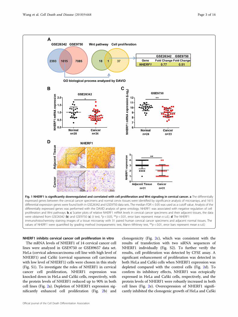
cancer, we compared the gene expression profilesbetween cervical cancer and normal cervix tissuesthrough microarray data obtained from GEO database.The data sets of GSE26342 and GSE9750 were analyzedby significance analysis of microarrays with the medianFDR < 0.05 as a cutoff value24. A total of 1615 genes wereidentified that differed significantly in expression in bothdata sets. Functional clustering analysis of these 1615genes revealed that 19 genes were involved in Wntpathway, and 38 genes participated in negative regulationof cell proliferation. NHERF1 was the only gene associatedwith both cell proliferation and Wnt signaling (Fig. 1a).To verify these findings, the levels of NHERF1 mRNA incervical cancer and their adjacent tissues were analyzed,and the results showed that NHERF1 mRNA was sig-nificantly decreased in the above two independent datasets (Fig. 1b, c). To further analyze the protein levels ofNHERF1 in cervical cancer, a tissue microarray contain-ing 31 paired cervical cancer and adjacent tissue speci-mens were used to analyze the expression level ofNHERF1. The protein levels of NHERF1 were alsorobustly reduced in cervical cancer specimens (Fig. 1d).Therefore, these findings suggest a tumor-suppressiverole of NHERF1 in cervical cancer. Wnt signaling and cellproliferation associated with downregulation of NHERF1might contribute to cervical cancer development andprogression.
Wang et al. Cell Death and Disease (2018) 9:668 Page 2 of 14
Official journal of the Cell Death Differentiation Association
NHERF1 inhibits cervical cancer cell proliferation in vitroThe mRNA levels of NHERF1 of 14 cervical cancer cell
lines were analyzed in GSE9750 or GSE89657 data set.HeLa (cervical adenocarcinoma cell line with high level ofNHERF1) and CaSki (cervical squamous cell carcinomawith low level of NHERF1) cells were chosen in this study(Fig. S1). To investigate the roles of NHERF1 in cervicalcancer cell proliferation, NHERF1 expression wasknocked down in HeLa and CaSki cells, respectively, withthe protein levels of NHERF1 reduced up to 90% in bothcell lines (Fig. 2a). Depletion of NHERF1 expression sig-nificantly enhanced cell proliferation (Fig. 2b) and
clonogenicity (Fig. 2c), which was consistent with theresults of transfection with two siRNA sequences ofNHERF1 individually (Fig. S2). To further verify theresults, cell proliferation was detected by CFSE assay. Asignificant enhancement of proliferation was detected inboth HeLa and CaSki cells when NHERF1 expression wasdepleted compared with the control cells (Fig. 2d). Toconfirm its inhibitory effects, NHERF1 was ectopicallyexpressed in HeLa and CaSki cells, respectively, and theprotein levels of NHERF1 were robustly increased in bothcell lines (Fig. 2e). Overexpression of NHERF1 signifi-cantly inhibited the clonogenic growth of HeLa and CaSki
Fig. 1 NHERF1 is significantly downregulated and correlated with cell proliferation and Wnt signaling in cervical cancer. a The differentiallyexpressed genes between the cervical cancer specimens and normal cervix tissues were identified by significance analysis of microarrays, and 1615differential expression genes were found both in GSE26342 and GSE9750 data sets. The median FDR < 0.05 was used as a cutoff value. Analysis of thedifferentially expressed genes was performed with the DAVID analysis of gene ontology. NHERF1 was associated with negative regulation of cellproliferation and Wnt pathways. b, c Scatter plots of relative NHERF1 mRNA levels in cervical cancer specimens and their adjacent tissues, the datawere obtained from GSE26342 (b) and GSE9750 (c) (t test, *p < 0.05, **p < 0.01, error bars represent mean ± s.d.). d The NHERF1immunohistochemistry staining images of a tissue microarray with 31 paired human cervical cancer specimens and adjacent normal tissues. Thevalues of NHERF1 were quantified by grading method (nonparametric test, Mann–Whitney test, **p < 0.01, error bars represent mean ± s.d.)
Wang et al. Cell Death and Disease (2018) 9:668 Page 3 of 14
Official journal of the Cell Death Differentiation Association
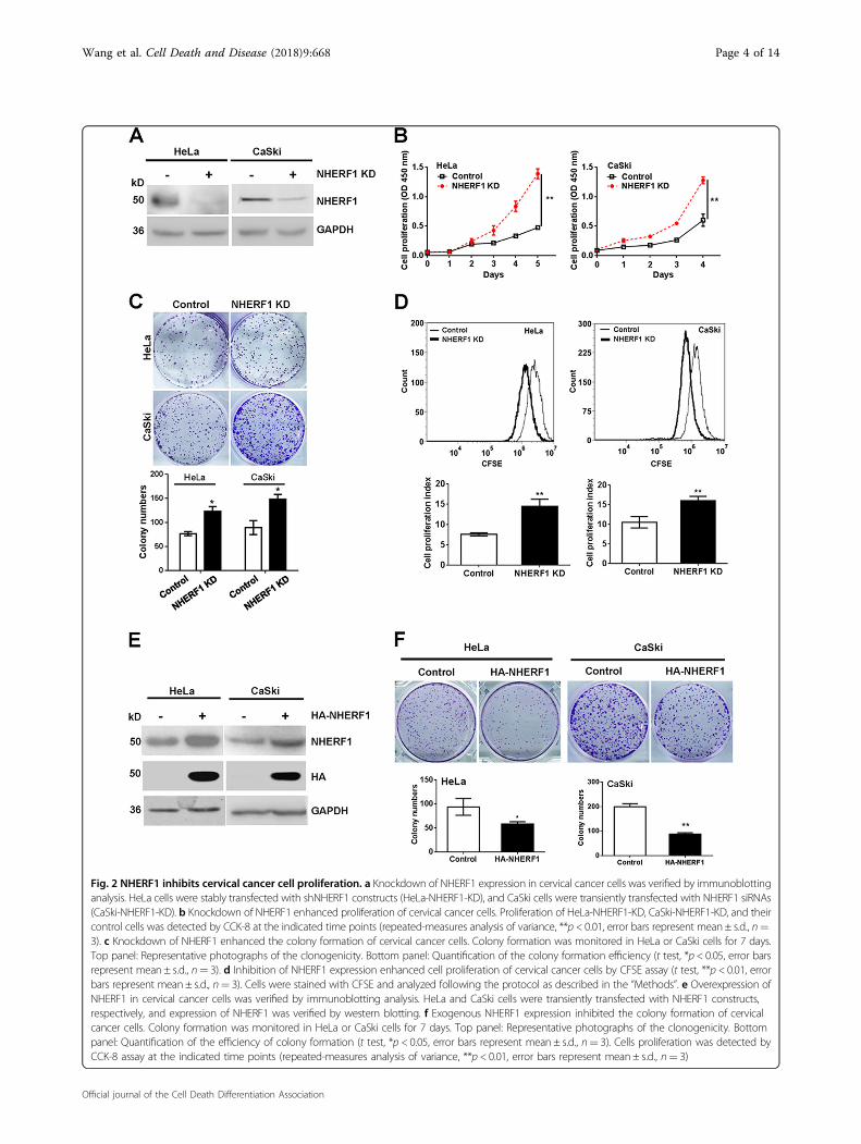
Fig. 2 NHERF1 inhibits cervical cancer cell proliferation. a Knockdown of NHERF1 expression in cervical cancer cells was verified by immunoblottinganalysis. HeLa cells were stably transfected with shNHERF1 constructs (HeLa-NHERF1-KD), and CaSki cells were transiently transfected with NHERF1 siRNAs(CaSki-NHERF1-KD). b Knockdown of NHERF1 enhanced proliferation of cervical cancer cells. Proliferation of HeLa-NHERF1-KD, CaSki-NHERF1-KD, and theircontrol cells was detected by CCK-8 at the indicated time points (repeated-measures analysis of variance, **p < 0.01, error bars represent mean ± s.d., n=3). c Knockdown of NHERF1 enhanced the colony formation of cervical cancer cells. Colony formation was monitored in HeLa or CaSki cells for 7 days.Top panel: Representative photographs of the clonogenicity. Bottom panel: Quantification of the colony formation efficiency (t test, *p < 0.05, error barsrepresent mean ± s.d., n= 3). d Inhibition of NHERF1 expression enhanced cell proliferation of cervical cancer cells by CFSE assay (t test, **p < 0.01, errorbars represent mean ± s.d., n= 3). Cells were stained with CFSE and analyzed following the protocol as described in the “Methods”. e Overexpression ofNHERF1 in cervical cancer cells was verified by immunoblotting analysis. HeLa and CaSki cells were transiently transfected with NHERF1 constructs,respectively, and expression of NHERF1 was verified by western blotting. f Exogenous NHERF1 expression inhibited the colony formation of cervicalcancer cells. Colony formation was monitored in HeLa or CaSki cells for 7 days. Top panel: Representative photographs of the clonogenicity. Bottompanel: Quantification of the efficiency of colony formation (t test, *p < 0.05, error bars represent mean ± s.d., n= 3). Cells proliferation was detected byCCK-8 assay at the indicated time points (repeated-measures analysis of variance, **p < 0.01, error bars represent mean ± s.d., n= 3)
Wang et al. Cell Death and Disease (2018) 9:668 Page 4 of 14
Official journal of the Cell Death Differentiation Association
cells (Fig. 2f), and these data were consistent with theproliferation results from HeLa cells (Fig. S3). Takentogether, these findings indicate that NHERF1 inhibitsproliferation of cervical cancer cells.
NHERF1 inhibits cervical cancer cell proliferation throughdownregulation of ACTN4We previously reported that NHERF1 downregulated
ACTN4 protein expression levels by promoting ACTN4ubiquitination and proteasomal degradation25. ACTN4could promote cervical cancer cell proliferation26. Thus, itis highly possible that NHERF1 may inhibit proliferationof cervical cancer cells through regulation of ACTN4protein expression. In order to explore this possibility, theendogenous levels of NHERF1 and ACTN4 in CaSki andHeLa cells were analyzed. We found that CaSki expressedrelatively low levels of NHERF1 and high levels of ACTN4compared with HeLa cells (Fig. S4A), whereas CaSki cells,as expected, exhibited higher proliferation ability thanHeLa cells (Fig. S4B–D), implying a potential role ofNHERF1 in cervical cancer cell proliferation via regula-tion of ACTN4. To further verify this hypothesis, pro-liferation of cervical cancer cells was analyzed aftercombined depletion of ACTN4 and NHERF1 expression.Data showed that knockdown of NHERF1 expressionupregulated ACTN4 protein levels, which were consistentwith our previous report25, and promoted proliferation ofHeLa (Fig. 3a) and CaSki cells (Fig. 3b) as compared withthe control. However, when ACTN4 expression wasknocked down by siRNA, NHERF1 had less effect on thecervical cancer cell proliferation (Fig. 3a, b and Fig. S5).These results were further confirmed with colony for-mation assay. Upon depletion of ACTN4 expression,NHERF1 failed to inhibit the clonogenic capacity of cer-vical cancer cells (Fig. 3c). Taken together, these dataindicate that NHERF1 inhibits cervical cancer cell pro-liferation via suppression of ACTN4 expression.
NHERF1 suppresses proliferation of cervical cancer cells viainhibition of ACTN4-mediated Wnt/β-catenin signalingactivationRecently, Ko et al. reported that ACTN4 increased
Wnt/β-catenin pathway activation by inhibition of β-catenin degradation, leading to promotion of cervicalcancer cell proliferation26. NHERF1 was found toassociate with Wnt pathway in this study (Fig. 1a). Thus,the regulation of β-catenin protein levels by NHERF1 wasfurther analyzed in cervical cancer cells. Western blotanalysis showed that knockdown of NHERF1 indeedincreased ACTN4 and β-catenin protein levels in HeLaand CaSki cells (Fig. 4a). Whereas, ACTN4 and β-cateninlevels were significantly downregulated when ectopicNHERF1 was expressed (Fig. 4b). To demonstratewhether regulation of β-catenin by NHERF1 required
ACTN4, β-catenin levels were analyzed after combinedknockdown of NHERF1 and ACTN4. The results showedthat silencing NHERF1 expression increased β-cateninlevels when ACTN4 existed. However, NHERF1 had lesseffect on β-catenin levels when ACTN4 was knockeddown (Fig. 4c). These data indicated that inhibition of β-catenin expression by NHERF1 required ACTN4. Since β-catenin acts as the central component in canonical Wntsignaling, we further investigated whether NHERF1 couldregulate Wnt/β-catenin pathway activation. The expres-sion of c-Myc and TCF-1, downstream target genes ofWnt/β-catenin signaling, was analyzed after treating thecervical cancer cells with IWR-1-endo, a Wnt/β-cateninsignaling inhibitor, in the presence or absence of NHERF1expression. Results showed that knockdown of NHERF1increased the expression of c-Myc and TCF-1, as weexpected. Meanwhile, it failed to regulate the expressionof c-Myc and TCF-1 when Wnt inhibitor was applied(Fig. 4d). To illuminate whether NHERF1 inhibited cer-vical cancer cell proliferation through Wnt pathway,CCK-8 assay was performed by treating the HeLa orCaSki cells with IWR-1-endo combined with/withoutknockdown of NHERF1 expression. Results showed thatdepletion of NHERF1 failed to promote cell proliferationwhen Wnt/β-catenin signaling was blocked by IWR-1-endo in both cells (Fig. 4e). These observations werefurther verified in colony formation assay. The clonogeniccapacity was increased when NHERF1 was depleted.However, clonogenic growth was not affected by NHERF1when IWR-1-endo was applied (Fig. 4f), indicating thatNHERF1 inhibition of cervical cancer cell proliferationwas mediated by suppression of Wnt/β-catenin signaling.Taken together, these data suggest that NHERF1 inhibitscervical cancer cells proliferation through reduction ofβ-catenin levels by regulation of ACTN4 expression.
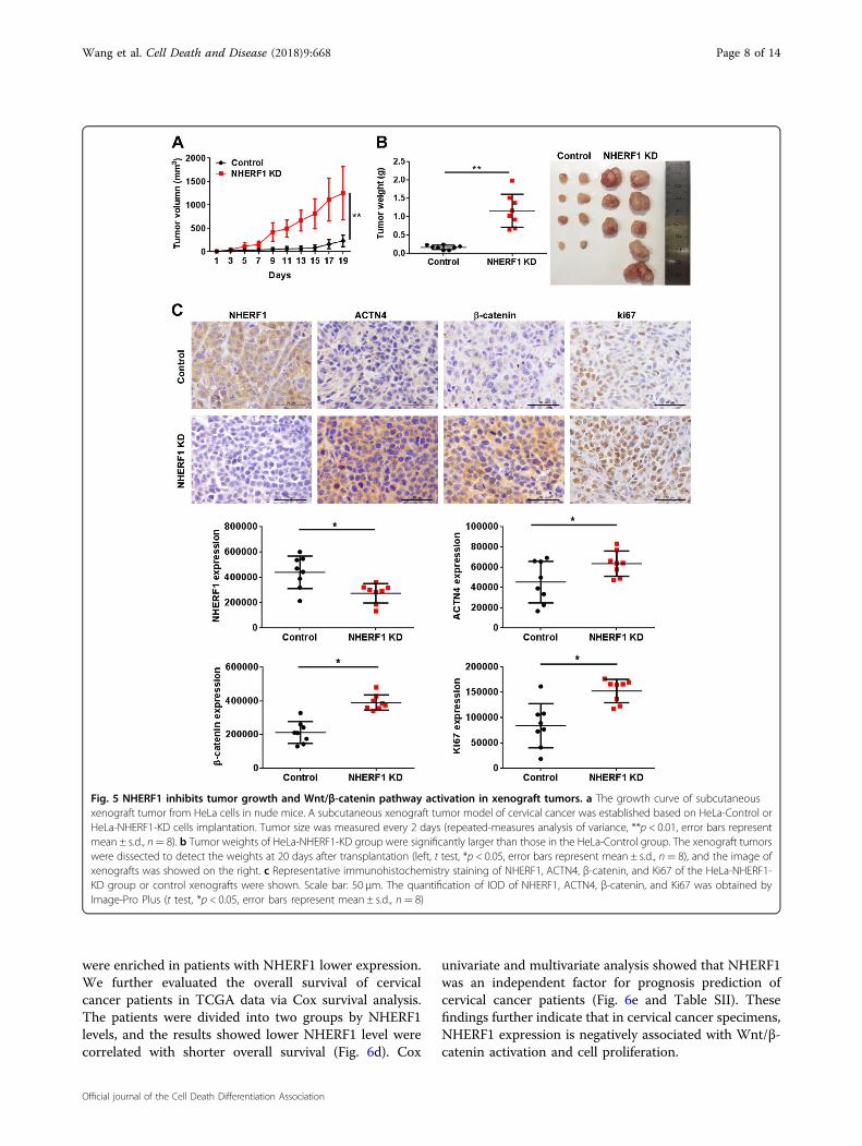
NHERF1 inhibits cervical cancer cell proliferation in vivowith the attenuation of Wnt/β-catenin signaling activationTo examine the roles of NHERF1 in the activation of
Wnt/β-catenin signaling and cervical cancer proliferationin vivo, HeLa cells were subcutaneously injected intoBALB/c nude mice. Knockdown of NHERF1 robustlyenhanced the growth of HeLa xenografts in nude micewithin 20 days (Fig. 5). Accordingly, both the volume(Fig. 5a) and weight (Fig. 5b) of tumors were significantlyenhanced in NHERF1 knocked down xenografts ascompared with the control group. Suppression ofNHERF1 in cervical cancer xenograft tumor increased itsACTN4 and β-catenin protein levels, and enhanced thelevels of Ki67 by immunohistochemical staining (Fig. 5c).These data suggest that NHERF1 depletion promotesproliferation of cervical cancer xenograft tumor byincreasing ACTN4 levels and activation of Wnt/β-cateninpathway.
Wang et al. Cell Death and Disease (2018) 9:668 Page 5 of 14
Official journal of the Cell Death Differentiation Association
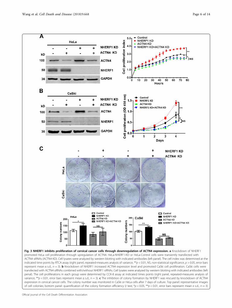
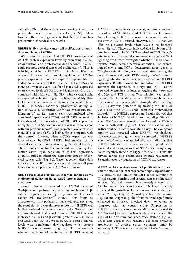
Fig. 3 NHERF1 inhibits proliferation of cervical cancer cells through downregulation of ACTN4 expression. a Knockdown of NHERF1promoted HeLa cell proliferation through upregulation of ACTN4. HeLa-NHERF1-KD or HeLa-Control cells were transiently transfected withACTN4 siRNAs (ACTN4 KD). Cell lysates were analyzed by western blotting with indicated antibodies (left panel). The cell index was determined at theindicated time points by RTCA assay (right panel, repeated-measures analysis of variance, **p < 0.01, NS, non-statistical significance, p > 0.05, error barsrepresent mean ± s.d., n= 3). b Knockdown of NHERF1 increased ACTN4 expression level and promoted CaSki cell proliferation. CaSki cells weretransfected with ACTN4 siRNAs combined with/without NHERF1 siRNAs. Cell lysates were analyzed by western blotting with indicated antibodies (leftpenal). The cell proliferations in each group were determined by CCK-8 assay at indicated times points (right panel, repeated-measures analysis ofvariance, **p < 0.01, error bars represent mean ± s.d., n= 3). c The inhibition of colony formation by NHERF1 was rescued by knockdown of ACTN4expression in cervical cancer cells. The colony number was monitored in CaSki or HeLa cells after 7 days of culture. Top panel: representative imagesof cell colonies; bottom panel: quantification of the colony formation efficiency (t test; *p < 0.05, **p < 0.01, error bars represent mean ± s.d., n= 3)
Wang et al. Cell Death and Disease (2018) 9:668 Page 6 of 14
Official journal of the Cell Death Differentiation Association
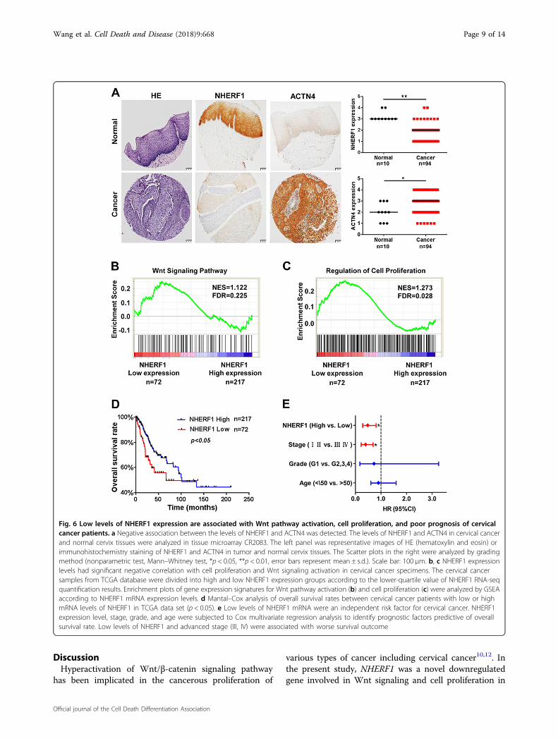
To further analyze the association of NHERF1, ACTN4,and Wnt/β-catenin activation in clinical cervical cancerspecimens, protein levels of NHERF1 and ACTN4 wereexamined with immunohistochemical staining. As com-pared with normal cervix tissues, NHERF1 protein levelswere markedly decreased in cervical cancer tissues, whichwas consistent with results of Fig. 1d, whereas ACTN4levels were significantly increased (Fig. 6a). Accordingly,the levels of ACTN4, β-catenin, c-Myc, and Ki67 were allincreased and NHERF1 levels was deceased in cervicalcancer specimens from THPA (www.proteinatlas.org)when compared with normal cervix tissues (Fig. S6).
These findings indicate that downregulation of NHERF1was associated with ACTN4 upregulation and Wnt/β-catenin activation in cervical cancer specimens. To verifythe relevance of NHERF1 expression with cell growth andbiologic pathways in cervical cancer pathogenesis, GSEAwas performed using TCGA cervical cancer data set withthe characteristics of patients shown in SupplementalTable I. The cervical cancer patients were stratified by thelower quartile of NHERF1 level in the specimens as high-and low-expression groups. Enrichment plots of GSEAshowed that the gene signatures of Wnt/β-catenin sig-naling activation (Fig. 6b) and cell proliferation (Fig. 6c)
Fig. 4 NHERF1 suppresses proliferation of cervical cancer cells through inhibition of Wnt/β-catenin pathway activation via ACTN4. aKnockdown of NHERF1 expression increased β-catenin protein level. Cell lysates of HeLa-NHERF1-KD/HeLa-Control or CaSki-NHERF1-KD/CaSki-Control cells were analyzed by western blotting with indicated antibodies. b NHERF1 overexpression reduced β-catenin protein level. HeLa and CaSkicells were transiently transfected with NHERF1 constructs, respectively, and the whole cell lysates were immunoblotted with the indicated antibodies.c Inhibition of β-catenin expression by NHERF1 required ACTN4 expression. HeLa-NHERF1-KD/HeLa-Control or CaSki-NHERF1-KD/CaSki-Control cellswere transfected with siRNAs of ACTN4 or control. Cell lysates were analyzed by western blot with indicated antibodies. d Wnt signaling inhibitorabolished NHERF1 inhibition of downstream genes activation of Wnt/β-catenin pathway. HeLa-NHERF1-KD/HeLa-Control or CaSki-NHERF1-KD/CaSki-Control cells were treated with or without IWR-1-endo (IWR-1, 20 μM, 24 h). The cell lysates were subjected to western blot by using specificantibodies. e NHERF1 inhibited cervical cancer cell proliferation through Wnt pathway. HeLa-NHERF1-KD/HeLa-Control and CaSki-NHERF1-KD/CaSki-Control cells were treated in the presence or absence of Wnt inhibitor IWR-1 (20 μM) for 72 h, then the proliferation of cells was assessed by CCK-8assay. Values were represented as relative value after comparing the absorbance at day 3 with that at day 0 (t test, *p < 0.05, error bars representmean ± s.d., n= 3). f NHERF1 inhibited the colony formation of cervical cancer cells via Wnt/β-catenin pathway. The clonogenicity of HeLa-NHERF1-KD/HeLa-Control and CaSki-NHERF1-KD/CaSki-Control cells was analyzed by colony formation assay in the presence or absence of Wnt inhibitor, IWR-1 (20 μM for 7 days). Top panel: typical images of cell colonies; bottom panel: quantification of the colony formation efficiency (t test, *p < 0.05, **p <0.01, error bars represent mean ± s.d., n= 3)
Wang et al. Cell Death and Disease (2018) 9:668 Page 7 of 14
Official journal of the Cell Death Differentiation Association
were enriched in patients with NHERF1 lower expression.We further evaluated the overall survival of cervicalcancer patients in TCGA data via Cox survival analysis.The patients were divided into two groups by NHERF1levels, and the results showed lower NHERF1 level werecorrelated with shorter overall survival (Fig. 6d). Cox
univariate and multivariate analysis showed that NHERF1was an independent factor for prognosis prediction ofcervical cancer patients (Fig. 6e and Table SII). Thesefindings further indicate that in cervical cancer specimens,NHERF1 expression is negatively associated with Wnt/β-catenin activation and cell proliferation.
Fig. 5 NHERF1 inhibits tumor growth and Wnt/β-catenin pathway activation in xenograft tumors. a The growth curve of subcutaneousxenograft tumor from HeLa cells in nude mice. A subcutaneous xenograft tumor model of cervical cancer was established based on HeLa-Control orHeLa-NHERF1-KD cells implantation. Tumor size was measured every 2 days (repeated-measures analysis of variance, **p < 0.01, error bars representmean ± s.d., n= 8). b Tumor weights of HeLa-NHERF1-KD group were significantly larger than those in the HeLa-Control group. The xenograft tumorswere dissected to detect the weights at 20 days after transplantation (left, t test, *p < 0.05, error bars represent mean ± s.d., n= 8), and the image ofxenografts was showed on the right. c Representative immunohistochemistry staining of NHERF1, ACTN4, β-catenin, and Ki67 of the HeLa-NHERF1-KD group or control xenografts were shown. Scale bar: 50 μm. The quantification of IOD of NHERF1, ACTN4, β-catenin, and Ki67 was obtained byImage-Pro Plus (t test, *p < 0.05, error bars represent mean ± s.d., n= 8)
Wang et al. Cell Death and Disease (2018) 9:668 Page 8 of 14
Official journal of the Cell Death Differentiation Association
DiscussionHyperactivation of Wnt/β-catenin signaling pathway
has been implicated in the cancerous proliferation of
various types of cancer including cervical cancer10,12. Inthe present study, NHERF1 was a novel downregulatedgene involved in Wnt signaling and cell proliferation in
Fig. 6 Low levels of NHERF1 expression are associated with Wnt pathway activation, cell proliferation, and poor prognosis of cervicalcancer patients. a Negative association between the levels of NHERF1 and ACTN4 was detected. The levels of NHERF1 and ACTN4 in cervical cancerand normal cervix tissues were analyzed in tissue microarray CR2083. The left panel was representative images of HE (hematoxylin and eosin) orimmunohistochemistry staining of NHERF1 and ACTN4 in tumor and normal cervix tissues. The Scatter plots in the right were analyzed by gradingmethod (nonparametric test, Mann–Whitney test, *p < 0.05, **p < 0.01, error bars represent mean ± s.d.). Scale bar: 100 μm. b, c NHERF1 expressionlevels had significant negative correlation with cell proliferation and Wnt signaling activation in cervical cancer specimens. The cervical cancersamples from TCGA database were divided into high and low NHERF1 expression groups according to the lower-quartile value of NHERF1 RNA-seqquantification results. Enrichment plots of gene expression signatures for Wnt pathway activation (b) and cell proliferation (c) were analyzed by GSEAaccording to NHERF1 mRNA expression levels. d Mantal–Cox analysis of overall survival rates between cervical cancer patients with low or highmRNA levels of NHERF1 in TCGA data set (p < 0.05). e Low levels of NHERF1 mRNA were an independent risk factor for cervical cancer. NHERF1expression level, stage, grade, and age were subjected to Cox multivariate regression analysis to identify prognostic factors predictive of overallsurvival rate. Low levels of NHERF1 and advanced stage (III, IV) were associated with worse survival outcome
Wang et al. Cell Death and Disease (2018) 9:668 Page 9 of 14
Official journal of the Cell Death Differentiation Association
cervical cancer (Fig. 1). The molecular mechanisms ofNHERF1 in the regulation of Wnt/β-catenin signaling andthe proliferation of cervical cancer were largely unknown.NHERF1 is extensively expressed in the epithelium of
tissues and has been found to be implicated in varioustypes of cancer. Extensive studies suggest an oncogenicrole of NHERF1 in breast cancer27–29, ovarian mucinouscarcinoma30, hepatocellular carcinoma31, and glio-blastoma32. On the contrary, several reports show thatNHERF1 acts as a tumor suppressor in esophageal squa-mous cell carcinoma33 and triple-negative breast cancer34.In the present study, we found that NHERF1 inhibitedcervical cancer cell proliferation from in vitro (Fig. 2 andFig. S2,3) and in vivo models (Fig. 5), suggesting an anti-proliferation and tumor-suppressive effect of NHERF1 incervical cancer. NHERF1 has emerged as a key regulatorof cancer signaling network via assembling cancer-associated proteins35. In previous study, we demon-strated that NHERF1 interacted with ACTN4 anddownregulated the expression levels of ACTN425. Studyfrom other groups showed that upregulation of ACTN4enhanced proliferation of cervical cancer cells in cellullarand xenograft models by promoting stability of β-cateninthrough phosphorylation of Akt and GSK3β26,36. In thepresent study, we further revealed that NHERF1 inhibi-tion of cervical cancer cell proliferation was mediated viaACTN4 (Fig. 3 and Fig. S5). These findings have providedfurther insights into the role of ACTN4 in cancer cellproliferation apart from its roles in maintaining cytoske-letal integrity37.Activation of Wnt/β-catenin signaling pathway is sig-
nificantly associated with the cell proliferation and poorprognosis of cervical cancer17,21. The present in vitro datashowed that NHERF1 downregulated the levels ofβ-catenin by suppression of ACTN4 expression (Fig. 4a–c).Blocking Wnt/β-catenin signaling abolished the enhance-ment of cervical cancer cell proliferation induced byknockdown of NHERF1 (Fig. 4d–h). Data from in vivomouse models and clinical specimens showed prominentdownregulation of NHERF1 and upregulation of ACTN4,β-catenin, and its downstream targets (Figs. 1, 5, 6a andFig. S6). Further analysis revealed that lower levels ofNHERF1 were prominently correlated with activation ofWnt/β-catenin signaling and cell proliferation (Fig. 6b, c),and were an independent risk factor for worse prognosis ofcervical cancer patients (Fig. 6d, e). NHERF1 loss has alsobeen reported to associate with the activation of otheroncogenic pathways, such as the ERK38 and Akt signaling39
in cervical cancer cells. However, there was no associationbetween ERK or Akt signaling activation and the overallsurvival of cervical cancer patients in TCGA database (datanot shown). All these findings suggest that NHERF1 maysuppress Wnt/β-catenin signaling activation via a decreasein ACTN4 levels to elicit anti-proliferation and tumor-
suppressive effects in cervical cancer. It is likely thatdownregulation of NHERF1 may result in development ofcervical cancer by promotion of β-catenin-mediated pro-liferation. Therefore, NHERF1 may potentially serve as abiomarker for prognosis evaluation or a therapeutic targetof cervical cancer.Cisplatin-based chemotherapy is the standard treatment
for the advanced stage and recurrent cervical cancer1.However, chemoresistance seriously compromises theefficacy of cisplatin40. Therefore, cisplatin resistance hasbecome a major clinical challenge. Recently, increasingevidences indicate that overactivation of Wnt signalingpathway has been implicated in resistance to che-motherapy41,42. In the present study, results showed thatcisplatin resistance was associated with dysregulation ofWnt signaling in HeLa cells (Fig. S7A), which furtherindicated that Wnt signaling may play a key role in cis-platin resistance in cervical cancer. However, the detailedmechanisms of Wnt signaling in cisplatin resistance arestill far from clear. In this study, we showed that bothgene signatures of cisplatin resistance and Wnt signalingwere enriched in NHERF1 low-expression cervical cancerpatients (Fig. S7B,C). Further results showed that activa-tion of downstream genes of cisplatin resistance and Wntsignaling was more profound in cisplatin-resistantpatients (Fig. S7D,E). We previously reported that lowlevels of NHERF1 expression were associated with cis-platin resistance in cervical cancer39. Taken together, weproposed a possible molecular mechanism for cisplatinresistance in cervical cancer by which downregulation ofNHERF1 promotes overactivation of Wnt/β-catenin sig-naling and results in cisplatin resistance. Further study isneeded to confirm this hypothesis.In the present study, significant worse prognosis was
found in cervical cancer patients with lower levels ofNHERF1 (Fig. 6d). It was surprising to find that propor-tion of HPV-inactive patients was significantly higherthan HPV-active group in NHERF1 low-expression cer-vical cancer patients (Fig. S8A and Table SIII). HPV-inactive cervical cancer patients had worse prognosis thanHPV-active group (Fig. S8B and Table SIV), in accordancewith findings from Banister et al. that activation ofWnt/β-catenin signaling was associated with worseprognosis of HPV-inactive cervical cancer patients16.However, the molecular mechanisms underlying thedevelopment of HPV-inactive cervical carcinoma are stillelusive. Our data further showed that the mRNA levels ofNHERF1 in HPV-inactive cervical cancer patients weresignificantly lower and activation of Wnt/β-catenin sig-naling and proliferation genes were more prominent inthis subgroup of patients (Fig. S8C–E). Therefore, it islikely that the worse prognosis of HPV-inactive cervicalcancer patients may be attributed to robustly low levels ofNHERF1. These findings suggest that overactivation of
Wang et al. Cell Death and Disease (2018) 9:668 Page 10 of 14
Official journal of the Cell Death Differentiation Association
Wnt/β-catenin signaling in response to the significantdownregulation of NHERF1 at mRNA levels may con-tribute to the development and progression of HPV-inactive cervical cancer.In HPV-active cervical cancer, activation of Wnt/β-
catenin signaling and cellular proliferation was moreremarkable in patients with poor prognosis (Fig. S9B,C).However, there was no difference of the mRNA levels ofNHERF1 between patients with good or poor prognosis(Fig. S9A). This seems in contradiction with the resultsfrom present study. Several studies reported thatoncogenic E6 and E7 proteins of HR-HPV coulddownregulate the levels of target proteins via regulationof the target stability43,44. For example, HPV16-E6protein has been found to degrade NHERF1 protein atposttranslational level45. Therefore, it is reasonable tospeculate that higher the oncogenic activities of HR-HPV-E6, the more powerful degradation of NHERF1protein. Downregulation of NHERF1 protein then leadsto oncogenenic proliferation upon upregulation ofACTN4 and activation of Wnt/β-catenin signaling inHPV-active cervical cancer. This possibility needs to beverified by comparison of the mRNA and protein levelsof NHERF1, activation status of Wnt/β-catenin signal-ing, and prognosis of HPV-active cervical cancer in thefuture study.In summary, the present study provides novel evidences
for a tumor-suppressive role of NHERF1 in cervical can-cer cell proliferation by attenuation of Wnt/β-cateninsignaling via a decrease in ACTN4 expression. Low levelof NHERF1 may contribute to the development of cervicalcancer and indicated poor prognosis of cervical cancerpatients. These findings could also improve our under-standing for the molecular mechanisms of cisplatinresistance, and the development and prognosis of HPV-inactive and HPV-active cervical cancer.
Materials and methodsCell culture and transfectionCells were purchased from the National Infrastructure
of Cell Line Resource (Beijing, China). HeLa and CaSkicervical cancer cells were cultured in DMEM, RPMI 1640medium (Gibico, Cleveland, TN), respectively, with 10%FBS (Gibico, Cleveland, TN) at 37℃ in an atmosphere of5% CO2. The stable transfection of HeLa cells was per-formed as previously described25.
RNA interference and plasmid constructionssiRNAs were purchased from Invitrogen (Carlsbad, CA)
and the sequences were shown as follows:NHERF1 siRNA1#: 5′-GCUAUGGCUUCAACCUGCA
TT-3′.NHERF1 siRNA2#: 5′-GUCGACCACCAGCAGGCGC
ACGGCGUUG-3′.
ACTN4 siRNA1#: 5′-CCUGAACAAUGCCUUCGAATT-3′.ACTN4 siRNA2#: 5′-CCUGAACAAUGCCUUCGAA
TT-3′.Negative control siRNA1#: 5′-UUCUUCGAACGUGU
CACG-3′.Negative control siRNA2#: 5′-UCCAGACGGCGCAGU
GGGCGACCGCUAC-3′.Cells were transfected with a mixture of two siRNA
sequences for relevant RNA interference experiments.NHERF1 was stably knocked down by shNHERF1 inHeLa (Hela-NHERF1-KD) cells and transiently knockeddown by siRNA in CaSki (CaSki-NHERF1-KD) cells.pCMV-HA and pCMV-HA-NHERF1 plasmids were
kindly provided by Dr. Randy Hall (EmoryUniversity,Atlanta, GA). pSuper.puro luciferase control and pSuper.puro-shNHERF1 plasmids were kind gifts of Dr. MargaretJ. Wheelock (University of Nebraska Medical Center,Omaha, NE).
Western blotting and reagentsWestern blotting assay was performed as previously
described46. The anti-NHERF1 was purchased fromSigma-Aldrich (HPA009672, St. Louis, MO) and BectonDickinson Labware (#611161, Billerica, MA), respectively.Anti-HA (#561) was purchased from Medical & BiologicalLaboratories (Nagoya, Japan). Anti-c-Myc (#ab32072) andanti-β-catenin (#ab22656) were purchased from Abcam(Cambridge, UK). Anti-GAPDH (#5174), anti-β-catenin(#9581), and anti-TCF-1 (#2206) were purchased fromCell Signaling Technology (Danvers, MA). Anti-ACTN4was purchased from Enzo Life Sciences (#ALX-210-356,Shanghai, China) and Santa Cruz Biotechnology (#sc-134236, Santa Cruz, CA), respectively. Anti-Ki67 (#zm-0166) and horse radish peroxidase-conjugated secondaryantibodies were purchased from ZSGB-BIO (Beijing,China). Infrared fluorescent dyes-conjugated secondaryantibodies were purchased from LI-COR Biosciences(Lincoln, NE). IWR-1-endo (#S7086) was purchased fromSelleck (Houston, TX).
Cell proliferation assayFor CCK-8 assay, cells were seeded in 96-well plates at a
density of 3000 per well and cultured for 1–5 days, andCCK-8 (Dojindo, Kumamoto, Japan) was added accordingto the manufacturer’s instructions and absorbance wasmeasured at 450 nm with an EnSpire label microplatereader (PerkinElmer, Waltham, MA).For CFSE (carboxy fluorescein succinimidyl ester) assay,
single-cell suspension at a density of 1 × 10⁶ cells per mlwas stained with CFSE Cell Proliferation Kit (#C34554,Life Technologies, Carlsbad, CA) at day 1 and continuedthe culture for 3 days. The labeling cells were analyzed byflow cytometry at day 1 and day 3, respectively. The
Wang et al. Cell Death and Disease (2018) 9:668 Page 11 of 14
Official journal of the Cell Death Differentiation Association
proliferation index of each group was analyzed by com-paring the fluorescence value of day 1 and day 3 by ModfitLT (Verity Software House, Topsham, ME).For RTCA (real-time cell analysis) assay, cells were
cultured in 16-well plates (3000 cells per well, E-Plate 16,ACEA Biosciences Inc) and proliferation index wasmonitored by the xCELLigence system (ACEA Bios-ciences Inc, San Diego, CA) for 72 h.For colony formation assay, cells were cultured in 6-well
plates (1000 cells per well) for 7 days. The number ofcolonies (> 50 cells) were counted after staining with 0.5%crystal violet.
In vivo xenograft formation assayThis study was performed following the Guide for the
Care and Use of Laboratory Animals by National Insti-tutes of Health, and all procedures were approved by theAnimal Care and Use Committee of Capital MedicalUniversity.BALB/c nude mice (5 weeks, female) were sub-
cutaneously injected with HeLa-NHERF1-KD or controlcells (1 × 10⁵ cells in 0.1 mL PBS per mouse, 8 mice pergroup) and monitored every 2 days for the growth oftumors (volume= (length × width2)/2). Mice were killed20 days after the injection. The tumor xenografts wereembedded in paraffin for further studies.
Immunohistochemical stainingThe tissue microarrays were immunostained with anti-
NHERF1 (HPA009672) and anti-ACTN4 (sc-134236),respectively. The intensity of immunostaining in indivi-dual tumor tissue (0, no staining; 1, weak staining; 2,moderate staining; 3, strong staining), and the stainingpercentage of tumor tissue (0, none; 1, 1%–25%; 2,26%–50%; 3, 51%–75%; 4, >75%) were scored. The abso-lute value of the protein expression levels was thus clas-sified into four grades after multiplying the correspondingintensity value with percentage scores: I (grades 0–3), II(grades 4–6), III (grades 7–9), and IV (grades 10–12).Xenograft tumor tissues were stained by anti-NHERF1,
anti-ACTN4, anti-β-catenin (#ab22656), or anti-Ki67antibody, respectively. The average IOD (integratedoptical density) of five fields of each sample was analyzedby Image-Pro Plus (Rockville, MD) and represented theexpression level of protein.
Data set collection and cervical cancer patient samplesThe tissue microarrays (CR2083, containing biopsies
from 94 cases of cervical cancer and 10 normal cervicaltissues) were purchased from Biomax, Xi’an Alenabio(Xi’an, Shanxi, China), and the tissue microarrays (OD-CT-RpUtr03-006, containing cervical cancer and adjacentnormal tissues biopsies from 31 stage III cervical cancerpatients) were purchased from Shanghai outdo biotech
co., LTD. GSE26342 and GSE9750 from GEO (two of thelargest cervical cancer data sets with more than 20 casesof normal cervix tissues and 30 cases of cervical cancerpatients) were used for the screening of differentialexpression genes by significance analysis of microarrays(http://statweb.stanford.edu/~tibs/SAM/) and DAVIDanalysis (https://david.ncifcrf.gov). GSE151120 was usedfor GO (gene ontology) analysis by protein analysisthrough evolutionary relationships (http://pantherdb.org/webservices/go/overrep.jsp). GSE89657 and GSE9750from GEO were used to analyze the NHERF1 mRNAexpression level in cervical cancer cell lines. RNA-Seqdata from TCGA cervical cancer patients were collectedfrom Synapse website (http://synapse.org;syn1571569).Clinical data were downloaded from cBioPortal database(http://www.cbioportal.org). Data of HPV-infected statuswere obtained from the study by Banister et al. HPV-active: with high levels of E6/E7 expression; HPV-inactive:with low or zero E6/E7 expression16. All the samplesobtained from GSE26342, GSE9750, and TCGA data setswere untreated primary cervical cancer.The immunohistochemistry-based protein expression
images of cervical cancer and normal cervix tissues weredownloaded from THPA (The Human Protein Atlas)(www.proteinatlas.org) and analyzed by grading method.
Gene set enrichment analysis (GSEA)GSEA (www.broad.mit.edu/gsea) protocol was per-
formed as previously described47. The association betweenphenotypes, biological processes/pathway, and proteinmRNA expression level were analyzed. Pre-defined geneset were obtained from the Molecular Signatures Data-base, MSigDB (http://software.broadinstitute.org/gsea/msigdb). Gene sets: GO_CANONICAL_WNT_SIGNA-LING_PATHWAY (M12752), GO_REGULATION_OF_EPITHELIAL_CELL_PROLIFERATION (M12114),KANG_CISPLATIN_RESISTANCE_UP (M2767). TheFDR (false discovery rate) score smaller than 0.25 wasconsidered significant enrichment of a gene set48.
Statistical analysisAll statistical analyses were carried out using the
GraphPad prism 5 (Graphpad Software, Inc., La Jolla, CA)or SPSS (SPSS Inc, Chicago, IL). It was considered asstatistical significant when p < 0.05 (p < 0.05 and p < 0.01are designated by * and **, respectively).
AcknowledgementsThis work was supported by the National Natural Science Foundation of thePeople’s Republic of China (81772707, 81572333, 81302373, and 81272887);Beijing Natural Science Foundation Program and Scientific Research KeyProgram of Beijing Municipal Commission of Education (KZ201710025015);Scientific Research Common Program of Beijing Municipal Commission ofEducation (KM201410025001); Support Project of High-level Teachers in BeijingMunicipal Universities in the Period of 13th Five-year Plan (IDHT20170516);BaiQianWan Talents Program (2017–2018).
Wang et al. Cell Death and Disease (2018) 9:668 Page 12 of 14
Official journal of the Cell Death Differentiation Association
Author details1Department of Biochemistry and Molecular Biology, Capital MedicalUniversity, Beijing, China. 2Beijing Key Laboratory for Tumor Invasion andMetastasis, Beijing, China. 3Core Facilities Center, Capital Medical University,Beijing, China. 4Department of Histology and Embryology, Capital MedicalUniversity, Beijing, China
Conflict of interestThe authors declare that they have no conflict of interest.
Publisher's noteSpringer Nature remains neutral with regard to jurisdictional claims inpublished maps and institutional affiliations.
Supplementary Information accompanies this paper at https://doi.org/10.1038/s41419-018-0711-x.
Received: 5 February 2018 Revised: 9 April 2018 Accepted: 10 May 2018
References1. Koh, W. J. et al. Cervical cancer, version 2.2015. J. Natl. Compr. Cancer Netw. 13,
395–404 (2015).2. Steenbergen, R. D., Snijders, P. J., Heideman, D. A. & Meijer, C. J. Clinical
implications of (epi)genetic changes in HPV-induced cervical precancerouslesions. Nat. Rev. Cancer 14, 395–405 (2014).
3. Kisseljov, F., Sakharova, O. & Kondratjeva, T. Cellular and molecular biologicalaspects of cervical intraepithelial neoplasia. Int. Rev. Cell Mol. Biol. 271, 35–95(2008).
4. Manzo-Merino, J. et al. The role of signaling pathways in cervical cancer andmolecular therapeutic targets. Arch. Med. Res. 45, 525–539 (2014).
5. Conesa-Zamora, P., Torres-Moreno, D., Isaac, M. A. & Perez-Guillermo, M. Geneamplification and immunohistochemical expression of ERBB2 and EGFR incervical carcinogenesis. Correlation with cell-cycle markers and HPV presence.Exp. Mol. Pathol. 95, 151–155 (2013).
6. Lee, M. S. et al. PI3K/AKT activation induces PTEN ubiquitination and desta-bilization accelerating tumourigenesis. Nat. Commun. 6, 7769 (2015).
7. Oh, M. J. et al. Detection of epidermal growth factor receptor in the serum ofpatients with cervical carcinoma. Clin. Cancer Res. 6, 4760–4763 (2000).
8. Longatto-Filho, A. et al. Molecular characterization of EGFR, PDGFRAand VEGFR2 in cervical adenosquamous carcinoma. BMC Cancer 9, 212(2009).
9. Yoshida, S., Kajitani, N., Satsuka, A., Nakamura, H. & Sakai, H. Ras modifiesproliferation and invasiveness of cells expressing human papilloma virusoncoproteins. J. Virol. 82, 8820–8827 (2008).
10. Ford, C. E., Henry, C., Llamosas, E., Djordjevic, A. & Hacker, N. Wnt signalling ingynaecological cancers: a future target for personalised medicine? Gynecol.Oncol. 140, 345–351 (2016).
11. Bahrami, A. et al. Clinical significance and prognosis value of Wnt signalingpathway in cervical cancer. J. Cell. Biochem. 118, 3028–3033 (2017).
12. Tomaic, V. Functional roles of E6 and E7 oncoproteins in HPV-inducedmalignancies at diverse anatomical sites. Cancers 8, https://doi.org/10.3390/cancers8100095 (2016).
13. Lichtig, H. et al. HPV16 E6 augments Wnt signaling in an E6AP-dependentmanner. Virology 396, 47–58 (2010).
14. Uren, A. et al. Activation of the canonical Wnt pathway during genital kera-tinocyte transformation: a model for cervical cancer progression. Cancer Res.65, 6199–6206 (2005).
15. Bulut, G. et al. Beta-catenin accelerates human papilloma virus type-16mediated cervical carcinogenesis in transgenic mice. PLoS ONE 6, e27243(2011).
16. Banister, C. E., Liu, C., Pirisi, L., Creek, K. E. & Buckhaults, P. J. Identification andcharacterization of HPV-independent cervical cancers. Oncotarget 8,13375–13386 (2017).
17. Ramachandran, I. et al. Wnt inhibitory factor 1 induces apoptosis and inhibitscervical cancer growth, invasion and angiogenesis in vivo. Oncogene 31,2725–2737 (2012).
18. Chang, B. et al. Klotho inhibits the capacity of cell migration and invasion incervical cancer. Oncol. Rep. 28, 1022–1028 (2012).
19. Asem, M. S., Buechler, S., Wates, R. B., Miller, D. L. & Stack, M. S. Wnt5a signalingin cancer. Cancers 8, https://doi.org/10.3390/cancers8090079 (2016).
20. Pereira-Suarez, A. L. et al. Frequent alterations of the beta-catenin protein incancer of the uterine cervix. Tumour Biol. 23, 45–53 (2002).
21. Liang, J. et al. Beta-catenin expression negatively correlates with WIF1 andpredicts poor clinical outcomes in patients with cervical cancer. Biomed. Res.Int. 2016, 4923903 (2016).
22. Klaus, A. & Birchmeier, W. Wnt signalling and its impact on development andcancer. Nat. Rev. Cancer 8, 387–398 (2008).
23. Segditsas, S. & Tomlinson, I. Colorectal cancer and genetic alterations in theWnt pathway. Oncogene 25, 7531–7537 (2006).
24. Poliseno, L. et al. Distinguishing between nodular and superficial spreadingmelanoma using specific microRNA alterations. J. Clin. Oncol. 29, 8540(2011).
25. Sun, L. et al. NHERF1 regulates actin cytoskeleton organization throughmodulation of alpha-actinin-4 stability. FASEB J. 30, 578–589 (2016).
26. An, H. T., Yoo, S. & Ko, J. Alpha-actinin-4 induces the epithelial-to-mesenchymaltransition and tumorigenesis via regulation of snail expression and beta-catenin stabilization in cervical cancer. Oncogene 35, 5893–5904 (2016).
27. Yao, W. et al. EBP50 inhibits EGF-induced breast cancer cell proliferation byblocking EGFR phosphorylation. Amino Acids 43, 2027–2035 (2012).
28. Cardone, R. A. et al. The NHERF1 PDZ2 domain regulates PKA-RhoA-p38-mediated NHE1 activation and invasion in breast tumor cells. Mol. Biol. Cell 18,1768–1780 (2007).
29. Malfettone, A., Saponaro, C., Paradiso, A., Simone, G. & Mangia, A. Peritumoralvascular invasion and NHERF1 expression define an immunophenotype ofgrade 2 invasive breast cancer associated with poor prognosis. BMC Cancer12, 106 (2012).
30. Tabrizi, A. D. et al. Primary ovarian mucinous carcinoma of intestinal type:significance of pattern of invasion and immunohistochemical expressionprofile in a series of 31 cases. Int. J. Gynecol. Pathol. 29, 99–107 (2010).
31. Shibata, T., Chuma, M., Kokubu, A., Sakamoto, M. & Hirohashi, S. EBP50, a beta-catenin-associating protein, enhances Wnt signaling and is over-expressed inhepatocellular carcinoma. Hepatology 38, 178–186 (2003).
32. Kislin, K. L., McDonough, W. S., Eschbacher, J. M., Armstrong, B. A. & Berens, M.E. NHERF-1: modulator of glioblastoma cell migration and invasion. Neoplasia11, 377–387 (2009).
33. Wang, L. et al. Reduced EBP50 expression or mis-localization of theEBP50 protein is associated with the malignant progression of esophagealsquamous cell carcinoma. Eur. Rev. Med. Pharmacol. Sci. 18, 3854–3863(2014).
34. Wang, Y. et al. NHERF1 inhibits proliferation of triple-negative breast cancercells by suppressing GPER signaling. Oncol. Rep. 38, 221–228 (2017).
35. Vaquero, J., Nguyen Ho-Bouldoires, T. H., Claperon, A. & Fouassier, L. Role of thePDZ-scaffold protein NHERF1/EBP50 in cancer biology: from signaling reg-ulation to clinical relevance. Oncogene 36, 3067–3079 (2017).
36. Vandermoere, F. et al. Proteomics exploration reveals that actin is a signalingtarget of the kinase Akt. Mol. Cell Proteomics 6, 114–124 (2007).
37. Hsu, K. S. & Kao, H. Y. Alpha-actinin 4 and tumorigenesis of breast cancer.Vitam. Horm. 93, 323–351 (2013).
38. Peng, Z., Wang, Q., Zhang, Y., He, J. & Zheng, J. EBP50 interacts with EGFR andregulates EGFR signaling to affect the prognosis of cervical cancer patients. Int.J. Oncol. 49, 1737–1745 (2016).
39. Tao, T. et al. NHERF1 enhances cisplatin sensitivity in human cervical cancercells. Int. J. Mol. Sci. 18, https://doi.org/10.3390/ijms18010005 (2017).
40. Eskander, R. N. & Tewari, K. S. Chemotherapy in the treatment of metastatic,persistent, and recurrent cervical cancer. Curr. Opin. Obstet. Gynecol. 26,314–321 (2014).
41. Xu, H. et al. Targeting the eIF4E/beta-catenin axis sensitizes cervical carcinomasquamous cells to chemotherapy. Am. J. Transl. Res. 9, 1203–1212 (2017).
42. Wang, T., Liu, Z., Shi, F. & Wang, J. Pin1 modulates chemo-resistance by up-regulating FoxM1 and the involvements of Wnt/beta-catenin signalingpathway in cervical cancer. Mol. Cell Biochem. 413, 179–187 (2016).
43. Scheffner, M., Werness, B., Huibregtse, J., Levine, A. & Howley, P. The E6oncoprotein encoded by human papillomavirus types 16 and 18 promotesthe degradation of p53. Cell 63, 1129–1136 (1990).
44. Thomas, M. et al. Oncogenic human papillomavirus E6 proteins target theMAGI-2 and MAGI-3 proteins for degradation. Oncogene 21, 5088–5096(2002).
Wang et al. Cell Death and Disease (2018) 9:668 Page 13 of 14
Official journal of the Cell Death Differentiation Association

45. Accardi, R. et al. E6 and E7 from human papillomavirus type 16 cooperate totarget the PDZ protein Na/H exchange regulatory factor 1. J. Virol. 85,8208–8216 (2011).
46. Ma, Q. et al. MAGI3 negatively regulates Wnt/beta-catenin signaling andsuppresses malignant phenotypes of glioma cells. Oncotarget 6, 35851–35865(2015).
47. Tao, T. et al. PDZK1 inhibits the development and progression of renal cellcarcinoma by suppression of SHP-1 phosphorylation. Oncogene 36,6119–6131 (2017).
48. Subramanian, A. et al. Gene set enrichment analysis: a knowledge-basedapproach for interpreting genome-wide expression profiles. Proc. Natl Acad.Sci. USA 102, 15545–15550 (2005).
Wang et al. Cell Death and Disease (2018) 9:668 Page 14 of 14
Official journal of the Cell Death Differentiation Association