Paclitaxel inhibits cervical cancer via autophagy · Cervical cancer, Paclitaxel, LncRNA,...
8
3010 Abstract. – OBJECTIVE: To observe the ef- fect of paclitaxel on the autophagy of human cervical cancer cell lines by the expression reg- ulation of lncRNARP11-381N20.2 as well as to explore the interaction and relationship between autophagy, proliferation, and apoptosis of cervi- cal cancer cells. MATERIALS AND METHODS: Genome-wide expression profiles of tumors and their suscep- tibility to drugs were downloaded through TCGA database to find differentially expressed lncRNA RP11-381N20.2 in chemosensitive sensitive and insensitive groups. Expression of RP11-381N20.2 in 60 cervical cancer tissues and 30 normal tis- sues was detected by qRT-PCR. The relationship between the expression of RP11-381N20.2 and the clinicopathological parameters of lung can- cer was statistically analyzed. The recombinant plasmid pcDNA-RP11-381N20.2 and pcDNA-NC of RP11-381N20.2 were transfected into SiHa cells by lipofectamine, respectively. The autoph- agy and phenotypic effects were observed. Cell proliferation was determined by colony formation assay. Apoptosis was detected by flow cytome- try. Western blot was conducted to detect expres- sions of autophagy-related proteins. RESULTS: Genome-wide expression profiles of chemotherapy-sensitive and insensitive da- ta in patients with cervical cancer in TCGA da- tabase were analyzed by edger package, results showed that the expression of lncRNA RP11- 381N20.2 was significantly lower in the che- motherapy-insensitive group. qRT-PCR results showed that the expression of RP11-381N20.2 in cervical cancer was decreased, and the total survival time of patients was positively correlat- ed with the expression of RP11-381N20.2. RP11- 381N20.2 was associated with TNM (tumor node metastasis) staging and tumor size. Biological functions of SiHa cells showed that the expres- sion of RP11-381N20.2 was negatively correlated with the treatment time and dose of paclitaxel. Colony formation assay showed that paclitaxel could inhibit the proliferation of cervical cancer cells in a dose-dependent manner. Flow cytome- try showed that paclitaxel induced apoptosis of cervical cancer cells, which was more promot- ed after combination with RP11-381N20.2. West- ern blot results suggested that paclitaxel could induce autophagy in cervical cancer cells in a time- and dose-dependent manners. Paclitaxel combined with RP11-381N20.2 could significant- ly increase apoptosis of cervical cancer cells. CONCLUSIONS: During the killing process of paclitaxel on cervical cancer SiHa cells, cell au- tophagy would affect the efficacy, after overex- pression of RP11-381N20.2 in SiHa cells, autoph- agy induced by paclitaxel was inhibited, thereby enhancing the killing effect of paclitaxel on tu- mor cells. Key Words: Cervical cancer, Paclitaxel, LncRNA, RP11-381N20.2, Autophagy, Proliferation, Apoptosis. Introduction Cervical cancer is a common malignant tumor of the female reproductive system. With the gene- ral survey and public treatment of cervical cancer has been developed, the prevalence of cervical can- cer has dropped significantly and the 5-year survi- val rate has also increased 1 . However, as a preven- table disease, the incidence of cervical cancer still remains the seventh in all cancers worldwide 2 , the mortality rate ranks the fourth 3,4 . It is worth noting that studies found that patients with cervical can- cer presented a younger age of onset 5,6 ; therefore, the prevention and treatment of cervical cancer is urgent. It is an important issue in clinical research to further study the pathogenesis and look for new therapeutic targets of cervical cancer. Paclitaxel has been well applied in treating cer- vical cancer, especially in neoadjuvant chemothe- European Review for Medical and Pharmacological Sciences 2018; 22: 3010-3017 S.-H. ZOU, X. DU, H. LIN, P.-C. WANG, M. LI Department of Pharmacy, Yantai Yuhuangding Hospital, Yantai, China Shaohua Zou and Xing Du contributed equally to this work Corresponding Author: Pengcheng Wang, BM; e-mail: [email protected] Min Li, BM; e-mail: [email protected] Paclitaxel inhibits the progression of cervical cancer by inhibiting autophagy via lncRNARP11-381N20.2
Transcript of Paclitaxel inhibits cervical cancer via autophagy · Cervical cancer, Paclitaxel, LncRNA,...
3010
Abstract. – OBJECTIVE: To observe the ef-fect of paclitaxel on the autophagy of human cervical cancer cell lines by the expression reg-ulation of lncRNARP11-381N20.2 as well as to explore the interaction and relationship between autophagy, proliferation, and apoptosis of cervi-cal cancer cells.
MATERIALS AND METHODS: Genome-wide expression profiles of tumors and their suscep-tibility to drugs were downloaded through TCGA database to find differentially expressed lncRNA RP11-381N20.2 in chemosensitive sensitive and insensitive groups. Expression of RP11-381N20.2 in 60 cervical cancer tissues and 30 normal tis-sues was detected by qRT-PCR. The relationship between the expression of RP11-381N20.2 and the clinicopathological parameters of lung can-cer was statistically analyzed. The recombinant plasmid pcDNA-RP11-381N20.2 and pcDNA-NC of RP11-381N20.2 were transfected into SiHa cells by lipofectamine, respectively. The autoph-agy and phenotypic effects were observed. Cell proliferation was determined by colony formation assay. Apoptosis was detected by flow cytome-try. Western blot was conducted to detect expres-sions of autophagy-related proteins.
RESULTS: Genome-wide expression profiles of chemotherapy-sensitive and insensitive da-ta in patients with cervical cancer in TCGA da-tabase were analyzed by edger package, results showed that the expression of lncRNA RP11-381N20.2 was significantly lower in the che-motherapy-insensitive group. qRT-PCR results showed that the expression of RP11-381N20.2 in cervical cancer was decreased, and the total survival time of patients was positively correlat-ed with the expression of RP11-381N20.2. RP11-381N20.2 was associated with TNM (tumor node metastasis) staging and tumor size. Biological functions of SiHa cells showed that the expres-sion of RP11-381N20.2 was negatively correlated with the treatment time and dose of paclitaxel. Colony formation assay showed that paclitaxel could inhibit the proliferation of cervical cancer cells in a dose-dependent manner. Flow cytome-
try showed that paclitaxel induced apoptosis of cervical cancer cells, which was more promot-ed after combination with RP11-381N20.2. West-ern blot results suggested that paclitaxel could induce autophagy in cervical cancer cells in a time- and dose-dependent manners. Paclitaxel combined with RP11-381N20.2 could significant-ly increase apoptosis of cervical cancer cells.
CONCLUSIONS: During the killing process of paclitaxel on cervical cancer SiHa cells, cell au-tophagy would affect the efficacy, after overex-pression of RP11-381N20.2 in SiHa cells, autoph-agy induced by paclitaxel was inhibited, thereby enhancing the killing effect of paclitaxel on tu-mor cells.
Key Words:Cervical cancer, Paclitaxel, LncRNA, RP11-381N20.2,
Autophagy, Proliferation, Apoptosis.
Introduction
Cervical cancer is a common malignant tumor of the female reproductive system. With the gene-ral survey and public treatment of cervical cancer has been developed, the prevalence of cervical can-cer has dropped significantly and the 5-year survi-val rate has also increased1. However, as a preven-table disease, the incidence of cervical cancer still remains the seventh in all cancers worldwide2, the mortality rate ranks the fourth3,4. It is worth noting that studies found that patients with cervical can-cer presented a younger age of onset5,6; therefore, the prevention and treatment of cervical cancer is urgent. It is an important issue in clinical research to further study the pathogenesis and look for new therapeutic targets of cervical cancer.
Paclitaxel has been well applied in treating cer-vical cancer, especially in neoadjuvant chemothe-
European Review for Medical and Pharmacological Sciences 2018; 22: 3010-3017
S.-H. ZOU, X. DU, H. LIN, P.-C. WANG, M. LI
Department of Pharmacy, Yantai Yuhuangding Hospital, Yantai, China
Shaohua Zou and Xing Du contributed equally to this work
Corresponding Author: Pengcheng Wang, BM; e-mail: [email protected] Li, BM; e-mail: [email protected]
Paclitaxel inhibits the progression of cervical cancer by inhibiting autophagy via lncRNARP11-381N20.2
Paclitaxel inhibits cervical cancer via autophagy
3011
rapy for locally advanced or recurrent cervical cancer. Recently, neoadjuvant chemotherapy has become the common treatment for locally ad-vanced cervical cancer, which has achieved good clinical efficacy, but at present, there is no uni-fied chemotherapy regimen in clinical applica-tion. The application of paclitaxel in neoadjuvant chemotherapy for cervical cancer was still in the early stage of exploration. Although there are few reports and clinical data in this area, and the long-term follow-up results are lacked, short-term ef-fects of paclitaxel on neoadjuvant chemotherapy is certain and the total effective rate is higher than other chemotherapy regimens. Additionally, its side effects are small, patients are easily tolerated, so there is a very promising application of pacli-taxel on neoadjuvant chemotherapy for cervical cancer. However, the best regimen of neoadjuvant chemotherapy for cervical cancer remains to be further determined.
LncRNAs (long non-coding RNA) are a class of RNA molecules with a transcript of over 200 nt (nucleotide) in length and they do not encode proteins. Instead, they regulate the expressions of genes at various levels such as epigenetics, tran-scription, and transcription7-9. LncRNA is widely involved in the process of cell proliferation, dif-ferentiation, and apoptosis, and plays an impor-tant role in the pathogenesis of many diseases10. Abnormally expressed lncRNAs are proved to be closely related to the recurrence, metastasis, and prognosis of cervical cancer. Currently, no repor-ts focused on lncRNA RP11-381N20.2.
Autophagy is a process that is highly conserved in eukaryotic cells, which functions as degrading and recycling the long-lived biological macromo-lecules and damaged organelles. It includes au-tophagy, micro-autophagy, and chaperone-media-ted autophagy11. Physiologically, the body removes senescent, degenerative and necrotic cells, and subcellular structures through autophagy to main-tain homeostasis12. Under disease conditions, the body’s autophagy changes. Autophagy has been considered as an approach of cell death induced by death program, and recent studies have found that autophagy can be used as a defense mechanism to clear cytoplasmic damaged organelles and meta-bolites thus protecting cells13. Besides, the protein level of Atg7 and LC-3 cascade are symbols of au-tophagy. There is little research on the effect of au-tophagy on cervical cancer.
This study first investigated the effect of RP11-381N20.2 on the autophagy mediated by paclita-xel in the treatment of cervical cancer.
Materials and Methods
Expression Analysis of LncrnaRP11-381N20.2
The TCGA (The Cancer Genome Atlas) da-tabase was used to download the information about tumor genome-wide expression and its sensitivity to drugs. The differential lncR-NAs in normal and tumor tissues, as well as the differential lncRNAs in chemosensitive and insensitive groups, were analyzed by ed-ger package. qRT-PCR (quantitative real-time polymerase chain reaction) was performed to analyze RP11-381N20.2 expression in patients with cervical cancer. This study was approved by the Ethics Committee of Yantai Yuhuang-ding Hospital.
Cell CultureThe cervical cancer cell line SiHa was cultu-
red in RP-MI1640 medium (Gibco, Rockville, MD, USA) supplemented with 10% fetal bovine serum (FBS, HyClone, South Logan, UT, USA) and penicillin/streptomycin (Thermo Fisher Scientific, Waltham, MA, USA), and maintai-ned at 37°C and 5% CO2 in a humidified atmo-sphere. Passage for about 4,5 generations, cells with good growth and strong proliferation were selected for further research. Cells were tran-sfected with Lipofectamine 2000 (Invitrogen, Carlsbad, CA, USA) at a cell density of about 80% in a 6-well plate, and 8 μL of Lipofectamine 2000 and 4 μg of pcDNA-RP11-381N20.2 dissol-ved in 500 μL of 1640 medium (Gibco, Rockvil-le, MD, USA) was added to each well. The same amount of Lipofectamine 2000 and pcDNA-NC was added in each well from the experimental group; the medium was replaced 6 h after tran-sfection.
Colony Formation AssayTransfection time point was considered as the
0 h, the medium was replaced 6 h later, 3×10³
cells per well were seeded in 60-mm dishes for transfection with siRNA 24 h later and cultured at 37°C and 5% CO2 in a humidified atmosphere. After cell culture for two weeks, the medium was replaced and cells were washed with phosphate buffered saline (PBS, BD Biosciences, San Jose, CA, USA) twice, colonies were fixed with metha-nol and stained by 0.2% crystal violet (Beyotime, Shanghai, China). Colonies were washed with PBS until the eluent was clear, images were taken for counting.
S.-H. Zou, X. Du, H. Lin, P.-C. Wang, M. Li
3012
Cell Proliferation Assay by CCK8Transfection time point was considered as the 0
h, medium was replaced 6 h later, 2×10³ cells per well were seeded in 96-well plates, after cultured for 24, 48, 72, and 92 h, cell counting kit-8 (CCK-8, Dojindo, Kumamoto, Japan) assay was perfor-med. At the time of detection, serum-free medium was replaced, 10 μL of CCK8 was added in each well and incubated at 37°C and 5% CO2 for 1 h. Absorbance values at 450 nm were detected by the microplate reader (Bio-Rad, Hercules, CA, USA). All experiments were performed in quadruplicate.
Flow Cytometric AnalysisThe supernatant was aspirated to the centrifu-
ge tube, the treated cells were trypsinized, and the cells were transferred to the appropriate cen-trifuge tube. They were centrifuged, and the su-pernatant was removed, washed twice with cold PBS, and resuspended with an AnnexinV binding solution (Bio-Rad, Hercules, CA, USA). 5 μL of AnnexinV-FITC and 5 μL of PI (Propidium Iodide) (Bio-Rad, Hercules, CA, USA) stain were added, protected from light for 15 min, and then diluted with 400 μL of 1 × binding buffer. They were me-asured with the machine. Cells were then analyzed by flow cytometry (Partec AG, Arlesheim, Swit-zerland). Each experiment was in triplicate.
Western BlottingThe total protein was extracted from treated
SiHa cells and the protein concentration was determined using the bicinchoninic acid (BCA, Abcam, Cambridge, MA, USA) kit. 40 μg pro-tein was used to perform gel electrophoresis. The proteins in the gel was transferred to polyvinyli-
dene fluoride (PVDF, Merck Millipore, Billerica, MA, USA) membrane using a wet film transfer apparatus, blocked with 5% skim milk for 1 h, and incubated with the primary antibodies (Cell Signaling Technology, Danvers, MA, USA) over-night at 4°C. Next, they were incubated with the corresponding secondary antibodies (Cell Signa-ling Technology, Danvers, MA, USA) for 1 h. The ECL (enhanced chemiluminescence) developer (Thermo Fisher Scientific, Waltham, MA, USA) was used for images.
Statistical AnalysisStatistic package for social science (SPSS) 22.0
software (IBM, Armonk, NY, USA) was used for statistical analysis, Kaplan-Meier survival curves for survival analysis. Flow cytometry results were analyzed using Win MDI software. Measurement data was analyzed by t-test and expressed as mean ± standard deviation (̀ x±SD). All p-values <0.05 were considered statistically significant; *p<0.05, **p <0.01, and *** p <0.001.
Results
Relationship Between the Expression of LncRNA RP11-381N20.2 in Cervical Cancer and Clinical Data
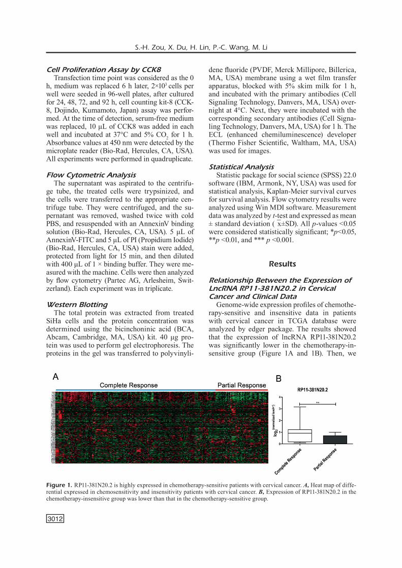
Genome-wide expression profiles of chemothe-rapy-sensitive and insensitive data in patients with cervical cancer in TCGA database were analyzed by edger package. The results showed that the expression of lncRNA RP11-381N20.2 was significantly lower in the chemotherapy-in-sensitive group (Figure 1A and 1B). Then, we
Figure 1. RP11-381N20.2 is highly expressed in chemotherapy-sensitive patients with cervical cancer. A, Heat map of diffe-rential expressed in chemosensitivity and insensitivity patients with cervical cancer. B, Expression of RP11-381N20.2 in the chemotherapy-insensitive group was lower than that in the chemotherapy-sensitive group.
Paclitaxel inhibits cervical cancer via autophagy
3013
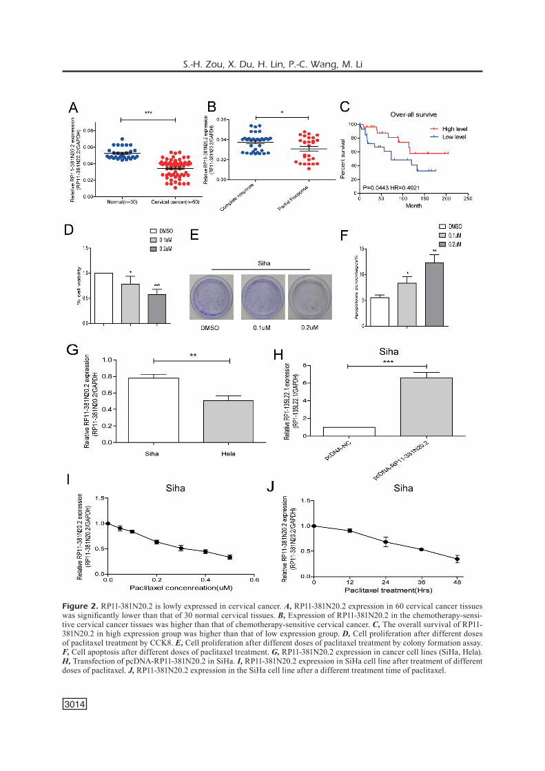
selected 60 cervical cancer tissues and 30 nor-mal tissues, qRT-PCR results revealed that the expression of lncRNA RP11-381N20.2 was lower in cervical cancer tissues (Figure 2A), and it was significantly lower in the chemotherapy-insensiti-ve group than the chemotherapy-sensitive group (Figure 2B). The clinical data of the patients were analyzed and the results showed that the overall survival time of patients was positively correlated with the expression of RP11-381N20.2 (Figure 2C, p = 0.0443, HR = 0.4021). In addition, we found that the tumor staging of patients with lower RP11-381N20.2 expression was positively corre-lated with tumor volume (Table I). These results indicated that RP11-381N20.2 had an inhibitory effect on cervical cancer and was closely related to chemosensitivity.
Paclitaxel Inhibits the Proliferation of Cervical Cancer Cells While Promoting Apoptosis
To determine the effect of paclitaxel on the via-bility of cervical cancer cells, CCK8 and colony formation assays were carried out. The CCK8 assay showed that the proliferation of SiHa cells treated with different concentrations of paclitaxel was gradually decreased with the increasing dose of paclitaxel (Figure 2D). The colony formation assay showed that, with gradually increased do-ses of paclitaxel culturing with cervical cancer cells, the number of observed colonies was signi-ficantly decreased (Figure 2E). It suggested that paclitaxel was capable of inhibiting the prolifera-
tion of cervical cancer cells. Next, to further stu-dy the effect of paclitaxel on apoptosis of cervical cancer cells, we conducted the flow cytometry for detecting apoptosis. The results showed that paclitaxel induced apoptosis in a dose-dependent manner (Figure 2F).
Expression of RP11-381N20.2 Decreases After Paclitaxel Treatment
The expression of RP11-381N20.2 was found to be relatively higher in SiHa cells detected by qRT-PCR (Figure 2G), so we selected SiHa cells for overexpression experiments. After transfection of pcDNA-NC and pcDNA-RP11-381N20.2 into SiHa cells, the expression of RP11-381N20.2 was significantly increased, the transfection efficiency was proved to be good (Figure 2H). To explore the correlation between paclitaxel and RP11-381N20.2, SiHa cells were cultured in medium containing different doses of paclitaxel and we found that the expression of RP11-381N20.2 was reduced with the increased paclitaxel dose (Figu-re 2I). Similarly, in the medium containing the same dose of paclitaxel, the longer the culturing time, the lower the expression of RP11-381N20.2 (Figure 2J). These results indicated that RP11-381N20.2 can promote the inhibitory effect of pa-clitaxel on cervical cancer.
RP11-381N20.2 Promotes the Antitumor Effect of Paclitaxel
After transfection of RP11-381N20.2 into cells containing paclitaxel medium, apoptosis of cervi-
Table I. Correlation between the expression of RP11-381N20.2 and clinicopathological characteristics in patients with cervical cancer (n = 60).
RP11-381N20.2 expressionClinicopathologic features Number of cases Low (n=30) High (n=30) p-value Age (years) 0.1904<50 35 20 15 ≥50 25 10 15 Histology 0.4383Squamous 31 14 17 Other type 29 16 13 Tumor size 0.0200*<4 cm 29 10 19 ≥4 cm 31 20 11 FIGO stage 0.0371*I-II 26 9 17 III-IV 34 21 13 Lymph node metastasis 0.1079No 22 14 8 Yes 38 16 22
*p<0.05
S.-H. Zou, X. Du, H. Lin, P.-C. Wang, M. Li
3014
Figure 2. RP11-381N20.2 is lowly expressed in cervical cancer. A, RP11-381N20.2 expression in 60 cervical cancer tissues was significantly lower than that of 30 normal cervical tissues. B, Expression of RP11-381N20.2 in the chemotherapy-sensi-tive cervical cancer tissues was higher than that of chemotherapy-sensitive cervical cancer. C, The overall survival of RP11-381N20.2 in high expression group was higher than that of low expression group. D, Cell proliferation after different doses of paclitaxel treatment by CCK8. E, Cell proliferation after different doses of paclitaxel treatment by colony formation assay. F, Cell apoptosis after different doses of paclitaxel treatment. G, RP11-381N20.2 expression in cancer cell lines (SiHa, Hela). H, Transfection of pcDNA-RP11-381N20.2 in SiHa. I, RP11-381N20.2 expression in SiHa cell line after treatment of different doses of paclitaxel. J, RP11-381N20.2 expression in the SiHa cell line after a different treatment time of paclitaxel.
Paclitaxel inhibits cervical cancer via autophagy
3015
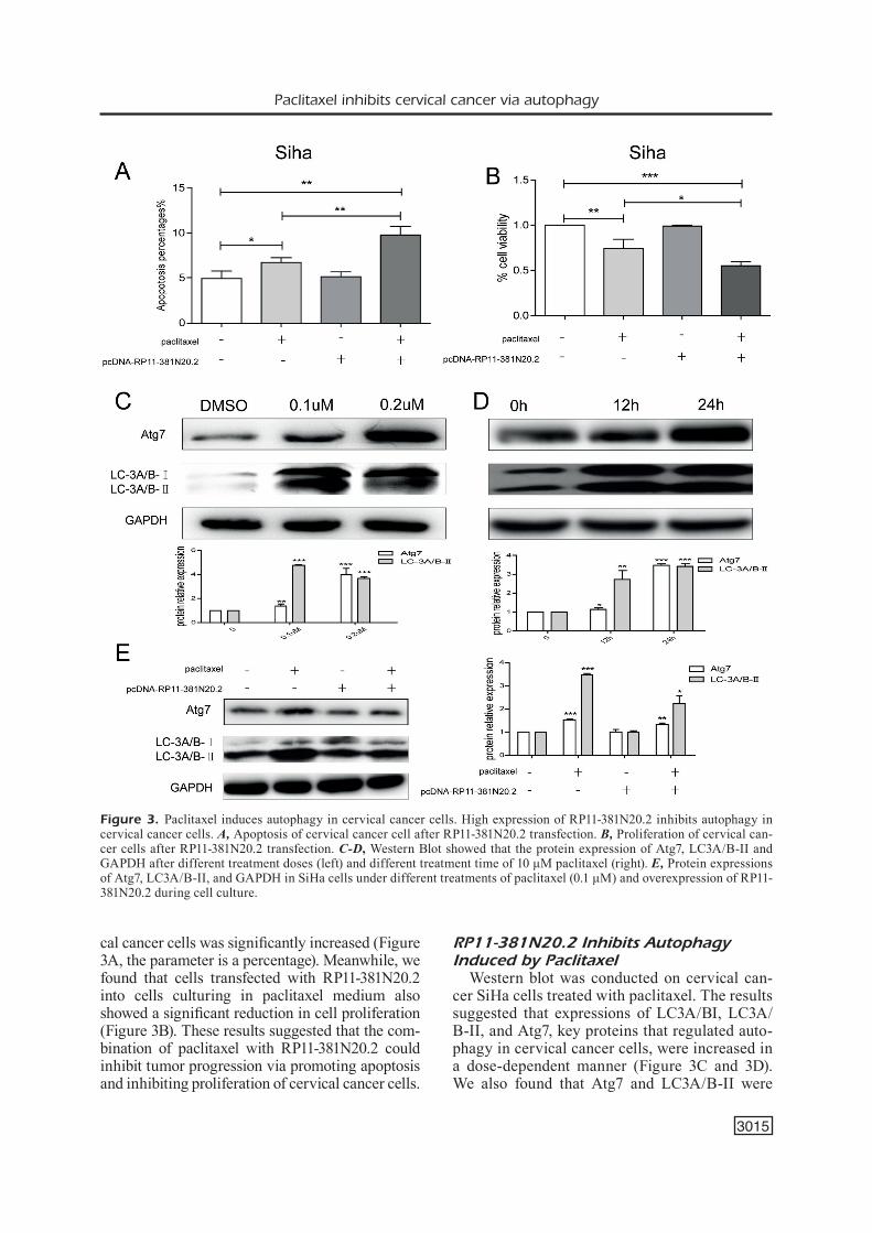
cal cancer cells was significantly increased (Figure 3A, the parameter is a percentage). Meanwhile, we found that cells transfected with RP11-381N20.2 into cells culturing in paclitaxel medium also showed a significant reduction in cell proliferation (Figure 3B). These results suggested that the com-bination of paclitaxel with RP11-381N20.2 could inhibit tumor progression via promoting apoptosis and inhibiting proliferation of cervical cancer cells.
RP11-381N20.2 Inhibits Autophagy Induced by Paclitaxel
Western blot was conducted on cervical can-cer SiHa cells treated with paclitaxel. The results suggested that expressions of LC3A/BI, LC3A/B-II, and Atg7, key proteins that regulated auto-phagy in cervical cancer cells, were increased in a dose-dependent manner (Figure 3C and 3D). We also found that Atg7 and LC3A/B-II were
Figure 3. Paclitaxel induces autophagy in cervical cancer cells. High expression of RP11-381N20.2 inhibits autophagy in cervical cancer cells. A, Apoptosis of cervical cancer cell after RP11-381N20.2 transfection. B, Proliferation of cervical can-cer cells after RP11-381N20.2 transfection. C-D, Western Blot showed that the protein expression of Atg7, LC3A/B-II and GAPDH after different treatment doses (left) and different treatment time of 10 μM paclitaxel (right). E, Protein expressions of Atg7, LC3A/B-II, and GAPDH in SiHa cells under different treatments of paclitaxel (0.1 μM) and overexpression of RP11-381N20.2 during cell culture.
S.-H. Zou, X. Du, H. Lin, P.-C. Wang, M. Li
3016
up-regulated after paclitaxel treatment, whilst Atg7 and LC3A/B-II were down-regulated af-ter treatment with RP11-381N20.2 (Figure 3E), indicating that RP11-381N20.2 could inhibit pa-clitaxel-induced autophagy. These experimental results showed that paclitaxel induced autopha-gy in cervical cancer cells and thus reducing its antitumor effect. RP11-381N20.2 inhibited pacli-taxel-induced autophagy.
Discussion
Cervical cancer is the most common gyneco-logic malignant tumor with an annual increase of about 500,000 cases, there have been high mor-bidity and mortality. Continuous high-risk human papillomavirus (HPV) infection is the major cause of cervical cancer14. However, the pathogenesis is not yet clear. Scholars15,16 found that macrophage played a dual role in tumor development, it not only promoted the death of cancer cells in the therapy but also enhanced the resistance of can-cer cells to chemotherapy. Some studies17,18 have shown that overexpression of Beclin1 in cervical cancer cells Hela can promote autophagic cell death; the opposite result was found when silenc-ing Beclin1. Therefore, to clarify the effect of au-tophagy on the development and treatment of cer-vical cancer is of great significance, and control of autophagy is expected to become a new target for the treatment of cervical cancer.
With the extensive application of bioinformatics and the comprehensive data sharing, TCGA data-base has been more and more used in medical re-search because of its complete data, large sample size, and full-grade prognosis information. There-fore, in this study, Genome-wide expression profiles of tumors and their susceptibility to drugs were downloaded through TCGA database to select the lowly expressed lncRNA RP11-381N20.2 in cervi-cal cancer by the edger package to analyze lncRNA map of normal and tumor tissues. Furthermore, we found that RP11-381N20.2 was downregulated in cervical cancer tissues compared with that of nor-mal cervical tissues, which was positively correlat-ed with the overall survival rate. The expression of RP11-381N20.2 was confirmed to be correlated with tumor size and stage in patients with cervical cancer. It suggested that RP11-381N20.2 had an an-titumor effect on cervical cancer, and can inhibit the formation of tumor resistance.
As we all know, there have been a large num-ber of clinical data focused on paclitaxel in the
treatment of ovarian cancer, the combination che-motherapy with cisplatin or carboplatin regimen proved to have the best efficacy. However, the ap-plication of paclitaxel in the treatment of cervical cancer is still in an exploration period. Current-ly, studies19-21 have shown that paclitaxel induced autophagy in cancer therapy. This study further studied the effect of paclitaxel on the autophagy in the treatment of cervical cancer, and its inter-action with RP11-381N20.2. We conducted the colony formation assay and flow cytometry. The results showed that paclitaxel had the ability to inhibit proliferation but increase the apoptosis of cervical cancer cells in a time- and dose-depen-dent manner. Western blot was utilized to study the correlation between paclitaxel and autoph-agy. The results suggested that paclitaxel could induce autophagy in cervical cancer cells in a time- and dose-dependent manners. Autophagy was significantly reduced after treatment with RP11-381N20.2. Further study found that pacli-taxel combined with RP11-381N20.2 significantly increased apoptosis of cervical cancer cells. It im-plied that paclitaxel had significant antitumor po-tential in cervical cancer, a combination of which with RP11-381N20.2 could inhibit autophagy but promote apoptosis of cervical cancer cells.
Conclusions
In summary, paclitaxel has a profound antitu-mor effect on cervical cancer and can induce auto-phagy. Our study first investigated that paclitaxel could inhibit the autophagy of cervical cancer via RP11-381N20.2, thereby inhibiting its progress. Our findings provided important insight into the usage of anti-cancer agents in combination with autophagy inhibitors. It provides new references in the treatment of cervical cancer to help develop new drugs.
Conflict of InterestThe Authors declare that they have no conflict of interest.
References
1) Jemal a, Siegel R, WaRd e, Hao Y, Xu J, THun mJ. Cancer statistics, 2009. CA Cancer J Clin 2009; 59: 225-249.
2) PaRkin dm, BRaY F, FeRlaY J, PiSani P. Global cancer statistics, 2002. CA Cancer J Clin 2005; 55: 74-108.

Paclitaxel inhibits cervical cancer via autophagy
3017
3) Jemal a, BRaY F, CenTeR mm, FeRlaY J, WaRd e, FoR-man d. Global cancer statistics. CA Cancer J Clin 2011; 61: 69-90.
4) Xu J, ZHang W, lv Q, ZHu d. Overexpression of miR-21 promotes the proliferation and migration of cervical cancer cells via the inhibition of PTEN. Oncol Rep 2015; 33: 3108-3116.
5) Cook ga, dRaPeR gJ. Trends in cervical cancer and carcinoma in situ in Great Britain. Br J Cancer 1984; 50: 367-375.
6) Wang Pd, lin RS. Epidemiology of cervical cancer in Taiwan. Gynecol Oncol 1996; 62: 344-352.
7) Quinn JJ, CHang HY. Unique features of long non-coding RNA biogenesis and function. Nat Rev Genet 2016; 17: 47-62.
8) di geSualdo F, CaPaCCioli S, lulli m. A pathophy-siological view of the long non-coding RNA world. Oncotarget 2014; 5: 10976-10996.
9) Pan JX. LncRNA H19 promotes atherosclerosis by regulating MAPK and NF-kB signaling pathway. Eur Rev Med Pharmacol Sci 2017; 21: 322-328.
10) meliSSaRi mT, gRoTe P. Roles for long non-coding RNAs in physiology and disease. Pflugers Arch 2016; 468: 945-958.
11) maJeSki ae, diCe JF. Mechanisms of chapero-ne-mediated autophagy. Int J Biochem Cell Biol 2004; 36: 2435-2444.
12) Wang l, laW Hk. The role of autophagy in lupus nephritis. Int J Mol Sci 2015; 16: 25154-25167.
13) moReau k, luo S, RuBinSZTein dC. Cytoprotective roles for autophagy. Curr Opin Cell Biol 2010; 22: 206-211.
14) Hanning Je, Saini Hk, muRRaY mJ, CaFFaRel mm, van dongen S, WaRd d, BaRkeR em, SCaRPini Cg, gRo-
veS iJ, STanleY ma, enRigHT aJ, PeTT mR, Coleman n. Depletion of HPV16 early genes induces autopha-gy and senescence in a cervical carcinogenesis model, regardless of viral physical state. J Pathol 2013; 231: 354-366.
15) WHiTe e. Deconvoluting the context-dependent role for autophagy in cancer. Nat Rev Cancer 2012; 12: 401-410.
16) Xu Y, Yu H, Qin H, kang J, Yu C, ZHong J, Su J, li H, Sun l. Inhibition of autophagy enhances cisplatin cytotoxicity through endoplasmic reticulum stress in human cervical cancer cells. Cancer Lett 2012; 314: 232-243.
17) Wang ZH, Xu l, duan Zl, Zeng lQ, Yan nH, Peng Zl. Beclin 1-mediated macroautophagy involves regulation of caspase-9 expression in cervical cancer HeLa cells. Gynecol Oncol 2007; 107: 107-113.
18) Sun Y, liu JH, Sui YX, Jin l, Yang Y, lin Sm, SHi H. Beclin1 overexpression inhibitis proliferation, in-vasion and migration of CaSki cervical cancer cells. Asian Pac J Cancer Prev 2011; 12: 1269-1273.
19) Yu YF, Hu PC, Wang Y, Xu Xl, RuSHWoRTH gm, ZHang Z, Wei l, ZHang JW. Paclitaxel induces autophagy in gastric cancer BGC823 cells. Ultrastruct Pathol 2017; 41: 284-290.
20) guo Y, Yuan J, Yin S, Wang X, SHuai R, kang J. MAP2K6-FP enhances the sensitiveness of pacli-taxel for ovarian cancer via inducing autophagy. Int J Gynecol Cancer 2017; 27: 1082-1087.
21) Wang m, liao C, Hu Y, QinWen P, Jiang J. Sensitiza-tion of breast cancer cells to paclitaxel by dichlo-roacetate through inhibiting autophagy. Biochem Biophys Res Commun 2017; 489: 103-108.
![Original Article Potential biomarkers for paclitaxel ... · Potential biomarkers for paclitaxel sensitivity in ... larynx and oropharynx cancer [5, 15]. ... Biomarkers for paclitaxel](https://static.fdocuments.in/doc/165x107/5af0f1e17f8b9a572b901a03/original-article-potential-biomarkers-for-paclitaxel-biomarkers-for-paclitaxel.jpg)