NewTricksofanOldPattern - Journal of Biological Chemistry · scribed using a mMESSAGE mMACHINE in...
11
New Tricks of an Old Pattern STRUCTURAL VERSATILITY OF SCORPION TOXINS WITH COMMON CYSTEINE SPACING * □ S Received for publication, December 1, 2011, and in revised form, December 21, 2011 Published, JBC Papers in Press, January 10, 2012, DOI 10.1074/jbc.M111.329607 Alma Leticia Saucedo ‡1,2 , David Flores-Solis ‡ , Ricardo C. Rodríguez de la Vega §3 , Belén Ramírez-Cordero ‡1 , Rogelio Hernández-López ¶1,4 , Patricia Cano-Sánchez ‡ , Roxana Noriega Navarro , Jesús García-Valdés , Fredy Coronas-Valderrama**, Adolfo de Roodt ‡‡ , Luis G. Brieba §§ , Lourival Domingos Possani**, and Federico del Río-Portilla ‡5 From the ‡ Instituto de Química, Universidad Nacional Autónoma de México, Ciudad Universitaria, México, D.F., 04510, México, the § UMR 7138, Département Systématique et Evolution, Muséum National d’Histoire Naturelle, Paris 75005, France, the ¶ Department of Chemistry and Chemical Biology, Harvard University, Cambridge, Massachusetts 02138, the Facultad de Química, Universidad Nacional Autónoma de México, Ciudad Universitaria, México, D.F., 04510, México, the **Instituto de Biotecnología, Universidad Nacional Autónoma de México, Avenida Universidad 2001, Apartado Postal 510-3, Cuernavaca México, the ‡‡ Centro de Patología Experimental y Aplicada, Facultad de Medicina, Universidad de Buenos Aires and Administracio ´n Nacional de Laboratorios e Institutos de Salud “Dr. Carlos G. Malbra ´n,” Ministerio de Salud, Av. Ve ´lez Sarsfield 563, CP 1281, CABA, Buenos Aires, Argentina, and the §§ Laboratorio de Genómica para la Biodiversidad, CINVESTAV Unidad Irapuato, 36821 Irapuato, Guanajuato, México Background: Most scorpion venom peptides adopt a single structural scaffold around four strictly conserved cysteines. Results: Two K channel-blocking peptides from Tityus venoms share this cysteine spacing but fold into a distinct cystine- stabilized helix-loop-helix scaffold. Conclusion: These peptides define a new structural group of scorpion venom peptides. Significance: Cysteine spacing does not dictate the three-dimensional fold of small disulfide-rich proteins. Scorpion venoms are a rich source of K channel-blocking peptides. For the most part, they are structurally related small disulfide-rich proteins containing a conserved pattern of six cys- teines that is assumed to dictate their common three-dimen- sional folding. In the conventional pattern, two disulfide bridges connect an -helical segment to the C-terminal strand of a dou- ble- or triple-stranded -sheet, conforming a cystine-stabilized / scaffold (CS/). Here we show that two K channel-block- ing peptides from Tityus scorpions conserve the cysteine spac- ing of common scorpion venom peptides but display an uncon- ventional disulfide pattern, accompanied by a complete rearrangement of the secondary structure topology into a CS helix-loop-helix fold. Sequence and structural comparisons of the peptides adopting this novel fold suggest that it would be a new elaboration of the widespread CS/ scaffold, thus reveal- ing an unexpected structural versatility of these small disulfide- rich proteins. Acknowledgment of such versatility is important to understand how venom structural complexity emerged on a limited number of molecular scaffolds. Scorpion venoms are complex mixtures of biomolecules, products of millions years of evolution (1, 2). Venoms contain, among others components, peptide toxins capable of interact- ing specifically with potassium, sodium, and calcium channels (3–5). These toxic peptides have contributed considerably to the understanding of the structure and functional mechanism of the ion channels (6). Some of them are considered good can- didates for the design and development of new drugs (7), although the number of structural and functional analyses of these peptides is still limited (8). Potassium channels blocker toxins (KTx) 6 can be classified into four subfamilies accordingly to the accepted nomencla- ture: , , , and (9, 10). -KTx are peptides of 23– 43 amino acids stabilized by three to four disulfide bonds, two of which are strictly conserved and link an -helix and one strand of a -sheet within the so-called cystine-stabilized / scaffold (CS/), the most common among scorpion toxins (10, 11). -KTx toxins, known as “long chain,” are three disulfide- bridged peptides with 60 amino acids that also contain the cysteine pattern of peptides adopting the CS/ scaffold, although no structure is available yet (12). -KTxs are blockers of the ether-á-go-go-related gene family of K channels; they are 36 – 47 amino acids long with three or four disulfide bridges, * This work was supported in part by Direccio ´ n General de Servicios de Co ´m- puto Acade ´ mico y Direccio ´ n General de Asuntos del Personal Acade ´ mico, Universidad Nacional Auto ´ noma de Me ´ xico Projects IN205110 (to F. R. P.) and IN204110 (to L. D. P.) and Consejo Nacional de Ciencia y Tecnologı ´a Project 59297 (to F. R. P.). □ S This article contains supplemental Figs. SM1–SM4. The atomic coordinates and structure factors (codes 2LI3 and 2LKA) have been deposited in the Protein Data Bank, Research Collaboratory for Structural Bioinformatics, Rutgers University, New Brunswick, NJ (http://www.rcsb.org/). 1 Recipients of scholarship support from Consejo Nacional de Ciencia y Tecnologı ´a. 2 Supported by the Programa de Doctorado en Ciencias Biome ´ dicas, Univer- sidad Nacional Autónoma de México. 3 Invited researcher at the Muse ´ um National d’Histoire Naturelle sponsored by the Marie de Paris. 4 Supported by the Fundación México en Harvard. 5 To whom correspondence should be addressed: Dept. de Química de Mac- romoléculas, Instituto de Química, Universidad Nacional Autónoma de México, Circuito Exterior s/n, Ciudad Universitaria, México, D.F. 04510, México. Tel.: 52-55-5622-4613; Fax: 52-55-5616-2203; E-mail: jfrp@unam. mx. 6 The abbreviations used are: KTx, K channel toxin; H, hydrogen in posi- tion; H, hydrogen in position; HN, amide proton; RMSD, root mean square deviation; CS, cystine-stabilized. THE JOURNAL OF BIOLOGICAL CHEMISTRY VOL. 287, NO. 15, pp. 12321–12330, April 6, 2012 © 2012 by The American Society for Biochemistry and Molecular Biology, Inc. Published in the U.S.A. APRIL 6, 2012 • VOLUME 287 • NUMBER 15 JOURNAL OF BIOLOGICAL CHEMISTRY 12321 by guest on October 3, 2018 http://www.jbc.org/ Downloaded from
Transcript of NewTricksofanOldPattern - Journal of Biological Chemistry · scribed using a mMESSAGE mMACHINE in...

New Tricks of an Old PatternSTRUCTURAL VERSATILITY OF SCORPION TOXINS WITH COMMON CYSTEINE SPACING*□S
Received for publication, December 1, 2011, and in revised form, December 21, 2011 Published, JBC Papers in Press, January 10, 2012, DOI 10.1074/jbc.M111.329607
Alma Leticia Saucedo‡1,2, David Flores-Solis‡, Ricardo C. Rodríguez de la Vega§3, Belén Ramírez-Cordero‡1,Rogelio Hernández-López¶1,4, Patricia Cano-Sánchez‡, Roxana Noriega Navarro�, Jesús García-Valdés�,Fredy Coronas-Valderrama**, Adolfo de Roodt‡‡, Luis G. Brieba§§, Lourival Domingos Possani**,and Federico del Río-Portilla‡5
From the ‡Instituto de Química, Universidad Nacional Autónoma de México, Ciudad Universitaria, México, D.F., 04510, México, the§UMR 7138, Département Systématique et Evolution, Muséum National d’Histoire Naturelle, Paris 75005, France, the ¶Departmentof Chemistry and Chemical Biology, Harvard University, Cambridge, Massachusetts 02138, the �Facultad de Química, UniversidadNacional Autónoma de México, Ciudad Universitaria, México, D.F., 04510, México, the **Instituto de Biotecnología, UniversidadNacional Autónoma de México, Avenida Universidad 2001, Apartado Postal 510-3, Cuernavaca México, the ‡‡Centro de PatologíaExperimental y Aplicada, Facultad de Medicina, Universidad de Buenos Aires and Administracion Nacional de Laboratorios eInstitutos de Salud “Dr. Carlos G. Malbran,” Ministerio de Salud, Av. Velez Sarsfield 563, CP 1281, CABA, Buenos Aires, Argentina,and the §§Laboratorio de Genómica para la Biodiversidad, CINVESTAV Unidad Irapuato, 36821 Irapuato, Guanajuato, México
Background:Most scorpion venom peptides adopt a single structural scaffold around four strictly conserved cysteines.Results: Two K� channel-blocking peptides from Tityus venoms share this cysteine spacing but fold into a distinct cystine-stabilized helix-loop-helix scaffold.Conclusion: These peptides define a new structural group of scorpion venom peptides.Significance: Cysteine spacing does not dictate the three-dimensional fold of small disulfide-rich proteins.
Scorpion venoms are a rich source of K� channel-blockingpeptides. For the most part, they are structurally related smalldisulfide-richproteins containing a conservedpatternof six cys-teines that is assumed to dictate their common three-dimen-sional folding. In the conventional pattern, twodisulfide bridgesconnect an �-helical segment to the C-terminal strand of a dou-ble- or triple-stranded �-sheet, conforming a cystine-stabilized�/� scaffold (CS�/�). Herewe show that twoK� channel-block-ing peptides from Tityus scorpions conserve the cysteine spac-ing of common scorpion venom peptides but display an uncon-ventional disulfide pattern, accompanied by a completerearrangement of the secondary structure topology into a CShelix-loop-helix fold. Sequence and structural comparisons ofthe peptides adopting this novel fold suggest that it would be anew elaboration of the widespread CS�/� scaffold, thus reveal-
ing an unexpected structural versatility of these small disulfide-rich proteins. Acknowledgment of such versatility is importantto understand how venom structural complexity emerged on alimited number of molecular scaffolds.
Scorpion venoms are complex mixtures of biomolecules,products of millions years of evolution (1, 2). Venoms contain,among others components, peptide toxins capable of interact-ing specifically with potassium, sodium, and calcium channels(3–5). These toxic peptides have contributed considerably tothe understanding of the structure and functional mechanismof the ion channels (6). Some of them are considered good can-didates for the design and development of new drugs (7),although the number of structural and functional analyses ofthese peptides is still limited (8).Potassium channels blocker toxins (KTx)6 can be classified
into four subfamilies accordingly to the accepted nomencla-ture: �, �, �, and � (9, 10). �-KTx are peptides of 23–43 aminoacids stabilized by three to four disulfide bonds, two of whichare strictly conserved and link an �-helix and one strand of a�-sheet within the so-called cystine-stabilized �/� scaffold(CS�/�), the most common among scorpion toxins (10, 11).�-KTx toxins, known as “long chain,” are three disulfide-bridged peptides with �60 amino acids that also contain thecysteine pattern of peptides adopting the CS�/� scaffold,although no structure is available yet (12). �-KTxs are blockersof the ether-á-go-go-related gene family of K� channels; theyare 36–47 amino acids longwith three or four disulfide bridges,
* This work was supported in part by Direccion General de Servicios de Com-puto Academico y Direccion General de Asuntos del Personal Academico,Universidad Nacional Autonoma de Mexico Projects IN205110 (to F. R. P.)and IN204110 (to L. D. P.) and Consejo Nacional de Ciencia y TecnologıaProject 59297 (to F. R. P.).
□S This article contains supplemental Figs. SM1–SM4.The atomic coordinates and structure factors (codes 2LI3 and 2LKA) have been
deposited in the Protein Data Bank, Research Collaboratory for StructuralBioinformatics, Rutgers University, New Brunswick, NJ (http://www.rcsb.org/).
1 Recipients of scholarship support from Consejo Nacional de Ciencia yTecnologıa.
2 Supported by the Programa de Doctorado en Ciencias Biomedicas, Univer-sidad Nacional Autónoma de México.
3 Invited researcher at the Museum National d’Histoire Naturelle sponsoredby the Marie de Paris.
4 Supported by the Fundación México en Harvard.5 To whom correspondence should be addressed: Dept. de Química de Mac-
romoléculas, Instituto de Química, Universidad Nacional Autónoma deMéxico, Circuito Exterior s/n, Ciudad Universitaria, México, D.F. 04510,México. Tel.: 52-55-5622-4613; Fax: 52-55-5616-2203; E-mail: [email protected].
6 The abbreviations used are: KTx, K� channel toxin; H�, hydrogen in � posi-tion; H�, hydrogen in � position; HN, amide proton; RMSD, root meansquare deviation; CS, cystine-stabilized.
THE JOURNAL OF BIOLOGICAL CHEMISTRY VOL. 287, NO. 15, pp. 12321–12330, April 6, 2012© 2012 by The American Society for Biochemistry and Molecular Biology, Inc. Published in the U.S.A.
APRIL 6, 2012 • VOLUME 287 • NUMBER 15 JOURNAL OF BIOLOGICAL CHEMISTRY 12321
by guest on October 3, 2018
http://ww
w.jbc.org/
Dow
nloaded from

which also adopt the CS�/� scaffold (13). The last and smallestfamily, the �-KTxs, displays purely helical structure stabilizedby two disulfide bridges and fold into an �-hairpin fold knownasCS�/� (cystine-stabilized helix-loop-helix) (11, 14–17). Pep-tides belonging to this family have been isolated from only twoscorpion genera: Heterometrus (14, 15) and Opisthacanthus(16, 17). �-KTxs are relatively poor blockers of K� channels,despite presence of the typical functional dyad, Tyr and Lys,important for other activity of the toxin (3). It is still neitherclear whether members of the �-KTx family might all workthrough the same mechanism on potassium channels (16, 17),nor whether K� channels are indeed their cognate receptors. Inpart this may be due to the lack of structural and functionalcharacterization of peptides in this family.This work describes the native isolation, recombinant pro-
duction and structural characterization of a newly identified 28amino acid K� channel-blocking peptide from Tityus trivitta-tus venom, named �-buthitoxin-Tt2b, �-BUTX-Tt2b for short,following the nomenclature proposal of King et al. (18). Thesequence shows high identity with peptides classified withinthe �-KTx subfamily 20 (19), including the six cysteines of thesequence signature of the CS�/� scaffold. Unexpectedly, thepeptide adopts aCS�/� fold in solution. The sameoverall struc-ture was determined for a recombinant version of another pep-tide fromTityus serrulatus classified within the same subfamily(UniProt P86271, Ts16 in Ref. 46). Peptides of the �-Ktx sub-family 20 were assumed to adopt the conventional CS�/� scaf-fold on the basis of their common cysteine spacing. However,both �-BUTX-Tt2b and Ts16 adopt an unexpected fold even ifsharing the cysteine pattern of conventional CS�/� peptides,thus offering new insights on the structural versatility of scor-pion venom peptides. Acknowledging this versatility is impor-tant if the full potential of animal venoms as natural peptidelibraries is to be realized.
EXPERIMENTAL PROCEDURES
Purification and Sequence Determination of the Novel Peptide
Venom from the scorpion Tityus trivitattus collected in theProvince of Santa Fe, Argentina, was obtained by electricalstimulation and separated as earlier described (20). Only twosteps of HPLC fractionation were necessary to obtain the purepeptide under study. An analytical C18 reverse phase column(Vydac, Hisperia) was used with a linear gradient from solventA (water in 0.12% TFA, Sigma-Aldrich; all of the chemicalswere purchased from this company, unless otherwise specified)to 60% solvent B (acetonitrile in 0.10% TFA), run for 60 min.The second separation used the same system with a gradientfrom 10 to 35% solvent B, run for 45 min. The amino acidsequence determination of the pure peptide was obtained usingautomatic Edman degradationwith a Beckman LF 3000 proteinsequencer, and MS measurements were obtained with an ion-trap equipment from Finnigan LCQDuo as earlier described(20). The peptide was named �-buthitoxin-Tt2b.
Disulfide Bridge Determination
Pure peptide (18 �g) was digested with tosylphenylalanylchloromethyl ketone-trypsin and endopeptidase V8 fromStaphylococcus aureus (Roche Applied Science). Initially, 1 �g
of trypsin was added in presence of an ammonium bicarbonatebuffer (pH8) andhydrolyzed for 5 h; then the endopeptidaseV8(1 �g) was added, and the reaction was incubated for 12 h at36 °C. The peptides were separated using the same HPLC con-ditions used initially for the whole venom (see above) andsequenced.
Design and Cloning of the Synthetic �-BUTX-Tt2b Gene
The �-BUTX-Tt2b synthetic gene coding for the peptidewasassembled based on the amino acid sequence obtained andoptimized for Escherichia coli codon usage. The �-BUTX-Tt2bgene (see supplemental data Fig. SM1) was cloned into theexpression vector pET32a (Novagen). The resulting plasmidpET32-�-BUTX-Tt2bwas confirmed byDNA sequencing. TheTs16 synthetic gene used was based on the �-BUTX-Tt2b gene(sequence available in supplemental data Fig. SM1).
Protein Expression
Recombinant-�-BUTX-Tt2b protein was overexpressed intuner E. coli cells (Novagen) transformed with the expressionvector pET32-�-BUTX-Tt2b. LB medium supplemented withampicillin was used to grown cells at 37 °C. Protein expressionwas induced at an A600 between 0.7–0.8 with isopropyl thio-�-galactopyranoside 0.5 mM. The cells were incubated for 6 h at30 °C and harvested by centrifugation.
Recombinant Protein Purification
For �-BUTX-Tt2b, the cell pellets were resuspended in lysisbuffer (150mMNaCl, 0.1mg/ml lisozyme, 50mMTris/HCl, pH8) and sonicated using (Misonix Sonicator 3000). The solublefraction was separated by centrifugation at 32,000 � g for 30min at 4 °C. Recombinant His6-tagged fusion toxin (20.3 kDa)was purified by metal-chelate affinity chromatography using aHiTrap Ni2� column (GE Healthcare) and eluted with two col-umn volumes of elution buffer (50 mM Tris/HCl, pH 8, 150 mM
NaCl, 500 mM imidazole). Thrombin was used to release the�-BUTX-Tt2b recombinant toxin from the N terminus thiore-doxin fusion protein. Cleavage reaction was performed for 6 hat 18 °C according to themanufacturer’s instructions. A secondround of isolation by metal-chelate affinity chromatographywas required to remove �-BUTX-Tt2b from cleavage reactionsubproducts. Final �-BUTX-Tt2b purification was performedwith reverse phase HPLC in a Pro-Star Varian instrumentequippedwith a binary gradient solvent systemon a Jupiter C18column (250 mm � 4.6 mm; Phenomenex) using a linear gra-dient from 12 to 30% of water-acetonitrile run for 30 min (con-taining 0.05% TFA) at a flow rate of 1 ml/min.
Protein Characterization
Mass Spectrometry—MALDI-TOFMSon aBrukerDaltonicsMicroflex LT equipment was used to determinate the molecu-lar mass of the recombinant proteins. The data were acquiredusing the reflector operation mode acquiring 150 shots. Thesamples were prepared with �-cyano-4-hydroxycinnamic acidin a 1:10 ratio (21).Electrophysiological Measurements—cRNAs for voltage-de-
pendent potassium channels: Shaker-IR (Shaker channel withinactivation domain removed) and hKv1.2 subunits were tran-
New Fold for Scorpion Venom Peptides
12322 JOURNAL OF BIOLOGICAL CHEMISTRY VOLUME 287 • NUMBER 15 • APRIL 6, 2012
by guest on October 3, 2018
http://ww
w.jbc.org/
Dow
nloaded from
scribed using a mMESSAGE mMACHINE in vitro transcrip-tion kit (Ambion). cRNAs were injected into Xenopus laevisoocytes using 1–2 ng (in 50 nl) of K�-subunits. Potassium cur-rents were recorded 2–4 days after cRNA injection.Two-electrode Voltage Clamp—The bath solution was
ND-96: 96 mM NaCl, 2 mM KCl, 1.8 mM CaCl2, 1 mM MgCl2, 5mM HEPES, pH 7.4, adjusted with NaOH. Resistance for thevoltage and current electrodes were 1.0–1.5 and 0.3–0.5 M�,respectively, when filled with 3 M KCl. Holding potential was�60 mV, and pulse protocol was applied from �60 mV to �60mV with increments of 20 mV for 200 ms. Macroscopic cur-rents were recorded using the CA1B high performance oocyteclamp amplifier (Dagan Co.), the Digidata 1440 digitizer, andthe pClamp10 software (MolecularDevices). The currentswerefiltered at 1⁄5 of the sampling frequency (2 kHz). The volume ofwashing solution was �20 times greater than that of the cham-ber containing the oocytes. Inhibition was evaluated everyminute until steady state was reached, usually from 8 to 10min.
Nuclear Magnetic Resonance Spectroscopy
Sample Preparation—Native and recombinant �-BUTX-Tt2b were prepared at 0.08 and 2.3 mM concentrations, respec-tively, by dissolving lyophilized protein in 95% H2O, 5% D2O(Cambridge Isotope Laboratories). The recombinant proteinspectra were obtained at pH 3.5. No pH adjustment was donewith the native sample. NMR data were recorded at 25 °C on an800 MHz Varian spectrometer equipped with a HCN indirectdetection probe. TOCSY spectra were recorded with isotropicmixing time of 75 and 20 ms. NOESY spectra were acquiredusing mixing times of 150 and 300 ms. The two-dimensionalNMR spectra were conducted using a scheme of water suppres-sion by gradient-tailored excitation for solvent suppression
(22). All of the experiments were acquired using 2048 and 1024complex points in direct and indirect dimensions, respectively.Ts16 recombinant protein (rTs16) spectra were recorded in a500 MHz Varian instrument at 3.0 mM; experiments wereobtained as before.Data Processing and Analysis—NMRDraw and NMRpipe
software were used for processing data (23). CARA1.5 softwarewas used for NMR data analysis (24). Semi-automated assign-ment and distance geometry calculations were performed withCYANA 2.1 (25).
Molecular Dynamics Refinement
Structure refinement was performed using moleculardynamics AMBER 9 (26) suite with an explicit solvent model.Lowest CYANA target function values were used for furtherrefinement. Molecular dynamics simulations and the energyminimizations were carried out with the topology and param-eters of AMBER-99SB force field. Distance and dihedral angleconstraints were used for AMBER calculations following themethod proposed by Xia et al. (27). Geometric quality of struc-tures was assessed using PROCHECK utility in the validationserver of the Protein Data Bank.
RESULTS
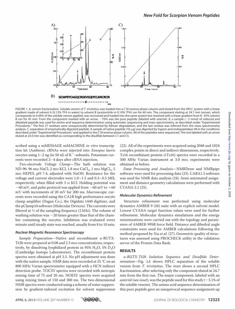
�-BUTX-Tt2b Isolation Sequence and Disulfide Deter-mination—Fig. 1A shows HPLC separation of the solublevenom from T. trivitattus. The inset shows a second HPLCfractionation, after selecting only the component eluted at 24.7min from the first run. The major component, labeled with anasterisk (see inset), was the peptide used for this study (�3.1%ofthe soluble venom). The amino acid sequence determination ofthis pure peptide gave an unequivocal sequence assignment up
FIGURE 1. A, venom fractionation. Soluble venom of T. trivitattus was loaded into a C18 reverse phase column and eluted from the HPLC system with a lineargradient made of solvent A (0.12% TFA in water) to solvent B (acetonitrile in 0.10% TFA) run for 60 min. The component eluting at 24.7 min (arrow), whichcorresponds to 4.44% of the soluble venom applied, was recovered and loaded into the same system but resolved with a linear gradient from 8 –35% solventB run for 45 min. From the component marked with an arrow, �70% was the pure peptide (labeled with asterisk). B, a sample (�2 nmol) of reduced andalkylated peptide was used for amino acid sequence determination using automatic sequencing and mass spectrometry, as described under “ExperimentalProcedures.” The first 27 residues were unequivocally determined by Edman degradation, and the last residue was inferred from the mass spectrometryanalysis. C, separation of enzymatically digested peptide. A sample of native peptide (18 �g) was digested by trypsin and endopeptidase V8 in the conditionsdescribed under “Experimental Procedures” and applied to the C18 reverse phase column. All of the peptides were sequenced. The one labeled with an arroweluted at 23.0 min was identified as corresponding to the disulfide between C1 and C5.
New Fold for Scorpion Venom Peptides
APRIL 6, 2012 • VOLUME 287 • NUMBER 15 JOURNAL OF BIOLOGICAL CHEMISTRY 12323
by guest on October 3, 2018
http://ww
w.jbc.org/
Dow
nloaded from

to residue number 27 (Fig. 1B). The last amino acid, Arg inposition 28, was inferred by mass spectrometry determination.The experimental mass determined for the native peptide was3179.75 Da, and the theoretical mass expected for the first 27residues directly determined was 3023.57, leaving a molecularmass of 156.18, which was identified as being that of arginine.This sequence was deposited in the UniProt Knowledgebaseunder the accession number B3A0L5. Sequence similaritysearches revealed high identity with only two Tityus sp. venompeptides: Ts16 fromT. serrulatus (P86271, 89%) and Tt28 fromT. trivittatus (P0C183 (18), 65%). No disulfide pairing for any ofthese homologous peptides has been reported.Fig. 1C shows the profile of HPLC separation after enzymatic
hydrolysis. The peptide eluted at 23.0 min was compatible witha disulfide paring between the first (Cys2) and fifth cysteines(Cys24), C1 and C5. The automatic sequencer showed twoamino acid residues for the first cycles: Gly and Asn. Position 2was a blank, butMetwas in position 3 and Prowas in position 4.It implies that endopeptidase V8 cleaved the peptide bond atthe Glu5, and trypsin cleaved Lys22 and Arg25. The remainingpeptides gave always more than two amino acid residues percycle of the sequencer, because the core fragment of peptideafter the enzymatic hydrolysis did not allow separating theother peptides containing the two remaining disulfide bridges.In this way, only one of the disulfide bridges of the toxin wasdirectly determined.Fusion Protein Expression and Purification—Most of the
fusion protein was located in the soluble cellular lysate, and itwas efficiently retained by the HiTrap Ni2� column with puri-fication yield of 36mg/liter. Thrombin recognizes the sequenceX3X2R/L—GSX1X2; therefore, the product of the hydrolysisreaction is the �-BUTX-Tt2b sequence with additional glycineand serine at theN-terminal segment, r�-BUTX-Tt2b.Optimalcleavage conditions were obtained at 18 °C after 6 h of reaction.Finally, another isolation by metal-chelate affinity chromatog-raphy purification step was performed.Reverse Phase HPLC Purification and Protein Char-
acterization—Under chromatographic conditions selected,r�-BUTX-Tt2b elutes at 23.0 min with a MALDI-TOF mass of
3325.1 for [M�H]�, which is in good agreement with the the-oretical mass of 3323.9 Da for an oxidized peptide formingthree disulfide bridges, calculatedwith the ProtParam tool fromExPASy server (28). The purification procedures indicatedabove yielded 0.7 mg of r�-BUTX-Tt2b per liter in LB culturemedium.Electrophysiological Measurements—Electrophysiological
assays were done on Shaker-IR and hKv1.2 channels. Fig. 2shows the effect of r�-BUTX-Tt2b on both channels, using theexperimental conditions described below. The protocol of testpulses was applied every 60 s to verify the current stability pre-vious to the toxin addition. Only oocytes showing a minimumrun-down were chosen for experimental assays. The controlcurrents (black line) through Shaker-IR or hKv1.2 channelsunder whole cell recording are plotted. Fig. 2 shows represent-ative traces for both channels only at �60 mV pulse. The addi-tion of r�-BUTX-Tt2b (200 �mol/liter) blocked partially out-ward currents (gray line). Recombinant toxin did not changebasically the original shape of currents, suggesting thatr�-BUTX-Tt2b acts as a pore blocker, although, further exper-iments are needed to test this hypothesis. Current reductionwas 88.7 � 9.9 and 74.9 � 7.1 (n � 3) for Shaker-IR and Kv1.2,respectively.Nuclear Magnetic Resonance Analysis—NMR experiments
for the �-BUTX-Tt2b native protein were obtained with poorsignal to noise ratio at a very low concentration. The TOCSYexperiment was obtained with sufficient sensitivity. However,the NOESY experiments displayed low sensitivity that madedifficult to complete the sequential assignments. In contrast,NMR spectroscopic studies using higher concentration ofr�-BUTX-Tt2b revealed well resolved resonances with goodchemical shift dispersion between 6.3 and 10.3 ppm.A compar-ison of the chemical shifts between the amide region signals ofthe recombinant (black) and the native (red) toxins shows justslight differences mainly caused by different pH levels and con-centrations used for the experiments (see Fig. 3). Aliphaticregion is almost identical. A NOESY HN region spectrumshows the HN-HN correlations consistent with the presence of
FIGURE 2. Effect of �-BUTX-Tt2b toxin on Shaker-IR (left panel) and Kv1.2 channels (right panel). Macroscopic K� currents were recorded using two-electrode voltage-clamp technique in ND96 solution. Currents were elicited by depolarizing the membrane for 200 ms, from a holding potential of �60 mV toa pulse step of �60 mV. Pulse protocol was applied at 60 s each. Traces for control conditions are shown with a black line, and those in the presence ofrecombinant toxin are shown with a gray line (after 8 min of application at 200 �mol/liter). The current percentages of blockade for Shaker IR and Kv1.2 were88.7 � 9.9 and 74.9 � 7.1 (mean � S.E.), respectively. The inhibition of currents was not fully reversible (not shown).
New Fold for Scorpion Venom Peptides
12324 JOURNAL OF BIOLOGICAL CHEMISTRY VOLUME 287 • NUMBER 15 • APRIL 6, 2012
by guest on October 3, 2018
http://ww
w.jbc.org/
Dow
nloaded from

helical structures (data not shown). The assignments wereachieved following the standard protocol (29).r�-BUTX-Tt2b Disulfide Bond Connectivities and Three-di-
mensional Structure—All 30 r�-BUTX-Tt2b spin systems wereidentified, and most proton resonance signals were unambigu-ously assigned (chemical shift completeness of 98.7%). Theposition of the disulfide bridges was established by analyzingthe NMR experimental results independently. A NOE signalbetween H�-H� from Cys7 and Cys20 (Fig. 3, inset), which cor-responds to C2-C4 cystine pair (residue numbers are indicatedas in the native protein), was found. Also, there were observedNOE correlations between HN of Cys24 with H� of Cys2 andH� of Met3; all of these signals are in agreement with the for-mation of the C1-C5 pair. These results confirm a new cystinepairing for scorpion toxins when compared with the conven-tional pairs of CS�/� scorpion venom peptides (C1-C4, C2-C5,and C3-C6 (11)). To further support the assigned disulfidebonds, three CYANA calculations were performed with differ-ent input parameters following a protocol described by Fadel etal. (30). The first calculation included data without disulfidebond restrictions. This result provides theNOE constraint con-tributions for the structure, which was used as a reference. Thesecond calculation was performed using as restrictions themost common disulfide pattern observed in KTxs toxins:C1-C4, C2-C5, and C3-C6. Finally, another set of structureswere calculated considering the alternative C1-C5, C2-C4, andC3-C6 cystine pairing pattern, as suggested by the NOE NMRdata assignment and mass spectrometry of a digested peptide.Table 1 shows the statistics and RMSD values of these threecalculations. After comparison of backbone for the residues3–30 of the lowest energy structure for each set of calculations,the RMSD against the reference structure gave 1.75 Å for theconventional disulfide bridges and 0.78 Å for the nonconven-tional disulfide bridges.
A total of 200 structures where generated with CYANA; only20 structures (Fig. 4A) with the lowest target function areshown. The proton shift index is in good agreement with thesecondary structure found (31). NOE correlations betweenHNi/HNi�1, H�i/HNi�3, and H�i/HNi�4 indicate thatr�-BUTX-Tt2b is mainly shaped by helical elements. Ther�-BUTX-Tt2b structure consists of an antiparallel helix-loop-helix topology with a new fold pattern stabilized by three non-conventional disulfide bonds between C1-C5, C2-C4, andC3-C6 for scorpion toxins. The helices are from residues Pro4–Gln10 and Val15–Asn23 and three disulfide bridges Cys2–Cys24,Cys7–Cys20, and Cys11–Cys26. A loop of four residues, Cys11–Lys14, connects the two helices. r�-BUTX-Tt2b toxin adopts acystine-stabilized helix-loop-helix fold (CS�/�). This is the firstscorpion venom CS�/� peptide, which contains three disulfidebonds.The structure with the lowest energy is shown in Fig. 4C, and
its correspondent electrostatic potential surface diagram is
3.5 3.4 3.3 3.2 3.1 3.0 2.9 2.8
3.55
3.45
3.35
3.25
3.15
3.05
2.95
2.85
2.75
ppm
ppm
Cys20 HB2Cys20 HB3
Cys20 HB2Cys7 HB2
Cys20 HB3Cys20 HB2
Cys7 HB2Cys20 HB3
Cys7 HB2Cys20 HB2
Cys7 HB3Cys7 HB2
0.001.002.003.004.00
-0.50
0.00
0.50
1.00
1.50
2.00
2.50
3.00
3.50
4.00
FIGURE 3. NOESY spectra showing the aliphatic region of native and recombinant �-BUTX-Tt2b spectra obtained with 0. 1 mM native (48 h ofacquisition in red) and with 2.3 mM recombinant (in black). Differences are attributed to concentration and the glycine and serine in the N termini. The insetshows the correlations between Cys7 and Cys20 forming the cystine pair C2-C4 for the recombinant protein (Cys7 HB2–Cys20 HB2, Cys7 HB2–Cys20 HB3, Cys7
HB3–Cys20 HB2, and Cys7 HB3–Cys20 HB3 with distances found of 4.9, 4.0, 3.4, and 4.0 Å).
TABLE 1RMSD between 20 CYANA calculated structures for �-BUTX-Tt2b vary-ing the disulfide bridge patternsThe initial data set of NOE restraints for all calculations was the same. The first setwas calculated using only NOE restrains, and no disulfide bridge constrains (NoDSB) were used. A second structure calculation set was done considering conven-tional DSB (cDSB): C1–C4, C2–C5, and C3–C6 pairs. Last calculations were doneincluding novel DSB (nDSB): C1–C5, C2–C4, andC3–C6 pairs. The reported num-bers under the upper distance limit are the final set of NOEs that were included byCYANA, which automatically rejects or accepts NOEs to improve the target func-tion value (25).
�-BUTX-Tt2b No DSB cDSB nDSB
Upper distance limitsTotal 367 342 365Short range 228 221 223Medium range 64 55 64Long range 75 66 78
RMSD (3–30)Average backbone (Å) 0.43 0.52 0.39Average heavy atom (Å) 1.09 1.19 1.01Target function value (Å) 0.0 0.08 0.01
New Fold for Scorpion Venom Peptides
APRIL 6, 2012 • VOLUME 287 • NUMBER 15 JOURNAL OF BIOLOGICAL CHEMISTRY 12325
by guest on October 3, 2018
http://ww
w.jbc.org/
Dow
nloaded from

shown in Fig. 4E. The r�-BUTX-Tt2b atom coordinates andchemical shifts were deposited in the Protein Data Bank andBiological Magnetic Resonance Bank, with access codes 2LI3and 17876, respectively.Native versus Recombinant �-BUTX-Tt2b—Using the NMR
assignments of r�-BUTX-Tt2b as a reference, it was possible toidentify all 28 spin systems for native �-BUTX-Tt2b. However,it was not possible to solve the tridimensional structure fromthe native sample because of the absence of a significant num-ber of NOE signals. Chemical shift assignments were used tocompare HN and aliphatic region between native and recom-
binant �-BUTX-Tt2b. Little difference in chemical shift wasobserved between native and recombinant peptides. Similaroverlapped NOE peaks were used as spectroscopic evidencethat both peptides have the same fold (Fig. 3). Furthermore,secondary structure prediction based on proton chemical shiftsof native �-BUTX-Tt2b shows helical patterns as described forthe recombinant protein.rTs16 Three-dimensional Structure—The high identity
between �-BUTX-Tt2b and Ts16 strongly suggests that the lat-ter would also adopt a CS�/� structure. To verify this hypoth-esis, the r�-BUTX-Tt2b gene was used as a template for Ts16
FIGURE 4. NMR solution structure of r�-BUTX-Tt2b and rTs16. A and B, superimposed 20 lowest energy structures, only backbone atoms are displayed (44).C and D, ribbon representation of both minimized structure; backbone trace and disulfide bridges are shown. E and F, electrostatic surface potential diagramshowing the positive region around the Tyr19 is in blue, and negative charge is printed in red. The ribbon shows the orientation of the molecule. N and C terminiare labeled in panels A–D. A, C, and E correspond to �-BUTX-Tt2b, and B, D, and F correspond to Ts16.
New Fold for Scorpion Venom Peptides
12326 JOURNAL OF BIOLOGICAL CHEMISTRY VOLUME 287 • NUMBER 15 • APRIL 6, 2012
by guest on October 3, 2018
http://ww
w.jbc.org/
Dow
nloaded from

cloning. Following the experimental procedures reportedabove, it was possible to obtain the NMR solution structure ofrTs16.All 31 rTs16 spin systems were identified (chemical shift
completeness of 96.8%). The disulfide bridges positions forr�-BUTX-Tt2b were determined based on the presence ofCys2–Cys24 and Cys7–Cys20 H�-H� NOE cross-peaks; neitherCys2–Cys20 nor Cys7–Cys24 showed any NOESY correlationbetween � protons.Final CYANA calculations were performed as for r�-BUTX-
Tt2b considering 809 NOE distance restraints. rTs16 peptidehas helical conformations between Lys4–Gln10 and Lys14–His23 NOE correlations between HNi/HNi�1, H�i/HNi�3, andH�i/HNi�4 indicate that Ts16 is mainly shaped by helical ele-ments (see the supplemental data Fig. SM4). The rTs16 struc-ture consists of an antiparallel helix-loop-helix topology withsame fold pattern stabilized by three nonconventional disulfidebonds for scorpion toxins as for �-BUTX-Tt2b. A loop of threeresidues, Cys11–Gly13, is connecting the two helices. Ts16 toxinthus also belongs to theCS�/� (Fig. 4,B,D, and F), which showsthe prevalence of the three disulfide bridged CS�/� peptides indifferent scorpion venoms. Atom coordinates and chemicalshifts were deposited in the Protein Data Bank (2LKA) and theBiological Magnetic Resonance Bank (17987). Table 2 showsthe statistics and RMSD values for Ts16.
DISCUSSION
New Structural Group of Scorpion Venom Peptides—Thevenom of the Argentinean scorpion T. trivitattus is quite toxic,having caused human fatalities (32). Although the interest onstudying this venom started several years ago (33), very little isknown about its components structure and function. There areonly two publications describing components of this venom,both dealing with peptides that affect K� ion permeability (19,20). The former publication describes a new subfamily of�-KTx toxins. The reported peptide was called Tt28, with thesystematic name �-KTx20.1. It has 29 amino acid residues withthree disulfide bridges and is a fairly good blocker of K� chan-nels Kv1.2 (EC50 � �100 nM) and Kv1.3 (EC50 � �7 nM) butfailed to have any effect on Kv1.1, Kv1.4, Shaker IR, or humanether-a-go-go related K� channels. The three-dimensionalstructure of this toxin is not solved yet, and the positions of itsdisulfide bonds remain to be determined, but they were sug-gested to be similar to other �-KTx scorpion toxins based onthe presence of the common cysteine spacing of the CS�/�proteins (19). The sequences of the two structures reported
here share large identitywithTt28 (Fig. 5), so theywould also beaccordingly classified as �-KTx 20 peptides; however, enzy-matic studies, NMR assignments, and molecular calculationswith both peptides showed that they do not correspond to thetypical CS�/� scaffold of most scorpion toxins known to date.Rather, both peptides adopt a CS�/� scaffold in solution.All � Variation of CS�/� Scaffold—The CS�/� fold is very
uncommon among scorpion venom peptides; all of thedescribed examples are highly similar and have been collec-tively named �-KTx (9, 11, 14–17). Disulfide pairing of �-KTx,C1-C4, and C2-C3 is different from the one assigned to�-BUTX-Tt2b and Ts16, C1-C5, C2-C4, and C3-C6; thus it isnot possible to align the sequences based on Tytgat criteria (9),which establish that toxins must be aligned based on their cys-teine position. Likewise, structure-based alignments of�-BUTX-Tt2b and Ts16 with �-KTx produce rather poormatches, thus indicating that their structure define a novelstructural fold of scorpion venom peptides. The foundingmember of this group, although so far unnoticed (see below),would be Tt28 (19).Database searches with �-BUTX-Tt2b and Ts16 sequences
retrieved only Tt28 as a significant match. It was nonethelesspossible to find other CS�/� peptides, mainly �-KTx, byremote homology searching within three rounds of PSI-BLASTagainst SwissProt database (34). The six cysteines could be sat-isfactorily aligned (Fig. 5). Similarly, Ts16 and Tt28 wereretrieved within three PSI-BLAST rounds starting with typical�-KTx. Their low sequence similarity with �-KTx prevents anunambiguous homology assignment; thus further evidence,such as conserved gene structure, is needed to resolve whetheror not there is an evolutionary link between these CS�/� pep-tides and the typical �-KTx.Structural comparison with TopMatch (35) revealed fairly
good overlaps for two regions of �-BUTX-Tt2b and Ts16 withthe �-helix and second �-strand of several �-KTx retrievedduring PSI-BLAST searches. Fig. 6A shows the structural align-ment of �-BUTX-Tt2b (green) and a chimeric peptide based ontwo �-KTx subfamily 6 peptides (Protein Data Bank code 1wpd(36) in gray); the structural alignment ofmain chain atoms over15 residues has a RMSD of 1.8 Å. Detailed analysis of this struc-tural superposition unveiled a tight overlap, 1.4Å over themainchain atoms, between the four cysteines defining the sequencesignature of the CS�/� scaffold in both structures (Cys7, Cys11,Cys24, and Cys26 in �-BUTX-Tt2b and Cys9, Cys13, Cys29, andCys31 for the chimera). Although we favor the noncanonicaldisulfide pattern for Ts16 on the basis of NOE cross-peaks,structure calculations with rTs16 suggest that it could form thetypical CS�/� disulfide pattern, while maintaining the CS�/�scaffold. Moreover, it has been previously shown that anotherscorpion venom peptide, maurotoxin, could accommodate twodifferent disulfide patterns without major changes in its CS�/�three-dimensional structure (37). It thus seems that the samecysteine pattern could accommodate more than one disulfideconnectivity, as previously shown for the cyclic peptide kalataB1 (38).From the structural comparison it is also evident that the
main difference between �-BUTX-Tt2b and Ts16 with respectto CS�/� peptides is the second �-helix of the newly described
TABLE 2RMSD between 20 CYANA calculated structures for Ts16 using onlyNOE restrains considering the novel DSB (nDSB): C1–C5, C2–C4, andC3–C6 pairs.
Ts16 nDSBC
Upper distance limitsTotal 421Short range 251Medium range 85Long range 85
RMSD (3–30)Average backbone (Å) 0.35Average heavy atom (Å) 1.00
New Fold for Scorpion Venom Peptides
APRIL 6, 2012 • VOLUME 287 • NUMBER 15 JOURNAL OF BIOLOGICAL CHEMISTRY 12327
by guest on October 3, 2018
http://ww
w.jbc.org/
Dow
nloaded from

structures. It occupies the place of the first �-strand of CS�/�peptides; therefore the CS�/� fold of �-BUTX-Tt2b and Ts16seems to be an all � version of the CS�/� scaffold where thesecond � strand is conserved as an extended conformation ofthe C-terminal residues. A thought-provoking possibility is theexistence of a structural switch betweenCS�/� andCS�/� con-formations. Indeed, two chromatographic peaks were consis-tently found during the reverse phase chromatography of therecombinant peptides described here; each peak presents thesamemolecularmass inMALDI-TOF results (data not shown).Such structural transitions have been previously found in anumber of proteins that undergo major shifts in secondarystructure (39). Further characterization ought to be done to testthis hypothesis.Hint about Convergent Molecular Determinants for K�
Channel Blockade—Like many other peptides targeting K�
channels, most scorpion �-KTx blocks ion conduction througha common pharmacophore composed by a pore-plugginglysine and an aromatic residue located �7 Å apart, which has
been dubbed as the “functional dyad” (11, 40). Neither the pep-tides described here nor the homologous Tt28 present themostcommon variant of the functional dyad among �-KTx (see dotsabove and below the alignment in Fig. 5). However, structuralsuperposition showed that the functional dyad residues of cha-rybdotoxin (ChTX), Lys27 and Tyr36 (Protein Data Bank code2crd) (41), overlap with the nonhomologous residues Lys22 andTyr19 of �-BUTX-Tt2b (Fig. 6B), two residues conserved inTs16 and Tt28. Thus, although mutagenesis studies would beneeded to corroborate this hypothesis, it might be possible thatconvergent evolution has lead to the emergence of a novel var-iant of the functional dyad.Note on Toxin Nomenclature and Classification—Here we
followed the proposal of King et al. (18) about naming of noveltoxins. Their classification scheme has the advantage of beingsystematic, although it intentionally let out the informationabout three-dimensional folding of the toxins. These authorsstressed the fact that the knowledge about the three-dimen-sional scaffolds recruited into venoms is still “rudimentary”(18); the unexpected folding of peptides described here rein-forces their cautionary remark. The newly described peptidewas named �-BUTX-Tt2b, acknowledging its activity againstK� channels (�), the taxonomic classification of the originalsource (buthitoxin-Tt), and the fact that it is the second exam-ple of the second type of K� channel blockers to be found in thisspecies (2b, the first one would be Tt28).Alternatively we could have followed the “KTx” classification
scheme for scorpion toxins active on K� channels proposed byan international panel of experts (9). This nomenclature has thevirtues of recapitulating what is known about the phylogeny ofmany scorpion toxins and of mirroring to a certain extent theirpharmacology (10). With the exception of �-KTx, all otherscorpionKTx are supposed to fold according to theCS�/� scaf-fold, a conjecture largely supported by all of the structuresknown until now and the strict conservation of the cysteinepattern (11). Following this scheme, the amino acid sequence ofthe novel peptide described here would lead to classify it asanother member of the �-KTx 20 subfamily, along with Tt28(19) and Ts16. However, it was only until the r�-BUTX-Tt2b
FIGURE 5. Multiple sequence alignment of scorpion venom peptides. Representative sequences retrieved during PSI-BLAST search (34) with �-BUTX-Tt2bas query were aligned with COBALT (45). Conserved residues in �-BUTX-Tt2b, Ts16, and Tt28 are highlighted (dark blue, complete conservation in all fourthround hits; light blue, partial conservation). PSI-BLAST columns indicate the expected values after a given number of rounds against SwissProt release of June2011. The values in bold type indicate that the sequences were used for the next PSI-BLAST round. The values in italics indicate that the sequences were not usedfor the next round. Lines above the alignment connect the Cys residues according to the disulfides determined for �-BUTX-Tt2b. The solid lines below thealignment connect the Cys residues according to the typical CS�/� scaffold. The dotted lines indicate the extra disulfide bridge of �-KTx subfamily 6 members.The gray dotted lines depict the unusual disulfide pattern of MTX (11).
FIGURE 6. Structural comparison of �-BUTX-Tt2b with �-KTx peptides.The structure of �-BUTX-Tt2b (green) was aligned with the structures of achimeric peptide based on HsTx1 and MTX (Protein Data Bank code 1wpd ingray) (A) and charybdotoxin (Protein Data Bank code 2crd in pink) (B); bothpanels have the same orientation. Cysteine residues are depicted as sticks inboth panels; notice the almost perfect overlap of the four cysteines, whichdefine the sequence signature of typical CS�/� peptides (RMSD of 0.76 Å overC-� atoms of the chimeric peptide), even if they are making different disulfidebridges. Functional dyad residues of charybdotoxin are shown as stick repre-sentation in B. In the superposition the distance between the side chainamino group of charybdotoxin Lys27 and �-BUTX-Tt2b Lys21 is 1.43 Å, and theaverage distance of all side chain atoms of charybdotoxin Tyr36 and �-BUTX-Tt2b Tyr19 is 1.3 Å.
New Fold for Scorpion Venom Peptides
12328 JOURNAL OF BIOLOGICAL CHEMISTRY VOLUME 287 • NUMBER 15 • APRIL 6, 2012
by guest on October 3, 2018
http://ww
w.jbc.org/
Dow
nloaded from

structure was obtained that it was possible to realize that a newstructural group of scorpion venom peptides has been found, aresult corroborated by the solution structure of Ts16.Assuming that Tt28 also folds into a CS�/� structure, the
three similar peptides could be transferred to the only othergroup of scorpion venompeptides, which adopt the same struc-tural pattern, the �-KTx subfamily. However, we hesitate to doso because the helix-loop-helix topology of r�-BUTX-Tt2b andTs16 appear to be an elaboration of the most common CS�/�scaffold, rather than a variation of the �-KTx �-hairpin. Suchstructural elaboration based on different cysteine patterns hasbeen previously proposed to explain the emergence of theinhibitory cystine knot motif over an “ancestral” disulfidedirected �-hairpin (42).Concluding Remarks—The three-dimensional structures
reported here highlight the structural versatility that could beattained by peptides sharing a common cysteine pattern, thusrevealing a new trick of this old pattern. Further researchwouldshowwhether or not they are unique examples of their kind, butproviding that animal venoms are arsenal-like mixtures of bio-active compounds, among which a small number of disulfiderich scaffolds are highly prevalent (11, 43), we expect that otherCS�/� peptides will be found in the future, thus helping toclarify their biological function in the venom and their relation-ships with other venom components.
Acknowledgments—We acknowledge the technical assistance given tothe project by Dr. Fernando Zamudio Zúñiga, CarmenMarquez, andEréndira García. We are grateful to Professors E. Wanke (Universitàdi Milano-Bicocca) and L. Toro (David Geffen School of Medicine atUCLA) for Kv1.2 and Shaker-IR clones, respectively.
REFERENCES1. Froy, O., Sagiv, T., Poreh,M., Urbach, D., Zilberberg, N., and Gurevitz, M.
(1999) Dynamic diversification from a putative common ancestor of scor-pion toxins affecting sodium, potassium, and chloride channels. J. Mol.Evol. 48, 187–196
2. Rodríguez de la Vega, R. C., Schwartz, E. F., and Possani, L. D. (2010)Mining on scorpion venom biodiversity. Toxicon 56, 1155–1161
3. Mouhat, S., Andreotti, N., Jouirou, B., and Sabatier, J. M. (2008) Animaltoxins acting on voltage-gated potassium channels. Curr. Pharm. Des. 14,2503–2518
4. Billen, B., Bosmans, F., and Tytgat J. (2008) Animal peptides targetingvoltage-activated sodium channels. Curr. Pharm. Des. 14, 2492–2502
5. Norton, R. S., and McDonough, S. I. (2008) Peptides targeting voltage-gated calcium channels. Curr. Pharm. Des. 14, 2480–2491
6. Dutertre, S., and Lewis, R. J. (2010) Use of venom peptides to probe ionchannel structure and function. J. Biol. Chem. 285, 13315–13320
7. Lewis, R. J., and Garcia, M. L. (2003) Therapeutic potential of venompeptides. Nat. Rev. Drug Discov. 2, 790–802
8. Jenkinson, D. H. (2006) Potassium channels. Multiplicity and challenges.Br. J. Pharmacol. 147, S63–S71
9. Tytgat, J., Chandy, K. G., Garcia, M. L., Gutman, G. A., Martin-Eauclaire,M. F., van derWalt, J. J., and Possani, L. D. (1999) A unified nomenclaturefor short-chain peptides isolated from scorpion venoms.�-KTxmolecularsubfamilies. Trends. Pharmacol. Sci. 20, 444–447
10. Rodríguez de la Vega, R. C., and Possani, L. D. (2004) Current views onscorpion toxins specific for K�-channels. Toxicon 43, 865–875
11. Mouhat, S., Jouirou, B., Mosbah, A., De Waard, M., and Sabatier, J. M.(2004) Diversity of folds in animal toxins acting on ion channels. Biochem.J. 378, 717–726
12. Diego-García, E., Abdel-Mottaleb, Y., Schwartz, E. F., Rodríguez de la
Vega, R. C., Tytgat, J., and Possani, L. D. (2008) Cytolytic and K� channelblocking activities of �-KTx and scorpine-like peptides purified fromscorpion venoms. Cell. Mol. Life Sci. 65, 187–200
13. Rodríguez de la Vega, R. C., Barraza, O., Restano-Cassulini, R., and Pos-sani, L. D. (2009) Animal Toxins: State of the Art. Perspectives in Healthand Biotechnology (M. E. Lima, ed), pp. 193–204, Editora UFMG, BeloHorizonte, Brazil
14. Srinivasan, K. N., Sivaraja, V., Huys, I., Sasaki, T., Cheng, B., Kumar, T. K.,Sato, K., Tytgat, J., Yu, C., San, B. C., Ranganathan, S., Bowie, H. J., Kini,R.M., andGopalakrishnakone, P. (2002) �-Hefutoxin1, a novel toxin fromthe scorpion Heterometrus fulvipes with unique structure and function.Importance of the functional diad in potassium channel selectivity. J. Biol.Chem. 277, 30040–30047
15. Nirthanan, S., Pil, J., Abdel-Mottaleb, Y., Sugahara, Y., Gopalakrishna-kone, P., Joseph, J. S., Sato, K., and Tytgat, J. (2005) Assignment of voltage-gated potassium channel blocking activity to �-KTx1.3, a non-toxic ho-mologue of �-hefutoxin-1, from Heterometrus spinifer venom. Biochem.Pharmacol. 69, 669–678
16. Chagot, B., Pimentel, C., Dai, L., Pil, J., Tytgat, J., Nakajima, T., Corzo, G.,Darbon, H., and Ferrat, G. (2005) An unusual fold for potassium channelblockers. NMR structure of three toxins from the scorpion Opisthacan-thus madagascariensis. Biochem. J. 388, 263–271
17. Camargos, T. S., Restano-Cassulini, R., Possani, L. D., Peigneur, S., Tytgat,J., Schwartz, C. A., Alves, E.M., de Freitas, S.M., and Schwartz, E. F. (2011)The new �-KTx 2.5 from the scorpion Opisthacanthus cayaporum. Pep-tides 32, 1509–1517
18. King, G. F., Gentz, M. C., Escoubas, P., and Nicholson, G. M. (2008) Arational nomenclature for naming peptide toxins from spiders and othervenomous animals. Toxicon 52, 264–276
19. Abdel-Mottaleb, Y., Coronas, F. V., de Roodt, A. R., Possani, L. D., andTytgat, J. (2006) A novel toxin from the venom of the scorpion Tityustrivittatus, is the first member of a new alpha-KTX subfamily. FEBS Lett.580, 592–596
20. Coronas, F. V., de Roodt, A. R., Portugal, T. O., Zamudio, F. Z., Batista,C. V., Gómez-Lagunas, F., and Possani, L. D. (2003) Disulfide bridges andblockage of Shaker B K�-channels by another butantoxin peptide purifiedfrom the Argentinean scorpion Tityus trivittatus. Toxicon 41, 173–179
21. Gobom, J., Schuerenberg, M., Mueller, M., Theiss, D., Lehrach, H., andNordhoff, E. (2001) �-Cyano-4-hydroxycinnamic acid affinity samplepreparation. A protocol for MALDI-MS peptide analysis in proteomics.Anal. Chem. 73, 434–438
22. Piotto, M., Saudek, V., and Sklenár, V. (1992) Gradient-tailored excitationfor single-quantum NMR spectroscopy of aqueous solutions. J. Biomol.NMR 2, 661–665
23. Delaglio, F., Grzesiek, S., Vuister, G. W., Zhu, G., Pfeifer, J., and Bax, A.(1995) NMRPipe. A multidimensional spectral processing system basedon UNIX pipes. J. Biomol. NMR 6, 277–293
24. Bartels, C., Xia, T., Billeter, M., Güntert, P., and Wüthrich, K. (1995)J. Biomol. NMR 6, 1–10
25. Güntert, P. (2004) Automated NMR structure calculation with CYANA.Methods Mol. Biol. 278, 353–378
26. Case, D. A., Cheatham, T. E., 3rd, Darden, T., Gohlke, H., Luo, R., Merz,K. M., Jr., Onufriev, A., Simmerling, C., Wang, B., andWoods, R. J. (2005)The AMBER biomolecular simulation programs. J. Comput. Chem. 26,1668–1688
27. Xia, B., Tsui, V., Case, D. A., Dyson, H. J., and Wright, P. E. (2002) Com-parison of protein solution structures refined bymolecular dynamics sim-ulation in vacuum,with a generalized Bornmodel, andwith explicit water.J. Biomol. NMR 22, 317–331
28. Gasteiger, E., Hoogland, C., Gattiker, A., Duvaud, S.,Wilkins,M. R., Appel,R. D., and Bairoch, A. (2005) “Protein Identification andAnalysis Tools onthe ExPASy Server,” in The Proteomics Protocols Handbook (John M.Walker, ed) 2nd Edition, pp. 571–607, Humana Press, Totowa, NJ
29. Wüthrich, K. (1986)NMR of Proteins andNucleic Acids,Wiley, New York30. Fadel, V., Bettendorff, P., Herrmann, T., de Azevedo, W. F., Jr., Oliveira,
E. B., Yamane, T., and Wüthrich, K. (2005) Automated NMR structuredetermination and disulfide bond identification of the myotoxin crota-mine from Crotalus durissus terrificus. Toxicon 46, 759–767
New Fold for Scorpion Venom Peptides
APRIL 6, 2012 • VOLUME 287 • NUMBER 15 JOURNAL OF BIOLOGICAL CHEMISTRY 12329
by guest on October 3, 2018
http://ww
w.jbc.org/
Dow
nloaded from

31. Wishart, D. S., Sykes, B. D., and Richards, F. M. (1992) The chemical shiftindex. A fast and simple method for the assignment of protein secondarystructure through NMR spectroscopy. Biochemistry 31, 1647–1651
32. de Roodt, A. R., Coronas, F. I., Lago, N., González, M. E., Laskowicz, R. D.,Beltramino, J. C., Saavedra, S., López, R. A., Reati, G. J., Vucharchuk,M.G.,Bazán, E., Varni, L., Salomón, O. D., and Possani, L. D. (2010) Generalbiochemical and immunological characterization of the venom from thescorpion Tityus trivittatus of Argentina. Toxicon 55, 307–319
33. de Roodt, A. R., Gimeno, E., Portiansky, E., Varni, L., Dolab, J. A., Segre, L.,Litwin, S., and Vidal, J. C. (2001) A study on the experimental envenoma-tion inmice with the venom ofTityus trivitattusKraepelin 1898 (Scorpio-nes, Buthidae) captured in Argentina. J. Nat. Toxins 10, 99–109
34. Altschul, S. F., Madden, T. L., Schäffer, A. A., Zhang, J., Zhang, Z., Miller,W., and Lipman, D. J. (1997) Gapped BLAST and PSI-BLAST. A newgeneration of protein database search programs. Nucleic Acids Res. 25,3389–3402
35. Sippl,M. J., andWiederstein,M. (2008) A note on difficult structure align-ment problems. Bioinformatics 24, 426–427
36. Regaya, I., Beeton, C., Ferrat, G., Andreotti, N., Darbon, H., DeWaard,M.,and Sabatier, J. M. (2004) Evidence for domain-specific recognition of SKand Kv channels by MTX and HsTx1 scorpion toxins. J. Biol. Chem. 279,55690–55696
37. Fajloun, Z., Mosbah, A., Carlier, E., Mansuelle, P., Sandoz, G., Fathallah,M., di Luccio, E., Devaux, C., Rochat, H., Darbon, H., De Waard, M., andSabatier, J. M. (2000) Maurotoxin versus Pi1/HsTx1 scorpion toxins. To-ward new insights in the understanding of their distinct disulfide bridgepatterns. J. Biol. Chem. 275, 39394–39402
38. Bryan, P. N., and Orban, J. (2010) Proteins that switch folds. Curr. Opin.Struct. Biol. 20, 482–488
39. Rosengren, K. J., Daly, N. L., Plan, M. R., Waine, C., Craik, D. J. (2003)
Twists, knots, and rings in proteins. Structural definition of the cyclotideframework. J. Biol. Chem. 278, 8606–8616
40. Dauplais,M., Lecoq,A., Song, J., Cotton, J., Jamin,N., Gilquin, B., Roumes-tand, C., Vita, C., de Medeiros, C. L., Rowan, E. G., Harvey, A. L., andMénez, A. (1997) On the convergent evolution of animal toxins. Conser-vation of a diad of functional residues in potassium channel-blocking tox-ins with unrelated structures. J. Biol. Chem. 272, 4302–4309
41. Bontems, F., Roumestand, C., Gilquin, B., Ménez, A., and Toma, F. (1991)Refined structure of charybdotoxin. Common motifs in scorpion toxinsand insect defensins. Science 254, 1521–1523
42. Smith, J. J., Hill, J. M., Little, M. J., Nicholson, G. M., King, G. F., andAlewood, P. F. (2011) Unique scorpion toxin with a putative ancestral foldprovides insight into evolution of the inhibitor cystine knot motif. Proc.Natl. Acad. Sci. U.S.A. 108, 10478–10483
43. Fry, B. G., Roelants, K., Champagne, D. E., Scheib, H., Tyndall, J. D., King,G. F., Nevalainen, T. J., Norman, J. A., Lewis, R. J., Norton, R. S., Renjifo, C.,and Rodríguez de la Vega, R. C. (2009) The toxicogenomic multiverse.Convergent recruitment of proteins into animal venoms. Annu. Rev.Genomics Hum. Genet. 10, 483–511
44. Koradi, R., Billeter,M., andWüthrich, K. (1996)MOLMOL: a program fordisplay and analysis of macromolecular structures. J. Mol. Graphics 14,29–32
45. Papadopoulos, J. S., and Agarwala, R. (2007) COBALT. Constraint-basedalignment tool for multiple protein sequences. Bioinformatics 23,1073–1079
46. Bordon, K. C. F., Cologna, C. T., Tytgat, J., and Arantes, E. C. (2011) 17thCongress of the European Section of the International Society of Toxinology,Valencia, September 11–15, 2011 (Calvete, J. J., chairman), Poster 128,SOM Ciencia, Valencia, Spain
New Fold for Scorpion Venom Peptides
12330 JOURNAL OF BIOLOGICAL CHEMISTRY VOLUME 287 • NUMBER 15 • APRIL 6, 2012
by guest on October 3, 2018
http://ww
w.jbc.org/
Dow
nloaded from

Brieba, Lourival Domingos Possani and Federico del Río-PortillaNavarro, Jesús García-Valdés, Fredy Coronas-Valderrama, Adolfo de Roodt, Luis G.
Ramírez-Cordero, Rogelio Hernández-López, Patricia Cano-Sánchez, Roxana Noriega Alma Leticia Saucedo, David Flores-Solis, Ricardo C. Rodríguez de la Vega, Belén
TOXINS WITH COMMON CYSTEINE SPACINGNew Tricks of an Old Pattern: STRUCTURAL VERSATILITY OF SCORPION
doi: 10.1074/jbc.M111.329607 originally published online January 10, 20122012, 287:12321-12330.J. Biol. Chem.
10.1074/jbc.M111.329607Access the most updated version of this article at doi:
Alerts:
When a correction for this article is posted•
When this article is cited•
to choose from all of JBC's e-mail alertsClick here
Supplemental material:
http://www.jbc.org/content/suppl/2012/01/10/M111.329607.DC1
http://www.jbc.org/content/287/15/12321.full.html#ref-list-1
This article cites 42 references, 8 of which can be accessed free at
by guest on October 3, 2018
http://ww
w.jbc.org/
Dow
nloaded from