Membrane Membrane receptors. Single-pass membrane receptors membrane.

fmicb-08-00775 May 2, 2017 Time: 15:17 # 1
ORIGINAL RESEARCHpublished: 04 May 2017
doi: 10.3389/fmicb.2017.00775
Edited by:Luis Cláudio Nascimento da Silva,
CEUMA University, Brazil
Reviewed by:Osmar Nascimento Silva,
Universidade Católica Dom Bosco,Brazil
Alessandra Romanelli,University of Naples Federico II, Italy
*Correspondence:Ana S. Veiga
Specialty section:This article was submitted to
Antimicrobials, Resistanceand Chemotherapy,
a section of the journalFrontiers in Microbiology
Received: 28 February 2017Accepted: 18 April 2017Published: 04 May 2017
Citation:Dias SA, Freire JM,
Pérez-Peinado C, Domingues MM,Gaspar D, Vale N, Gomes P,
Andreu D, Henriques ST,Castanho MARB and Veiga AS
(2017) New PotentMembrane-Targeting Antibacterial
Peptides from Viral Capsid Proteins.Front. Microbiol. 8:775.
doi: 10.3389/fmicb.2017.00775
New Potent Membrane-TargetingAntibacterial Peptides from ViralCapsid ProteinsSusana A. Dias1, João M. Freire1,2, Clara Pérez-Peinado3, Marco M. Domingues1,Diana Gaspar1, Nuno Vale4, Paula Gomes5, David Andreu3, Sónia T. Henriques6,Miguel A. R. B. Castanho1 and Ana S. Veiga1*
1 Instituto de Medicina Molecular, Faculdade de Medicina, Universidade de Lisboa, Lisbon, Portugal, 2 Department ofVirology, Institut Pasteur, Paris, France, 3 Department of Experimental and Health Sciences, Pompeu Fabra University,Barcelona Biomedical Research Park, Barcelona, Spain, 4 UCIBIO-REQUIMTE, Faculdade de Farmácia, Universidade doPorto, Porto, Portugal, 5 LAQV-REQUIMTE, Departamento de Química e Bioquímica, Faculdade de Ciências, Universidadedo Porto, Porto, Portugal, 6 Institute for Molecular Bioscience, The University of Queensland, Brisbane, QLD, Australia
The increasing prevalence of multidrug-resistant bacteria urges the development ofnew antibacterial agents. With a broad spectrum activity, antimicrobial peptides havebeen considered potential antibacterial drug leads. Using bioinformatic tools we havepreviously shown that viral structural proteins are a rich source for new bioactivepeptide sequences, namely antimicrobial and cell-penetrating peptides. Here, wetest the efficacy and mechanism of action of the most promising peptides amongthose previously identified against both Gram-positive and Gram-negative bacteria.Two cell-penetrating peptides, vCPP 0769 and vCPP 2319, have high antibacterialactivity against Staphylococcus aureus, MRSA, Escherichia coli, and Pseudomonasaeruginosa, being thus multifunctional. The antibacterial mechanism of action of thetwo most active viral protein-derived peptides, vAMP 059 and vCPP 2319, was studiedin detail. Both peptides act on both Gram-positive S. aureus and Gram-negativeP. aeruginosa, with bacterial cell death occurring within minutes. Also, these peptidescause bacterial membrane permeabilization and damage of the bacterial envelope ofP. aeruginosa cells. Overall, the results show that structural viral proteins are an abundantsource for membrane-active peptides sequences with strong antibacterial properties.
Keywords: antimicrobial peptides (AMPs), cell-penetrating peptides (CPPs), minimum inhibitory concentration(MIC), minimal bactericidal concentration (MBC), membrane permeabilization, atomic force microscopy (AFM)
INTRODUCTION
Bacterial resistance to conventional antibiotics has increased drastically during the last decades,making difficult the effective treatment of infections caused by drug-resistant bacteria (Tanwaret al., 2014; Brown, 2015). According to a recent report from the World Health Organization(WHO, 2014), it is estimated that each year, in the European Union alone, over two millionpeople become infected with resistant bacteria, of which 25,000 die. Also, it has been described thatmethicillin-resistant Staphylococcus aureus kills more Americans each year than HIV, Parkinson’sdisease, emphysema, and homicide combined (Ventola, 2015). This represents a real threat tohuman health worldwide and has driven the search for novel effective antibacterial agents.
Frontiers in Microbiology | www.frontiersin.org 1 May 2017 | Volume 8 | Article 775
fmicb-08-00775 May 2, 2017 Time: 15:17 # 2
Dias et al. Antibacterial Peptides from Viral Capsid Proteins
Antimicrobial peptides (AMPs) have emerged as potentialalternatives to currently used antibiotics (Zasloff, 2002; Baltzerand Brown, 2011). This group of molecules can be found inall life forms, from microorganisms to plants and animals, andare known to have broad spectrum activity against multiplemicroorganisms, including bacteria, fungi, viruses and parasites(Zasloff, 2002; Jenssen et al., 2006; Nguyen et al., 2011). AMPsare very diverse in their amino acid sequences and folding(Hancock, 2001; Powers and Hancock, 2003; Peters et al.,2010), and display mechanisms of action that are distinct fromthe ones used by conventional antibiotics (Sancho-Vaello andZeth, 2015). It has been proposed that the first step in theiraction involves the contact with bacterial membranes, whichoccurs through electrostatic and hydrophobic interactions withnegatively charged lipids on the cell membrane (Yeaman andYount, 2003; Teixeira et al., 2012). Membrane-targeting isusually followed by permeabilization and disruption of the lipidbilayer structure, leading to loss of integrity and ultimatelycell death (Shai, 2002). Alternatively, some AMPs can exerttheir antimicrobial activity by targeting intracellular metabolicprocesses without damaging membrane integrity (Park et al.,2000; Brogden, 2005; Bahar and Ren, 2013). Currently, someAMPs are already clinically available, such as the cationiclipopeptide polymyxin B (Sandri et al., 2013) and the cycliccationic peptide gramicidin S (Mogi and Kita, 2009), and manyare in clinical development (Mahlapuu et al., 2016).
Peptide sequences with antimicrobial properties have beenidentified using different approaches, such as, identificationof bioactive compounds on natural extracts (Bulet et al.,2004), in silico analysis of natural proteins (Torrent et al.,2012), de novo or structure-based design, or chimeras ofpeptide fragments (Piers et al., 1994; Wu and Hancock,1999; Rozek et al., 2003; Brogden and Brogden, 2011). Sinceenveloped viruses are abundant in multifunctional proteins,using bioinformatic tools we searched structural viral proteinsfor bioactive peptides sequences (Freire et al., 2015a), namelyAMPs and cell-penetrating peptides (CPPs). CPPs are a groupof peptides capable of crossing biological membranes withoutcausing significant membrane damage (Gräslund et al., 2011).Given the similarities in the structure and activity between thesetwo groups of peptides, it has been proposed that CPPs are nottotally distinct from AMPs (Henriques et al., 2006; Zhu et al.,2006; Splith and Neundorf, 2011).
In this study, we intended to investigate the antibacterialactivity and mechanism of action of previously identified viralprotein-derived peptides (Table 1) (Freire et al., 2015a). First, thepotential antibacterial activity of six selected viral protein-derivedpeptides proven to have cell-penetrating properties (vCPPs) wasevaluated against Gram-positive and Gram-negative bacteria.The results obtained allowed to identify two vCPPs with highantibacterial activity, vCPP 0769 and vCPP 2319, showing thatviral protein-derived sequences are a possible source for peptideswith dual action. Furthermore, the antibacterial mechanism ofaction of the two most active viral protein-derived peptidesequences, vAMP 059, previously identified (Freire et al., 2015a),and vCPP 2319, was investigated and the results show that thepeptides act mainly through a mechanism involving membrane
disruption. This study shows the potential of viral structuralproteins as a source of bioactive peptides sequences withantibacterial properties.
MATERIALS AND METHODS
Peptide Selection and SynthesisPeptide selection was performed based on a previous study(Freire et al., 2015a) in which viral proteins were searched forAMPs and CPPs using AMPA and CellPPD bioinformatic tools,respectively. The AMPA server (Torrent et al., 2012) is basedon an antimicrobial propensity scale that considers the physicalchemical properties of each amino acid, such as hydrophobicityand amphipathicity, and the relevance of amino acid positionfor antimicrobial activity. An antimicrobial index (AI) < 0.225was considered a positive hit for an AMP. CellPPD is a SupportVector Machine (SVM) that scores each amino acid residuesequence with a SVM score (Gautam et al., 2013). A SVM > 0was considered a positive CPP hit. Several peptide sequences wereselected for experimental validation, based on their sequencenovelty (when compared to the AMP/CPP sequences listed onthe existing databases) and best ranking in AI and SVM score.The selection covered a broad range of scores and peptidesequence physical–chemical properties such as hydrophobicityand amphipathicity. The most active peptides were selected forthe present study.
All the peptides have an amidated C-terminus and a freeamine N-terminus. The viral protein-derived CPPs (vCPPs)used in this study, Table 1, were synthesized by Bachem AG(Bubendorf, Switzerland) with a purity of >95%. The viralprotein-derived AMP used, vAMP 059 (Table 1), was synthesizedon a standard Fmoc-Rink amide MBHA resin, using a Liberty 1Microwave Peptide Synthesizer (CEM Corporation, Mathews,NC, USA). Standard Fmoc/tBu SPPS protocols were appliedfollowing procedures previously reported by us (Monteiroet al., 2015; Barbosa et al., 2017). The crude peptide waspurified by preparative HPLC to a final 100% purity, asevaluated by analytical HPLC. The pure peptide was analyzedby electrospray-ionization/ion-trap mass spectrometry (ESI-ITMS). The average calculated mass and observed mass values ofall vCPPs and vAMP 059 are shown in Supplementary Table S1.
To prepare stock solutions lyophilized peptides were weighedout on a high precision analytical microbalance (0.01 mg± 0.02),dissolved in sterile Milli-Q water and stored at –20◦C.
Antibacterial Activity AssayThe antibacterial activity of the vCPPs was tested usinga standard broth microdilution procedure (Wikler et al.,2006; Wiegand et al., 2008) to evaluate the bacterial growthinhibition and determine the minimal inhibitory concentration(MIC). The assay was performed using the following strains:Gram-positive Staphylococcus aureus (S. aureus) ATCC 25923and methicillin-resistant Staphylococcus aureus (MRSA) ATCC33591, and Gram-negative Escherichia coli (E. coli) ATCC 25922and Pseudomonas aeruginosa (P. aeruginosa) ATCC 27853,obtained from American Type Culture Collection (ATCC)
Frontiers in Microbiology | www.frontiersin.org 2 May 2017 | Volume 8 | Article 775

fmicb-08-00775 May 2, 2017 Time: 15:17 # 3
Dias et al. Antibacterial Peptides from Viral Capsid Proteins
TABLE 1 | Viral protein-derived peptides used in this study.
Name Sequence Length Net charge Source (protein:position)
vCPP 0275 KKRYKKKYKAYKPYKKKKKF-NH2 20 +14 Cauliflower mosaic virus (Capsid: aa367–387)
vCPP 0417 SPRRRTPSPRRRRSQSPRRR-NH2 20 +11 Hepatitis B virus genotype C (Capsid: aa155–175)
vCPP 0667 RPRRRATTRRRITTGTRRRR-NH2 20 +12 Human Adenovirus C serotype 1 (Minor Core Protein – Capsid: aa314–334)
vCPP 0769 RRLTLRQLLGLGSRRRRRSR-NH2 20 +10 Fowl adenovirus A serotype 1 (Major Capsid Protein: aa17–37)
vCPP 1779 GRRGPRRANQNGTRRRRRRT-NH2 20 +11 Barley Virus (Capsid: aa5–25)
vCPP 2319 WRRRYRRWRRRRRWRRRPRR-NH2 20 +16 Torque teno douroucouli vírus (Capsid: aa16–36)
vAMP 059 INWKKWWQVFYTVV-NH2 14 +3 Rotavirus VP7 (Capsid: aa94–107)
(Manassas, VA, USA). Bacterial suspensions were preparedby direct suspension of morphologically similar colonies onMueller Hinton Broth (MHB) from BD (Franklin Lakes, NJ,USA) to a final concentration of 1 × 10⁶ CFU/mL, andadded to a sterile 96-well microtiter polypropylene plate fromCorning (Corning, NY, USA) containing two-fold dilutionsof each peptide. Final bacterial concentration was 5 × 10⁵
CFU/mL whereas peptide concentrations ranged between 100and 0.78 µM. The 96-well plate was incubated at 37◦C for18 h. The MIC was defined as the lowest peptide concentrationrequired to inhibited visible bacterial growth. The assay wasperformed in triplicate.
Bactericidal Activity AssayTo study the bactericidal activity of the most active viralprotein-derived peptides, the minimal bactericidal concentration(MBC) was determined by a colony count assay (Barryet al., 1999). After the MIC assay, aliquots were removedfrom the wells with no visible bacterial growth, seriallydiluted in MHB, plated on nutrient-rich trypcase soy agar(TSA) plates from bioMérieux (Marcy l’Etoile, France), andincubated at 37◦C for 24 h. After incubation, bacterialcolonies were counted and the MBC was defined as thelowest peptide concentration that caused ≥99.9% cell deathof the initial bacterial inoculum. The assay was performed intriplicate.
Time-Kill AssayFor viral protein-derived peptides displaying bactericidal activity,bacterial killing kinetics was determined by a conventionalmethod based on colony counts obtained after different timesof exposure of the bacterial cells to the peptides (Barry et al.,1999). The assay was performed using S. aureus and P. aeruginosaas representative strains of Gram-positive and Gram-negativebacteria, respectively. Bacterial suspensions prepared in MHBat 5 × 10⁵ CFU/mL were treated with each peptide at finalconcentrations corresponding to their MBC, and incubated at37◦C and 200 rpm. A control of bacterial suspensions withoutpeptide was performed under the same conditions. Aliquotsof untreated and peptide-treated bacterial suspensions werewithdrawn at 0, 15, 30, 60, 120, and 180 min, serially diluted inMHB, and plated on TSA plates. Bacterial colonies were countedafter 24 h of incubation at 37◦C. Viable bacteria (in CFU/mL) arereported as percentage of the control. The assay was performedin triplicate.
SYTOX Green Uptake AssaySYTOX R© Green (Invitrogen, Carlsbad, CA, USA) is ahigh-affinity nucleic acid stain that only penetrates cellswith compromised plasma membranes and emits fluorescencewhen bound to DNA (Roth et al., 1997). A SYTOX Green uptakeassay (Torcato et al., 2013; Sani et al., 2015) was performedusing P. aeruginosa to study the effect of viral protein-derivedpeptides on those bacterial membranes. Bacterial suspensionsat 1 × 10⁸ CFU/mL in MHB were centrifuged for 10 min at4000× g and the pellet resuspended in 10 mM HEPES buffer, pH7.4, containing 150 mM NaCl, to a final concentration of 5× 10⁵
CFU/mL. The bacterial suspensions were incubated for 1 h at37◦C and 200 rpm, with twofold dilutions of each bactericidalviral protein-derived peptide in a range of concentrations startingat their corresponding MBC. Peptide-treated bacterial cells wereincubated with 0.1 µM of SYTOX Green for 10 min, on iceand protected from light. The fluorescence emission intensitysignal was measured by flow cytometry (excitation with 488 nmlaser and detection at 530 nm with 30 nm bandpass) and a totalof 50 000 events were recorded. Flow cytometry experimentswere performed using a BD LSR Fortessa from BD Biosciences(San Jose, CA, USA). The percentage of permeabilized cells wasdetermined considering that untreated bacterial cells (negativecontrol) are 0% permeabilized and bacterial cells treated withisopropyl alcohol (positive control) are 100% permeabilized. Tostudy the correlation between bacterial cell permeabilizationand viability, a viability assay using a colony count methodwas used. Using the same conditions described for the SYTOXGreen uptake assay, untreated and peptide-treated bacterialsuspensions were incubated, aliquots were serially diluted inHEPES buffer, and plated on TSA plates. Plates were incubated at37◦C for 24 h. From the number of bacterial colonies obtained,viable bacteria (in CFU/mL) are reported as percentage of thecontrol. The assays were performed in triplicate.
Atomic Force Microscopy (AFM) ImagingAtomic force microscopy (AFM) imaging was used to studythe effect of the bactericidal viral protein-derived peptides onP. aeruginosa cells. Bacterial suspensions at 1 × 10⁸ CFU/mLin MHB were centrifuged for 10 min at 4000 × g, and thepellet resuspended in HEPES buffer to a final concentration of1 × 107 CFU/mL. The bacterial suspensions were treated witheach peptide at concentrations corresponding to their MBC,as well as the concentrations above and below their MBC,and incubated for 1 h at 37◦C and 200 rpm. As control,
Frontiers in Microbiology | www.frontiersin.org 3 May 2017 | Volume 8 | Article 775

fmicb-08-00775 May 2, 2017 Time: 15:17 # 4
Dias et al. Antibacterial Peptides from Viral Capsid Proteins
untreated bacterial suspension was also incubated using thesame conditions. A 200 µL droplet of each test sample wasapplied on a poly-L-lysine-coated glass slide and allowed todeposit for 20 min at room temperature. Each sample was thenrinsed 10 times with sterile Milli-Q water and allowed to dryfor 30 min at room temperature. AFM images were acquiredusing a JPK NanoWizard II (Berlin, Germany) mounted on aZeiss Axiovert 200 inverted microscope (Göttingen, Germany).Measurements were carried out in air and in intermittentcontact mode using uncoated silicon ACL cantilevers fromAPPNano (Mountain View, CA, USA). ACL cantilevers hadtypical resonance frequencies of 190 kHz and a spring constantof 45 N/m. Bacteria were first visualized through the opticalmicroscope before being selected for imaging. On average,14 untreated bacteria and 10 individual bacterial cells wereimaged for each peptide concentration, with a total area of4 µm × 4 µm from three independent bacterial preparationslides. Height images were recorded and treated with JPK SPMdata processing 5.1.8.
RESULTS
Antibacterial Activity of vCPPsThe antibacterial activity of six viral protein-derived CPPs(vCPPs; Table 1) was evaluated by determining the MICagainst S. aureus and MRSA, and E. coli and P. aeruginosa,as representative models of Gram-positive and Gram-negativebacteria, respectively. The MIC values obtained for each peptideare presented in Table 2. Among the vCPPs studied, two peptides,vCPP 0769 and vCPP 2319, exhibited high antibacterial activityagainst all bacterial strains tested. vCPP 0769 was particularlyactive against S. aureus, MRSA and P. aeruginosa, with a MIC of3.13 µM for these three bacteria, whereas for E. coli a MIC valueof 25 µM was obtained. vCPP 2319 displayed high antibacterialactivity against all bacterial strains tested, with a MIC of 1.56 µMfor S. aureus and MRSA, and 3.13 µM for E. coli and P.aeruginosa.
Bactericidal Activity of ViralProtein-Derived PeptidesTo assess whether vCPP 0769 and vCPP 2319 are bactericidalor bacteriostatic, their MBC was determined for all bacterialstrains tested. Peptides are considered bacteriostatic if their
TABLE 2 | Antibacterial activity of vCPPs.
Peptide MIC (µM)
S. aureus MRSA E. coli P. aeruginosa
vCPP 0275 25–50 50 12.5 100
vCPP 0417 >100 >100 25 100
vCPP 0667 50 100 12.5 25
vCPP 0769 3.13 3.13 25 3.13
vCPP 1779 100–>100 >100 25 25
vCPP 2319 1.56 1.56 3.13 3.13
MBC is more than fourfold their MIC, and bactericidal whentheir MBC is equal or lower than four-fold their MIC (Levisonand Levison, 2009). The MBC values obtained are presentedin Table 3. vCPP 2319 exhibited bactericidal activity againstall bacterial strains tested. On the other hand, vCPP 0769showed to be bactericidal only against E. coli and P. aeruginosa,whereas for S. aureus and MRSA it only inhibited bacterialgrowth. Additionally, the bactericidal activity of the peptidevAMP 059 was also studied. This peptide was identified in ourprevious screen for antimicrobial sequences from structural viralproteins showing high antibacterial activity against S. aureusand P. aeruginosa, with MIC values of 0.78 and 6.25 µM,respectively (Freire et al., 2015a). MBC values showed the peptideis bactericidal against both S. aureus and P. aeruginosa strainstested.
Bactericidal Kinetics of ViralProtein-Derived PeptidesThe above-mentioned results show that vAMP 059 and vCPP2319 are the most active peptides with a broad bactericidalactivity against the Gram-positive and Gram-negative bacterialstrains tested. Bacterial killing kinetics of S. aureus andP. aeruginosa by vAMP 059 and vCPP 2319 were determinedat the MBC (Figure 1). In the presence of both peptides, asignificant reduction in the number of viable bacteria occurredwithin minutes, although a faster bacterial killing was observedfor S. aureus (Figure 1A) when compared to P. aeruginosa(Figure 1B). Both vAMP 059 and vCPP 2319 exhibited a fastbactericidal activity against S. aureus with a reduction in bacterialviability of 99.9 and 90.9%, respectively, after 30 min of peptideactivity. Both peptides exhibited a slower bactericidal activityagainst P. aeruginosa. In this case, cell viability decreased moregradually over time with a 99.7 and 97% reduction of bacterialviability occurring after 180 min of exposure to vAMP 059 andvCPP 2319, respectively.
Viral Protein-Derived Peptides InducePermeabilization of P. aeruginosa CellsA fast bacterial killing kinetics is frequently observed for AMPsand is related to their membrane-disrupting mode of action.To investigate if peptides vAMP 059 and vCPP 2319 affectbacterial membrane integrity, P. aeruginosa permeability to theSYTOX Green dye was studied after treatment with increasingpeptide concentrations, with an increase in the dye fluorescenceintensity directly reflecting membrane permeabilization (Rothet al., 1997). Results (Figure 2) showed an increase in membrane
TABLE 3 | Bactericidal activity of vCPP 0769, vCPP 2319, and vAMP 059.
Peptide MBC (µM)
S. aureus MRSA E. coli P. aeruginosa
vCPP 0769 >100 >100 50 6.25
vCPP 2319 3.13 3.13 3.13 3.13
vAMP 059 1.56 – – 6.25
Frontiers in Microbiology | www.frontiersin.org 4 May 2017 | Volume 8 | Article 775

fmicb-08-00775 May 2, 2017 Time: 15:17 # 5
Dias et al. Antibacterial Peptides from Viral Capsid Proteins
FIGURE 1 | Killing kinetics of S. aureus (A) and P. aeruginosa (B). Bacterial cells were exposed to the peptides vAMP 059 ( ) and vCPP 2319 (◦) at their MBC.Viable bacteria (in CFU/mL) are reported as percentage of the control without peptide. Data correspond to mean ± SD of three independent experiments.
FIGURE 2 | Bacterial cells viability and membrane permeabilization after treatment with vAMP 059 (A) or vCPP 2319 (B). P. aeruginosa cells wereincubated with a range of concentrations of vAMP 059 or vCPP 2319 for 1 h at 37◦C. Viable bacteria (in CFU/mL) ( , �) and permeabilized bacteria (#, �) arereported as a percentage of the control. Data correspond to mean ± SD of three independent experiments.
permeabilization with increasing peptide concentrations.However, it was also noticed that at high concentrations ofboth peptides there was a slight reduction in permeabilization.Such result may be due to SYTOX Green displacementfrom nucleic acids by the peptides, which would cause areduction in its fluorescence emission intensity and consequently
an apparent reduction in the percentage of permeabilizedbacteria.
Simultaneously with the SYTOX Green uptake assay, aviability assay was performed for both peptides under thesame conditions. Results demonstrated a correlation betweenP. aeruginosa permeability and viability in the presence of vAMP
Frontiers in Microbiology | www.frontiersin.org 5 May 2017 | Volume 8 | Article 775

fmicb-08-00775 May 2, 2017 Time: 15:17 # 6
Dias et al. Antibacterial Peptides from Viral Capsid Proteins
059 (Figure 2A) and vCPP 2319 (Figure 2B). These results showthat both peptides kill bacteria through membrane-disruptionmechanisms.
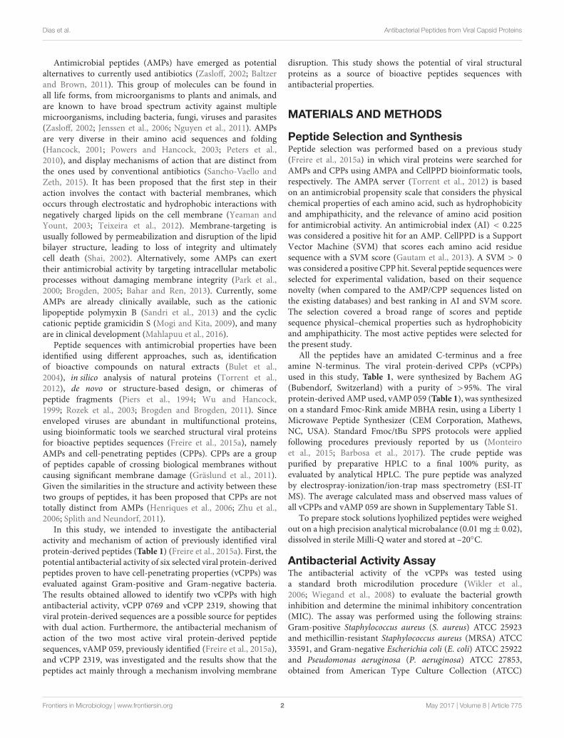
Bioimaging of the Viral Protein-DerivedPeptides Effect on P. aeruginosa CellsAtomic force microscopy imaging was used to visualize themorphological changes in P. aeruginosa after treatment withvAMP 059 and vCPP 2319. The images obtained in air foruntreated P. aeruginosa cells (Figure 3A) showed, in all cases,the characteristic rod shaped structure as well as a corrugatedsurface with no visible pores or ruptures. Treatment of bacteriawith increasing concentrations of both peptides revealed a cleareffect on cell envelope integrity when compared with untreatedbacteria, although the cells maintained their global rod-likeform. After treatment with vAMP 059, a pronounced collapse inP. aeruginosa cell envelope occurred at all concentrations tested(Figures 3B–D). For vCPP 2319 a similar result was observed, buta more gradual effect with increasing concentrations of peptidewas observed (Figures 3E–G). At 1.56 µM, a concentrationbelow MBC, only minor alterations on bacterial topography wereobserved.
DISCUSSION
The therapeutic options for treatment of bacterial infectionsare drastically decreasing due to a fast increase in drugresistance. Driven by the need to discover new and effectiveantimicrobial agents, this work has focused on a group ofpromising alternatives, the AMPs.
In a previous study performed in our lab, we becameaware that, among AMPs and CPPs derived from naturalsources and listed on existing databases, only a few of themwere related to viral proteins. Using bioinformatic tools, aset of peptide sequences with potential antimicrobial andcell-penetrating properties was identified, suggesting that virusesare an underexplored source for AMPs and CPPs (Freire et al.,2015a). One viral protein-derived AMP (vAMP 059) and tenviral protein-derived CPPs (vCPPs) were identified. In this study,from the six more active vCPPs, we were able to identifytwo peptides, vCPP 0769 and vCPP 2319, with concurrent,strong antibacterial activity. vCPP 2319 demonstrated to beactive at low micromolar range concentrations, being bactericidalagainst all the bacterial strains tested. vCPP 0769 revealedhigh antibacterial activity against the S. aureus, MRSA andP. aeruginosa strains, and a moderate activity against E. coli,but only displayed bactericidal activity against the Gram-negative bacterial strains tested. Therefore, both peptides reveala dual cell-penetrating and antibacterial activity, a peculiarcross functionality that has been reported for few otherpeptides (Takeshima et al., 2003; Nekhotiaeva et al., 2004;Bobone et al., 2011). It has been proposed that peptideswith dual antimicrobial and cell-penetrating properties mightconstitute potential candidates to target intracellular pathogenicbacteria (Bahnsen et al., 2013; Torcato et al., 2013) thatescape host defenses (Ernst et al., 1999) and are extremely
difficult to treat with conventional antibiotics (Clement et al.,2005).
In the present study, the results also show that thepreviously identified viral protein-derived peptide vAMP 059,with demonstrated activity against S. aureus and P. aeruginosa(Freire et al., 2015a), revealed a bactericidal mode of actionagainst these bacterial strains. It is interesting to notice thatthis peptide, as well as both vCPP 0769 and vCPP 2319, arederived from viral capsid proteins (Freire et al., 2015a). Despitethe fact that only few viral-derived AMPs are known, there areother examples of peptides with antimicrobial properties derivedfrom viral capsid proteins. HBc ARD, is a peptide derived fromthe capsid protein of the human hepatitis B virus (Chen et al.,2013), and pepR, derived from the putative RNA-binding domainof the dengue virus capsid protein (Alves et al., 2010). Viralcapsid proteins are known as multifunctional proteins and someof them can also be described as super charged proteins, aclass of proteins with high net charge to molecular mass ratioand are known to have cell-membrane translocating properties(Freire et al., 2015b). In fact, there are several examples ofcell-penetrating domains found in capsid proteins. With Denguevirus capsid protein as an example, we showed that one of thesedomains, the aforementioned pepR, also displays antibacterialactivity. This potential antibacterial activity of viral proteins mayhave conferred viruses an evolutionary advantage over bacteria(Freire et al., 2015b). Additionally, the presence of domains withboth antibacterial and cell-penetrating properties in viral capsidproteins demonstrates the potential use of viral proteins in drugdiscovery.
To get insights on the mode of action of the viralprotein-derived peptides, the most active peptides vAMP 059and vCPP 2319 were studied. Both peptides revealed a fastkilling kinetics against S. aureus and P. aeruginosa. Since itis known that antimicrobial agents that kill within minutes ofbacterial exposure, at concentrations similar to their MICs, act bydisrupting bacterial membranes (Zasloff, 2002; Melo et al., 2009),we can suggest that both viral protein-derived peptides inducebacterial death through membrane disruption. This behavior iscommon amongst AMPs; for instance, the antimicrobial peptidemelittin, derived from bee venom, exhibits an extremely rapidmembrane-disrupting action, killing bacteria within minutes(Kaplan et al., 2011). Additionally, when comparing vAMP 059and vCPP 2319 killing kinetics on both bacterial strains testedhere, faster rates were observed against S. aureus than againstP. aeruginosa. This behavior can be related to the differentialpeptide affinity for the molecular structures on Gram-negative orGram-positive bacteria envelope surfaces. Gram-positive bacteriaenvelope consists on a single lipid membrane followed by athick peptidoglycan layer enriched in negatively charged teichoicacids (Xia et al., 2010). In contrast, Gram-negative bacteriapossesses an inner cytoplasmic membrane surrounded by a thinpeptidoglycan layer and an outer membrane containing negativelipopolysaccharides (LPS) (Beveridge, 1999). For instance,the peptide rBPI21 exerts its antimicrobial activity throughinteractions with LPS of the outer membrane of Gram-negativebacteria, followed by fusion of the bacterial outer and innermembranes (Domingues et al., 2009). In turn, the antimicrobial
Frontiers in Microbiology | www.frontiersin.org 6 May 2017 | Volume 8 | Article 775
fmicb-08-00775 May 2, 2017 Time: 15:17 # 7
Dias et al. Antibacterial Peptides from Viral Capsid Proteins
FIGURE 3 | Effect of vAMP 059 and vCPP 2319 on P. aeruginosa cells imaged by AFM. (A) AFM height image (left image) and three-dimensional projection(right image) of P. aeruginosa cells incubated for 1 h in the absence of peptide (control). (B–G) AFM height images and three-dimensional projections of P. aeruginosacells incubated for 1 h with increasing concentrations of vAMP 059 (B–D) and vCPP 2319 (E–G). Total scanning area for each image was 4 µm × 4 µm.
peptide omiganan has high affinity for the peptidoglycan layer inGram-positive bacteria (Melo and Castanho, 2007). vAMP 059and vCPP 2319 may have faster interaction with peptidoglycanwhen compared to LPS, resulting on a faster killing kineticstowards S. aureus rather than P. aeruginosa. The hypothesisthat vAMP 059 and vCPP 2319 cause bacterial cell membranedisruption was confirmed in Gram-negative P. aeruginosa cells.Both peptides induce permeabilization of the outer and innermembranes, in a concentration-dependent manner, as shown
using the non-permeable SYTOX Green dye, which was ableto bind to the nucleic acids of bacterial cells treated withpeptide. A mechanism involving membrane disruption wasfurther confirmed by AFM imaging. Treatment of P. aeruginosacells with vAMP 059 induced a pronounced collapse at the septalregion of the cell envelope at all concentrations tested. Negativelycharged cardiolipin domains are located at the apical and septalregions of E. coli cells (Mileykovskaya and Dowhan, 2000).Assuming a similar cardiolipin-distribution for P. aeruginosa,
Frontiers in Microbiology | www.frontiersin.org 7 May 2017 | Volume 8 | Article 775
fmicb-08-00775 May 2, 2017 Time: 15:17 # 8
Dias et al. Antibacterial Peptides from Viral Capsid Proteins
the observed collapse in the mid-region could be explained byaccumulation of the peptides in these regions due to electrostaticattractions between the positively charged peptides and thesenegatively charged domains. Treatment of P. aeruginosa withvCPP 2319 at concentrations below the MBC did not causesubstantial alterations on cell membrane morphology. However,treatment with MBC, or higher, caused a marked collapse of themembrane structure. Overall, AFM imaging suggested that bothpeptides cause significant structural alterations on bacterial cellsurface, which is in agreement with the proposed hypothesis thatvAMP 059 and vCPP 2319 act at the membrane-level.
AUTHOR CONTRIBUTIONS
SD, CP-P, MD, DG, ST-H, MC, and ASV designed theexperiments. SD, CP-P, MD, DG, and ASV performed theexperimental work and data analysis. JF contributed with thedesign of the studied peptides. PG and NV contributed withpeptide synthesis. All authors contributed to data interpretationand discussion. SD, MC, and ASV wrote the manuscript withcontributions from all other authors.
FUNDING
This work was supported by Fundação para a Ciência e aTecnologia (FCT-MCTES, Portugal) projects PTDC/QEQ-MED/4412/2014 and UID/QUI/50006/2013, and by Marie
Skłodowska-Curie Research and Innovation Staff Exchange(RISE): call H2020-MSCA-RISE-2014, Grant agreement644167, 2015–2019. SD, JF, and DG acknowledge FCT forfellowships PD/BD/114425/2016, SFRH/BD/70423/2010, andSFRH/BPD/109010/2015, respectively, and MD for a grant(PTDC/BBB-BQB/3494/2014). ASV and NV acknowledgeFCT for funding within the FCT Investigator Programme,IF/00803/2012 and IF/00092/2014, respectively. CP-Packnowledges financial support from the Spanish Ministry ofEconomy and Competitiveness, through grant AGL2014-52395-C2-2-R and the “María de Maeztu” Programme for Unitsof Excellence in R&D (MDM-2014-0370). PG acknowledges“Comissão de Coordenação e Desenvolvimento Regional doNorte (CCDR-N)/NORTE2020/Portugal 2020” for fundingthrough project DESignBIOtechHealth (ref. Norte-01-0145-FEDER-000024). SH is the recipient of an Australian ResearchCouncil Future Fellowship (FT150100398).
ACKNOWLEDGMENT
The authors thank Tiago Figueira for the help with data analysis.
SUPPLEMENTARY MATERIAL
The Supplementary Material for this article can be foundonline at: http://journal.frontiersin.org/article/10.3389/fmicb.2017.00775/full#supplementary-material
REFERENCESAlves, C. S., Melo, M. N., Franquelim, H. G., Ferre, R., Planas, M., Feliu, L.,
et al. (2010). Escherichia coli cell surface perturbation and disruption inducedby antimicrobial peptides BP100 and pepR. J. Biol. Chem. 285, 27536–27544.doi: 10.1074/jbc.M110.130955
Bahar, A. A., and Ren, D. (2013). Antimicrobial peptides. Pharmaceuticals 6,1543–1575. doi: 10.3390/ph6121543
Bahnsen, J. S., Franzyk, H., Sandberg-Schaal, A., and Nielsen, H. M. (2013).Antimicrobial and cell-penetrating properties of penetratin analogs: effect ofsequence and secondary structure. Biochim. Biophys. Acta 1828, 223–232.doi: 10.1016/j.bbamem.2012.10.010
Baltzer, S. A., and Brown, M. H. (2011). Antimicrobial peptides-promisingalternatives to conventional antibiotics. J. Mol. Microbiol. Biotechnol. 20,228–235. doi: 10.1159/000331009
Barbosa, M., Vale, N., Costa, F. M. T. A., Martins, M. C. L., and Gomes, P. (2017).Tethering antimicrobial peptides onto chitosan: optimization of azide-alkyne“click” reaction conditions. Carbohydr. Polym. 165, 384–393. doi: 10.1016/j.carbpol.2017.02.050
Barry, A. L., Craig, W. A., Nadler, H., Reller, L. B., Sanders, C. C., and Swenson, J. M.(1999). Methods for Determining Bactericidal Activity of Antimicrobial Agents;Approved Guidelines. Wayne, PA: Clinical and Laboratory Standards Institute(CLSI).
Beveridge, T. J. (1999). Structures of gram-negative cell walls and their derivedmembrane vesicles. J. Bacteriol. 181, 4725–4733.
Bobone, S., Piazzon, A., Orioni, B., Pedersen, J. Z., Nan, Y. H., Hahm, K. S., et al.(2011). The thin line between cell-penetrating and antimicrobial peptides: thecase of Pep-1 and Pep-1-K. J. Pept. Sci. 17, 335–341. doi: 10.1002/psc.1340
Brogden, K. A. (2005). Antimicrobial peptides: pore formers or metabolicinhibitors in bacteria? Nat. Rev. Microbiol. 3, 238–250. doi: 10.1038/nrmicro1098
Brogden, N. K., and Brogden, K. A. (2011). Will new generations of modifiedantimicrobial peptides improve their potential as pharmaceuticals? Int. J.Antimicrob. Agents 38, 217–225. doi: 10.1016/j.ijantimicag.2011.05.004
Brown, D. (2015). Antibiotic resistance breakers: Can repurposed drugs fill theantibiotic discovery void? Nat. Rev. Drug Discov. 14, 821–832. doi: 10.1038/nrd4675
Bulet, P., Stöcklin, R., and Menin, L. (2004). Anti-microbial peptides: frominvertebrates to vertebrates. Immunol. Rev. 198, 169–184. doi: 10.1111/j.0105-2896.2004.0124.x
Chen, H. L., Su, P., Chang, Y., Wu, S., Liao, Y., Yu, H., et al. (2013). Identificationof a novel antimicrobial peptide from human hepatitis B virus core proteinarginine-rich domain (ARD). PLoS Pathog. 9:e1003425. doi: 10.1371/journal.ppat.1003425
Clement, S., Vaudaux, P., Francois, P., Schrenzel, J., Huggler, E., Kampf, S.,et al. (2005). Evidence of an intracellular reservoir in the nasal mucosa ofpatients with recurrent Staphylococcus aureus rhinosinusitis. J. Infect. Dis. 192,1023–1028. doi: 10.1086/432735
Domingues, M. M., Castanho, M. A. R. B., and Santos, N. C. (2009). rBPI21promotes lipopolysaccharide aggregation and exerts its antimicrobial effects by(hemi)fusion of PG-containing membranes. PLoS ONE 4:e8385. doi: 10.1371/journal.pone.0008385
Ernst, R. K., Guina, T., and Miller, S. I. (1999). How intracellular bacteria survive:surface modifications that promote resistance to host innate immune responses.J. Infect. Dis. 179, S326–S330. doi: 10.1086/513850
Freire, J. M., Dias, S. A., Flores, L., Veiga, A. S., and Castanho, M. A. R. B. (2015a).Mining viral proteins for antimicrobial and cell-penetrating drug deliverypeptides. Bioinformatics 31, 2252–2256. doi: 10.1093/bioinformatics/btv131
Freire, J. M., Santos, N. C., Veiga, A. S., Da Poian, A. T., and Castanho, M. A. R. B.(2015b). Rethinking the capsid proteins of enveloped viruses: multifunctionalityfrom genome packaging to genome transfection. FEBS J. 282, 2267–2278.doi: 10.1111/febs.13274
Frontiers in Microbiology | www.frontiersin.org 8 May 2017 | Volume 8 | Article 775

fmicb-08-00775 May 2, 2017 Time: 15:17 # 9
Dias et al. Antibacterial Peptides from Viral Capsid Proteins
Gautam, A., Chaudhary, K., Kumar, R., Sharma, A., Kapoor, P., Tyagi, A., et al.(2013). In silico approaches for designing highly effective cell penetratingpeptides. J. Transl. Med. 11:74. doi: 10.1186/1479-5876-11-74
Gräslund, A., Madani, F., Lindberg, S., Langel, Ü, and Futaki, S. (2011).Mechanisms of cellular uptake of cell-penetrating peptides. J. Biophys.2011:414729. doi: 10.1155/2011/414729
Hancock, R. E. W. (2001). Cationic peptides: effectors in innate immunityand novel antimicrobials. Lancet Infect. Dis. 1, 156–164. doi: 10.1016/S1473-3099(01)00092-5
Henriques, S. T., Melo, M. N., and Castanho, M. A. R. B. (2006). Cell-penetratingpeptides and antimicrobial peptides: How different are they? Biochem. J. 399,1–7. doi: 10.1042/BJ20061100
Jenssen, H., Hamill, P., and Hancock, R. E. W. (2006). Peptide antimicrobial agents.Clin. Microbiol. Rev. 19, 491–511. doi: 10.1128/CMR.00056-5
Kaplan, C. W., Sim, J. H., Shah, K. R., Kolesnikova-Kaplan, A., Shi, W., andEckert, R. (2011). Selective membrane disruption: mode of action of C16G2,a specifically targeted antimicrobial peptide. Antimicrob. Agents Chemother. 55,3446–3452. doi: 10.1128/AAC.00342-11
Levison, M. E., and Levison, J. H. (2009). Pharmacokinetics andpharmacodynamics of antibacterial agents. Infect. Dis. Clin. North Am.23, 791–819. doi: 10.1016/j.idc.2009.06.008.Pharmacokinetics
Mahlapuu, M., Håkansson, J., Ringstad, L., and Björn, C. (2016). Antimicrobialpeptides: an emerging category of therapeutic agents. Front. Cell. Infect.Microbiol. 6:194. doi: 10.3389/fcimb.2016.00194
Melo, M., Ferre, R., and Castanho, M. (2009). Antimicrobial peptides: linkingpartition, activity and high membrane-bound concentrations. Nat. Rev.Microbiol. 7, 245–250. doi: 10.1038/nrmicro2095
Melo, M. N., and Castanho, M. A. R. B. (2007). Omiganan interaction with bacterialmembranes and cell wall models. Assigning a biological role to saturation.Biochim. Biophys. Acta 1768, 1277–1290. doi: 10.1016/j.bbamem.2007.02.005
Mileykovskaya, E., and Dowhan, W. (2000). Visualization of phospholipid domainsin Escherichia coli by using the cardiolipin-specific fluorescent dye 10-N-nonylacridine orange. J Bacteriol 182, 1172–1175. doi: 10.1128/JB.182.4.1172-1175.2000
Mogi, T., and Kita, K. (2009). Gramicidin S and polymyxins: the revival of cationiccyclic peptide antibiotics. Cell. Mol. Life Sci. 66, 3821–3826. doi: 10.1007/s00018-009-0129-9
Monteiro, C., Fernandes, M., Pinheiro, M., Maia, S., Seabra, C. L., Ferreira-Da-Silva, F., et al. (2015). Antimicrobial properties of membrane-activedodecapeptides derived from MSI-78. Biochim. Biophys. Acta 1848, 1139–1146.doi: 10.1016/j.bbamem.2015.02.001
Nekhotiaeva, N., Elmquist, A., Rajarao, G. K., Hällbrink, M., Langel, U., andGood, L. (2004). Cell entry and antimicrobial properties of eukaryotic cell-penetrating peptides. FASEB J. 18, 394–396. doi: 10.1096/fj.03-0449fje
Nguyen, L. T., Haney, E. F., and Vogel, H. J. (2011). The expanding scope ofantimicrobial peptide structures and their modes of action. Trends Biotechnol.29, 464–472. doi: 10.1016/j.tibtech.2011.05.001
Park, C. B., Yi, K. S., Matsuzaki, K., Kim, M. S., and Kim, S. C. (2000). Structure-activity analysis of buforin II, a histone H2A-derived antimicrobial peptide: theproline hinge is responsible for the cell-penetrating ability of buforin II. PNAS97, 8245–8250. doi: 10.1073/pnas.150518097
Peters, B. M., Shirtliff, M. E., and Jabra-Rizk, M. A. (2010). Antimicrobial peptides:Primeval molecules or future drugs? PLoS Pathog. 6:e1001067. doi: 10.1371/journal.ppat.1001067
Piers, K. L., Brown, M. H., and Hancock, R. E. W. (1994). Improvement ofouter membrane-permeabilizing and lipopolysaccharide-binding activities ofan antimicrobial cationic peptide by C-terminal modification. Antimicrob.Agents Chemother. 38, 2311–2316. doi: 10.1128/AAC.38.10.2311
Powers, J. P. S., and Hancock, R. E. W. (2003). The relationship between peptidestructure and antibacterial activity. Peptides 24, 1681–1691. doi: 10.1016/j.peptides.2003.08.023
Roth, B. L., Poot, M., Yue, S. T., and Millard, P. J. (1997). Bacterial viability andantibiotic susceptibility testing with SYTOX green nucleic acid stain. Appl.Environ. Microbiol. 63, 2421–2431.
Rozek, A., Powers, J. P. S., Friedrich, C. L., and Hancock, R. E. W. (2003).Structure-based design of an indolicidin peptide analogue with increasedprotease stability. Biochemistry 42, 14130–14138. doi: 10.1021/bi035643g
Sancho-Vaello, E., and Zeth, K. (2015). Antimicrobial peptides: Has their timearrived? Future Microbiol. 10, 1103–1106. doi: 10.2217/fmb.15.45
Sandri, A. M., Landersdorfer, C. B., Jacob, J., Boniatti, M. M., Dalarosa, M. G.,Falci, D. R., et al. (2013). Pharmacokinetics of polymyxin B in patients oncontinuous venovenous haemodialysis. J. Antimicrob. Chemother. 68, 674–677.doi: 10.1093/jac/dks437
Sani, M. A., Henriques, S. T., Weber, D., and Separovic, F. (2015). Bacteria maycope differently from similar membrane damage caused by the Australian treefrog antimicrobial peptide maculatin 1.1. J. Biol. Chem. 290, 19853–19862.doi: 10.1074/jbc.M115.643262
Shai, Y. (2002). Mode of action of membrane active antimicrobial peptides.Biopolymers 66, 236–248. doi: 10.1002/bip.10260
Splith, K., and Neundorf, I. (2011). Antimicrobial peptides with cell-penetratingpeptide properties and vice versa. Eur. Biophys. J. 40, 387–397. doi: 10.1007/s00249-011-0682-7
Takeshima, K., Chikushi, A., Lee, K. K., Yonehara, S., and Matsuzaki, K. (2003).Translocation of analogues of the antimicrobial peptides magainin and buforinacross human cell membranes. J. Biol. Chem. 278, 1310–1315. doi: 10.1074/jbc.M208762200
Tanwar, J., Das, S., Fatima, Z., and Hameed, S. (2014). Multidrug resistance: anemerging crisis. Interdiscip. Perspect. Infect. Dis. 2014:541340. doi: 10.1155/2014/541340
Teixeira, V., Feio, M. J., and Bastos, M. (2012). Role of lipids in the interactionof antimicrobial peptides with membranes. Prog. Lipid Res. 51, 149–177.doi: 10.1016/j.plipres.2011.12.005
Torcato, I. M., Huang, Y. H., Franquelim, H. G., Gaspar, D. D., Craik, D. J.,Castanho, M. A. R. B., et al. (2013). The antimicrobial activity of sub3 isdependent on membrane binding and cell-penetrating ability.Chembiochem 14,2013–2022. doi: 10.1002/cbic.201300274
Torrent, M., Di Tommaso, P., Pulido, D., Nogués, M. V., Notredame, C., Boix, E.,et al. (2012). AMPA: an automated web server for prediction of proteinantimicrobial regions. Bioinformatics 28, 130–131. doi: 10.1093/bioinformatics/btr604
Ventola, C. L. (2015). The antibiotic resistance crisis: part 1: causes and threats.P T 40, 277–283.
WHO (2014). Antimicrobial Resistance: Global Report on Surveillance, Vol.61. Geneva: World Health Organization, 383–394. doi: 10.1007/s13312-014-0374-3
Wiegand, I., Hilpert, K., and Hancock, R. E. W. (2008). Agar and brothdilution methods to determine the minimal inhibitory concentration (MIC) ofantimicrobial substances. Nat. Protoc. 3, 163–175. doi: 10.1038/nprot.2007.521
Wikler, M. A., Low, D. E., Cockerill, F. R., Sheehan, D. J., Craig, W. A., Tenover,F. C., et al. (2006). Methods for Dilution Antimicrobial Susceptibility Tests forBacteria that Grow Aerobically: Approved Standard. Wayne, PA: Clinical andLaboratory Standards Institute (CLSI).
Wu, M., and Hancock, R. E. W. (1999). Improved derivatives of bactenecin, a cyclicdodecameric antimicrobial cationic peptide. Antimicrob. Agents Chemother. 43,1274–1276.
Xia, G., Kohler, T., and Peschel, A. (2010). The wall teichoic acid and lipoteichoicacid polymers of Staphylococcus aureus. Int. J. Med. Microbiol. 300, 148–154.doi: 10.1016/j.ijmm.2009.10.001
Yeaman, M. R., and Yount, N. Y. (2003). Mechanisms of antimicrobial peptideaction and resistance. Pharmacol. Rev. 55, 27–55. doi: 10.1124/pr.55.1.2
Zasloff, M. (2002). Antimicrobial peptides of multicellular organisms. Nature 415,389–395.
Zhu, W. L., Lan, H., Park, I. S., Kim, J. II, Jin, H. Z., Hahm, K. S., et al. (2006). Designand mechanism of action of a novel bacteria-selective antimicrobial peptidefrom the cell-penetrating peptide Pep-1. Biochem. Biophys. Res. Commun. 349,769–774. doi: 10.1016/j.bbrc.2006.08.094
Conflict of Interest Statement: The authors declare that the research wasconducted in the absence of any commercial or financial relationships that couldbe construed as a potential conflict of interest.
Copyright © 2017 Dias, Freire, Pérez-Peinado, Domingues, Gaspar, Vale, Gomes,Andreu, Henriques, Castanho and Veiga. This is an open-access article distributedunder the terms of the Creative Commons Attribution License (CC BY). The use,distribution or reproduction in other forums is permitted, provided the originalauthor(s) or licensor are credited and that the original publication in this journalis cited, in accordance with accepted academic practice. No use, distribution orreproduction is permitted which does not comply with these terms.
Frontiers in Microbiology | www.frontiersin.org 9 May 2017 | Volume 8 | Article 775