New perspectives on the molecular basis of the interaction ... · submit your manuscript | Hypoxia...
11
© 2015 Recalcati et al. This work is published by Dove Medical Press Limited, and licensed under Creative Commons Attribution – Non Commercial (unported, v3.0) License. The full terms of the License are available at http://creativecommons.org/licenses/by-nc/3.0/. Non-commercial uses of the work are permitted without any further permission from Dove Medical Press Limited, provided the work is properly attributed. Permissions beyond the scope of the License are administered by Dove Medical Press Limited. Information on how to request permission may be found at: http://www.dovepress.com/permissions.php Hypoxia 2015:3 93–103 Hypoxia Dovepress submit your manuscript | www.dovepress.com Dovepress 93 REVIEW open access to scientific and medical research Open Access Full Text Article http://dx.doi.org/10.2147/HP.S83537 New perspectives on the molecular basis of the interaction between oxygen homeostasis and iron metabolism Stefania Recalcati Elena Gammella Gaetano Cairo Department of Biomedical Sciences for Health, University of Milan, Milan, Italy Correspondence: Gaetano Cairo Department of Biomedical Sciences for Health, University of Milan, Via Mangiagalli 31, 20133 Milan, Italy Email [email protected] Abstract: Oxygen and iron are two elements closely related from a (bio)chemical point of view. Moreover, they share the characteristic of being indispensable for life, while also being potentially toxic. Therefore, their level is strictly monitored, and sophisticated pathways have evolved to face variations in either element. In addition, the expression of proteins involved in iron and oxygen metabolism is mainly controlled by a complex interplay of proteins that sense both iron levels and oxygen availability (ie, prolyl hydroxylases, hypoxia inducible factors, and iron regulatory proteins), and in turn activate feedback mechanisms to re-establish homeostasis. In this review, we describe how cells and organisms utilize these intricate networks to regulate responses to changes in oxygen and iron levels. We also explore the role of these pathways in some pathophysiological settings. Keywords: hypoxia, prolyl hydroxylases, hypoxia inducible factors, iron regulatory proteins, proteasome Introduction Iron and oxygen are two closely related elements: the large amount of iron that is present in the earth originated approximately 4.6 billion years ago from an oxygen-dependent process occurring during supernova explosions, the thermonuclear disruptions of a white dwarf star in which carbon and oxygen fused into radioactive nickel that decayed quickly into radioactive cobalt, which subsequently decayed into stable iron ( 56 Fe). Recently, proof of this process was provided by the INTEGRAL satellite that detected gamma ray lines from the radioactive decay of nickel into iron. 1 The interaction of iron and oxygen continued during the slow and long (1.5 billion years) accumulation in the atmosphere of the oxygen produced by photosynthesizing cyanobacteria that led to the present 20.9% atmospheric oxygen content. Over this period of time, living organisms adapted to exploit the higher metabolic efficiency of aerobic reactions and the chemical reactivity of iron for processes essential for life. In parallel, it has been necessary to evolve protection systems against the damage caused by the iron-catalyzed generation of highly reactive hydroxyl radicals from the less harmful superoxide and hydrogen peroxide (reactive oxygen species) continuously produced by every cell living in aero- bic conditions. 2 To this purpose, a complex regulatory pathway formed by a variety of proteins that bind, transport, and store iron has been developed, in order to maintain an appropriate iron balance in both the individual cells and the whole body. 3 In addition, an adaptive response to hypoxia, both at the cellular and organismal levels and in populations living at high altitude, has evolved in order to ensure the delivery of appropriate levels of oxygen to tissues. Paradoxically, while activation of hypoxia
Transcript of New perspectives on the molecular basis of the interaction ... · submit your manuscript | Hypoxia...

© 2015 Recalcati et al. This work is published by Dove Medical Press Limited, and licensed under Creative Commons Attribution – Non Commercial (unported, v3.0) License. The full terms of the License are available at http://creativecommons.org/licenses/by-nc/3.0/. Non-commercial uses of the work are permitted without any further
permission from Dove Medical Press Limited, provided the work is properly attributed. Permissions beyond the scope of the License are administered by Dove Medical Press Limited. Information on how to request permission may be found at: http://www.dovepress.com/permissions.php
Hypoxia 2015:3 93–103
Hypoxia Dovepress
submit your manuscript | www.dovepress.com
Dovepress 93
R e v i e w
open access to scientific and medical research
Open Access Full Text Article
http://dx.doi.org/10.2147/HP.S83537
New perspectives on the molecular basis of the interaction between oxygen homeostasis and iron metabolism
Stefania Recalcatielena GammellaGaetano CairoDepartment of Biomedical Sciences for Health, University of Milan, Milan, italy
Correspondence: Gaetano Cairo Department of Biomedical Sciences for Health, University of Milan, via Mangiagalli 31, 20133 Milan, italy email [email protected]
Abstract: Oxygen and iron are two elements closely related from a (bio)chemical point of
view. Moreover, they share the characteristic of being indispensable for life, while also being
potentially toxic. Therefore, their level is strictly monitored, and sophisticated pathways have
evolved to face variations in either element. In addition, the expression of proteins involved in
iron and oxygen metabolism is mainly controlled by a complex interplay of proteins that sense
both iron levels and oxygen availability (ie, prolyl hydroxylases, hypoxia inducible factors, and
iron regulatory proteins), and in turn activate feedback mechanisms to re-establish homeostasis.
In this review, we describe how cells and organisms utilize these intricate networks to regulate
responses to changes in oxygen and iron levels. We also explore the role of these pathways in
some pathophysiological settings.
Keywords: hypoxia, prolyl hydroxylases, hypoxia inducible factors, iron regulatory proteins,
proteasome
IntroductionIron and oxygen are two closely related elements: the large amount of iron that is present
in the earth originated approximately 4.6 billion years ago from an oxygen-dependent
process occurring during supernova explosions, the thermonuclear disruptions of a
white dwarf star in which carbon and oxygen fused into radioactive nickel that decayed
quickly into radioactive cobalt, which subsequently decayed into stable iron (56Fe).
Recently, proof of this process was provided by the INTEGRAL satellite that detected
gamma ray lines from the radioactive decay of nickel into iron.1 The interaction of iron
and oxygen continued during the slow and long (1.5 billion years) accumulation in the
atmosphere of the oxygen produced by photosynthesizing cyanobacteria that led to the
present 20.9% atmospheric oxygen content. Over this period of time, living organisms
adapted to exploit the higher metabolic efficiency of aerobic reactions and the chemical
reactivity of iron for processes essential for life. In parallel, it has been necessary to
evolve protection systems against the damage caused by the iron-catalyzed generation
of highly reactive hydroxyl radicals from the less harmful superoxide and hydrogen
peroxide (reactive oxygen species) continuously produced by every cell living in aero-
bic conditions.2 To this purpose, a complex regulatory pathway formed by a variety of
proteins that bind, transport, and store iron has been developed, in order to maintain
an appropriate iron balance in both the individual cells and the whole body.3
In addition, an adaptive response to hypoxia, both at the cellular and organismal levels
and in populations living at high altitude, has evolved in order to ensure the delivery
of appropriate levels of oxygen to tissues. Paradoxically, while activation of hypoxia

Hypoxia 2015:3submit your manuscript | www.dovepress.com
Dovepress
Dovepress
94
Recalcati et al
inducible factors (HIF) confers cellular adaptation to hypoxia,
in some highlanders (Tibetan and Ethiopians) variants in genes
of the HIF pathway (eg, EGLN1 and EPAS1 coding for PHD2
and HIF-2α, respectively, see section: The HIF-hydroxylase
system controls cellular oxygen homeostasis) that confer a
hypo-responsiveness to hypoxia seem to have been crucial
for adaptation to high altitude.4
HIF and IRPs are two interconnected sensors of both iron levels and oxygen availabilityThe interaction between oxygen and iron is not limited to
the oxygen transport function performed by proteins contain-
ing heme iron, such as hemoglobin and myoglobin. In fact,
the iron regulatory proteins (IRP1 and IRP2) that control
cellular iron homeostasis and the hydroxylases that control
the stability and activity of HIF sense both iron levels and
oxygen availability, and in turn regulate both pathways in a
coordinate and interconnected way.
The HiF-hydroxylase system controls cellular oxygen homeostasisIn this paragraph, we will offer only a brief summary of the
role of HIF (HIF-1α and HIF-2α) as regulators of oxygen
homeostasis, a topic comprehensively described in recent
excellent reviews.5,6 The cells respond to conditions of insuf-
ficient oxygen availability by increasing the amount and
activity of HIF transcription factors, which induce the expres-
sion of a large number of genes involved in the response
to hypoxia.5,6 The oxygen-regulated HIF-1α and HIF-2α
subunits form heterodimers with the constitutively expressed
HIF-1β subunit and bind hypoxia responsive elements
(HRE) in DNA regulatory regions that can be far from the
transcription start site. A third HIF-α (HIF-3α), structurally
and functionally distinct, is present in humans and mammals,
but its role and mode of action are not completely clear. One
reason for this is the presence of alternative splice variants
coding for at least six isoforms with structural and functional
differences that complicate the study of HIF-3α. Although
several studies have led to the view that HIF-3α is a negative
regulator of HIF-1α and HIF-2α, recent evidence suggests
that HIF-3α activates a distinct transcriptional response to
hypoxia, at least in zebrafish.7
The targets of HIF include genes with a direct role in
the adaptation to hypoxia, as well as regulatory molecules
with a secondary effect on gene expression, such as induc-
tion of transcriptional repressors that result in decreased
expression of many genes under hypoxic conditions.
Notably, genes coding for proteins directly or indirectly
involved in iron metabolism are among the variety of HIF
target genes (Table 1).8 Another connection between the
hypoxic response and iron metabolism is represented by
the iron requirement of the prolyl hydroxylases (PHDs) that
regulate HIF-α stability. PHDs are members of a family
of 2-oxoglutarate-dependent dioxygenases whose activity
requires 2-oxoglutarate and oxygen as obligatory substrates
and non-heme ferrous iron as cofactor.9,10 In addition to
other important functions, such as collagen biosynthesis,
these enzymes play an important role in gene expression. In
fact, they are involved in the control of both epigenetic (eg,
DNA demethylation and histone lysine demethylation) and
transcriptional (eg, HIF) regulation. PHDs catalyze prolyl
hydroxylation in the oxygen-dependent degradation domains
of HIF-α. After this modification, the affinity of HIF-α for
the E3 ubiquitin ligase von Hippel-Lindau (VHL) protein
increases several hundred fold, thereby leading to HIF-α
degradation by the proteasome (Figure 1).9,10 The mechanism
by which PHDs act as hypoxia sensors is both efficient and
sensitive to changes in oxygen availability, such that under
normoxia the inducible HIF-α are extremely short lived, but
as oxygen levels fall slightly, the decrease in PHDs activ-
ity allows HIF-α levels to rise and the hypoxic response to
start. PHDs are also sensors of energy metabolism, as the
Krebs cycle intermediates 2-oxoglutarate and succinate
and fumarate are an activator or inhibitors, respectively, of
2-oxoglutarate-dependent dioxygenases enzymes. Moreover,
PHDs activity is dependent on the availability of ferrous iron,
and therefore, HIF-dependent transcription is also induced
by iron deprivation, even under normoxia.9,10 Asparaginyl
hydroxylation of HIF-1/2α by factor inhibiting HIF, which
Table 1 Proteins of iron metabolism regulated by HiF
Proteins Functions
HIF-1 dependentTransferrin iron transportTransferrin receptor 1 iron uptakeCeruloplasmin iron oxidationFerrochelatase Heme synthesisFurin Regulation of iron homeostasisHeme oxygenase Heme catabolismMatriptase2 Regulation of iron homeostasisHIF-2 dependentDMT1 iron uptakeDcytB iron reductionepo erythropoiesisFerroportin iron exportFrataxin Assembly of iron sulfur-cluster
Abbreviations: DMT1, divalent metal transporter; epo, erythropoietin; HiF, hypoxia inducible factors.

Hypoxia 2015:3 submit your manuscript | www.dovepress.com
Dovepress
Dovepress
95
Crosstalk between oxygen and iron homeostasis
inhibits their transactivation activity, represents an additional
mechanism preventing the hypoxic response when sufficient
oxygen and cofactors are available.6
Despite extensive studies, the respective role of HIF-1α
and HIF-2α has not been fully clarified. They bind the same
HRE in vitro and share an overlapping set of target genes,
but they also activate the transcription of distinct genes. For
example, both HIF-α bind the HRE of erythropoietin (Epo),
but selective inactivation in mice revealed that only HIF-2α
is important for Epo production in vivo.11
In this context, the activity of the various components
of the HIF pathway can be modulated by various factors,
including tissue-specific expression and oxygen sensing. In
relation to the interdependence of iron and oxygen homeo-
stasis covered in this review, we would like to point out the
direct regulation of HIF-2α mRNA stability by iron (see
section: The IRE/IRP regulatory pathaway controls cellular
iron homeostasis).
The iRe/iRP regulatory pathway controls cellular iron homeostasisThe challenging task of maintaining intracellular iron
levels sufficient for essential cellular functions, but as low
as possible to avoid reactive oxygen species formation, is
mainly performed by IRP1 and IRP2, which tightly control
intracellular iron metabolism by regulating at the post-
transcriptional level the expression of genes coding for
proteins involved in iron uptake, utilization, storage, and
export.8,12–14
All these proteins are coded by mRNAs with conserved
25–30 nucleotide-long sequences in their untranslated
regions that are able to form stem-loop structures called iron
responsive elements (IREs).
IRP1 is a “moonlighting” protein that can perform
two entirely different functions; when sufficient iron is
available, IRP1 can assemble a [4Fe-4S] cluster becom-
ing the cytosolic counterpart of mitochondrial aconitase
and catalyzing the conversion of citrate and isocitrate,
a function that may affect NADPH levels and lipid bio-
synthesis.15 Conversely, in conditions of iron deficiency,
IRP1 cannot form the cluster and the apoform is able to
bind IREs, and thus control the expression of a variety of
mRNAs (Figure 2).
IRP2, which is highly homologous to IRP1, binds IRE
motifs with affinity and specificity similar to that of IRP1,
but is controlled by a different mechanism. In fact, IRP2
is not able to assemble a [4Fe-4S] cluster, and hence is not
regulated by the switch between an apoform and a cluster-
containing form; conversely, the protein accumulated in iron-
deficient cells and in iron-replete cells is rapidly targeted for
proteasomal degradation by an E3 ubiquitin ligase complex
that comprises FBXL5 (see section: The iron-oxygen axis:
regulation at the cellular level).16
When iron inside the cell is scarce, active IRPs bind to
IREs and stabilize the mRNA for transferrin receptor (TfR1)
and divalent metal transporter DMT1, thus increasing the
uptake of both transferrin-bound and unbound iron, while
also decreasing translation of mRNAs for the iron storage
HERC2
Fe + O2
Fe + O2
FBLX5 FBLX5
FBLX5
NCOA4 NCOA4
CITED2 CITED2
IRP2 IRP2
HIFα HIFα No hypoxic response
Cellular ironavailability
Hypoxic response
Ferritin(less autophagicdegradation)
(less degradation)IRP2
Proteasomaldegradation
Figure 1 Different levels of iron and oxygen promote the proteasomal degradation of key regulatory proteins of iron and oxygen metabolism.Notes: when iron and oxygen are plentiful, PHD-dependent hydroxylation targets for proteasomal degradation HiF-α, whereas FBXL5 accumulates, binds, and targets to degradation of iRP2 and CiTeD2. Conversely, the lack of iron and/or oxygen leads to FBXL5 ubiquitination and degradation by the proteasome, thus allowing iRP2 to function as a regulator of iron homeostasis. HeRC2 is responsible for the basal proteasomal degradation of FBXL5 at the steady state, but whether its activity is modulated by changes in iron and oxygen availability has not been defined (dotted arrows). Moreover, HERC2 induces iron-dependent proteasomal degradation of NCOA4, thus, preventing ferritin destruction in the autophagosome.Abbreviations: HiF, hypoxia inducible factors; iRP, iron regulatory protein.
Hypoxia 2015:3submit your manuscript | www.dovepress.com
Dovepress
Dovepress
96
Recalcati et al
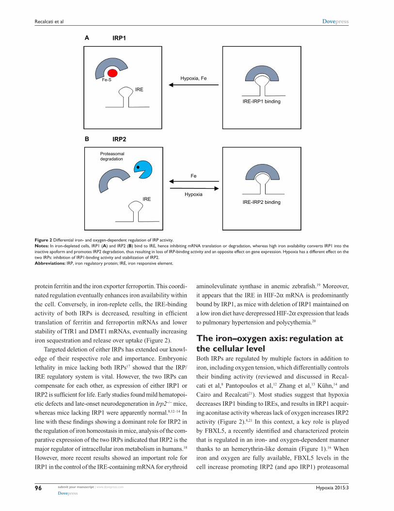
A IRP1
IRE
Hypoxia, Fe
IRE-IRP1 binding
Fe-S
B IRP2
IRE
Fe
Hypoxia
IRE-IRP2 binding
Proteasomaldegradation
Figure 2 Differential iron- and oxygen-dependent regulation of iRP activity.Notes: In iron-depleted cells, IRP1 (A) and IRP2 (B) bind to IRE, hence inhibiting mRNA translation or degradation, whereas high iron availability converts IRP1 into the inactive apoform and promotes iRP2 degradation, thus resulting in loss of iRP-binding activity and an opposite effect on gene expression. Hypoxia has a different effect on the two iRPs: inhibition of iRP1-binding activity and stabilization of iRP2.Abbreviations: iRP, iron regulatory protein; iRe, iron responsive element.
protein ferritin and the iron exporter ferroportin. This coordi-
nated regulation eventually enhances iron availability within
the cell. Conversely, in iron-replete cells, the IRE-binding
activity of both IRPs is decreased, resulting in efficient
translation of ferritin and ferroportin mRNAs and lower
stability of TfR1 and DMT1 mRNAs, eventually increasing
iron sequestration and release over uptake (Figure 2).
Targeted deletion of either IRPs has extended our knowl-
edge of their respective role and importance. Embryonic
lethality in mice lacking both IRPs17 showed that the IRP/
IRE regulatory system is vital. However, the two IRPs can
compensate for each other, as expression of either IRP1 or
IRP2 is sufficient for life. Early studies found mild hematopoi-
etic defects and late-onset neurodegeneration in Irp2−/− mice,
whereas mice lacking IRP1 were apparently normal.8,12–14 In
line with these findings showing a dominant role for IRP2 in
the regulation of iron homeostasis in mice, analysis of the com-
parative expression of the two IRPs indicated that IRP2 is the
major regulator of intracellular iron metabolism in humans.18
However, more recent results showed an important role for
IRP1 in the control of the IRE-containing mRNA for erythroid
aminolevulinate synthase in anemic zebrafish.19 Moreover,
it appears that the IRE in HIF-2α mRNA is predominantly
bound by IRP1, as mice with deletion of IRP1 maintained on
a low iron diet have derepressed HIF-2α expression that leads
to pulmonary hypertension and polycythemia.20
The iron–oxygen axis: regulation at the cellular levelBoth IRPs are regulated by multiple factors in addition to
iron, including oxygen tension, which differentially controls
their binding activity (reviewed and discussed in Recal-
cati et al,8 Pantopoulos et al,12 Zhang et al,13 Kühn,14 and
Cairo and Recalcati21). Most studies suggest that hypoxia
decreases IRP1 binding to IREs, and results in IRP1 acquir-
ing aconitase activity whereas lack of oxygen increases IRP2
activity (Figure 2).8,21 In this context, a key role is played
by FBXL5, a recently identified and characterized protein
that is regulated in an iron- and oxygen-dependent manner
thanks to an hemerythrin-like domain (Figure 1).16 When
iron and oxygen are fully available, FBXL5 levels in the
cell increase promoting IRP2 (and apo IRP1) proteasomal
Hypoxia 2015:3 submit your manuscript | www.dovepress.com
Dovepress
Dovepress
97
Crosstalk between oxygen and iron homeostasis
degradation. On the other hand, lack of iron, which impairs
the assembly of the di-iron center in the hemerythrin-like
domain, or hypoxia leads to FBXL5 polyubiquitination and
degradation by the proteasome, thus stabilizing IRP2 and
increasing iron availability. A recent study demonstrated that
FBXL5 itself is constitutively targeted to ubiquitin-dependent
proteasomal degradation through interaction with HERC2,22
a protein involved in the response to DNA damage. As iron
is a cofactor of several DNA repair proteins, these findings
reveal an interesting new link between iron and an essential
function required for cell survival.
The effects of hypoxia on IRPs binding activity are not
the only link between oxygen and iron homeostasis. As
reported above, the lack of oxygen and/or iron inhibits the
hydroxylation process that results in proteasomal degrada-
tion of HIF-1/2α, thereby increasing protein stability and
transcriptional capacity.5,6
Moreover, the finding of a functional 5′ IRE in the mRNA
for HIF-2α showed the existence of another physiologically
relevant connection between oxygen and iron sensing.13,23 In
normoxia, active IRP1 represses basal HIF-2α translation,
whereas in hypoxic conditions the IRP1/IRE interaction is
impaired and HIF-2α is efficiently translated, thus suggest-
ing that IRP1 acts as a direct or indirect sensor of hypoxia.
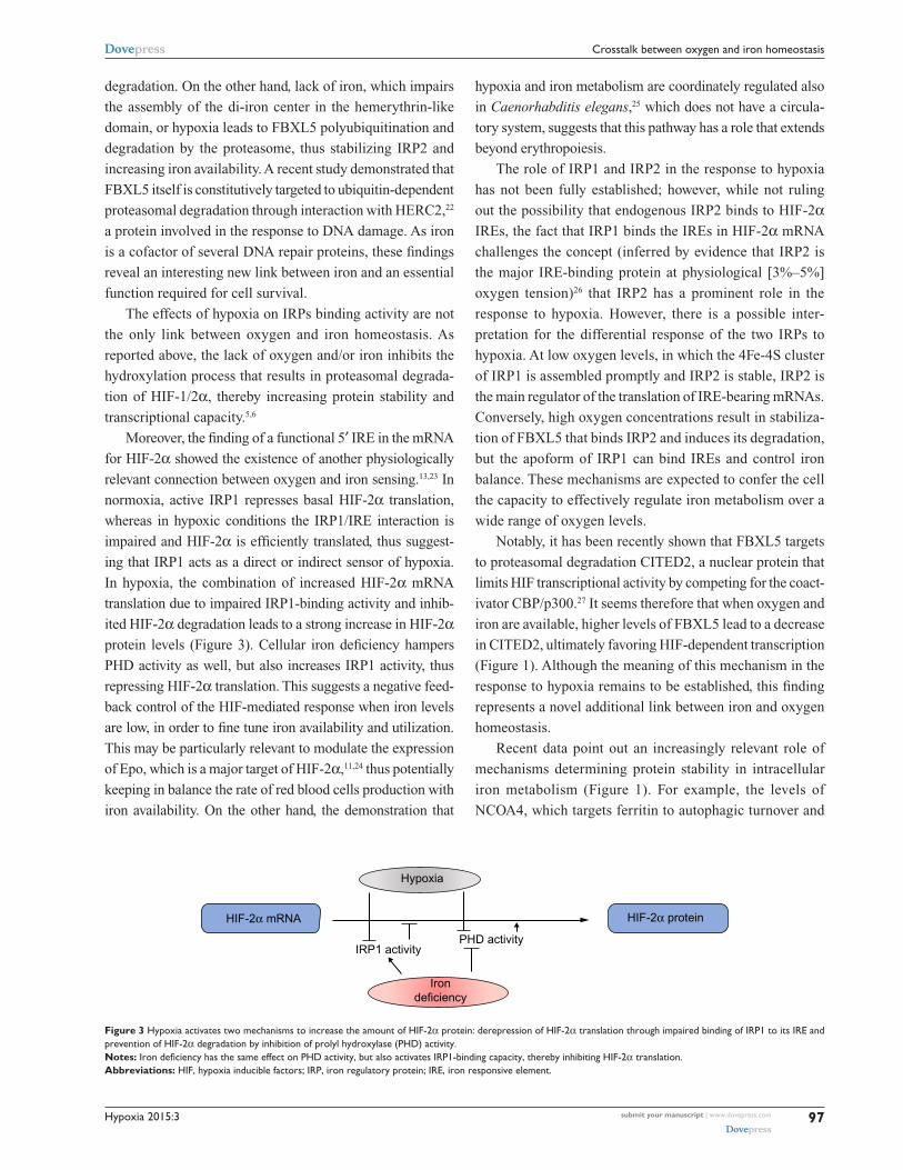
In hypoxia, the combination of increased HIF-2α mRNA
translation due to impaired IRP1-binding activity and inhib-
ited HIF-2α degradation leads to a strong increase in HIF-2α
protein levels (Figure 3). Cellular iron deficiency hampers
PHD activity as well, but also increases IRP1 activity, thus
repressing HIF-2α translation. This suggests a negative feed-
back control of the HIF-mediated response when iron levels
are low, in order to fine tune iron availability and utilization.
This may be particularly relevant to modulate the expression
of Epo, which is a major target of HIF-2α,11,24 thus potentially
keeping in balance the rate of red blood cells production with
iron availability. On the other hand, the demonstration that
hypoxia and iron metabolism are coordinately regulated also
in Caenorhabditis elegans,25 which does not have a circula-
tory system, suggests that this pathway has a role that extends
beyond erythropoiesis.
The role of IRP1 and IRP2 in the response to hypoxia
has not been fully established; however, while not ruling
out the possibility that endogenous IRP2 binds to HIF-2α
IREs, the fact that IRP1 binds the IREs in HIF-2α mRNA
challenges the concept (inferred by evidence that IRP2 is
the major IRE-binding protein at physiological [3%–5%]
oxygen tension)26 that IRP2 has a prominent role in the
response to hypoxia. However, there is a possible inter-
pretation for the differential response of the two IRPs to
hypoxia. At low oxygen levels, in which the 4Fe-4S cluster
of IRP1 is assembled promptly and IRP2 is stable, IRP2 is
the main regulator of the translation of IRE-bearing mRNAs.
Conversely, high oxygen concentrations result in stabiliza-
tion of FBXL5 that binds IRP2 and induces its degradation,
but the apoform of IRP1 can bind IREs and control iron
balance. These mechanisms are expected to confer the cell
the capacity to effectively regulate iron metabolism over a
wide range of oxygen levels.
Notably, it has been recently shown that FBXL5 targets
to proteasomal degradation CITED2, a nuclear protein that
limits HIF transcriptional activity by competing for the coact-
ivator CBP/p300.27 It seems therefore that when oxygen and
iron are available, higher levels of FBXL5 lead to a decrease
in CITED2, ultimately favoring HIF-dependent transcription
(Figure 1). Although the meaning of this mechanism in the
response to hypoxia remains to be established, this finding
represents a novel additional link between iron and oxygen
homeostasis.
Recent data point out an increasingly relevant role of
mechanisms determining protein stability in intracellular
iron metabolism (Figure 1). For example, the levels of
NCOA4, which targets ferritin to autophagic turnover and
HIF-2α mRNA HIF-2α protein
Hypoxia
IRP1 activityPHD activity
Irondeficiency
Figure 3 Hypoxia activates two mechanisms to increase the amount of HiF-2α protein: derepression of HiF-2α translation through impaired binding of iRP1 to its iRe and prevention of HiF-2α degradation by inhibition of prolyl hydroxylase (PHD) activity.Notes: Iron deficiency has the same effect on PHD activity, but also activates IRP1-binding capacity, thereby inhibiting HIF-2α translation.Abbreviations: HiF, hypoxia inducible factors; iRP, iron regulatory protein; iRe, iron responsive element.

Hypoxia 2015:3submit your manuscript | www.dovepress.com
Dovepress
Dovepress
98
Recalcati et al
thus affects iron availability at both the cellular and systemic
levels, are regulated by HERC2-mediated proteasomal
degradation.28 Clearly, in this context, the proteasome is
a key player; therefore, one possible question is how iron
homeostasis could be affected by changes in proteasomal
activity, which can occur in a variety of pathophysiologic
conditions including the immune response, neurodegenera-
tive diseases, and cancer.29 For instance, will proteasomal
inhibition lead (directly) to a higher amount of IRP2 or
(indirectly) to a decrease in IRP2 levels because of FBXL5
stabilization? Although this problem has not been experi-
mentally addressed in a specific way, a couple of recent
papers provide some information. Multiple myeloma cell
lines exposed to bortezomib, a proteasome inhibitor in use
for treatment of this type of tumor, showed a 50% reduction
in IRP-binding activity,30 an effect that the authors attrib-
uted to oxidative stress-mediated IRP2 degradation, but
could well be the result of lower proteasomal degradation
of FBXL5. Conversely, higher IRP2 levels were found in
neuronal cells exposed to a different proteasomal inhibitor
(lactacystin).31 These contrasting results may depend on the
remarkably different experimental models, but also suggest
that further studies are needed to investigate the role of the
proteasome in cellular iron metabolism.
The rapid emergence of microRNAs (miRNAs) as key
regulators of gene expression in a variety of biological pro-
cesses expanded recently to the connection between iron and
oxygen homeostasis. miRNA-210 was identified as a HIF
target, induced by both hypoxia and iron deprivation,32 which
regulates iron homeostasis through a complex mechanism. In
fact, miRNA-210 directly inhibits TfR1 expression by direct
binding to TfR1 mRNA. However, miRNA-210 concurrently
suppresses ISCU, a protein involved in iron–sulfur cluster
formation, thus switching IRP1 to the RNA-binding apoform,
and leading to TfR1 mRNA stabilization. The net result of
these contrasting mechanisms was decreased transferrin
internalization (and possibly iron uptake) in miRNA-210-
transfected breast cancer cells. On the other hand, previous
studies have shown that TfR1 transcription is induced by
hypoxia and iron deficiency.33–35 Therefore, it seems that
numerous mechanisms of regulation of gene expression
cooperate to amplify the range of control and response to
stimuli. These recent advances support previous evidence
indicating that precise regulation of TfR1 expression, and
more generally of iron levels, is a key for cell activity, and
probably depends on individual cell needs and specificities.
HIF-1-dependent transcriptional upregulation may cooperate
with IRP-dependent post-transcriptional control to expand
the extent of response of iron homeostasis genes to oxygen or
iron shortage. In fact, iron chelation induced TfR1 expression
less efficiently in HIF-1-deficient hepatoma cells, which only
depend on IRP-mediated upregulation, than in their wild-type
counterpart,34 and is strongly induced in VHL-deficient renal
carcinoma cells that overexpress HIF-1.36
The iron–oxygen axis: regulation at the systemic levelThe two hormones most important to regulate iron and
oxygen homeostasis at the systemic level are hepcidin and
Epo, respectively. Hepcidin controls body iron balance by
downregulating ferroportin, thus stopping iron efflux from
duodenal and reticuloendothelial cells.37 Given their related
physiological function, the idea that Epo could directly
regulate hepcidin was attractive and initially supported
by experimental data. For example, under conditions of
elevated erythropoiesis, such as hypoxia or anemia, the
enhanced iron absorption to face the higher demand of
erythropoietic cells is accompanied by a strong inhibition of
hepcidin expression.38,39 Therefore, the possibility of a direct
effect of Epo on hepcidin has been explored. Indeed, Epo
administration in mice (reviewed by Kautz and Nemeth39)
and humans40,41 resulted in strong repression of hepcidin
synthesis. Moreover, early studies found a direct repression of
hepcidin transcription in Epo-treated cell lines.42,43 However,
the idea of a direct effect of Epo was challenged by studies
showing that administration of Epo to mice with damaged
bone marrow due to exposure to cytotoxic agents or irradia-
tion did not result in hepcidin suppression.44 Furthermore, it
has been demonstrated that increasing liver iron content can
stimulate hepcidin expression in conditions of permanently
high Epo serum levels.45 More recently, the inhibitory effect
of Epo on hepcidin synthesis was observed in mice lacking
Epo receptor expression in hepatic cells, allowing us to
show that the direct binding of Epo to liver receptors is not
necessary to suppress hepcidin.46 In line with this conclusion,
the erythroid factor erythroferrone (ERFE), which hinders
hepcidin expression under conditions of high erythropoietic
activity, was recently identified.47 Altogether, the available
data indicate that the role of Epo is to stimulate the synthesis
of ERFE in erythroblasts, which eventually downregulates
hepcidin production in the liver (Figure 4).
Similarly, it was initially proposed that the increased lev-
els of HIF-1α present in livers of iron-deficient mice might
repress hepcidin transcription.48 However, subsequent studies
using liver-restricted deletion of various components of the
HIF complex49,50 did not confirm a direct transcriptional

Hypoxia 2015:3 submit your manuscript | www.dovepress.com
Dovepress
Dovepress
99
Crosstalk between oxygen and iron homeostasis
suppression of hepcidin, and showed that the real role of HIF
in this context is to induce Epo. The consequent stimulation
of erythropoiesis then leads to hepcidin inhibition (Figure 4).
In line with these findings, hepcidin was not suppressed in
Ethiopians living at high altitude, who do not have a higher
erythropoietic drive,51 as these populations chronically
adapted to hypoxia by a pattern not involving increased
hemoglobin concentration.4 Although in vivo the effect of
hypoxia on hepcidin is probably indirect, several studies
reported that activation of HIF by hypoxia, iron chelation,
or PHDs inhibitors resulted in hepcidin downregulation in
cultured cells. Also in this case, the effect appears to be
indirect, because proteins that negatively control hepcidin
expression are induced by HIF (Table 1). Iron-mediated
hepcidin induction depends on the assembly on the cell
surface of a complex consisting of bone morphogenetic
proteins (BMPs), in particular BMP6, their receptors and
hemojuvelin, a membrane protein that functions as a co-
receptor for BMPs.37 Hemojuvelin is cleaved by two pro-
teases, Matriptase2 and furin, resulting in the interruption of
the hepcidin activation pathway and the release of a soluble
form that acts as a decoy molecule for BMPs, respectively.
The demonstration that expression of both Matriptase252
and furin53,54 is increased in hypoxia via HIF-1/2α may thus
explain the hepcidin downregulation found in cell culture
experiments. Recently, it has been shown that hypoxia inhib-
its hepcidin expression by a novel pathway involving platelet
derived growth factor-BB.55 This growth factor is increased
by hypoxia and regulates hepcidin by interfering with the
CREB and CREB-H signaling pathways. As platelet derived
growth factor-BB is stored in several cell types (eg, platelets)
and promptly released in response to Epo,56 this finding may
explain that even low-dose Epo injections in humans lead
to early and considerable decrease in hepcidin levels that
preceded any change in potentials mediators.40,41,57
Implications in pathophysiologyThe IRP–HIF axis is important for various functions of spe-
cific tissues and organs, and its disruption is involved in sev-
eral pathophysiological settings, for example erythropoiesis,
which has been recently reviewed elsewhere.13,58 Here, we
will focus on the intestine, the adipose tissue, and the lungs.
iron/oxygen sensing and duodenal iron absorptionA number of recent studies provided convincing evidence
that, in the context of duodenum, which is a key for iron
absorption, the IRP 1/HIF-2α axis plays a predominant role.
Iron deficiency specifically induced intestinal HIF-2α,59 and
intestine-restricted inactivation indicated that HIF-2α is
required to activate the reductase DcytB and the iron trans-
porters DMT1 and ferroportin, and increase iron absorption
in the gut of mice kept on an iron-deficient diet.59–61 Notably,
it has been shown that dysregulation of the HIF-2α–iron axis
in absorbing enterocytes plays a role in animal models of both
primary50 and secondary62 iron overload. Moreover, experi-
Hypoxia
Epo
rEpo
ERFE
HIF-2αHepcidin
Iron absorption
Figure 4 Mechanisms of hepcidin downregulation by hypoxia and epo. Notes: endogenous epo produced by the kidney in response to hypoxia in a HiF-2α-dependent way or exogenous recombinant Epo (rEpo) stimulates erythropoiesis. Erythroid cells in the bone marrow produce erythroferrone (ERFE) that then leads to hepcidin inhibition and consequent increase in iron absorption.Abbreviations: epo, erythropoietin; HiF, hypoxia inducible factors.

Hypoxia 2015:3submit your manuscript | www.dovepress.com
Dovepress
Dovepress
100
Recalcati et al
ments in mice with IRP1 inactivation and increased duodenal
HIF-2α expression confirmed that the IRP 1/HIF-2α axis
coordinates duodenal iron absorption according to systemic
oxygen and iron availability.63
However, recent studies showing that FBXL5 also plays
a relevant role at the systemic level, including the gut (see
review Ruiz and Bruick16), led to reappraisal of the role
of IRP2 in this context. Regulation of IRP-mediated iron
homeostasis is disrupted in mice lacking FBXL5, and the
unregulated IRP2 expression results in embryonic lethality
due to iron overload in the early placenta. Mice with condi-
tional deletion in the liver showed hepatic iron accumulation
and died with acute liver damage when fed an iron-enriched
diet.64 Intriguingly, despite liver overload, these mice express
inappropriately low hepcidin levels, possibly because of
impaired BMP6 signaling, and thus present systemic iron
overload. Moreover, FBXL5 seems to have a special role in
the intestine, as mice heterozygous for FBXL5 deletion fed
an iron-deficient diet are able to maintain normal hematologi-
cal values and avoid iron deficiency anemia by increasing
DMT1-mediated iron absorption.
Role of iron and hypoxia in obesityBoth iron and oxygen homeostasis are modified in obesity.
Animal studies have demonstrated that adipose tissues
become hypoxic in obesity and, despite HIF-2α protective
role, the consequent HIF signaling (mainly mediated by
HIF-1α) contributes to the development of obesity-associated
inflammation, insulin resistance, and other metabolic dis-
orders that are strongly associated with obesity.65,66 The
modifications of iron metabolism in obesity are multifari-
ous and more difficult to put in a coherent framework, with
strong differences between cell autonomous and systemic
regulation.67 On the one hand, obese subjects often present
body iron deficiency, and possibly anemia, probably because
the chronic and low-grade inflammation that characterizes
obesity68 leads to hepcidin upregulation and decreased
circulating iron availability.69 On the other hand, in adipose
tissue, the picture is more complex, as iron overload may
play an important role in at least two cell types of obese
adipose tissue: adipocytes and macrophages. Iron is required
for adipogenesis (and adipocyte tissue expansion), probably
because these cells are rich in mitochondria, which are the
major iron sink of the cell.70,71 However, excess iron in adi-
pocytes leads to decreased adiponectin release and impaired
adipocyte insulin sensitivity, resulting in compromised sys-
temic glucose tolerance.70,72 Therefore, in adipocytes, like
in every other body cell, an optimal level of iron is required
for homeostasis. Adipose tissue also contains macrophages
that are mainly polarized toward a M2 anti-inflammatory
phenotype in lean subjects, whereas in obesity the number
of macrophages is greatly increased, and their polariza-
tion is shifted to the M1 classically activated phenotype.73
Interestingly, macrophage subtypes not only differ for the
expression of classical inflammatory markers, but also have
profound differences in iron handling.74 M1 macrophages
are characterized by increased iron levels, whereas M2
macrophages have a pattern of gene expressions that favors
uptake of heme iron and ferroportin-mediated iron release.75
As HIF-1α and HIF-2α have been mainly related to M1
and M2 phenotypes, respectively,76 the lower iron content
that characterizes M2 macrophages is expected to favor
HIF-2α stabilization and activity, with a favorable outcome
in obesity. In fact, activated macrophage HIF-2α will relieve
adipose tissue inflammation and insulin resistance.77 These
data reveal another aspect of the complex interplay between
iron and oxygen homeostasis and highlight its importance
in relevant pathologies. However, the situation may be more
complicated because the distinction of macrophages in the
M1 and M2 subclasses is probably an oversimplification,78
and indeed M2-like macrophages with either low or high
iron content have been isolated from adipose tissue.79
Role of iron in hypoxia-dependent pulmonary hypertensionBoth hypoxia and iron have been demonstrated to play a key
role in pulmonary hypertension, a common clinical problem
characterized by progressive pulmonary vasculature remodel-
ing leading to increased pulmonary arterial pressures.80
Both animal models, in which heterozygous deletion or
activating mutation of HIF-α genes leads to protection from
pulmonary hypertension and higher sensitivity to hypoxia,
respectively, and a human genetic condition, Chuvash poly-
cythemia, in which hypoxia sensing is increased because the
VHL protein is inactive, provided a strong demonstration
of the role of the HIF pathway in pulmonary hypertension
(discussed in Zhang et al13).
Several studies have reported that iron deficiency may
cooperate in the activation of HIF triggered by the condi-
tions of persistent or intermittent hypoxia that are associated
with pulmonary hypertension. Indeed, increasing iron bio-
availability by intravenous iron loading effectively reversed
the hypertensive response of the pulmonary vasculature to
hypoxia.81 The effect of intravenous iron on the pulmonary
circulation in human studies was observed both under
laboratory conditions (normobaric hypoxia) and in subjects

Hypoxia 2015:3 submit your manuscript | www.dovepress.com
Dovepress
Dovepress
101
Crosstalk between oxygen and iron homeostasis
exposed to high altitude hypobaric hypoxia. Given the high
prevalence of both nutritional and functional iron deficiency
(in chronic diseases characterized by cytokine-induced iron
sequestration in the reticuloendothelial compartment), the
effect of iron status on oxygen sensing and hypoxia-induced
pulmonary vasoconstriction would be potentially significant
for human health.
Notably, mice with IRP 1 inactivation and consequent
unrestricted HIF-2 activity develop spontaneous pulmonary
hypertension, thus underscoring the physiological importance
of the overlap between oxygen and iron sensing.20
Candidate HIF-dependent genes that may contribute
to the development of hypoxic pulmonary hypertension in
humans include endothelin and proteins involved in sodium
and potassium transport. Interestingly, a recent study showed
that hypoxia elevates miR-210, which in turn represses its
target ISCU1/2 and downregulates Fe-S cluster formation,
thus leading to mitochondrial dysfunction and promoting
pulmonary hypertension.82 Therefore, iron deficiency can
act both by impairing PHDs activity and by preventing Fe-S
cluster assembly.
ConclusionRecent results have advanced the identification of strong
associations between iron metabolism and oxygen
homeostasis. However, our understanding of this complex
interplay remains incomplete. Further studies will clarify
the mechanistic relationships between these pathways and
their relevance in pathophysiological conditions and disease,
possibly paving the way for more advanced therapeutic
approaches aimed at re-establishing iron and/or oxygen
homeostasis in clinical settings.
AcknowledgmentThe authors gratefully acknowledge the grant support of
MIUR (Project COFIN).
DisclosureThe authors report no conflicts of interest in this work.
References1. Churazov E, Sunyaev R, Isern J, et al. Cobalt-56 γ-ray emission lines from
the type Ia supernova 2014J. Nature. 2014;512(7515):406–408.2. Sheftel AD, Mason AB, Ponka P. The long history of iron in the Uni-
verse and in health and disease. Biochim Biophys Acta. 2012;1820(3): 161–187.
3. Andrews NC. Forging a field: the golden age of iron biology. Blood. 2008;112(2):219–230.
4. Beall CM. Human adaptability studies at high altitude: research designs and major concepts during fifty years of discovery. Am J Hum Biol. 2013;25(2):141–147.
5. Semenza GL. Hypoxia-inducible factors in physiology and medicine. Cell. 2012;148(3):399–408.
6. Bishop T, Ratcliffe PJ. Signaling hypoxia by hypoxia-inducible factor protein hydroxylases: a historical overview and fututre perspective. Hypoxia. 2014;2:197–213.
7. Zhang P, Yao Q, Lu L, Li Y, Chen PJ, Duan C. Hypoxia-inducible factor 3 is an oxygen-dependent transcription activator and regulates a distinct transcriptional response to hypoxia. Cell Rep. 2014;6(6):1110–1121.
8. Recalcati S, Minotti G, Cairo G. Iron regulatory proteins: from molecular mechanisms to drug development. Antioxid Redox Signal. 2010;13(10):1593–1616.
9. Markolovic S, Wilkins SE, Schofield CJ. Protein hydroxylation catalyzed by 2-oxoglutarate-dependent oxygenases. J Biol Chem. 2015;290(34):20712–20722.
10. Salminen A, Kauppinen A, Kaarniranta K. 2-Oxoglutarate-dependent dioxygenases are sensors of energy metabolism, oxygen availability, and iron homeostasis: potential role in the regulation of aging process. Cell Mol Life Sci. 2015;72(20):3897–3914.
11. Gale DP, Harten SK, Reid CD, Tuddenham EG, Maxwell PH. Autosomal dominant erythrocytosis and pulmonary arterial hypertension associ-ated with an activating HIF2 alpha mutation. Blood. 2008;112(3): 919–921.
12. Pantopoulos K, Porwal SK, Tartakoff A, Devireddy L. Mechanisms of mammalian iron homeostasis. Biochemistry. 2012;51(29):5705–5724.
13. Zhang DL, Ghosh MC, Rouault TA. The physiological functions of iron regulatory proteins in iron homeostasis – an update. Front Pharmacol. 2014;5:124.
14. Kühn LC. Iron regulatory proteins and their role in controlling iron metabolism. Metallomics. 2015;7(2):232–243.
15. Moreno M, Ortega F, Xifra G, Ricart W, Fernández-Real JM, Moreno-Navarrete JM. Cytosolic aconitase activity sustains adipogenic capacity of adipose tissue connecting iron metabolism and adipogenesis. FASEB J. 2015;29(4):1529–1539.
16. Ruiz JC, Bruick RK. F-box and leucine-rich repeat protein 5 (FBXL5): sensing intracellular iron and oxygen. J Inorg Biochem. 2014;133: 73–77.
17. Smith SR, Cooperman S, Lavaute T, et al. Severity of neurodegeneration correlates with compromise of iron metabolism in mice with iron regulatory protein deficiencies. Ann NY Acad Sci. 2004;1012:65–83.
18. Recalcati S, Alberghini A, Campanella A, et al. Iron regulatory proteins 1 and 2 in human monocytes, macrophages and duodenum: expression and regulation in hereditary hemochromatosis and iron deficiency. Haematologica. 2006;91(3):303–310.
19. Wingert RA, Galloway JL, Barut B, et al. Deficiency of glutaredoxin 5 reveals Fe-S clusters are required for vertebrate haem synthesis. Nature. 2005;436(7053):1035–1039.
20. Ghosh MC, Zhang DL, Jeong SY, et al. Deletion of iron regulatory protein 1 causes polycythemia and pulmonary hypertension in mice through translational derepression of HIF2α. Cell Metab. 2013;17(2): 271–281.
21. Cairo G, Recalcati S. Iron-regulatory proteins: molecular biology and pathophysiological implications. Expert Rev Mol Med. 2007;9(33):1–13.
22. Moroishi T, Yamauchi T, Nishiyama M, Nakayama KI. HERC2 targets the iron regulator FBXL5 for degradation and modulates iron metabolism. J Biol Chem. 2014;289(23):16430–16441.
23. Simpson RJ, McKie AT. Iron and oxygen sensing: a tale of 2 interacting elements? Metallomics. 2015;7(2):223–231.
24. Warnecke C, Zaborowska Z, Kurreck J, et al. Differentiating the functional role of hypoxia-inducible factor (HIF)-1alpha and HIF-2alpha (EPAS-1) by the use of RNA interference: erythropoietin is a HIF-2alpha target gene in Hep3B and Kelly cells. FASEB J. 2004;18(12):1462–1464.
25. Ackerman D, Gems D. Insulin/IGF-1 and hypoxia signaling act in concert to regulate iron homeostasis in Caenorhabditis elegans. PLoS Genet. 2012;8(3):e1002498.
26. Meyron-Holtz EG, Ghosh MC, Rouault TA. Mammalian tissue oxygen levels modulate iron-regulatory protein activities in vivo. Science. 2004; 306(5704):2087–2090.

Hypoxia 2015:3submit your manuscript | www.dovepress.com
Dovepress
Dovepress
102
Recalcati et al
27. Machado-Oliveira G, Guerreiro E, Matias AC, Facucho-Oliveira J, Pacheco-Leyva I, Bragança J. FBXL5 modulates HIF-1α transcrip-tional activity by degradation of CITED2. Arch Biochem Biophys. 2015;576:61–72.
28. Mancias JD, Pontano Vaites L, Nissim S, et al. Ferritinophagy via NCOA4 is required for erythropoiesis and is regulated by iron dependent HERC2-mediated proteolysis. Elife. 2015;4.
29. Grigoreva TA, Tribulovich VG, Garabadzhiu AV, Melino G, Barlev NA. The 26S proteasome is a multifaceted target for anti-cancer therapies. Oncotarget. 2015;6(28):24733–24749.
30. Campanella A, Santambrogio P, Fontana F, et al. Iron increases the susceptibility of multiple myeloma cells to bortezomib. Haematologica. 2013;98(6):971–979.
31. Li XP, Xie WJ, Zhang Z, Kansara S, Jankovic J, Le WD. A mechanistic study of proteasome inhibition-induced iron misregulation in dopamine neuron degeneration. Neurosignals. 2012;20(4):223–236.
32. Yoshioka Y, Kosaka N, Ochiya T, Kato T. Micromanaging Iron Homeostasis: hypoxia-inducible micro-RNA-210 suppresses iron homeostasis-related proteins. J Biol Chem. 2012;287(41): 34110–34119.
33. Tacchini L, Bianchi L, Bernelli-Zazzera A, Cairo G. Transferrin receptor induction by hypoxia. HIF-1-mediated transcriptional activa-tion and cell-specific post-transcriptional regulation. J Biol Chem. 1999;274(34):24142–24146.
34. Bianchi L, Tacchini L, Cairo G. HIF-1-mediated activation of transfer-rin receptor gene transcription by iron chelation. Nucleic Acids Res. 1999;27(21):4223–4227.
35. Lok CN, Ponka P. Identification of a hypoxia response element in the transferrin receptor gene. J Biol Chem. 1999;274(34):24147–24152.
36. Alberghini A, Recalcati S, Tacchini L, Santambrogio P, Campanella A, Cairo G. Loss of the von Hippel Lindau tumor suppressor disrupts iron homeostasis in renal carcinoma cells. J Biol Chem. 2005;280(34):30120–30128.
37. Hentze MW, Muckenthaler MU, Galy B, Camaschella C. Two to tango: regulation of Mammalian iron metabolism. Cell. 2010;142(1):24–38.
38. Camaschella C, Pagani A. Iron and erythropoiesis: a dual relationship. Int J Hematol. 2011;93(1):21–26.
39. Kautz L, Nemeth E. Molecular liaisons between erythropoiesis and iron metabolism. Blood. 2014;124(4):479–482.
40. Robach P, Recalcati S, Girelli D, et al. Alterations of systemic and muscle iron metabolism in human subjects treated with low-dose recombinant erythropoietin. Blood. 2009;113(26):6707–6715.
41. Ashby DR, Gale DP, Busbridge M, et al. Erythropoietin administration in humans causes a marked and prolonged reduction in circulating hepcidin. Haematologica. 2010;95(3):505–508.
42. Fein E, Merle U, Ehehalt R, Herrmann T, Kulaksiz H. Regulation of hepcidin in HepG2 and RINm5F cells. Peptides. 2007;28(5):951–957.
43. Pinto JP, Ribeiro S, Pontes H, et al. Erythropoietin mediates hepcidin expression in hepatocytes through EPOR signaling and regulation of C/EBPalpha. Blood. 2008;111(12):5727–5733.
44. Pak M, Lopez MA, Gabayan V, Ganz T, Rivera S. Suppression of hepcidin during anemia requires erythropoietic activity. Blood. 2006; 108(12):3730–3735.
45. Díaz V, Gammella E, Recalcati S, et al. Liver iron modulates hepcidin expression during chronically elevated erythropoiesis in mice. Hepatology. 2013;58(6):2122–2132.
46. Gammella E, Diaz V, Recalcati S, et al. Erythropoietin’s inhibiting impact on hepcidin expression occurs indirectly. Am J Physiol Regul Integr Comp Physiol. 2015;308(4):R330–R335.
47. Kautz L, Jung G, Valore EV, Rivella S, Nemeth E, Ganz T. Identification of erythroferrone as an erythroid regulator of iron metabolism. Nat Genet. 2014;46(7):678–684.
48. Peyssonnaux C, Zinkernagel AS, Schuepbach RA, et al. Regulation of iron homeostasis by the hypoxia-inducible transcription factors (HIFs). J Clin Invest. 2007;117(7):1926–1932.
49. Liu Q, Davidoff O, Niss K, Haase VH. Hypoxia-inducible factor regu-lates hepcidin via erythropoietin-induced erythropoiesis. J Clin Invest. 2012;122(12):4635–4644.
50. Mastrogiannaki M, Matak P, Mathieu JR, et al. Hepatic hypoxia-inducible factor-2 down-regulates hepcidin expression in mice through an erythropoietin-mediated increase in erythropoiesis. Haematologica. 2012;97(6):827–834.
51. Lundgrin EL, Janocha AJ, Koch CD, et al. Plasma hepcidin of Ethiopian highlanders with steady-state hypoxia. Blood. 2013;122(11): 1989–1991.
52. Lakhal S, Schödel J, Townsend AR, Pugh CW, Ratcliffe PJ, Mole DR. Regulation of type II transmembrane serine proteinase TMPRSS6 by hypoxia-inducible factors: new link between hypoxia signaling and iron homeostasis. J Biol Chem. 2011;286(6):4090–4097.
53. McMahon S, Grondin F, McDonald PP, Richard DE, Dubois CM. Hypoxia-enhanced expression of the proprotein convertase furin is mediated by hypoxia-inducible factor-1: impact on the bioactivation of proproteins. J Biol Chem. 2005;280(8):6561–6569.
54. Silvestri L, Pagani A, Camaschella C. Furin-mediated release of soluble hemojuvelin: a new link between hypoxia and iron homeostasis. Blood. 2008;111(2):924–931.
55. Sonnweber T, Nachbaur D, Schroll A, et al. Hypoxia induced down-regulation of hepcidin is mediated by platelet derived growth factor BB. Gut. 2014;63(12):1951–1959.
56. Janmaat ML, Heerkens JL, de Bruin AM, Klous A, de Waard V, de Vries CJ. Erythropoietin accelerates smooth muscle cell-rich vascular lesion formation in mice through endothelial cell activation involving enhanced PDGF-BB release. Blood. 2010;115(7):1453–1460.
57. Robach P, Recalcati S, Girelli D, et al. Serum hepcidin levels and muscle iron proteins in humans injected with low- or high-dose erythropoietin. Eur J Haematol. 2013;91(1):74–84.
58. Shah YM, Xie L. Hypoxia-inducible factors link iron homeostasis and erythropoiesis. Gastroenterology. 2014;146(3):630–642.
59. Shah YM, Matsubara T, Ito S, Yim SH, Gonzalez FJ. Intestinal hypoxia-inducible transcription factors are essential for iron absorption following iron deficiency. Cell Metab. 2009;9(2):152–164.
60. Mastrogiannaki M, Matak P, Keith B, Simon MC, Vaulont S, Peyssonnaux C. HIF-2alpha, but not HIF-1alpha, promotes iron absorption in mice. J Clin Invest. 2009;119(5):1159–1166.
61. Taylor M, Qu A, Anderson ER, et al. Hypoxia-inducible factor-2α mediates the adaptive increase of intestinal ferroportin during iron deficiency in mice. Gastroenterology. 2011;140(7):2044–2055.
62. Anderson ER, Taylor M, Xue X, et al. Intestinal HIF2α promotes tissue-iron accumulation in disorders of iron overload with anemia. Proc Natl Acad Sci U S A. 2013;110(50):E4922–E4930.
63. Anderson SA, Nizzi CP, Chang YI, et al. The IRP1-HIF-2α axis coordinates iron and oxygen sensing with erythropoiesis and iron absorption. Cell Metab. 2013;17(2):282–290.
64. Moroishi T, Nishiyama M, Takeda Y, Iwai K, Nakayama KI. The FBXL5-IRP2 axis is integral to control of iron metabolism in vivo. Cell Metab. 2011;14(3):339–351.
65. Trayhurn P. Hypoxia and adipocyte physiology: implications for adipose tissue dysfunction in obesity. Annu Rev Nutr. 2014;34:207–236.
66. Goossens GH, Blaak EE. Adipose tissue dysfunction and impaired metabolic health in human obesity: a matter of oxygen? Front Endocrinol (Lausanne). 2015;6:55.
67. Hubler MJ, Peterson KR, Hasty AH. Iron homeostasis: a new job for macrophages in adipose tissue? Trends Endocrinol Metab. 2015;26(2): 101–109.
68. Shoelson SE, Herrero L, Naaz A. Obesity, inflammation, and insulin resistance. Gastroenterology. 2007;132(6):2169–2180.
69. Simcox JA, McClain DA. Iron and diabetes risk. Cell Metab. 2013;17(3): 329–341.
70. Gabrielsen JS, Gao Y, Simcox JA, et al. Adipocyte iron regulates adiponectin and insulin sensitivity. J Clin Invest. 2012;122(10): 3529–3540.
71. Moreno-Navarrete JM, Ortega F, Moreno M, Ricart W, Fernández-Real JM. Fine-tuned iron availability is essential to achieve optimal adipocyte differentiation and mitochondrial biogenesis. Diabetologia. 2014;57(9):1957–1967.

Hypoxia
Publish your work in this journal
Submit your manuscript here: http://www.dovepress.com/hypoxia-journal
Hypoxia is an international, peer-reviewed, open access journal that aims to improve understanding of the biological response to hypoxia. The journal will publish original research articles, reviews, methodological advances, clinical studies, and expert opinions that identify developments in the regulation of the physiological and pathological responses to
hypoxia and in the therapeutic targeting of hypoxia-responsive pathways. The manuscript management system is completely online and includes a very quick and fair peer-review system, which is all easy to use. Visit http://www.dovepress.com/testimonials.php to read real quotes from published authors.
Hypoxia 2015:3 submit your manuscript | www.dovepress.com
Dovepress
Dovepress
Dovepress
103
Crosstalk between oxygen and iron homeostasis
72. Dongiovanni P, Ruscica M, Rametta R, et al. Dietary iron overload induces visceral adipose tissue insulin resistance. Am J Pathol. 2013;182(6):2254–2263.
73. Odegaard JI, Chawla A. Pleiotropic actions of insulin resistance and inflam-mation in metabolic homeostasis. Science. 2013;339(6116):172–177.
74. Cairo G, Recalcati S, Mantovani A, Locati M. Iron trafficking and metabolism in macrophages: contribution to the polarized phenotype. Trends Immunol. 2011;32(6):241–247.
75. Recalcati S, Locati M, Marini A, et al. Differential regulation of iron homeostasis during human macrophage polarized activation. Eur J Immunol. 2010;40(3):824–835.
76. Sica A, Mantovani A. Macrophage plasticity and polarization: in vivo veritas. J Clin Invest. 2012;122(3):787–795.
77. Choe SS, Shin KC, Ka S, Lee YK, Chun JS, Kim JB. Macrophage HIF-2α ameliorates adipose tissue inflammation and insulin resistance in obesity. Diabetes. 2014;63(10):3359–3371.
78. Martinez FO, Gordon S. The M1 and M2 paradigm of macrophage activation: time for reassessment. F1000Prime Rep. 2014;6:13.
79. Orr JS, Kennedy A, Anderson-Baucum EK, et al. Obesity alters adipose tissue macrophage iron content and tissue iron distribution. Diabetes. 2014;63(2):421–432.
80. Robinson JC, Graham BB, Rouault TC, Tuder RM. The crossroads of iron with hypoxia and cellular metabolism. Implications in the pathobiology of pulmonary hypertension. Am J Respir Cell Mol Biol. 2014;51(6):721–729.
81. Smith TG, Talbot NP, Privat C, et al. Effects of iron supplementation and depletion on hypoxic pulmonary hypertension: two randomized controlled trials. JAMA. 2009;302(13):1444–1450.
82. White K, Lu Y, Annis S, et al. Genetic and hypoxic alterations of the microRNA-210-ISCU1/2 axis promote iron-sulfur deficiency and pulmonary hypertension. EMBO Mol Med. 2015;7(6):695–713.