The Application of Clinical Genetics Dovepress
14
© 2012 Tasher and Dalal, publisher and licensee Dove Medical Press Ltd. This is an Open Access article which permits unrestricted noncommercial use, provided the original work is properly cited. The Application of Clinical Genetics 2012:5 67–80 e Application of Clinical Genetics The genetic basis of severe combined immunodeficiency and its variants Diana Tasher 1,2 Ilan Dalal 1,2 1 The Pediatric Infectious and Immunology Unit, E Wolfson Medical Center, Holon, Israel; 2 The Sackler School of Medicine, Tel Aviv University, Tel Aviv, Israel Correspondence: Ilan Dalal Pediatric Infectious and Immunology Unit, E Wolfson Medical Center, POB 5, Holon 58100, Israel Tel +972 3 5028302 Fax +972 3 5028164 Email [email protected] Abstract: Severe combined immunodeficiency (SCID) syndromes are characterized by a block in T lymphocyte differentiation that is variably associated with abnormal development of other lymphocyte lineages (B and/or natural killer [NK] cells), leading to death early in life unless treated urgently by hematopoietic stem cell transplant. SCID comprises genotypically and phe- notypically heterogeneous conditions, of which the genetic basis for approximately 85% of the underlying immunologic defects have been recently elucidated. A major obstacle in deciphering the pathogenesis of SCID syndromes is that different mutations in a single gene may give rise to distinct clinical conditions and that a similar clinical phenotype can result from mutations in different genes. Mutation analysis is now an important component of the complete evaluation of a patient with SCID since it has a dramatic impact on many aspects of this potentially life- threatening disease such as genetic counseling, prenatal diagnosis, modalities of treatment, and, eventually, prognosis. Dr Robert Good, one of the founders of modern immunology, described the SCID syndrome as “experiments of nature.” By understanding the cellular and genetic basis of these immunodeficiency diseases and, eventually, normal immunity, we optimize the “bedside to research laboratory and back again” approach to medicine. Keywords: severe combined immune deficiency, molecular defects, lymphocytes Human severe combined immunodeficiency (SCID) comprises a group of genotypi- cally and phenotypically heterogeneous diseases. The clinical presentation usually includes severe, recurrent, and potentially lethal infections early in infancy such as chronic diarrhea, failure to thrive, lymphopenia (particularly of T lymphocytes) with profound abnormalities of cell-mediated immunity, and antibody deficiency. Skin rashes might reflect graft-versus-host disease caused by maternal T-cell engraftment in infants with SCID or tissue damage caused by the infiltration of activated, autologous T lymphocytes, as is typically seen in Omenn syndrome (OS). In addition, some forms of SCID are associated with distinctive features in other systems. 1 SCID is a syndrome caused by mutations in different genes whose products are crucial for the development and function of both T and B cells. In some cases, the molecular defect prevents only T-cell function, while B cells are normal. Natural killer (NK) cells, a lymphocyte subset exhibiting cytotoxic activities, develop via a pathway that is distinct from B and T cells. NK cells are present in approximately 50% of patients with SCID, and provide a degree of protection against bacterial and viral infections in these patients, and, ultimately, a better prognosis. Dovepress submit your manuscript | www.dovepress.com Dovepress 67 REVIEW open access to scientific and medical research Open Access Full Text Article http://dx.doi.org/10.2147/TACG.S18693 The Application of Clinical Genetics downloaded from https://www.dovepress.com/ by 95.216.99.24 on 10-Apr-2019 For personal use only. 1 / 1
Transcript of The Application of Clinical Genetics Dovepress

© 2012 Tasher and Dalal, publisher and licensee Dove Medical Press Ltd. This is an Open Access article which permits unrestricted noncommercial use, provided the original work is properly cited.
The Application of Clinical Genetics 2012:5 67–80
The Application of Clinical Genetics
The genetic basis of severe combined immunodeficiency and its variants
Diana Tasher1,2
Ilan Dalal1,2
1The Pediatric Infectious and Immunology Unit, E Wolfson Medical Center, Holon, Israel; 2The Sackler School of Medicine, Tel Aviv University, Tel Aviv, Israel
Correspondence: Ilan Dalal Pediatric Infectious and Immunology Unit, E Wolfson Medical Center, POB 5, Holon 58100, Israel Tel +972 3 5028302 Fax +972 3 5028164 Email [email protected]
Abstract: Severe combined immunodeficiency (SCID) syndromes are characterized by a block
in T lymphocyte differentiation that is variably associated with abnormal development of other
lymphocyte lineages (B and/or natural killer [NK] cells), leading to death early in life unless
treated urgently by hematopoietic stem cell transplant. SCID comprises genotypically and phe-
notypically heterogeneous conditions, of which the genetic basis for approximately 85% of the
underlying immunologic defects have been recently elucidated. A major obstacle in deciphering
the pathogenesis of SCID syndromes is that different mutations in a single gene may give rise
to distinct clinical conditions and that a similar clinical phenotype can result from mutations in
different genes. Mutation analysis is now an important component of the complete evaluation
of a patient with SCID since it has a dramatic impact on many aspects of this potentially life-
threatening disease such as genetic counseling, prenatal diagnosis, modalities of treatment, and,
eventually, prognosis. Dr Robert Good, one of the founders of modern immunology, described
the SCID syndrome as “experiments of nature.” By understanding the cellular and genetic
basis of these immunodeficiency diseases and, eventually, normal immunity, we optimize the
“bedside to research laboratory and back again” approach to medicine.
Keywords: severe combined immune deficiency, molecular defects, lymphocytes
Human severe combined immunodeficiency (SCID) comprises a group of genotypi-
cally and phenotypically heterogeneous diseases. The clinical presentation usually
includes severe, recurrent, and potentially lethal infections early in infancy such
as chronic diarrhea, failure to thrive, lymphopenia (particularly of T lymphocytes)
with profound abnormalities of cell-mediated immunity, and antibody deficiency.
Skin rashes might reflect graft-versus-host disease caused by maternal T-cell
engraftment in infants with SCID or tissue damage caused by the infiltration of
activated, autologous T lymphocytes, as is typically seen in Omenn syndrome
(OS). In addition, some forms of SCID are associated with distinctive features in
other systems.1
SCID is a syndrome caused by mutations in different genes whose products are
crucial for the development and function of both T and B cells. In some cases, the
molecular defect prevents only T-cell function, while B cells are normal. Natural killer
(NK) cells, a lymphocyte subset exhibiting cytotoxic activities, develop via a pathway
that is distinct from B and T cells. NK cells are present in approximately 50% of patients
with SCID, and provide a degree of protection against bacterial and viral infections in
these patients, and, ultimately, a better prognosis.
Dovepress
submit your manuscript | www.dovepress.com
Dovepress 67
R E v I E W
open access to scientific and medical research
Open Access Full Text Article
http://dx.doi.org/10.2147/TACG.S18693
T
he A
pplic
atio
n of
Clin
ical
Gen
etic
s do
wnl
oade
d fr
om h
ttps:
//ww
w.d
ovep
ress
.com
/ by
95.2
16.9
9.24
on
10-A
pr-2
019
For
per
sona
l use
onl
y.
Powered by TCPDF (www.tcpdf.org)
1 / 1
The Application of Clinical Genetics 2012:5
SCID immunophenotypes can be classified according
to the presence (T–B+ SCID) or absence (T–B− SCID) of
B cells in the peripheral blood. Both main groups of SCID
include forms with or without NK lymphocytes.
SCID can also be categorized based on the cellular function
of the protein that is encoded by the defective gene. The five
functional categories include proteins involved in cytokine
signaling, antigen presentation, V(D)J recombination, T-cell
receptor (TCR) signaling, and basic cellular functions.
SCID syndromes have a prevalence of approximately
1:50,000 live births and are more common in male subjects,
reflecting the overrepresentation of X-linked SCID
(XL-SCID), the most common worldwide form (∼50%) of
SCID in human subjects.1–3
However, in cultures in which consanguineous marriage
is common, the incidence of autosomal recessive (AR) SCID
is higher than has been previously reported.4,5
Gene defects are present in approximately 85% of SCID
cases.1 Thus, mutation analysis in the characterization of SCID
is now an important component of the complete evaluation
of a patient, particularly as affected genes associated with
this group of diseases continue to increase. In addition, it has
a dramatic impact on many aspects of this potentially life-
threatening disease such as genetic counseling, prenatal diag-
nosis, modalities of treatment, and, eventually, prognosis.
Normal T-cell development and activationBefore discussing the different types of SCID phenotypes,
one should describe the normal steps involved in T-cell devel-
opment, maturation, differentiation, and activation. Each
step in the normal process that generates normal, functional
T cells is genetically controlled by many structural and regu-
latory genes, and, therefore, the potential for genetic defects
resulting in an abnormal number or function of T cells – and
subsequently, SCID phenotype – is great.
Pluripotential hematopoietic stem cells develop into
lymphoid stem cells, which differentiate into T, B, or NK
cells depending on the organs or tissues to which these stem
cells migrate. T-cell progenitors occur in the embryonic
thymus as early as 8 weeks of gestation and, by the age
of 10 weeks, 25% of thymocytes bear the mature, specific
T-cell receptor (TCR). TCRs consist of two chains (α and β)
that are coexpressed on the cell surface with CD3 – a mul-
tichain signaling complex of five polypeptides: γ, δ, ε, ζ,
and η. Together, the TCR and the CD3 molecule form the
TCR complex. This complex also includes tyrosine phos-
phatase CD45, which is found on all hematopoietic cells
and is essential for the normal maturation of T cells. Early
development of TCR in the thymus requires the expression
of specific markers such as CD1, CD2, and interleukin
2 receptor (IL-2R) molecules, which serve critical receptor–
ligand functions during the early stages of ontogeny. The next
step involves the lymphoid-specific recombinase activating
genes (RAG)1 and RAG2, which are responsible for the V(D)J
rearrangement process, and are critical for the normal devel-
opment of TCRs. As immature cortical thymocytes begin to
express TCRs, they move through the processes of positive
and negative selection. Mature T cells that survive positive
selection either express CD4 and are restricted to interact-
ing only with self class II molecules or express CD8 and are
restricted to interacting with self class I human histocompat-
ibility leukocyte antigens (HLAs). The purpose of negative
selection is to remove autoreactive T cells and, subsequently,
by the end of the process, 97% of all cortical thymocytes
die. During negative selection, mature single positive T cells
emigrate from the thymus to secondary lymphoid organs
such as the spleen, lymph nodes, tonsils, and appendix at 12
weeks of embryonic life. Immune cell interaction is crucial
for the adequate response and activation of T cells, since
TCRs can only recognize processed antigenic peptides,
presented to it by antigen-presenting cells (APCs) such as
B cells, macrophages, and dendritic cells, in the context
of class I or II HLA molecules. Normal activation of CD4
T cells by B cells requires a transient expression of CD40
ligand molecules on the surface of CD4 T cells, which bind to
CD40 molecules on B cells. The TCR then interacts with the
peptide-bearing HLA molecule and, through the multichain,
CD3 signaling complex, sends a signal to produce cytokines,
ultimately resulting in T-cell activation and proliferation. The
most important cytokine involved in the activation and pro-
liferation of T cells is IL-2, which binds to its high affinity,
multi-chain (α, β, γ) receptor; namely, IL-2R.
Activation of the TCR complex results in the following
activation events: (1) Production of lipid mediators such as
inositol triphosphate and diacylglycerol and activation of protein
kinase C. (2) Phosphorylation and activation of tyrosine kinases
such as Lck and ZAP-70. (3) Elevation of intracellular calcium
levels. All these activation events convey messages to the cell
nucleus resulting in the normal functioning of T cells.6,7
Although it is more appropriate to refer to SCID syn-
dromes according to the specific molecular defect (once it
has been identified), phenotypic classification is still viewed
as a simpler, more useful, and more intuitive approach.
Thus, we decided to take this approach in order to classify
the different types of SCID (Table 1). However, some forms
submit your manuscript | www.dovepress.com
Dovepress
Dovepress
68
Tasher and Dalal
The
App
licat
ion
of C
linic
al G
enet
ics
dow
nloa
ded
from
http
s://w
ww
.dov
epre
ss.c
om/ b
y 95
.216
.99.
24 o
n 10
-Apr
-201
9F
or p
erso
nal u
se o
nly.
Powered by TCPDF (www.tcpdf.org)
1 / 1
The Application of Clinical Genetics 2012:5
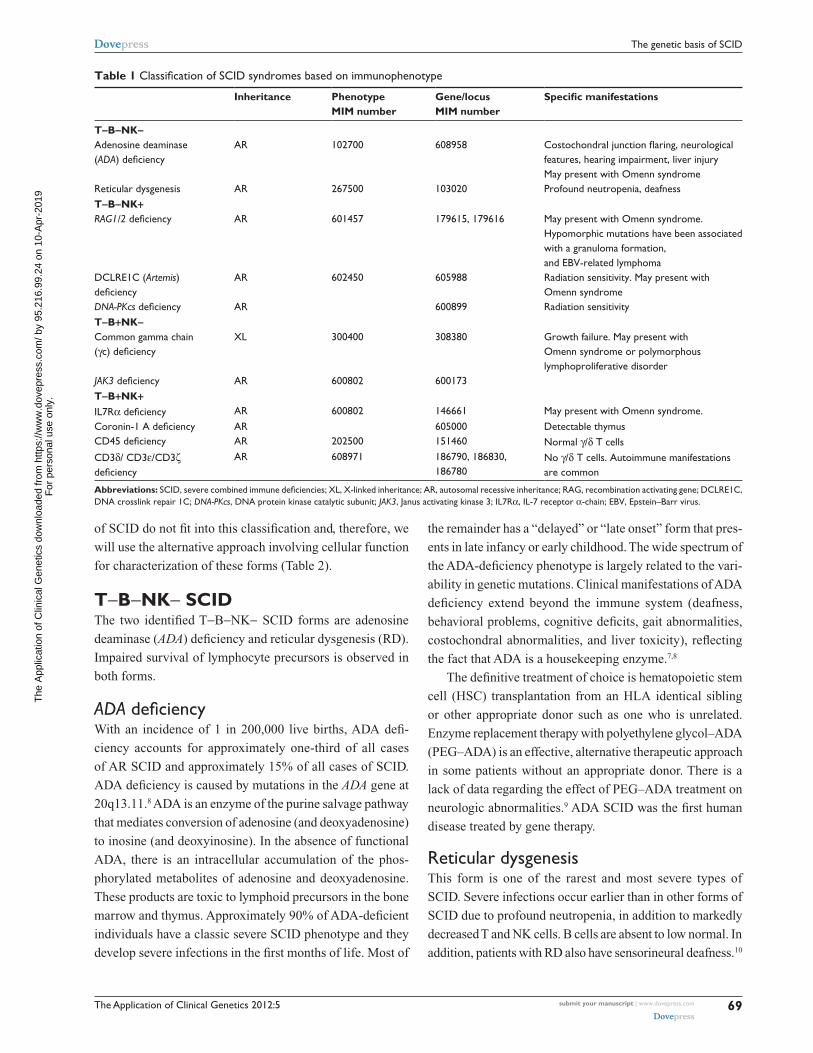
Table 1 Classification of SCID syndromes based on immunophenotype
Inheritance Phenotype MIM number
Gene/locus MIM number
Specific manifestations
T-B-NK-Adenosine deaminase (ADA) deficiency
AR 102700 608958 Costochondral junction flaring, neurological features, hearing impairment, liver injury May present with Omenn syndrome
Reticular dysgenesis AR 267500 103020 Profound neutropenia, deafnessT-B-NK+RAG1/2 deficiency AR 601457 179615, 179616 May present with Omenn syndrome.
Hypomorphic mutations have been associated with a granuloma formation, and EBv-related lymphoma
DCLRE1C (Artemis) deficiency
AR 602450 605988 Radiation sensitivity. May present with Omenn syndrome
DNA-PKcs deficiency AR 600899 Radiation sensitivityT-B+NK-Common gamma chain (γc) deficiency
XL 300400 308380 Growth failure. May present with Omenn syndrome or polymorphous lymphoproliferative disorder
JAK3 deficiency AR 600802 600173T-B+NK+IL7Rα deficiency AR 600802 146661 May present with Omenn syndrome.Coronin-1 A deficiency AR 605000 Detectable thymusCD45 deficiency AR 202500 151460 Normal γ/δ T cellsCD3δ/ CD3ε/CD3ζ deficiency
AR 608971 186790, 186830, 186780
No γ/δ T cells. Autoimmune manifestations are common
Abbreviations: SCID, severe combined immune deficiencies; XL, X-linked inheritance; AR, autosomal recessive inheritance; RAG, recombination activating gene; DCLRE1C, DNA crosslink repair 1C; DNA-PKcs, DNA protein kinase catalytic subunit; JAK3, Janus activating kinase 3; IL7Rα, IL-7 receptor α-chain; EBv, Epstein–Barr virus.
of SCID do not fit into this classification and, therefore, we
will use the alternative approach involving cellular function
for characterization of these forms (Table 2).
T−B−NK− SCIDThe two identified T−B−NK− SCID forms are adenosine
deaminase (ADA) deficiency and reticular dysgenesis (RD).
Impaired survival of lymphocyte precursors is observed in
both forms.
ADA deficiencyWith an incidence of 1 in 200,000 live births, ADA defi-
ciency accounts for approximately one-third of all cases
of AR SCID and approximately 15% of all cases of SCID.
ADA deficiency is caused by mutations in the ADA gene at
20q13.11.8 ADA is an enzyme of the purine salvage pathway
that mediates conversion of adenosine (and deoxyadenosine)
to inosine (and deoxyinosine). In the absence of functional
ADA, there is an intracellular accumulation of the phos-
phorylated metabolites of adenosine and deoxyadenosine.
These products are toxic to lymphoid precursors in the bone
marrow and thymus. Approximately 90% of ADA-deficient
individuals have a classic severe SCID phenotype and they
develop severe infections in the first months of life. Most of
the remainder has a “delayed” or “late onset” form that pres-
ents in late infancy or early childhood. The wide spectrum of
the ADA-deficiency phenotype is largely related to the vari-
ability in genetic mutations. Clinical manifestations of ADA
deficiency extend beyond the immune system (deafness,
behavioral problems, cognitive deficits, gait abnormalities,
costochondral abnormalities, and liver toxicity), reflecting
the fact that ADA is a housekeeping enzyme.7,8
The definitive treatment of choice is hematopoietic stem
cell (HSC) transplantation from an HLA identical sibling
or other appropriate donor such as one who is unrelated.
Enzyme replacement therapy with polyethylene glycol–ADA
(PEG–ADA) is an effective, alternative therapeutic approach
in some patients without an appropriate donor. There is a
lack of data regarding the effect of PEG–ADA treatment on
neurologic abnormalities.9 ADA SCID was the first human
disease treated by gene therapy.
Reticular dysgenesisThis form is one of the rarest and most severe types of
SCID. Severe infections occur earlier than in other forms of
SCID due to profound neutropenia, in addition to markedly
decreased T and NK cells. B cells are absent to low normal. In
addition, patients with RD also have sensorineural deafness.10
submit your manuscript | www.dovepress.com
Dovepress
Dovepress
69
The genetic basis of SCID
The
App
licat
ion
of C
linic
al G
enet
ics
dow
nloa
ded
from
http
s://w
ww
.dov
epre
ss.c
om/ b
y 95
.216
.99.
24 o
n 10
-Apr
-201
9F
or p
erso
nal u
se o
nly.
Powered by TCPDF (www.tcpdf.org)
1 / 1
The Application of Clinical Genetics 2012:5
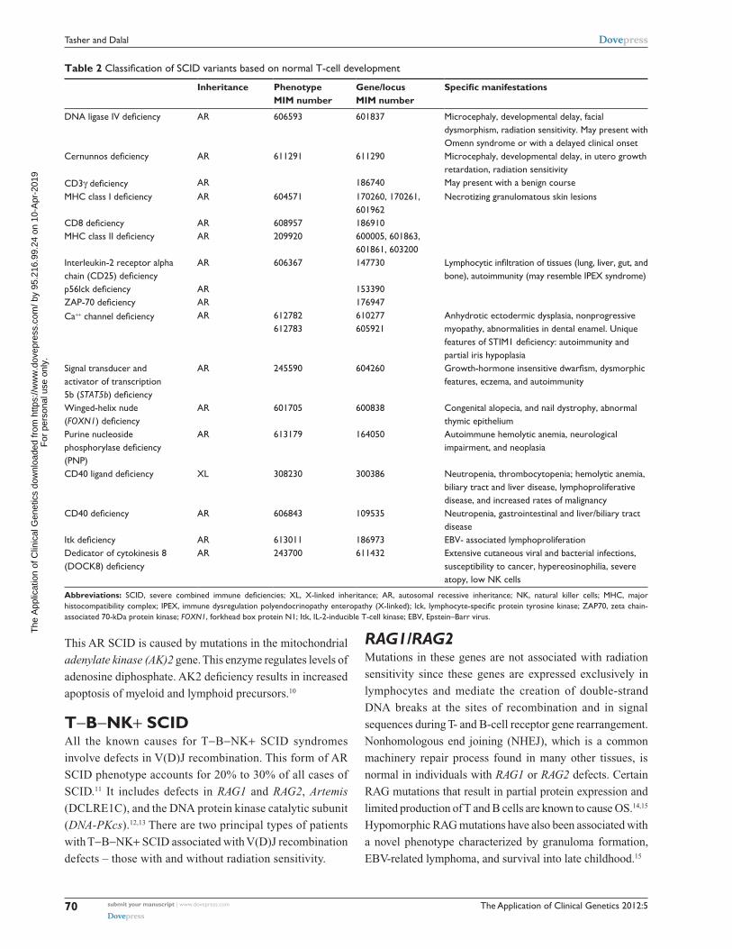
Table 2 Classification of SCID variants based on normal T-cell development
Inheritance Phenotype MIM number
Gene/locus MIM number
Specific manifestations
DNA ligase IV deficiency AR 606593 601837 Microcephaly, developmental delay, facial dysmorphism, radiation sensitivity. May present with Omenn syndrome or with a delayed clinical onset
Cernunnos deficiency AR 611291 611290 Microcephaly, developmental delay, in utero growth retardation, radiation sensitivity
CD3γ deficiency AR 186740 May present with a benign courseMHC class I deficiency AR 604571 170260, 170261,
601962Necrotizing granulomatous skin lesions
CD8 deficiency AR 608957 186910MHC class II deficiency AR 209920 600005, 601863,
601861, 603200Interleukin-2 receptor alpha chain (CD25) deficiency
AR 606367 147730 Lymphocytic infiltration of tissues (lung, liver, gut, and bone), autoimmunity (may resemble IPEX syndrome)
p56lck deficiency AR 153390ZAP-70 deficiency AR 176947Ca++ channel deficiency AR 612782
612783610277 605921
Anhydrotic ectodermic dysplasia, nonprogressive myopathy, abnormalities in dental enamel. Unique features of STIM1 deficiency: autoimmunity and partial iris hypoplasia
Signal transducer and activator of transcription 5b (STAT5b) deficiency
AR 245590 604260 Growth-hormone insensitive dwarfism, dysmorphic features, eczema, and autoimmunity
Winged-helix nude (FOXN1) deficiency
AR 601705 600838 Congenital alopecia, and nail dystrophy, abnormal thymic epithelium
Purine nucleoside phosphorylase deficiency (PNP)
AR 613179 164050 Autoimmune hemolytic anemia, neurological impairment, and neoplasia
CD40 ligand deficiency XL 308230 300386 Neutropenia, thrombocytopenia; hemolytic anemia, biliary tract and liver disease, lymphoproliferative disease, and increased rates of malignancy
CD40 deficiency AR 606843 109535 Neutropenia, gastrointestinal and liver/biliary tract disease
Itk deficiency AR 613011 186973 EBv- associated lymphoproliferationDedicator of cytokinesis 8 (DOCK8) deficiency
AR 243700 611432 Extensive cutaneous viral and bacterial infections, susceptibility to cancer, hypereosinophilia, severe atopy, low NK cells
Abbreviations: SCID, severe combined immune deficiencies; XL, X-linked inheritance; AR, autosomal recessive inheritance; NK, natural killer cells; MHC, major histocompatibility complex; IPEX, immune dysregulation polyendocrinopathy enteropathy (X-linked); lck, lymphocyte-specific protein tyrosine kinase; ZAP70, zeta chain-associated 70-kDa protein kinase; FOXN1, forkhead box protein N1; Itk, IL-2-inducible T-cell kinase; EBv, Epstein–Barr virus.
This AR SCID is caused by mutations in the mitochondrial
adenylate kinase (AK)2 gene. This enzyme regulates levels of
adenosine diphosphate. AK2 deficiency results in increased
apoptosis of myeloid and lymphoid precursors.10
T−B−NK+ SCIDAll the known causes for T−B−NK+ SCID syndromes
involve defects in V(D)J recombination. This form of AR
SCID phenotype accounts for 20% to 30% of all cases of
SCID.11 It includes defects in RAG1 and RAG2, Artemis
(DCLRE1C), and the DNA protein kinase catalytic subunit
(DNA-PKcs).12,13 There are two principal types of patients
with T−B−NK+ SCID associated with V(D)J recombination
defects – those with and without radiation sensitivity.
RAG1/RAG2Mutations in these genes are not associated with radiation
sensitivity since these genes are expressed exclusively in
lymphocytes and mediate the creation of double-strand
DNA breaks at the sites of recombination and in signal
sequences during T- and B-cell receptor gene rearrangement.
Nonhomologous end joining (NHEJ), which is a common
machinery repair process found in many other tissues, is
normal in individuals with RAG1 or RAG2 defects. Certain
RAG mutations that result in partial protein expression and
limited production of T and B cells are known to cause OS.14,15
Hypomorphic RAG mutations have also been associated with
a novel phenotype characterized by granuloma formation,
EBV-related lymphoma, and survival into late childhood.15
submit your manuscript | www.dovepress.com
Dovepress
Dovepress
70
Tasher and Dalal
The
App
licat
ion
of C
linic
al G
enet
ics
dow
nloa
ded
from
http
s://w
ww
.dov
epre
ss.c
om/ b
y 95
.216
.99.
24 o
n 10
-Apr
-201
9F
or p
erso
nal u
se o
nly.
Powered by TCPDF (www.tcpdf.org)
1 / 1

The Application of Clinical Genetics 2012:5
According to our local experience, XL-SCID is rare in
Israel, while the most common phenotype is T−B− SCID
due to RAG1/RAG2 mutations. This phenomenon is probably
due to the high rate of consanguinity among our Arab-origin
population, which accounts for approximately 20% of the
whole population, and for more than 50% of our SCID
patients.16,17
Artemis and DNA-PKcs lead to increased sensitivity to
ionizing radiation, since these gene products are important
in the process of NHEJ required for repair of double-strand
DNA breaks caused by radiation.
ArtemisArtemis deficiency is also known as Athabascan SCID
(SCIDA), since a founder mutation in Artemis is found with
increased frequency in Native Americans speaking one of
the Athabascan family languages (eg, Apache, Navajo).18
In addition to T-cell maturation arrest, mutations in the
Artemis gene result in B-cell differentiation arrest at the
pre-B-cell-receptor checkpoint.19
DNA-PKcsDNA-PKcs regulates Artemis by both phosphorylation and
complex formation to permit enzymatic activities that are
critical for V(D)J recombination and for NHEJ.
Intriguingly, the described functional missense DNA-
PKcs mutation does not affect either DNA-PKcs kinase
activity or autophosphorylation – an important event that
remodels the protein. Yet the clinical and cellular phenotype
is similar to that caused by Artemis deficiency, suggesting that
the mutation precludes Artemis activation, an event known
to be DNA-PKcs dependent.20
T−B+NK− SCIDGene mutations affecting the integrity of the common
gamma chain (γc)/JAK3 signaling pathway result in the
most common form of inherited T−B+NK− SCID. Despite
the presence of normal numbers of B cells, cell activation,
proliferation, and differentiation are impaired, resulting in
profound hypogammaglobulinemia.
X-linked SCIDThis form of SCID is due to defects in γc. Mutations in this
gene lead to profound derangement of the immune system
via the blockade of multiple cytokine pathways (IL-2Rγc
is shared by the receptors of IL-4, IL-7, IL-9, IL-15, and
IL-21), which are important for lymphocyte development
and function.21,22 In particular, IL-7 mediates expansion of
early thymocyte progenitors, whereas IL-15 plays a role in
NK cell development. Accordingly, patients with XL-SCID
lack both T and NK cells, whereas they have a normal number
of circulating B lymphocytes.23
XL-SCID patients usually have the classic clinical
SCID phenotype. However, some patients have an atypical
presentation such as an OS phenotype24 or a polymorphous
lymphoproliferative disorder with Hodgkin-like features.25
The γc subunit is also involved in growth hormone-receptor
signaling. Thus, growth failure seen in children with XL-
SCID may also be due to the underlying genetic defect.26
The γ chain mediates signal transduction via its physical
association with a member of the Janus family of protein
kinases, JAK3.27 The primary therapy currently available for
XL-SCID is hematopoietic cell transplantation. Of note is
that, in some cases, gene therapy has been successful.
JAK3 deficiencyJAK3 (encoded on chromosome 19p12-13.1) mediates
cytokine signal transduction and is essential for lymphoid
cell development. SCID due to JAK3 deficiency is rare,
accounting only for approximately 6% of all cases of SCID.
This AR form of T−B+NK− SCID is identical to XL-SCID
in cellular and clinical phenotypes. Rarely, partial JAK3
defects associated with low amounts of functional protein
can present with mild immunodeficiency.27
T−B+NK+ SCIDThis SCID phenotype includes defects in the IL-7 receptor
alpha chain (IL-7Rα; also called CD127), actin-regulating
protein, coronin 1A (CORO1A), CD45 (also called protein-
tyrosine phosphatase receptor-type C; PTPRC), and the CD3
chains: CD3 delta (CD3D), CD3 epsilon (CD3E), and CD3
zeta (CD3Z).
IL-7Rα chain deficiencyThis defect is the third most common type of SCID. IL-7Rα (encoded on chromosome 5p13) plays a critical role in
cytokine signaling that is necessary for T-cell development.
Although both T-cell and B-cell progenitors express the
functional IL-7 receptor that consists of IL-7Rα and a
γc chain, this lymphocyte-receptor system is critical for
the T lineage, but not for the B lineage, or for NK-cell
development. IL-15 promotes NK-cell differentiation and
maturation. Many patients have been identified with a
classic SCID phenotype and they lack the IL-7Rα chain.28,29
Mutations in the IL-7Rα gene can also present with an OS
phenotype.30
submit your manuscript | www.dovepress.com
Dovepress
Dovepress
71
The genetic basis of SCID
The
App
licat
ion
of C
linic
al G
enet
ics
dow
nloa
ded
from
http
s://w
ww
.dov
epre
ss.c
om/ b
y 95
.216
.99.
24 o
n 10
-Apr
-201
9F
or p
erso
nal u
se o
nly.
Powered by TCPDF (www.tcpdf.org)
1 / 1

The Application of Clinical Genetics 2012:5
Actin-regulating protein coronin 1A deficiencyCoronin 1A (encoded on chromosome 16p11.2) is involved
in actin cytoskeleton regulation and is essential for T-cell
immigration from the thymus to the secondary lymphoid
organs. Defects in Coronin 1A lead to an absence of normal
peripheral T cells and to a classic SCID phenotype. The
thymus is present, unlike in most other forms of SCID.31
CD45 deficiencyCD45, the leukocyte common antigen (encoded on chromo-
some 1q31-q32) is a transmembrane tyrosine phosphatase
involved in TCR signaling and T-cell development in the
thymus. Only a few patients have been identified with classic
SCID due to CD45 deficiency.32
CD3 complex component deficienciesThe CD3 complex plays a major role in signaling through
the TCR, which is essential for the normal maturation of
T cells. Mutations in the genes encoding CD3 chains (CD3
delta, CD3 epsilon, and CD3 zeta) appear to have an AR
pattern of inheritance and have been reported in several
patients. The clinical features are highly variable, depending
upon the affected chain and mutation. Hypomorphic muta-
tions in genes that are typically associated with SCID can
allow residual T-cell development. In these cases, impaired
cross-talk between thymocytes and thymic epithelial cells
might compromise mechanisms of central tolerance, with
failure to delete autoreactive T cells, and with impaired gen-
eration of regulatory T (Treg) cells. Accordingly, autoim-
mune manifestations are common, with infiltration of target
tissues by activated and oligoclonal T lymphocytes.33–35
Omenn syndromeOS is characterized by early postnatal diffuse exfoliative
erythroderma, protracted diarrhea, lymphadenopathy, and
hepatosplenomegaly, together with the typical recurrent
severe infections. Laboratory investigations reveal prominent
eosinophilia and lymphocytosis, mainly of T cells. These
lymphocytes exhibit activation markers despite their highly
restricted repertoire. Variable hypogammaglobulinemia with
elevated levels of immunoglobulin E (IgE) and skewing
toward TH2, the absence of B cells with minimal antibody
response, and the absence of normal mitogenic responses are
some of the common immune abnormalities in this syndrome.
This peculiar phenotype was found to be associated with
several different gene mutations including RAG1/2 and
Artemis, IL-7Rα, RNA-processing endoribonuclease, DNA
ligase IV, ADA, and γc.15,36
T+B+NK+ SCIDOther forms of combined immunodeficiency have many
of the elements of the clinical presentation of SCID,
including opportunistic infections. These forms of combined
immunodeficiency are usually distinguished by distinctive
laboratory features and other elements of the clinical
presentation. However, in some cases, the distinction
between SCID and combined immunodeficiency (CID) is
only made by molecular testing.
In this chapter, we took the approach of dissecting
the different types of SCID based on the normal process
of development, activation, and maturation of T cells
as described previously (Figure 1). Defects in T-cell
development that occur beyond the CD4+ CD8+ double-
positive cell stage result in SCID with residual numbers of T
lymphocytes. These SCID variants include: DNA ligase IV;
Cernunnos-XLF; CD3γ deficiency; MHC class I deficiency;
CD8 deficiency; MHC class II deficiency; IL-2 receptor
alpha chain (CD25) deficiency; p56lck deficiency; ZAP-
70 deficiency; ORAI1/CRACM1 and STIM1 deficiencies;
signal transducer and activator of transcription 5b (STAT5b)
deficiency; winged-helix nude (FOXN1) deficiency; PNP;
CD40 and CD40 ligand deficiencies; Itk; and dedicator of
cytokinesis 8 (DOCK8) deficiency.
DNA ligase IV (LIG4)The DNA ends must be ligated as part of the DNA repair
process and this is implemented by an LIG4-XRCC4 complex.
XRCC4 (X-ray-complementing Chinese hamster gene 4) has
no known enzymatic activity, but functions as a scaffolding
protein that helps attract other repair proteins to the DNA break.
XRCC4 stabilizes LIG4 and enhances its activity.
Patients with LIG4 syndrome present with microcephaly,
developmental delay, facial dysmorphism, increased suscep-
tibility to tumors, and a variable degree of immunodeficiency
that ranges from SCID/OS, to hypogammaglobulinemia with
impaired class-switch recombination, to moderate, or even
very modest defects in T- and B-cell immunity.37–39
Cernunnos–XLFDefects in the NHEJ factor 1 gene (NHEJ1 gene) that codes
for the protein Cernunnos, also known as the XRCC4-like
factor (XLF), lead to a T−B−NK+ radiation-sensitive
SCID phenotype with associated developmental delay
and microcephaly. T cells are absent, except for memory
submit your manuscript | www.dovepress.com
Dovepress
Dovepress
72
Tasher and Dalal
The
App
licat
ion
of C
linic
al G
enet
ics
dow
nloa
ded
from
http
s://w
ww
.dov
epre
ss.c
om/ b
y 95
.216
.99.
24 o
n 10
-Apr
-201
9F
or p
erso
nal u
se o
nly.
Powered by TCPDF (www.tcpdf.org)
1 / 1

The Application of Clinical Genetics 2012:5
Figure 1 Genes with mutations that cause monogenic, severe T-cell immunodeficiency in humans can be intrinsic to the thymic epithelium or to T cells. (A) Genetic defects that are intrinsic to thymic epithelial cells ultimately affect the antigen-presentation pathway. (B) Genetic defects that are intrinsic to T cells include those that affect T-cell receptor (TCR) signaling, cytokine signaling, somatic recombination, or basic cellular processes. Other genes that are important to these pathways or processes, but have not been linked to severe T-cell deficiency in humans are shown in grey. Copyright © 2008. Nature Publishing Group. Adapted with permission from Liston A, Enders A, Siggs OM. Unravelling the association of partial T-cell immunodeficiency and immune dysregulation. Nat Rev Immunol. 2008;8:546.80
Abbreviations: ADA, adenosine deaminase; ATM, ataxia-telangiectasia mutated; CBL, Casitas B-lineage lymphoma; CIITA, class II transactivator; DCLRE1C, DNA crosslink repair 1C; DNMT3β, DNA cytosine-5 methyltransferase 3β; ER, endoplasmic reticulum; FOXN1, forkhead box N1; GADS, GRB2-related adaptor protein; IL-2RΓ, IL-2 receptor Γ-chain; IL-7, interleukin-7; IL-7Rα, IL-7 receptor α-chain; JAK, Janus kinase; LAT, linker for activation of T cells; LIG4, ligase Iv; MRE11A, meiotic recombination 11 homolog A; Nibrin, Nijmegen breakage syndrome 1; NP, nucleoside phosphorylase; CRACM1, calcium release-activated calcium modulator 1; RAG, recombination-activating gene; RFX5, regulatory factor X5; RFXANK, RFX-associated ankyrin-containing protein; RFXAP, RFX-associated protein; RMRP, RNA component of mitochondrial RNA-processing endoribonuclease; SLP76, SRC-homology-2-domain-containing leukocyte protein of 76 kDa; SMARCAL1, SWI/SNF-related matrix-associated actin-dependent regulator of chromatin a-like 1; SP110, SP110 nuclear body protein; STAT5B, signal transducer and activator of transcription 5B; TAP, transporter associated with antigen processing; TAPBP, TAP-binding protein; TSAD, T-cell-specific adaptor protein; ZAP70, ζ-chain-associated protein kinase of 70 kDa.
T cells, and patients have a progressive loss of B cells.
XLF-Cernunnos is a core protein of the NHEJ pathway
of DNA double-strand break repair. The roles of the
Cernunnos-XLF protein are still being elucidated. Crystal-
lography suggests that it is part of the XRCC4-LIG4 complex.
It enhances the DNA ligation activity of the XRCC4-LIG4
complex by promoting its re-adenylation and appears to play
an essential role in gap filling by either polymerase during
NHEJ, suggesting that it plays a major role in aligning the
two DNA ends in the repair complex.40,41
CD3γ deficiencyCD3γ deficiency is an AR immunodeficiency caused by
mutations in the gene coding for T-cell surface glycoprotein
submit your manuscript | www.dovepress.com
Dovepress
Dovepress
73
The genetic basis of SCID
The
App
licat
ion
of C
linic
al G
enet
ics
dow
nloa
ded
from
http
s://w
ww
.dov
epre
ss.c
om/ b
y 95
.216
.99.
24 o
n 10
-Apr
-201
9F
or p
erso
nal u
se o
nly.
Powered by TCPDF (www.tcpdf.org)
1 / 1

The Application of Clinical Genetics 2012:5
CD3γ chain precursors. Affected patients have decreased
T-cell numbers and function. B cells are variably affected.
Patients with CD3γ deficiency have varying phenotypes,
with some having SCID-like symptoms and some having a
benign course.42
MHC class I deficiency (Bare lymphocyte syndrome type I)Most patients with MHC I expression abnormalities exhibit
decreased or absent MHC I expression on the cell surface,
low numbers of CD8+ T cells, and decreased NK cell-killing
activity. However, they have normal antibody-dependent
cell-mediated cytotoxicity.43 The clinical presentation
is highly variable, depending upon the amount of MHC
class I expression. Affected patients with a severe reduction
in MHC class I expression also tend to have necrotizing
granulomatous skin lesions.44 Some patients with MHC
class I deficiency have defects in the transporter associated
with antigen processing: (TAP) 1, TAP2, or tapasin. TAP1
and TAP2 are required for peptide transport from the cytosol
into the ER lumen, where the assembly of the MHC class I
complex takes place. Tapasin serves as a docking site that is
specific for interaction with class I MHC molecules, which
is essential for peptide loading and expression. The exact
defect has not been identified in other patients with MHC
class I deficiency. The inheritance pattern is unclear due to
its rarity.45
CD8 deficiencyCD8 is a TCR accessory molecule that binds to class I MHC.
CD8 is primarily expressed on cytotoxic T cells, but is also
found on NK cells. A single patient has been described with
symptomatic CD8 deficiency due to a homozygous muta-
tion in the gene encoding the CD8 alpha chain (CD8α) on
chromosome 2p12. He had recurrent sinopulmonary infec-
tions, which suggested a humoral deficiency. However, his
immunoglobulin levels and specific antibody titers were
normal. CD4+ T-cell, B-cell, and NK-cell percentages and
absolute counts were normal, but CD8+ T cells were com-
pletely absent. Two younger sisters, who also had absent
CD8+ T cells, were asymptomatic at the time of the report,
reflecting the importance of other factors (such as epigenet-
ics and infections) in these complex diseases.46
MHC class II deficiency (Bare lymphocyte syndrome type II)This is an AR disease. Both constitutive and induced MHC
class II expressions are compromised. The exact molecular
defects have not been identified in most patients, but
are suspected to involve mutations in MHCII promoter
complex DNA-binding regulatory factors (CIITA-class II
transactivator, RFX5, RFXAP, and RFXANK). The majority
of identified defects are RFXANK mutations (chromosome
19p12). The disease is more common in certain geographic
regions (North Africa47 and Palestinians [Broides A, email
communication, 2011]). Patients with MHC II expression
abnormalities exhibit complete absence of MHC II expres-
sion on B cells, a normal number of B cells in peripheral
blood, hypogammaglobulinemia, and poor specific-antibody
responses. In addition, CD4 lymphopenia, with a proportional
increase in CD8, a normal number of T cells in peripheral
blood, but with decreased in vitro T-cell response to antigens
are observed. Class II MHC deficiency generally results in
a clinical picture of severe combined immune deficiency.
However, milder cases have been described.48
Interleukin-2 receptor alpha chain (CD25) deficiencyCD25 deficiency is extremely rare. Only a few patients have
been described with lesions in the IL2-Rα gene encoding
the α-chain of the IL-2 receptor. This immunodeficiency is
characterized by normal to modestly decreased numbers
of circulating T cells, displaying abnormal proliferation,
but normal B-cell development. Extensive lymphocytic
infiltration of tissues including the lung, liver, gut, and
bone, is observed, accompanied by tissue atrophy, and
inflammation.49 A phenotype resembling the syndromes of
immune dysregulation, polyendocrinopathy, enteropathy,
and X-linked (IPEX) – a syndrome that is more often
associated with mutations of the transcription factor
FOXP3 – has also been described.50
Lymphocyte-specific protein tyrosine kinase (Lck or p56lck) deficiencyInitial transduction of antigen-binding signals from the
TCR/CD3 complex is primarily dependent upon two
protein tyrosine kinases: Lck and ZAP-70. Several patients
have been reported with a deficiency of Lck, which was
activated upon engagement of the TCR-CD3 complex.
Lck is a 56-kDa member of the Src-family tyrosine
kinase family. Deficiency of Lck is reported to result in
an immunodeficiency characterized by selective CD4
lymphopenia, decreased CD28 expression on CD8 cells,
and low, to absent immunoglobulins. Patients demonstrate
the typical failure to thrive that is seen in SCID.51
submit your manuscript | www.dovepress.com
Dovepress
Dovepress
74
Tasher and Dalal
The
App
licat
ion
of C
linic
al G
enet
ics
dow
nloa
ded
from
http
s://w
ww
.dov
epre
ss.c
om/ b
y 95
.216
.99.
24 o
n 10
-Apr
-201
9F
or p
erso
nal u
se o
nly.
Powered by TCPDF (www.tcpdf.org)
1 / 1

The Application of Clinical Genetics 2012:5
ZAP-70 deficiencyZAP-70 deficiency is a rare combined immunodeficiency
with an AR pattern of inheritance. Zeta chain-associated
70-kDa protein kinase (ZAP-70) is a signaling molecule
associated with the TCR complex, which is expressed
primarily in T cells. Most mutations occur within the kinase
domain of the ZAP-70 protein, and significantly affect both
protein stability, and catalytic activity. Deficiency of ZAP-70
(which was originally described in a Mennonite family) is
characterized by the selective absence of circulating CD8+
T cells and by abundant CD4+ T cells in the peripheral blood
that are unresponsive to TCR-mediated stimuli in vitro.52 All
patients with ZAP-70 deficiency have normal to elevated
numbers of circulating lymphocytes.53
ORAI1/CRACM1 and STIM1 deficienciesLymphocyte activation after antigen stimulation is dependent
upon store-operated entry of Ca2+ across the plasma membrane
via Ca2+ release-activated Ca2+ (CRAC) channels. OraI1 or
calcium release-activated calcium modulator 1 (CRACM1)
is the pore-forming subunit of the CRAC channel. Stromal
interaction molecule 1 (STIM1) senses release of Ca2+
from endoplasmic reticulum stores and activates CRAC
channels in the plasma membrane. Sustained Ca2+ influx
induces expression of IL-2 and other cytokines. The clini-
cal phenotypes of the ORAI1 defect and of the STIM defect
are similar. Mutations in ORAI1 on chromosome 12q24
and STIM1 on chromosome 11q15.5 appear to have AR
inheritance. The laboratory findings are similar for both of
these defects. Lymphocyte counts are normal to slightly
reduced, and immunoglobulin levels are normal to elevated.
However, the T-cell proliferative response to mitogens and
antigens is compromised and specific antibody response to
vaccination is impaired. Non-progressive muscular hypotonia
and abnormalities in dental enamel are seen in both ORAI1
and STIM1 deficiencies. Extensive ectodermal defects
(ectodermal dysplasia and anhydrosis) are more prominent
in patients with ORAI1 deficiency. Unique features of
STIM1 deficiency are severe autoimmunity and partial iris
hypoplasia.54,55
Signal transducer and activator of transcription 5b (STAT5b) deficiencySeveral patients have been identified with mutations in the
STAT5b gene that cause a defect in the signaling pathways
of receptors for IL-2, IL-4, macrophage colony-stimulating
factor, and growth hormone. CD4+ CD25+ regulatory T cells
are decreased in number in patients with STAT5b deficiency.
Other immunologic parameters are variable, including T-, B-,
and NK-cell levels, and T-cell proliferation. Patients present
with signs of growth-hormone insensitivity, including short
stature, a prominent forehead and saddle nose, and a high-
pitched voice. Associated manifestations include eczema and
autoimmunity as well.56
Winged-helix nude (FOXN1) deficiencyThis extremely rare disorder is caused by a homozygous
defect in the forkhead box N1 (FOXN1) gene, which is the
gene mutated in “nude” SCID mice. FOXN1 is a member
of the winged-helix domain family of transcription factors.
This protein is involved in the development of the thymus as
well as formation of hair and nails. It also may be involved
in brain and neural tube development. Patients with defects
in this gene have decreased circulating T cells, mainly due
to low levels of CD4+ T cells. T-cell proliferative responses
to mitogens were abnormal. Congenital alopecia and nail
dystrophy are characteristic features. Additional defects,
including spina bifida and anencephaly, have been described
in a fetus.57,58
Purine nucleoside phosphorylase (PNP) deficiencyPNP follows ADA in the purine salvage pathway, and PNP
deficiency causes SCID because excess deoxyguanosine
and deoxyguanosine triphosphate lead to increased apop-
tosis of lymphocytes, mainly immature T lymphocytes.59
PNP-deficient cells do not produce uric acid and, therefore,
low uric acid in serum (,2 mg/dL) supports the diagnosis.
PEG-PNP is not commercialized and gene therapy is still
experimental in mice. HSCT is the only available therapy,
but it does not correct the severe neurological manifestations
usually present (hypertonia, hypotonia, ataxia, psychomo-
tor delay). Autoimmunity (hemolytic anemia, autoimmune
thrombocytopenia, neutropenia, arthritis, etc) and neoplasia
are also frequent.59,60
CD40 and CD40 ligand deficienciesCD40 is a molecule on the surface of B cells and mononuclear
cells (including dendritic cells) that interacts with its CD40
ligand (CD40L) on the surface of activated CD4+ T cells.
Defects in CD40L or CD40 cause defective isotype switching
and impaired dendritic cell signaling. Mutations in CD40L
submit your manuscript | www.dovepress.com
Dovepress
Dovepress
75
The genetic basis of SCID
The
App
licat
ion
of C
linic
al G
enet
ics
dow
nloa
ded
from
http
s://w
ww
.dov
epre
ss.c
om/ b
y 95
.216
.99.
24 o
n 10
-Apr
-201
9F
or p
erso
nal u
se o
nly.
Powered by TCPDF (www.tcpdf.org)
1 / 1

The Application of Clinical Genetics 2012:5
(CD154) are responsible for most cases of hyper-IgM (HIGM)
syndrome.61 The principal laboratory findings of HIGM syn-
drome are low serum levels of IgG and IgA, in combination
with normal or elevated concentrations of IgM. Circulating
B cells are normal in number, but exclusively express cell-
surface IgM and IgD. Vaccination may elicit specific IgM
responses. No other isotypes are produced and there is no
B-cell memory. CD40L is located on the X chromosome,
while the gene encoding CD40 (TNF-receptor superfamily
member 5 or TNFRSF5) is located on chromosome 20q12-
q13.2. Mutations in this gene underlie a clinically indis-
tinguishable disorder with AR inheritance.62 Patients with
HIGM usually present within the first two years of life with
the combination of recurrent respiratory tract infections with
encapsulated bacteria and opportunistic infections. Lymph-
adenopathy and hepatosplenomegaly are often present. They
are also prone to neutropenia, thrombocytopenia, anemia,
sclerosing cholangitis, cirrhosis, and lymphoproliferative
disease. Increased rates of malignancy such as hepatocel-
lular, bile duct, and neuroendocrine carcinomas are also
reported.63
Interleukin-2 tyrosine kinase (Itk) deficiencyItk deficiency is characterized by lymphoproliferation and
severe immune dysregulation following EBV infection and
is inherited as an AR disorder. Itk is a member of the TEC
kinase family. These proteins are important mediators of
antigen-receptor signaling in lymphocytes, and Itk is believed
to be the predominant TEC kinase in T cells, and one of
the key molecules involved in NKT cell maturation and
survival.64 Itk deficiency was recently identified in female
siblings who presented with EBV-associated lymphopro-
liferation, an absence of NKT cells, and hemophagocytic
lymphohistiocytosis.65
Dedicator of cytokinesis 8 (DOCK8) deficiencySeveral patients have been identified with loss of functional
homozygous or compound heterozygous mutations in an
uncharacterized gene, DOCK8, which is expressed in lym-
phocytes. Most patients have low absolute lymphocyte counts,
including low T cells; B and NK cells are also low in many
patients. Most patients also have eosinophilia and elevated
IgE. IgM levels are low, but many patients have increased
IgG levels. IgG antibody responses to bacterial and viral
antigens are variable.66 Common clinical features include
recurrent respiratory tract infections, extensive cutaneous
viral and bacterial (Staphylococcus aureus) infections,
severe atopy, and susceptibility to cancer (vulvar, facial, and
anal squamous-cell dysplasia and carcinomas, and T-cell
lymphoma/leukemia).66
Other well-defined immunodeficiency syndromesIn addition to the above-described syndromes, there are other
well-defined primary immunodeficiency disease (PID) syn-
dromes with a variable degree of immune dysfunction such
as Wiskott–Aldrich syndrome, DNA repair defects (ataxia
telangiectasia, Bloom and Nijmegen syndromes), and oth-
ers. Some of them can present with the typical clinical and
laboratory features of SCID/CID. Herein, we discuss some
of these syndromes in detail.
DiGeorge syndrome (DGS)DGS (OMIM 188400) results from dysmorphogenesis of the
third and fourth pharyngeal pouches during early embryo-
genesis, leading to hypoplasia or aplasia of the thymus and
parathyroid glands. Other structures forming at the same age
are also frequently affected, resulting in anomalies of maxilla,
mandible, aortic arch, cardiac outflow tract, and external/
middle ear. The classical presentation of DGS is the triad
of conotruncal cardiac anomalies, hypoplastic thymus, and
hypocalcemia, although the phenotype is variable. Palatal
abnormalities and developmental delay are common.
Approximately 90% of patients with DGS have spontane-
ously arising heterozygous microdeletions in chromosome
22q11.2. Defects in the gene encoding the T-box tran-
scription family member, TBX1, are responsible for the
DGS phenotype. The basis for the syndrome without the
characteristic deletion is often unknown. Some patients
will have a known exposure to toxins such as isotretinoin or
maternal alcohol ingestion. There is also a small subset that
shows the deletion of chromosome 10p13-14.
The immunodeficiency has been characterized as either
complete or partial, based on the level of immunologic
function. The minority of patients (approximately 1%)
have a complete absence of thymic tissue and profound
immunodeficiency. Patients with complete DGS resemble
patients with SCID. Atypical cases phenotypically appear
to be similar to OS. Complete DGS is fatal if not recognized
and treated with a thymic or HSC transplant.67
Cartilage-hair hypoplasia (CHH)CHH (OMIM 250250) is an AR, predominantly T-cell defi-
ciency associated with metaphyseal chondrodysplasia, a form
submit your manuscript | www.dovepress.com
Dovepress
Dovepress
76
Tasher and Dalal
The
App
licat
ion
of C
linic
al G
enet
ics
dow
nloa
ded
from
http
s://w
ww
.dov
epre
ss.c
om/ b
y 95
.216
.99.
24 o
n 10
-Apr
-201
9F
or p
erso
nal u
se o
nly.
Powered by TCPDF (www.tcpdf.org)
1 / 1
The Application of Clinical Genetics 2012:5
of short-limb dwarfism. Associated features may include
fine sparse hair, cellular and humoral immunodeficiencies,
Hirschsprung disease, hematologic and skin malignancies,
autoimmune disease, and bronchiectasis. The responsible
gene for CHH has been mapped to 9p21-p12 and shown to
be the gene encoding the ribonuclease mitochondrial RNA-
processing (RMRP) protein.
Phenotypic heterogeneity is great and the immunode-
ficiency is variable. Cell-mediated immunodeficiency is
most commonly reported. SCID is known to occur in the
setting of CHH. SCID may present with severe T- and B-cell
functional defects, but with quantifiable cell counts, or with
an OS phenotype. Stem-cell transplantation has resulted in
immunologic reconstitution in some CHH patients with the
SCID phenotype.68
Hereditary folate malabsorption (HFM)HFM (OMIM 229050) is an extremely rare AR cause of
folic acid deficiency. It presents early in life with diarrhea,
failure to thrive, and megaloblastic anemia often accompa-
nied by leukopenia, and/or thrombocytopenia. HFM patients
can develop progressive neurologic deterioration (ataxia,
seizures, and mental retardation) unless they are treated
promptly and aggressively. Family studies have shown this
disorder to be due to mutations in a proton-coupled folate
transporter (PCFT), resulting in abnormal folate transport
across the gastrointestinal tract and the blood–brain barrier.
HFM can present with life-threatening infections due to
profound immunological defects that may mimic SCID. The
immune defects are fully reversible with adequate folate
repletion.69
Hoyeraal–Hreidarsson (HH)HH syndrome (OMIM 300240) represents a severe variant
of dyskeratosis congenita (DC). This rare, progressive,
multisystemic disorder is characterized by intrauterine
growth retardation, microcephaly, cerebellar hypopla-
sia, mental retardation, early onset bone marrow failure,
progressive combined immune deficiency, and aplastic
anemia. DC and HH result from heterogeneous telomere
defects.70
Gene therapyGene therapy for SCID is still in its infancy. Virus-based
vectors are widely used in HSC gene therapy, and have
the ability to integrate permanently into genomic DNA,
thus driving long-term expression of corrective genes in all
hematopoietic lineages. To date, HSC gene therapy has been
successfully employed in the clinic for improving clinical
outcomes in small numbers of patients with ADA SCID and
XL-SCID.
ADA deficiency was the first genetic disorder to be treated
by gene therapy; however, the first cases failed to achieve
sustained T-cell and normal ADA levels.71 Since 2000, over
30 patients with ADA deficiency have been treated with
gene therapy throughout the world. Most cases have shown
successful outcomes, achieving immune reconstitution,
restoration of thymic activity, systemic detoxification, and
long-term engraftment.72
The possible role of gene therapy continues to evolve
and a second trial was carried out on XL-SCID patients. To
date, gene therapy results are available for 20 patients with
a typical XL-SCID. Most patients have shown substantial
improvements in clinical and immunological features.
However, five of the 20 patients developed a T-cell acute
leukemia 2 to 5.5 years after gene therapy. In all cases, it
was found that the abnormal clone had one or two provirus
integrations within a proto-oncogene locus. Many other
genomic abnormalities were found. Accordingly, the clini-
cal trials were discontinued. Today, gene therapy is allowed
to continue on a case-by-case basis if no other treatment is
available.73,74
Numerous studies have been performed to understand the
molecular basis of vector-mediated genotoxicity, with the
aim of developing safer vectors and lower risk gene-therapy
protocols. One such approach involves self-inactivating
gammaretrovirus and lentivirus-based vectors, which, when
integrated, lack enhancer and promoter sequences from the
long terminal repeat.75 These new vectors should decrease or
alleviate the risks of proto-oncogene activation by insertional
mutagenesis.76
Meanwhile, vector-mediated genotoxicity has fostered
a growing interest for alternative strategies, referred to as
“targeted approaches,” that consist of replacing random inte-
gration of therapeutic transgenes by targeted insertion or even
correction of the deleterious mutation. Results are beginning
to accumulate for clinical trials using new viral vectors77
and the first clinical trial using a targeted approach has been
initiated.78 Although additional time is needed to evaluate,
more precisely, the potentials and limits of each method,
targeted approaches remain today, at least conceptually, more
attractive than classical gene-transfer techniques.79
DisclosureThe authors report no conflicts of interest in this work.
submit your manuscript | www.dovepress.com
Dovepress
Dovepress
77
The genetic basis of SCID
The
App
licat
ion
of C
linic
al G
enet
ics
dow
nloa
ded
from
http
s://w
ww
.dov
epre
ss.c
om/ b
y 95
.216
.99.
24 o
n 10
-Apr
-201
9F
or p
erso
nal u
se o
nly.
Powered by TCPDF (www.tcpdf.org)
1 / 1

The Application of Clinical Genetics 2012:5
References 1. Bonilla FA. Severe combined immunodeficiency (SCID): An overview.
In: Stiehm ER, editor. UpToDate. Waltham, MA: UpToDate; 2012. 2. International Union of Immunological Societies Expert Committee
on Primary Immunodef iciencies; Notarangelo LD, Fischer A, Geha RS, et al. Primary immunodeficiencies: 2009 update. J Allergy Clin Immunol. 2009;124(6):1161–1178.
3. Lee PP, Chan KW, Chen TX, et al. Molecular diagnosis of severe combined immunodeficiency: Identification of IL2RG, JAK3, IL7R, DCLRE1C, RAG1, and RAG2 mutations in a cohort of Chinese and southeast Asian children. J Clin Immunol. 2011;31(2):281–296.
4. Suliaman F, Al-Ghonaium A, Harfi H. High incidence of severe com-bined immune deficiency in the Eastern Province of Saudi Arabia. Pediatr Asthma Allergy Immunol. 2006;19(1):14–18.
5. Golan H, Dalal I, Garty BZ, et al. The incidence of primary immunode-ficiency syndromes in Israel. Isr Med Assoc J. 2002;4(11):868–871.
6. Fischer A, Notarangelo LD. Combined immunodeficiencies. In: Stiehm ER, Ochs HD, Winkelstein JA, editors. Immunologic Disorders in Infants and Children, 5th ed. Philadelphia, PA: Elsevier Saunders; 2004:3–16.
7. Buckley RH. T lymphocytes, B lymphocytes, and natural killer cells. In: Behrman RE, Kliegman RM, Jenson HB, editors. Nelson Textbook of Pediatrics, 19th ed. Philadelphia, PA: W.B. Saunders; 2011:722.
8. Hershfield MS. Genotype is an important determinant of phenotype in adenosine deaminase deficiency. Curr Opin Immunol. 2003;15(5): 571–577.
9. Gaspar HB. Bone marrow transplantation and alternatives for adenosine deaminase deficiency. Immunol Allergy Clin North Am. 2010;30(2):221–236.
10. Lagresle-Peyrou C, Six EM, Picard C, et al. Human adenylate kinase 2 deficiency causes a profound hematopoietic defect associated with sensorineural deafness. Nat Genet. 2009;41(1):106–111.
11. de Villartay JP. V(D)J recombination deficiencies. Adv Exp Med Biol. 2009;650:46–58.
12. Schwarz K, Hansen-Hagge TE, Knobloch C, Friedrich W, Kleihauer E, Bartram CR. Severe combined immunodeficiency (SCID) in man: B cell-negative (B−) SCID patients exhibit an irregular recombination pattern at the JH locus. J Exp Med. 1991;174(5):1039–1048.
13. Nicolas N, Moshous D, Cavazzana-Calvo M, et al. A human severe combined immunodeficiency (SCID) condition with increased sensitivity to ionizing radiations and impaired V(D)J rearrangements defines a new DNA recombination/repair deficiency. J Exp Med. 1998;188(4):627–634.
14. Villa A, Sobacchi C, Notarangelo LD, et al. V(D)J recombination defects in lymphocytes due to RAG mutations: Severe immunodeficiency with a spectrum of clinical presentations. Blood. 2001;97(1):81–88.
15. Niehues T, Perez-Becker R, Schuetz C. More than just SCID-the phenotypic range of combined immunodeficiencies associated with mutations in the recombinase activating genes (RAG) 1 and 2. Clin Immunol. 2010;135(2):183–192.
16. Tabori U, Mark Z, Amariglio N, et al. Detection of RAG mutations and prenatal diagnosis in families presenting with either T−B− severe combined immunodeficiency or Omenn’s syndrome. Clin Genet. 2004;65(4):322–326.
17. Dalal I, Tasher D, Somech R, et al. Novel mutations in RAG1/2 and ADA genes in Israeli patients presenting with T−B− SCID or Omenn syndrome. Clin Immunol. 2011;140(3):284–290.
18. Li L, Moshous D, Zhou Y, et al. Founder mutation in Artemis, an SNM1-like protein, causes SCID in Athabascan-speaking Native Americans. J Immunol. 2002;168(12):6323–6329.
19. Noordzij JG, Verkaik NS, van der Burg M, et al. Radiosensitive SCID patients with Artemis gene mutations show a complete B-cell differentiation arrest at the pre-B-cell receptor checkpoint in bone marrow. Blood. 2003;101(4):1446–1452.
20. van der Burg M, Ijspeert H, Verkaik NS, et al. A DNA-PKcs mutation in a radiosensitive T−B− SCID patient inhibits Artemis activation and nonhomologous end-joining. J Clin Invest. 2009;119(1):91–98.
21. Noguchi M, Yi H, Rosenblatt HM, et al. Interleukin-2 receptor gamma chain mutation results in X-linked severe combined immunodeficiency in humans. Cell. 1993;73(1):147–157.
22. Kovanen PE, Leonard WJ. Cytokines and immunodeficiency diseases: Critical roles of the gamma(c)-dependent cytokines interleukins 2, 4, 7, 9, 15, and 21, and their signaling pathways. Immunol Rev. 2004;202: 67–83.
23. Rochman Y, Spolski R, Leonard WJ. New insights into the regulation of T cells by gamma(c) family cytokines. Nat Rev Immunol. 2009;9(7): 480–490.
24. Wada T, Yasui M, Toma T, et al. Detection of T lymphocytes with a second-site mutation in skin lesions of atypical X-linked severe combined immunodeficiency mimicking Omenn syndrome. Blood. 2008;112(5):1872–1875.
25. Slatter MA, Angus B, Windebank K, et al. Polymorphous lymphoproliferative disorder with Hodgkin-like features in common γ-chain-deficient severe combined immunodeficiency. J Allergy Clin Immunol. 2011;127(2):533–535.
26. De Ravin SS, Shum E, Zarember KA, et al. Short stature in partially corrected X-linked severe combined immunodeficiency-suboptimal response to growth hormone. J Pediatr Endocrinol Metab. 2008;21(11): 1057–1063.
27. O’Shea JJ, Notarangelo LD, Johnston JA, Candotti F. Advances in the understanding of cytokine signal transduction: The role of Jaks and STATs in immunoregulation and the pathogenesis of immunodeficiency. J Clin Immunol. 1997;17(6):431–447.
28. Puel A, Ziegler SF, Buckley RH, Leonard WJ. Defective IL7R expression in T(−)B(+)NK(+) severe combined immunodeficiency. Nat Genet. 1998;20(4):394–397.
29. Roifman CM, Zhang J, Chitayat D, Sharfe N. A partial deficiency of interleukin-7R alpha is sufficient to abrogate T-cell development and cause severe combined immunodeficiency. Blood. 2000;96(8): 2803–2807.
30. Villa A, Notarangelo LD, Roifman CM. Omenn syndrome: Inflammation in leaky severe combined immunodeficiency. J Allergy Clin Immunol. 2008;122(6):1082–1086.
31. Shiow LR, Roadcap DW, Paris K, et al. The actin regulator coronin 1A is mutant in a thymic egress-deficient mouse strain and in a patient with severe combined immunodeficiency. Nat Immunol. 2008;9(11): 1307–1315.
32. Kung C, Pingel JT, Heikinheimo M, et al. Mutations in the tyrosine phosphatase CD45 gene in a child with severe combined immunodeficiency disease. Nat Med. 2000;6(3):343–345.
33. Recio MJ, Moreno-Pelayo MA, Kiliç SS, et al. Differential biological role of CD3 chains revealed by human immunodeficiencies. J Immunol. 2007;178(4):2556–2564.
34. Roberts JL, Lauritsen JP, Cooney M, et al. T−B+NK+ severe combined immunodeficiency caused by complete deficiency of the CD3ζ subunit of the T-cell antigen receptor complex. Blood. 2007;109(8): 3198–3206.
35. Dadi HK, Simon AJ, Roifman CM. Effect of CD3 delta deficiency on maturation of alpha/beta and gamma/delta T-cell lineages in severe com-bined immunodeficiency. N Engl J Med. 2003;349(19):1821–1828.
36. Poliani PL, Facchetti F, Ravanini M, et al. Early defects in human T-cell development severely affect distribution and maturation of thymic stromal cells: Possible implications for the pathophysiology of Omenn syndrome. Blood. 2009;114(1):105–108.
37. van der Burg M, van Veelen LR, Verkaik NS, et al. A new type of radiosensitive T−B−NK+ severe combined immunodeficiency caused by a LIG4 mutation. J Clin Invest. 2006;116(1):137–145.
38. Buck D, Moshous D, de Chasseval R, et al. Severe combined immunodeficiency and microcephaly in siblings with hypomorphic mutations in DNA ligase IV. Eur J Immunol. 2006;36(1):224–235.
39. Omenn syndrome is associated with mutations in DNA ligase IV. Grunebaum E, Bates A, Roifman CM. J Allergy Clin Immunol. 2008;122(6):1219–1220.
submit your manuscript | www.dovepress.com
Dovepress
Dovepress
78
Tasher and Dalal
The
App
licat
ion
of C
linic
al G
enet
ics
dow
nloa
ded
from
http
s://w
ww
.dov
epre
ss.c
om/ b
y 95
.216
.99.
24 o
n 10
-Apr
-201
9F
or p
erso
nal u
se o
nly.
Powered by TCPDF (www.tcpdf.org)
1 / 1

The Application of Clinical Genetics 2012:5
40. Buck D, Malivert L, de Chasseval R, et al. Cernunnos, a novel nonhomologous end-joining factor, is mutated in human immunodeficiency with microcephaly. Cell. 2006;124(2):287–299.
41. Ahnesorg P, Smith P, Jackson SP. XLF interacts with the XRCC4-DNA ligase IV complex to promote DNA nonhomologous end-joining. Cell. 2006;124(2):301–313.
42. Arnaiz-Villena A, Timon M, Rodriguez-Gallego C, et al. T lymphocyte signalling defects and immunodeficiency due to the lack of CD3 gamma. Immunodeficiency. 1993;4(1–4):121–129.
43. Schuurman RK, van Rood JJ, Vossen JM, et al. Failure of lymphocyte-membrane HLA-A and -B expression in two siblings with combined immunodef iciency. Clin Immunol Immunopathol. 1979;14(4): 418–434.
44. Moins-Teisserenc HT, Gadola SD, Cella M, et al. Association of a syndrome resembling Wegener’s granulomatosis with low surface expression of HLA class-I molecules. Lancet. 1999;354(9190): 1598–1603.
45. Furukawa H, Murata S, Yabe T, et al. Splice acceptor site mutation of the transporter associated with antigen processing-1 gene in human bare lymphocyte syndrome. J Clin Invest. 1999;103(5):755–758.
46. de la Calle-Martin O, Hernandez M, Ordi J, et al. Familial CD8 deficiency due to a mutation in the CD8 alpha gene. J Clin Invest. 2001;108(1):117–123.
47. Masternak K, Barras E, Zufferey M, et al. A gene encoding a novel RFX-associated transactivator is mutated in the majority of MHC class II deficiency patients. Nat Genet. 1998;20(3):273–277.
48. Wiszniewski W, Fondaneche MC, Le Deist F, et al. Mutation in the class II trans-activator leading to a mild immunodeficiency. J Immunol. 2001;167(3):1787–1794.
49. Sharfe N, Dadi HK, Shahar M, Roifman CM. Human immune disorder arising from mutation of the alpha chain of the interleukin-2 receptor. Proc Natl Acad Sci U S A. 1997;94(7):3168–3171.
50. Torgerson TR, Ochs HD. Immune dysregulation, polyendocrinopathy, enteropathy, X-linked syndrome: A model of immune dysregulation. Curr Opin Allergy Clin Immunol. 2002;2(6):481–487.
51. Goldman FD, Ballas ZK, Schutte BC, et al. Defective expression of p56lck in an infant with severe combined immunodeficiency. J Clin Invest. 1998;102(2):421–429.
52. Roifman CM, Hummel D, Martinez-Valdez H, et al. Depletion of CD8+ cells in human thymic medulla results in selective immune deficiency. J Exp Med. 1989;170(6):2177–2182.
53. Arpaia E, Shahar M, Dadi H, Cohen A, Roifman CM. Defective T cell receptor signaling and CD8+ thymic selection in humans lacking zap-70 kinase. Cell. 1994;76(5):947–958.
54. Feske S, Gwack Y, Prakriya M, et al. A mutation in OraI1 causes immune deficiency by abrogating CRAC channel function. Nature. 2006;441(7090):179–185.
55. Picard C, McCarl CA, Papolos A, et al. STIM1 mutation associated with a syndrome of immunodeficiency and autoimmunity. N Engl J Med. 2009;360(19):1971–1980.
56. Kofoed EM, Hwa V, Little B, et al. Growth hormone insensitivity associated with a STAT5b mutation. N Engl J Med. 2003;349(12): 1139–1147.
57. Frank J, Pignata C, Panteleyev AA, et al. Exposing the human nude phenotype. Nature. 1999;398(6727):473–474.
58. Amorosi S, D’Armiento M, Calcagno G, et al. FOXN1 homozygous mutation associated with anencephaly and severe neural tube defect in human athymic Nude/SCID fetus. Clin Genet. 2008;73(4):380–384.
59. Markert ML. Purine nucleoside phosphorylase deficiency. Immunodefic Rev. 1991;3(1):45–81.
60. Dalal I, Grunebaum E, Cohen A, Roifman CM. Two novel mutations in a purine nucleoside phosphorylase (PNP)-deficient patient. Clin Genet. 2001;59(6):430–437.
61. Allen RC, Armitage RJ, Conley ME, et al. CD40 ligand gene defects responsible for X-linked hyper-IgM syndrome. Science. 1993;259(5097):990–993.
62. Lanzi G, Ferrari S, Vihinen M, et al. Different molecular behavior of CD40 mutants causing hyper-IgM syndrome. Blood. 2010;116(26): 5867–5874.
63. Ferrari S, Giliani S, Insalaco A, et al. Mutations of CD40 gene cause an autosomal recessive form of immunodeficiency with hyper IgM. Proc Natl Acad Sci U S A. 2001;98(22):12614–12619.
64. Felices M, Berg LJ. The TEC kinases Itk and Rlk regulate NKT cell maturation, cytokine production, and survival. J Immunol. 2008;180(5): 3007–3018.
65. Huck K, Feyen O, Niehues T, et al. Girls homozygous for an IL-2-inducible T cell kinase mutation that leads to protein deficiency develop fatal EBV-associated lymphoproliferation. J Clin Invest. 2009;119(5): 1350–1358.
66. Zhang Q, Davis JC, Lamborn IT, et al. Combined immunodeficiency associated with DOCK8 mutations. N Engl J Med. 2009;361(21): 2046–2055.
67. Sullivan KE. DiGeorge syndrome and chromosome 22q11.2 deletion syndrome. In: Immunologic Disorders in Infants and Children, 5th ed. Ochs HD, Stiehm ER, Winkelstein JA, editors. Philadelphia, PA: Elsevier; 2004:523.
68. Roifman CM, Gu Y, Cohen A. Mutations in the RNA component of RNase mitochondrial RNA processing might cause Omenn syndrome. J Allergy Clin Immunol. 2006;117(4):897–903.
69. Borzutzky A, Crompton B, Bergmann AK, et al. Reversible severe combined immunodeficiency phenotype secondary to a mutation of the proton-coupled folate transporter. Clin Immunol. 2009;133(3):287–294.
70. Touzot F, Gaillard L, Vasquez N, et al. Heterogeneous telomere defects in patients with severe forms of dyskeratosis congenita. J Allergy Clin Immunol. 2012;129(2):473–482.
71. Blaese RM, Culver KW, Miller AD, et al. T lymphocyte-directed gene therapy for ADA-SCID: Initial trial results after 4 years. Science. 1995;270(5235):475–480.
72. Ferrua F, Brigida I, Aiuti A. Update on gene therapy for adenosine deaminase-deficient severe combined immunodeficiency. Curr Opin Allergy Clin Immunol. 2010;10(6):551–556.
73. Hacein-Bey-Abina S, Fischer A, Cavazzana-Calvo M. Gene therapy of X-linked severe combined immunodeficiency. Int J Hematol. 2002;76(4):295–298.
74. Hacein-Bey-Abina S, Hauer J, Lim A, et al. Efficacy of gene therapy for X-linked severe combined immunodeficiency. N Engl J Med. 2010;363(4):355–364.
75. Zhou S, Mody D, Deravin SS, et al. A self-inactivating lentiviral vector for SCID-X1 gene therapy that does not activate LMO2 expression in human T cells. Blood. 2010;116(6):900–908.
76. Montini E, Cesana D, Schmidt M, et al. The genotoxic potential of retroviral vectors is strongly modulated by vector design and integration site selection in a mouse model of HSC gene therapy. J Clin Invest. 2009;119:964–975.
77. Cartier N, Hacein-Bey-Abina S, Bartholomae CC, et al. Hematopoietic stem cell gene therapy with a lentiviral vector in X-linked adrenoleukodystrophy. Science. 2009;326:818–823.
78. Perez EE, Wang J, Miller JC, et al. Establishment of HIV-1 resistance in CD4+ T cells by genome editing using zinc-finger nucleases. Nat Biotechnol. 2008;26:808–816.
79. Silva G, Poirot L, Galetto R, et al. Meganucleases and other tools for targeted genome engineering: Perspectives and challenges for gene therapy. Curr Gene Ther. 2011;11(1):11–27.
80. Liston A, Enders A, Siggs OM. Unravelling the association of partial T-cell immunodeficiency and immune dysregulation. Nat Rev Immunol. 2008;8:546.
submit your manuscript | www.dovepress.com
Dovepress
Dovepress
79
The genetic basis of SCID
The
App
licat
ion
of C
linic
al G
enet
ics
dow
nloa
ded
from
http
s://w
ww
.dov
epre
ss.c
om/ b
y 95
.216
.99.
24 o
n 10
-Apr
-201
9F
or p
erso
nal u
se o
nly.
Powered by TCPDF (www.tcpdf.org)
1 / 1

The Application of Clinical Genetics
Publish your work in this journal
Submit your manuscript here: http://www.dovepress.com/the-application-of-clinical-genetics-journal
The Application of Clinical Genetics is an international, peer-reviewed open access journal that welcomes laboratory and clinical findings in the field of human genetics. Specific topics include: Population genetics; Functional genetics; Natural history of genetic disease; Management of genetic disease; Mechanisms of genetic disease; Counselling and
ethical issues; Animal models; Pharmacogenetics; Prenatal diagnosis; Dysmorphology. The manuscript management system is completely online and includes a very quick and fair peer-review system, which is all easy to use. Visit http://www.dovepress.com/testimonials.php to read real quotes from published authors.
The Application of Clinical Genetics 2012:5submit your manuscript | www.dovepress.com
Dovepress
Dovepress
Dovepress
80
Tasher and Dalal
The
App
licat
ion
of C
linic
al G
enet
ics
dow
nloa
ded
from
http
s://w
ww
.dov
epre
ss.c
om/ b
y 95
.216
.99.
24 o
n 10
-Apr
-201
9F
or p
erso
nal u
se o
nly.
Powered by TCPDF (www.tcpdf.org)
1 / 1