New Insights in Anorexia Nervosa - Maison des … · National de la Santé et de la Recherche...
21
REVIEW published: 29 June 2016 doi: 10.3389/fnins.2016.00256 Frontiers in Neuroscience | www.frontiersin.org 1 June 2016 | Volume 10 | Article 256 Edited by: Mitsuhiro Kawata, Kyoto Prefectural University of Medicine, Japan Reviewed by: Tatsushi Onaka, Jichi Medical University, Japan Mari Szuki, National Graduate Institute for Policy Studies, Japan *Correspondence: Philip Gorwood [email protected] Specialty section: This article was submitted to Neuroendocrine Science, a section of the journal Frontiers in Neuroscience Received: 25 February 2016 Accepted: 23 May 2016 Published: 29 June 2016 Citation: Gorwood P, Blanchet-Collet C, Chartrel N, Duclos J, Dechelotte P, Hanachi M, Fetissov S, Godart N, Melchior J-C, Ramoz N, Rovere-Jovene C, Tolle V, Viltart O and Epelbaum J on behalf of the GIR-AFDAS-TCA Group (2016) New Insights in Anorexia Nervosa. Front. Neurosci. 10:256. doi: 10.3389/fnins.2016.00256 New Insights in Anorexia Nervosa Philip Gorwood 1, 2, 3 *, Corinne Blanchet-Collet 4 , Nicolas Chartrel 5, 6, 7 , Jeanne Duclos 8, 9, 10 , Pierre Dechelotte 11, 12 , Mouna Hanachi 13, 14 , Serguei Fetissov 11 , Nathalie Godart 8, 9 , Jean-Claude Melchior 13, 14 , Nicolas Ramoz 2, 3 , Carole Rovere-Jovene 15 , Virginie Tolle 2, 3 , Odile Viltart 16 , and Jacques Epelbaum 2, 3 on behalf of the GIR-AFDAS-TCA Group 1 Centre Hospitalier Sainte-Anne (CMME), Paris, France, 2 UMR-S 894, Institut National de la Santé et de la Recherche Médicale, Centre de Psychiatrie et Neurosciences, Paris, France, 3 Université Paris Descartes, Sorbonne Paris Cité, Paris, France, 4 CB Maison de Solenn-Maison des Adolescents, Cochin Hospital, Paris, France, 5 Institut National de la Santé et de la Recherche Médicale U982, Laboratory of Neuronal and Neuroendocrine Differentiation and Communication, Institute for Research and Innovation in Biomedicine, Rouen, France, 6 Normandy University, Caen, France, 7 University of Rouen, Rouen, France, 8 Adolescents and Young Adults Psychiatry Department, Institut Mutualiste Montsouris, Paris, France, 9 CESP, Institut National de la Santé et de la Recherche Médicale, Université Paris-Descartes, USPC, Paris, France, 10 University Reims, Champagne-Ardenne, Laboratoire Cognition, Santé, Socialisation (C2S)-EA 6291, Reims, France, 11 Institut National de la Santé et de la Recherche Médicale U1073 IRIB Normandy University, Rouen, France, 12 Faculté de Médecine-Pharmacie, Rouen, France, 13 Université de Versailles Saint-Quentin-en-Yvelines, Institut National de la Santé et de la Recherche Médicale U1179, équipe Thérapeutiques Innovantes et Technologies Appliquées aux Troubles Neuromoteurs, UFR des Sciences de la Santé Simone Veil, Montigny-le-Bretonneux, France, 14 Département de Médecine (Unité de Nutrition), Hôpital Raymond Poincaré, Assistance Publique-Hôpitaux de Paris, Garches, France, 15 Institut de Pharmacologie Moléculaire et Cellulaire, UMR6097, Centre National de la Recherche Scientifique, Valbonne, France, 16 Université Lille, Inserm, CHU Lille, UMR-S 1172 - JPArc - Centre de Recherche Jean-Pierre AUBERT Neurosciences et Cancer, Lille, France Anorexia nervosa (AN) is classically defined as a condition in which an abnormally low body weight is associated with an intense fear of gaining weight and distorted cognitions regarding weight, shape, and drive for thinness. This article reviews recent evidences from physiology, genetics, epigenetics, and brain imaging which allow to consider AN as an abnormality of reward pathways or an attempt to preserve mental homeostasis. Special emphasis is put on ghrelino-resistance and the importance of orexigenic peptides of the lateral hypothalamus, the gut microbiota and a dysimmune disorder of neuropeptide signaling. Physiological processes, secondary to underlying, and premorbid vulnerability factors—the “pondero-nutritional-feeding basements”- are also discussed. Keywords: eating disorders, reward system adaptations, microbiota, autoantibodies, susceptibility factors, mental homeostasis INTRODUCTION Anorexia nervosa (AN) is an eating disorder defined as an abnormally low body weight associated with intense fear of gaining weight and distorted cognitions regarding weight, shape, and drive for thinness (American Psychiatric Association, 2013). This disorder has a 12-month prevalence rate of 0.4% among females, and is characterized by the highest mortality rate of all psychiatric disorders (Harris and Barraclough, 1998) and exceptionally high relapse rates (Zipfel et al., 2000). The research on anorexia nervosa has historically focused on pituitary glands (Gull, 1888), psychiatric aspects associated with a minimum of 12 kg voluntary loss (Bliss and Branch, 1960), or hormones such as oestradiol, progesterone, and LHRH (Boyar et al., 1974), but the aetiology of AN is still unknown, while its development, progression and outcome are considered as clearly influenced by biological, sociocultural, and psychological factors. At the biological level, increased
Transcript of New Insights in Anorexia Nervosa - Maison des … · National de la Santé et de la Recherche...
REVIEWpublished: 29 June 2016
doi: 10.3389/fnins.2016.00256
Frontiers in Neuroscience | www.frontiersin.org 1 June 2016 | Volume 10 | Article 256
Edited by:
Mitsuhiro Kawata,
Kyoto Prefectural University of
Medicine, Japan
Reviewed by:
Tatsushi Onaka,
Jichi Medical University, Japan
Mari Szuki,
National Graduate Institute for Policy
Studies, Japan
*Correspondence:
Philip Gorwood
Specialty section:
This article was submitted to
Neuroendocrine Science,
a section of the journal
Frontiers in Neuroscience
Received: 25 February 2016
Accepted: 23 May 2016
Published: 29 June 2016
Citation:
Gorwood P, Blanchet-Collet C,
Chartrel N, Duclos J, Dechelotte P,
Hanachi M, Fetissov S, Godart N,
Melchior J-C, Ramoz N,
Rovere-Jovene C, Tolle V, Viltart O and
Epelbaum J on behalf of the
GIR-AFDAS-TCA Group (2016) New
Insights in Anorexia Nervosa.
Front. Neurosci. 10:256.
doi: 10.3389/fnins.2016.00256
New Insights in Anorexia Nervosa
Philip Gorwood 1, 2, 3*, Corinne Blanchet-Collet 4, Nicolas Chartrel 5, 6, 7, Jeanne Duclos 8, 9, 10,
Pierre Dechelotte 11, 12, Mouna Hanachi 13, 14, Serguei Fetissov 11, Nathalie Godart 8, 9,
Jean-Claude Melchior 13, 14, Nicolas Ramoz 2, 3, Carole Rovere-Jovene 15, Virginie Tolle 2, 3,
Odile Viltart 16, and Jacques Epelbaum 2, 3 on behalf of the GIR-AFDAS-TCA Group
1Centre Hospitalier Sainte-Anne (CMME), Paris, France, 2UMR-S 894, Institut National de la Santé et de la Recherche
Médicale, Centre de Psychiatrie et Neurosciences, Paris, France, 3Université Paris Descartes, Sorbonne Paris Cité, Paris,
France, 4CB Maison de Solenn-Maison des Adolescents, Cochin Hospital, Paris, France, 5 Institut National de la Santé et de
la Recherche Médicale U982, Laboratory of Neuronal and Neuroendocrine Differentiation and Communication, Institute for
Research and Innovation in Biomedicine, Rouen, France, 6Normandy University, Caen, France, 7University of Rouen, Rouen,
France, 8 Adolescents and Young Adults Psychiatry Department, Institut Mutualiste Montsouris, Paris, France, 9CESP, Institut
National de la Santé et de la Recherche Médicale, Université Paris-Descartes, USPC, Paris, France, 10University Reims,
Champagne-Ardenne, Laboratoire Cognition, Santé, Socialisation (C2S)-EA 6291, Reims, France, 11 Institut National de la
Santé et de la Recherche Médicale U1073 IRIB Normandy University, Rouen, France, 12 Faculté de Médecine-Pharmacie,
Rouen, France, 13Université de Versailles Saint-Quentin-en-Yvelines, Institut National de la Santé et de la Recherche
Médicale U1179, équipe Thérapeutiques Innovantes et Technologies Appliquées aux Troubles Neuromoteurs, UFR des
Sciences de la Santé Simone Veil, Montigny-le-Bretonneux, France, 14Département de Médecine (Unité de Nutrition), Hôpital
Raymond Poincaré, Assistance Publique-Hôpitaux de Paris, Garches, France, 15 Institut de Pharmacologie Moléculaire et
Cellulaire, UMR6097, Centre National de la Recherche Scientifique, Valbonne, France, 16Université Lille, Inserm, CHU Lille,
UMR-S 1172 - JPArc - Centre de Recherche Jean-Pierre AUBERT Neurosciences et Cancer, Lille, France
Anorexia nervosa (AN) is classically defined as a condition in which an abnormally
low body weight is associated with an intense fear of gaining weight and distorted
cognitions regarding weight, shape, and drive for thinness. This article reviews recent
evidences from physiology, genetics, epigenetics, and brain imaging which allow to
consider AN as an abnormality of reward pathways or an attempt to preserve mental
homeostasis. Special emphasis is put on ghrelino-resistance and the importance of
orexigenic peptides of the lateral hypothalamus, the gut microbiota and a dysimmune
disorder of neuropeptide signaling. Physiological processes, secondary to underlying,
and premorbid vulnerability factors—the “pondero-nutritional-feeding basements”- are
also discussed.
Keywords: eating disorders, reward system adaptations, microbiota, autoantibodies, susceptibility factors, mental
homeostasis
INTRODUCTION
Anorexia nervosa (AN) is an eating disorder defined as an abnormally low body weight associatedwith intense fear of gaining weight and distorted cognitions regarding weight, shape, and drive forthinness (American Psychiatric Association, 2013). This disorder has a 12-month prevalence rate of0.4% among females, and is characterized by the highest mortality rate of all psychiatric disorders(Harris and Barraclough, 1998) and exceptionally high relapse rates (Zipfel et al., 2000).
The research on anorexia nervosa has historically focused on pituitary glands (Gull, 1888),psychiatric aspects associated with a minimum of 12 kg voluntary loss (Bliss and Branch, 1960),or hormones such as oestradiol, progesterone, and LHRH (Boyar et al., 1974), but the aetiologyof AN is still unknown, while its development, progression and outcome are considered as clearlyinfluenced by biological, sociocultural, and psychological factors. At the biological level, increased

Gorwood et al. New Insights in Anorexia Nervosa
levels of AgRP, NPY, and Ghrelin were, for example, consideredas driving the rewarding aspects of thinness, while decreasedlevels of BDNF, Oxytocin, TRH, VP, Leptin, and PYY havebeen related to the abnormal satiety feedback observed in AN(Tortorella et al., 2014). But with the progress of brain imagingtechniques (especially functional MRI), GWAS and epigeneticapproaches, animal models, discovery of neuropeptides (such as26RFa), and increased knowledge in the role of gut microbiota,it now appears possible to propose more comprehensive modelsof AN, which also take into account the improved knowledgeof psychological risk factors during (or even before) childhood,and the new insights given by the high psychiatric co-morbidityrate of AN. The present review collection proposes seven shortcontributions, based on these different approaches, in orderto propose a tentatively holistic model of AN, based on theconcept of homeostasis disruption both at the level of the body(neuroimmunoendocrine approaches), the brain (imaging), andthe mind (psychological and clinical approaches).
MODEL 1: ANOREXIA NERVOSA IS ANABNORMALITY OF REWARD PATHWAY:EVIDENCES FROM PHYSIOLOGY,GENETICS, EPIGENETICS, AND BRAINIMAGING
Since three decades, physiological evidences support thehypothesis that anorexia nervosa (AN) can be considered as astarvation addiction, driven by abnormalites of the food reward
OPRD1 (52kb)
DAT1 (64kb)
DRD2 (65kb)
Rewarding effects of feed, hunger and exercice
feeding, feeling hunger and doing exercice
ce
Epigenetic
Regulation
(methylation)
Single Nucleotide Polymorphism
Variable Number of Tandem Repeat Methyl (CH3)
CpG island
fe
Genetic
vulnerability
ce
Alteration of brain reward circuits
FIGURE 1 | How genetic and epigenetic factors could influence the risk and/or the maintenance of anorexic behaviors (driving for further thinness
while underweight).
pathway. Novel tools from molecular genetics and brain imagingsupplied more evidence supporting this pathophysiologicalhypothesis.
Numerous opioid neuropeptides have been identified andcharacterized as being involved in the regulation of vitalfunctions, such as appetite and reproduction, conferring themreward properties, apparently as highly addictive as theexogenous opiates (Le Merrer et al., 2009). The addiction theoryin eating disorders is therefore supported by the fact thatboth appetite dysfunction (starvation and bingeing) and intensephysical activity stimulate endorphin activity in 80% of ANpatients (Kaye et al., 1989). In 1982, Kaye et al. reported thatthe opioid activity from the cerebrospinal fluid was significantlyhigher in underweight AN patients compared to controls. Incontrast, this activity decreases in AN patients with a restoredweight or in recovered cases compared to controls (Kaye et al.,1982). Furthermore, the plasma levels of codeine and morphinewere reported as significantly elevated in AN patients comparedto a control group (Marrazzi et al., 1997). These endogenousopioids could be released during the first diets, the feedbackcontrol of opioids then reinforcing the associated starvationprocess in some at-risk subjects. Indeed, peripheral endorphinsmay foster survival in starvation conditions by conservationof nutrients and water and by decreasing energy-expendingactivities (Margules, 1979).
Functional brain imaging study analyzing a simple monetaryreward task has shown that healthy women had different striatalactivity for positive vs. negative feedbacks, while recovered ANpatients did not (Wagner et al., 2007). This study supported
Frontiers in Neuroscience | www.frontiersin.org 2 June 2016 | Volume 10 | Article 256

Gorwood et al. New Insights in Anorexia Nervosa
the hypothesis of an altered reward process in AN. Anotherfunctional brain imaging study demonstrated the existence ofan increased salience attribution to rewarding and aversivefood stimuli in recovered AN patients (Cowdrey et al., 2011).Structural brain imaging also disclose alteration in the brainregions that are involved in reward circuitry in AN patients, andeven in recovered ones (Frank et al., 2013). Recent evidence moreprecisely showed that patients with AN differ from controls asthey favor delayed rewards (larger and later instead of smallerand sooner monetary rewards), being the only one to havean increased activity of the ventral striatum for such rewards(Decker et al., 2015). Finally, two functional brain imagingstudies based on the evaluation of visual stimuli depicting afemale body with underweight, normal weight, and overweightcanonical whole-body features, assessed brain activity with a“feel” task (in a self-referring way) and a “weight” task, as acontrol task (Fladung et al., 2010, 2013). No difference betweenAN patients and controls was reported for the “weight” task.In contrast, opposite score for the “feel” task was observed forboth adult and adolescent AN patients compared to healthysubjects. Most interestingly, the functional activity of the ventralstriatum was significantly higher in AN patients for underweightfeatures (compared to controls) and dramatically reduced fornormal body features in adults (Fladung et al., 2010). Similartrends were observed when adolescent were analyzed (Fladunget al., 2013). These studies support once again the existenceof an alteration of the reward circuitry in AN, likely due toa reinforcing effect of starvation, due to an increased hedonicfeeling of underweight, and a decreased positive feeling of normalbodyweight. Another candidate to explain abnormal rewardprocess is excessive exercise, which is observed in 80% of patientswith AN (Davis et al., 1997). In the largest GWAS performedup to now on AN, two SNPs were marginally associated withthe disorder (p = 5 × 10⁻⁶), one of them (rs17030795) beinglocated in PPP3CA, a calcineurin gene which might be involvedin human variations in endurance exercise capacity and tolerance(He et al., 2010b). Furthermore, in a rodent study, leptin wasinfluencing the motivational effects of running via dopaminetone (Fernandes et al., 2015), leptin being more depending ofDNA methylation (i.e., under epigenetic regulation) rather thandirect genetic control (Tremolizzo et al., 2014).
Interestingly, one of the first AN genome-wide linkage study,performed in 37 AN families, identified a significant peak onchromosome 1p33-36, containing HTR1D and OPRD1 genes,respectively encoding for 1D serotonin receptor and opioiddelta receptor. Furthermore, individual variants and haplotypeswithin both HTR1D and OPRD1 genes were associated withAN in a candidate gene study performed on 191 AN patientsand 98 controls (Bergen et al., 2003). The role of the OPRD1gene in anorexia nervosa was replicated in an independentstudy comparing 226 AN patients to 678 controls (Brownet al., 2007). Lastly, a genome-wide association study performedon 1033 AN patients vs. 3733 controls confirmed that acommon variant of OPRD1 gene is indeed associated withAN (Wang et al., 2011), although not in the more recentGWAS which was performed on a larger sample (Boraska et al.,2014).
The level of opioids in the nucleus accumbens (NA)critically regulates the release of dopamine. A positron emissiontomography performed on 10 recovered AN patients and 12controls reported an increased binding of dopamine D2/D3receptors in the anterior ventral striatum, which contains theNA (Frank et al., 2005). This difference could be due to eitheran increase of density and/or affinity of the D2/D3 receptorsin the NA, or a decrease of dopamine level in AN. It is thuspossible that AN patients reduce their food intake because ofabnormalities of dopamine dysfunction, i.e., of the reward braincircuit, transmitting an anxiogenic signal instead of an expectedhedonic one. Genetic variants of the DRD2 gene, including -141C Ins/Del previously shown to modify transcription level,were associated with AN (Bergen et al., 2005). Furthermore, asignificant higher percentage of methylated promoter of DAT1and DRD2 genes was observed in AN patients compared tocontrols, and such methylation were associated with a increasedexpression of the DAT1 gene and a decreased expression ofthe DRD2 gene (Frieling et al., 2010). Thus, potential geneticand epigenetic dysregulations of the dopamine reward circuitin patients reinforce its role in the pathophysiology of AN. Inaddition, the receptor of the hunger hormone ghrelin is ableto form heterodimers with DRD2 in hypothalamic neurons,potentially promoting anorexigenic behavior (Kern et al., 2012).
To conclude (Figure 1), we propose that anorexia nervosaresults from dysregulation(s) of the balance between input(feeding/hunger) and output (excessive exercise), also at thegenetic and/or epigenetic levels, of the dopamine genes involvedin (1) the reward circuitry, located in the ventral striatum, and (2)the food regulatory mechanism, located in the hypothalamus, toalter these processes and confer starvation dependence.
MODEL 2: ANOREXIA NERVOSA IS AGHRELIN-SPECIFIC RESISTANCE?
AN patients exhibit changes in the release of hormones involvedin energy metabolism and regulation of feeding behavior(Germain et al., 2007; Hasan and Hasan, 2011). In particularplasma levels of ghrelin, an orexigenic hormone mostly releasedfrom the stomach (Cummings et al., 2001), are increased(Germain et al., 2009, 2010). Such an increase seems paradoxicalin light of the restrained eating adopted by these patients, butmay be adaptive by a feedback mechanism due to the lack ofnutrients. Several groups have proposed the concept of ghrelino-resistance that reflects the inability of increased ghrelin to induceappetite in AN patients, thereby creating a metabolic viciouscircle maintained by their food restriction behavior. In thiscontext, the ghrelin system should be considered as a valuabletherapeutic target in eating disorders.
Ghrelin Is Derived from a UniqueProhormone Coding Various PeptidesInvolved in Feeding-Oriented BehaviorsAmongst peripheral factors sensitive to nutritional, hedonicand emotional signals, preproghrelin is a unique prohormoneencoding several peptides with structural and functional
Frontiers in Neuroscience | www.frontiersin.org 3 June 2016 | Volume 10 | Article 256
Gorwood et al. New Insights in Anorexia Nervosa
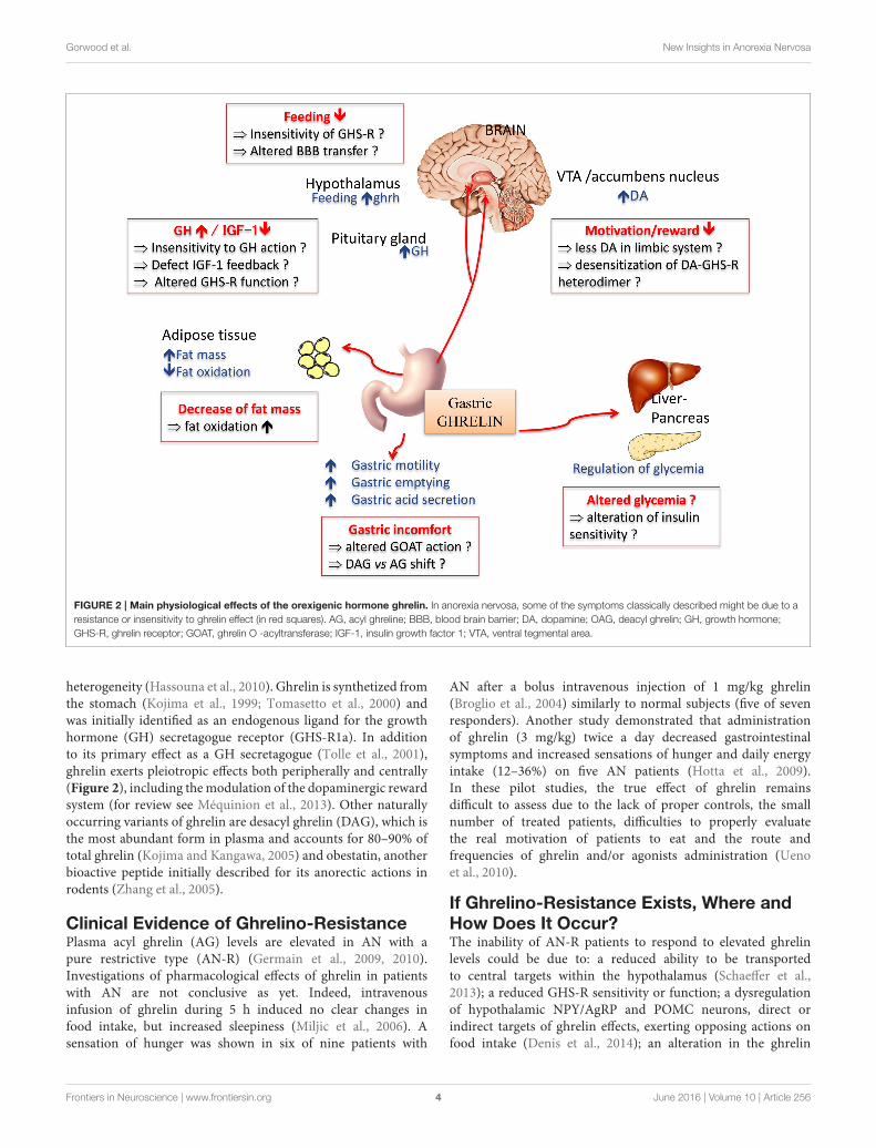
FIGURE 2 | Main physiological effects of the orexigenic hormone ghrelin. In anorexia nervosa, some of the symptoms classically described might be due to a
resistance or insensitivity to ghrelin effect (in red squares). AG, acyl ghreline; BBB, blood brain barrier; DA, dopamine; OAG, deacyl ghrelin; GH, growth hormone;
GHS-R, ghrelin receptor; GOAT, ghrelin O -acyltransferase; IGF-1, insulin growth factor 1; VTA, ventral tegmental area.
heterogeneity (Hassouna et al., 2010). Ghrelin is synthetized fromthe stomach (Kojima et al., 1999; Tomasetto et al., 2000) andwas initially identified as an endogenous ligand for the growthhormone (GH) secretagogue receptor (GHS-R1a). In additionto its primary effect as a GH secretagogue (Tolle et al., 2001),ghrelin exerts pleiotropic effects both peripherally and centrally(Figure 2), including themodulation of the dopaminergic rewardsystem (for review see Méquinion et al., 2013). Other naturallyoccurring variants of ghrelin are desacyl ghrelin (DAG), which isthe most abundant form in plasma and accounts for 80–90% oftotal ghrelin (Kojima and Kangawa, 2005) and obestatin, anotherbioactive peptide initially described for its anorectic actions inrodents (Zhang et al., 2005).
Clinical Evidence of Ghrelino-ResistancePlasma acyl ghrelin (AG) levels are elevated in AN with apure restrictive type (AN-R) (Germain et al., 2009, 2010).Investigations of pharmacological effects of ghrelin in patientswith AN are not conclusive as yet. Indeed, intravenousinfusion of ghrelin during 5 h induced no clear changes infood intake, but increased sleepiness (Miljic et al., 2006). Asensation of hunger was shown in six of nine patients with
AN after a bolus intravenous injection of 1 mg/kg ghrelin(Broglio et al., 2004) similarly to normal subjects (five of sevenresponders). Another study demonstrated that administrationof ghrelin (3 mg/kg) twice a day decreased gastrointestinalsymptoms and increased sensations of hunger and daily energyintake (12–36%) on five AN patients (Hotta et al., 2009).In these pilot studies, the true effect of ghrelin remainsdifficult to assess due to the lack of proper controls, the smallnumber of treated patients, difficulties to properly evaluatethe real motivation of patients to eat and the route andfrequencies of ghrelin and/or agonists administration (Uenoet al., 2010).
If Ghrelino-Resistance Exists, Where andHow Does It Occur?The inability of AN-R patients to respond to elevated ghrelinlevels could be due to: a reduced ability to be transportedto central targets within the hypothalamus (Schaeffer et al.,2013); a reduced GHS-R sensitivity or function; a dysregulationof hypothalamic NPY/AgRP and POMC neurons, direct orindirect targets of ghrelin effects, exerting opposing actions onfood intake (Denis et al., 2014); an alteration in the ghrelin
Frontiers in Neuroscience | www.frontiersin.org 4 June 2016 | Volume 10 | Article 256

Gorwood et al. New Insights in Anorexia Nervosa
signaling on dopaminergic neurons, in the ventral tegmentalarea (VTA) (Abizaid et al., 2006), a structure involved in thereward/locomotion behavior, and hypothalamus to modulateappetite, respectively (Jiang et al., 2006; Kern et al., 2012; Sharpeet al., 2016).
Ghrelino-Resistance: Antagonism by OtherGhrelin-Derived Peptides?The hypothesis of a ghrelino-resistance in AN-R requiresfurther evidence that (1) endogenous ghrelin is important instimulating appetite and (2) other ghrelin-derived peptidesmight be modulators of AG actions in animal models and ANpatients.
In mice with total ghrelin deficiency or selective AGdeficiency, food intake is not reduced (Sun et al., 2003, 2004;Zigman et al., 2005; Pfluger et al., 2008; Zhao et al., 2010). Thisdoes not seem to support a key role of AG in homeostatic eating.However, suppressed intake of rewarding food in a free choicefood paradigm, lack of cue-potentiated feeding and suppressedmotivation for food in an operant responding model rathersupport a role of the endogenous peptide(s) in hedonic eating(Uchida et al., 2013).
DAG may be an important modulator of appetite bycounteracting AG actions as demonstrated after central orperipheral injections (Asakawa et al., 2005; Inhoff et al.,2008). Interestingly, transgenicmice overexpressing DAGdisplaysignificantly reduced body weight, reduced food intake and bodyfat mass and moderately decreased linear growth compared tonon-transgenice mice (Asakawa et al., 2005). In addition, DAGexerts several biological actions, independent of the GHS-Rpathway, including regulation of glucose homeostasis and fatmetabolism (Delhanty et al., 2012, 2013). Although obestatinwas initially reported to decrease appetite and weight gain thusopposing ghrelin’s effects by acting on an orphan receptor, GPR39(Zhang et al., 2005), controversies rapidly arose concerning theanorexigenic actions of this peptide through GPR39 (Lauwerset al., 2006; Chartrel et al., 2007; Holst et al., 2007; Zizzariet al., 2007; Hassouna et al., 2010, 2012). In AN-R patients,obestatin, and the ghrelin/obestatin ratio were found decreasedwhereas this ratio was not altered in constitutionally thin women(Germain et al., 2009, 2010). Such changes may participate inthe eating restriction despite hyperghrelinemia observed in thesepatients.
Other Possible Mechanisms to ExplainResistance to FeedingGhrelin might also impact non-homeostatic systems that involvemore particularly the meso-cortico-limbic system. Skibicka et al.(2013) demonstrated that the significant increase for foodmotivation/reward behavior observed after a ghrelin injectionin the VTA is abolished by a pretreatment with D1-like orD2 receptor antagonist, injected in the nucleus accumbens,the main target of VTA dopaminergic neurons. This brainstructure is also involved in the integration of messages fromthe prefrontal inhibitory circuitry. Indeed, prefrontal cortexis strongly activated in AN patient following presentation of
food images as observed in a fMRI study (Zhu et al., 2012),and alterations in the serotoninergic/dopaminergic signalingare also described in these patients (Bailer et al., 2007, 2013).In AN patients, an altered impact of ghrelin on the rewardsystems might modify the integration of informations relatedto emotional process associated with food. Due to the paucityof data and the lack of consistent clinical information, the useof animal models to analyze more finely the mechanisms ofghrelin action at different levels of the brain system appearsessential.
From Clinics to Bench: What Can We Learnfrom Animal Models?A complete animal model of AN does not exist due in part to theneuropsychiatric aspects of the disease (Méquinion et al., 2015b).Such an ideal model of AN would include adolescent onset of thedisease, predominance in females, high activity, chronic stress,decreased food intake, and body weight. Moreover, AN is achronic disease, and most of the murine models described so farmaintain a protocol on a short period (few days). Neverheless,some aspects like food restriction associated with voluntaryphysical activity or chronic stress can be reproduced in micemodels. The model of separation with restricted access to food(separation-based anorexia, SBA), first developed in mice byvan Leeuwen et al. (1997), induces a chronic stress that resultsin an increase of energy expenditure without hyperactivity.Recent data demonstrated that, when this protocol is maintainedon the long-term, it induced AN like alterations such as adecrease in bone mineral density (osteoporosis) associated witha dysregulation of leptin signaling (Zgheib et al., 2014). Themodel of hyperactivity associated with food restriction (activity-based anorexia, ABA), first developed in rats (Hall et al., 1953;Routtenberg and Kuznesof, 1967) and subsequently adapted tomice (Gelegen et al., 2007) is considered as a good animalmodel of AN as it mimics important features of the disease(Routtenberg and Kuznesof, 1967; de Rijke et al., 2005). The ABAmodel also emphasizes the involvement of the ghrelin/GHS-Rpathway in food anticipatory activities, a behavior also describedin AN-R patients (Blum et al., 2009; Scheurink et al., 2010;Verhagen et al., 2011). On a modified long-term ABA model,where food was quantitatively restricted (50% during 10 weeks),symptoms classically observed in AN patients (body weight loss,endocrine changes, oestral cycle loss...) as well as alterations inthe integration of the ghrelin message at the hypothalamic levelswere observed (Méquinion et al., 2015a).
Conclusion: Is Anorexia Nervosa aGhrelino-Resistance or an AdaptativeMetabolic Response to Denutrition?In human pathology, the question remains whether ghrelinresistance (or decreased sensitivity) is only limited to the balancebetween patients’ hunger and satiety and motivation to therewarding aspect of food. Indeed, such hyperghrelinemia canalso serve as an adaptive neuroendocrine metabolic response tothe disease since ghrelin appears required for the maintenanceof blood glucose homeostasis during severe calorie restriction in
Frontiers in Neuroscience | www.frontiersin.org 5 June 2016 | Volume 10 | Article 256

Gorwood et al. New Insights in Anorexia Nervosa
mice (Zhao et al., 2010). Thus, up-regulation in ghrelin synthesismay be needed to maintain survival in the restrictive type ofAN. However, ghrelin is differentially regulated according tothe anorexic subtype as plasma levels are found unchanged inbinge-purging anorexic patients, suggesting that both subtypesengage in differential adaptive process to starvation and/ora difference in the perception of hunger and the motivationto eat. Extrapolation from these fundamental observationsto the human pathology should be carefully interpreted andwill require further exploration and validation of experimentalmodels. Animal models, despite their weakness on some aspectsof the disease might permit to assess long term peripheraland/or central alterations due to chronic and drastic foodrestriction associated or not with activity or chronic stress.Anxiety and hyperactivity behaviors have a significant rolein the pathogenesis and progression of the disease. One ofthe major advantages of using mice as an animal model isthat many well-described inbred lines are available to assessthe part of genetic or environmental component of anorexia.These animal models will be useful to precisely determine themechanisms involved in the impaired integration of the ghrelinsignal.
MODEL 3: ANOREXIA NERVOSA (AN) IS ACHRONIC STIMULATION OF THE REWARDSYSTEM BY OREXIGENICNEUROPEPTIDES OF THE LATERALHYPOTHALAMIC AREA
The hypothalamus plays a pivotal role in behavioral andemotional reactions and contains the major center that regulatesfeeding behavior. All patients are unable to adapt their feedingbehavior to energy demand and costs, raising the possibility of adysfunction of the orexigenic and/or anorexigenic neuropeptidesregulating appetite within the hypothalamus. Besides, recentevidence support the view that AN might be a pathologyof abnormally high reward process for starvation or purgingbehaviors. For instance, patients recovered from AN still showan increased neural response to both pleasant and aversivefood stimuli in brain circuitry mediating reward (Cowdreyet al., 2011). Dopamine is the neurotransmitter of the rewardsystem and several lines of evidence suggest the occurrence of adysfunctional dopamine-striatal system in AN that might explainthe lack of pleasure associated with food intake and the generalanhedonia of anorectic patients (Kaye et al., 2009). Supportingthis notion, amphetamine-induced dopamine release increasesanxiety in individuals recovered from AN (Bailer et al., 2012) andthat could explain why food-related dopamine release generatesanxiety in AN patients whereas feeding is pleasurable in healthysubjects. Interestingly, the lateral hypothalamic area (LHA), thatcontains the orexigenic neurons expressing orexins, melanin-concentrating hormone (MCH), and 26RFa, sends projections tothe brain reward circuitry (Aston-Jones et al., 2010), suggestingthat dysfunction of orexigenic hypothalamic neuropeptides mayalter the dopamine reward system in AN.
FIGURE 3 | Schematic representation of the hypothetical chronic
stimulation of orexigenic neuropeptides on the reward circuitry in
anorexia nervosa. Briefly, decrease of food ingestion induces a stimulation of
the neuronal activity of the lateral hypothalamic area (LHA) that will release the
orexigenic neuropeptides orexins (ORX), melanin-concentrating hormone
(MCH), and 26RFain the ventral tegmental area (VTA). This results in an
increase of dopamine (DA) release in the accumbens nucleus (NAc). In
anorectic patients, this stimulation of the reward system results in food
aversion associated with enhanced anxiety that will, in turn, reinforce the
decrease in food intake.
Herein, we will briefly present (Figure 3) the currentknowledge on three orexigenic neuropeptides produced byneurons of the LHA in AN, i.e., the orexins, MCH, and 26RFa.
OrexinsOrexins are produced by <80,000 neurons exclusively locatedin the LHA (Sakurai et al., 1998). Orexin A (OxA) and orexinB (OxB) are two neuropeptides that share 46% identity andare in tandem on the same precursor (Sakurai et al., 1998).Orexins are the endogenous ligands of two G-protein coupledreceptors termed OxR1 and OxR2 (Sakurai et al., 1998). Centraladministration of OxA or OxB stimulates food consumption(Sakurai et al., 1998). Prepro-orexin mRNA is up-regulatedfollowing fasting, and conversely down-regulated in ob/ob anddb/db obese mice (Sakurai et al., 1998; Yamamoto et al., 1999),whereas orexin deficient mice show robust hypophagia (Haraet al., 2001). Furthermore, systemic administration of the OxR1antagonist SB-334867 reduces feeding by selectively enhancingthe behavioral satiety (Ishii et al., 2004).
Two distinct studies have evaluated the evolution of plasmaOxA levels in patients with AN during 2, 3, or 6 months ofrealimentation (Bronsky et al., 2011; Janas-Kozik et al., 2011).The data obtained are conflicting as the first study (Bronskyet al., 2011) reports a significant increase of fasting plasmaOxA concentration in untreated AN patients whereas the secondone describes a decrease of circulating OxA in AN subjectsbefore refeeding (Janas-Kozik et al., 2011). However, the twostudies observe a progressive decrease of circulating OxA during
Frontiers in Neuroscience | www.frontiersin.org 6 June 2016 | Volume 10 | Article 256

Gorwood et al. New Insights in Anorexia Nervosa
realimentation, rather suggesting that the orexin system is up-regulated in AN.
Accumulating data indicate that orexins are involved inreward-based feeding. As a matter of fact, activation of orexinneurons in the LHA is strongly linked to preferences forcues associated with drug and food reward (Harris et al.,2005). Consistent with this, orexin injected into the VTA, thatcontains the dopamine neurons, increases dopamine releasein the accumbens nucleus (NAc), which provides enjoymentand reinforcement to motivate repeated activity, and stimulatesintake of a rewarding high fat diet (Zheng et al., 2007).Conversely, OXR1 antagonism in VTA attenuates high-fatfeeding in sated rat (for review, Cason et al., 2010). Additionally,central administration of orexin increases self-administration forsweet, an effect that is blocked by OXR1 antagonists, suggestingthat orexins promote the reinforcing properties of at least sometypes of food (Cason et al., 2010).
MCHMCH is a cyclic peptide predominantly expressed in the LHA.MCH induces food intake after central injections (Qu et al.,1996), and its gene expression is modulated by fasting and inhypoleptinemic ob/ob mice (Qu et al., 1996). Two G protein-coupled (GPCR) MCH receptors (MCH-R1 and MCH-R2) arepresent in human, of which only MCH-R1 appears functionalin rodents (Forray, 2003). Several genetically-modified mice thatoverexpress or lack MCH (Shimada et al., 1998) or MCH-R1 (Marsh et al., 2002) genes were generated and displayedmarked alterations in the regulation of appetite and/or energybalance. At present, the MCH system has never been investigatedin AN. However, it is interesting to note that the orexigeniceffect of MCH is largely dependent upon its action in theNAc of the reward system. Indeed, injections of MCH andMCH antagonists into the NAc are orexigenic and anorexigenicrespectively (Georgescu et al., 2005). MCH injections in this areadecrease neuronal firing of spiny neurons and increase hedonicvalue of sweet foods (Lopez et al., 2011). This suggest that MCHsignaling pathways play a complex but important role in feeding-reward control and could provide important information aboutmechanisms underlying the development of AN.
26RFa26RFa is a 26-residue RFamide peptide characterized in variousvertebrate species including Man (Chartrel et al., 2003, 2011).26RFa has been identified as the cognate ligand of the humanorphan G protein-coupled receptor, GPR103 (Takayasu et al.,2006; Chartrel et al., 2011). Neuroanatomical observationsrevealed that 26RFa- and GPR103-expressing neurons areprimarily localized in hypothalamic nuclei involved in the controlof feeding behavior (Chartrel et al., 2003; Takayasu et al., 2006;Bruzzone et al., 2007). 26RFa-containing neurons are exclusivelylocated in the LHA and the ventromedial hypothalamic nucleus(Chartrel et al., 2003). Central administration of 26RFa stimulatesfood intake and causes obesity in rodents (Chartrel et al., 2003;Moriya et al., 2006; Takayasu et al., 2006). In addition, 26RFamRNA is up-regulated in the hypothalamus of obese ob/ob and
db/db mice (Takayasu et al., 2006) indicating that the expressionof the 26RFa gene is modulated in condition of energy imbalance.
Circadian profile of plasma 26RFa was investigated in ANpatients. The data showed that 26RFa levels are significantlyincreased all over the day in patients with restrictive AN ascompared to healthy subjects (Galusca et al., 2012).
Detailed mapping of GPR103 mRNA in the brain revealedthat the 26RFa receptor is expressed in the VTA, the amygdala,and the NAc (Bruzzone et al., 2007), suggesting that, in as muchthe same way as orexins and MCH, 26RFa may be involved inreward-based food intake.
ConclusionOrexigenic neuropeptides including ghrelin, orexins and 26RFaare up-regulated in AN and it is thought that this orexigenicprofile reflects an adaptive mechanism of the organism topromote food intake and thus to counteract undernutrition.However, this adaptive mechanism is ineffective in increasingfood consumption leading to the concept of a global resistanceof AN patients to orexigenic signals. Here we present an alternatehypothesis. We speculate that a chronic increase of the activityof LHA orexigenic neurons expressing orexins, MCH, or 26RFacould reinforce dopamine-induced anxiety in the reward systemof AN patients and thus the aversion to ingest food.
MODEL 4: GUT MICROBIOTA IS ACENTRAL FACTOR IN ANOREXIANERVOSA
Anorexia nervosa (AN), the most frequent and the most seriouseating disorder, is often associated with severe proteino-energeticmalnutrition (PEM). Marasmus, the adaptative form of semi-prolonged fasting, is the predominant form of malnutritionassociated with AN. However, the kwashiorkor type is presentin some cases, and is characterized by a constellation of featuresincluding peripheral edema, hypoalbuminemia, fatty liver, skinand hair lesions, apathy, high a relative immuno-depression withhigh risk of infections. It is associated with a lot of deleterioussecondary complications and a high rate of morbidity/mortality.Some patients have a mixed picture of marasmic-kwashiokormalnutrition.
In the last decades, the role of gut microbiota in differentdiseases was demonstrated thanks to the development of newmethods. The most accurate ones are metagenomics whichaims to study all genetic content of prokaryote cells by highthroughput sequencing.
With such new tools, the role of gut microbiome wasdefined on nutriments absorption and energy regulation (harvest,storage, and expenditure of energy). It is now established thatthe gut microbiome contributes to the risk and pathogenesis ofmalnutrition through nutrient metabolism and immune function(Krajmalnik-Brown et al., 2012). A clinical trial, comparing 16srRNA PCR fecal samples of seven malnourished children withseven healthy ones, showed that the former have a significantlylower number of operational taxonomic units in their gutthan healthy subjects (310 vs. 546) (Monira et al., 2011). A
Frontiers in Neuroscience | www.frontiersin.org 7 June 2016 | Volume 10 | Article 256

Gorwood et al. New Insights in Anorexia Nervosa
FIGURE 4 | Gut microbiota as an important contributing factor in Anorexia Nervosa.
metagenomic comparison of gut microbiota, studying 9 well-nourished twin pairs to 13 twin pairs discordant for kwashiorkor,showed a significant difference on gut microbiota profile.Indeed malnourished twins (kwashiorkor) had a considerablyless differentiated and immature gut microbiome than healthyor marasmic children (Smith et al., 2013). When stool samplesfrom kwashiorkor-malnourished children were transplanted tognotobiotis mice, these animals developed weight loss and severemalnutrition (Smith et al., 2013). In a recently randomizedcontrolled clinical trial in 2767 Malawian malnourished children,a 1-week oral antibiotic (which modulates gut microbiota)treatment improved the nutritional status of malnourishedchildren and decreased mortality (Trehan et al., 2013).
Modifications of the gut microbiota have also been describedin AN patients. Armougom et al. (2009) measured bacterialdivisions of Bacteriodetes, Firmicutes, Lactobacillus, andMethanobrevibacter smithii. They compared the feces of 20obeses subjects, 9 patients with AN and 20 normal weighthealthy controls. AN patients were moderately undernourished,with an average BMI at 16. All AN patients had an increase ofM.smithii (Armougom et al., 2009). This Archeon, which comprisesup to 10% of all anaerobes in the colon of healthy adults,plays a central role in bacterial digestion of polysaccharides. Itrecycles hydrogen in methane, allowing for an increase in thetransformation of nutrients into calories and thereby increasingenergy efficiency (Samuel et al., 2007). It is possible that chronicconstipation, which is common in AN patients, could also bepresent before weight loss and causes the increase ofM smithii intheir gut (Kim et al., 2012). In addition, by using electrophoreticprofile of the 16S RNAmicrobiota, a deviation of microbiota wasfound in 8 AN undernourished patients (average BMI at 12) incomparison to nine healthy subjects (Hanachi et al., 2013).
All these data show that there are modifications ingut microbiota of AN patients, not necessarily severelymalnourished. These changes include an increase of methanogenarchea directly involved in energy efficiency. Indeed the increaseofM. smithii can lead to the optimisation of food transformationof nutrients into calories. This increase of M. smithii could havepreceded undernutrition and could play a central role in theoutbreak of anorexia nervosa. In order to explore this pathway
the gut microbiota of patients with AN should be assessed beforethe beginning of weight loss, during nutritional therapy and afterrecovery of weight loss.
In summary (Figure 4), at least two hypotheses can beproposed concerning the role of gut microbiota in anorexianervosa: (1) some pre-existing changes in gut microbiota induceand/or participate to food restriction in these patients andchronic constipation could induce and/or facilitate this changein gut microbiota. (2) The gut microbiota determines for eachindividual patient the type of PEM, i.e., marasmus type vs.kwashiorkor. In such case, manipulation of gut microbiotaduring AN (with antibiotics for example), might improve theissue of malnutrition during nutritional therapy.
MODEL 5: ANOREXIA NERVOSA AS ADYSIMMUNE DISORDER OFNEUROPEPTIDE SIGNALING
We review here recent evidence from experimental and clinicalstudies explaining how stressors could result in the dysregulationof eating behavior by the means of specific immunoglobulins(Igs) altering neuropeptide signaling (Figure 5). We also discussa key role of intestinal bacteria and gut barrier dysfunction inthis dysregulation. Although anorexia nervosa (AN) is classifiedas a psychiatric disease, the clinical observations that it involvesmultiple organ dysfunction, in addition to the core behavioraland psychiatric symptoms, underlines the need for an integrativepathophysiological approach. Anorexia is generally consideredas the driving mechanism leading to malnutrition and itsmultiple organ consequences, leading in turn, to the perpetuationof anorexia and psychiatric comorbidity in a vicious circle.However, this classical approach does not clarify how anorexiaand related symptoms may occur in so far healthy adolescentsor adults. It is widely accepted that the risk of developing ANis markedly increased by different types of stressors: physical(such as puberty, trauma, abuse, or infections) and/or mentalstress, including for example intellectual or physical exhaustionor repeated dieting (Hilbert et al., 2014). Familial history ofstress events or stress-related disorders is another risk factor of
Frontiers in Neuroscience | www.frontiersin.org 8 June 2016 | Volume 10 | Article 256

Gorwood et al. New Insights in Anorexia Nervosa
FIGURE 5 | Implication of microbial protein and specific Immunoglobulins in the dysfunction of neuropeptide signaling during anorexia nervosa.
AN, suggesting that genetic or epigenetic factors, so far poorlyidentified, may contribute to the maladjusted response to stressin patients developing AN (Trace et al., 2013). Finally, over thepast 10–20 years, rapidly growing knowledge on the physiology ofeating behavior identified neuropeptides and peptide hormonesas major signaling molecules, in addition to monoamines, toregulate food intake, satiety, as well pleasure and other behaviorsand functions that may be altered during AN, e.g., sleep,anxiety, digestive motility, and sensitivity, endocrine functionsand bone metabolism (Berthoud, 2011). This led to the conceptof neuropeptide signaling dysfunction in AN (Inui, 2001), but anapproach based only on peptide concentrations measurementsand classical hormone-receptor interaction provided conflictingor paradoxical findings (Prince et al., 2009), e.g., that food intakeremains low in anorectic patients despite increased orexigenicghrelin (Otto et al., 2001) and decreased anorexigenic leptin(Hebebrand et al., 1995) levels.
We have performed a series of clinical and experimentalstudies indicating that neurobiological mechanisms of eatingdisorders and obesity may involve Igs reactive with peptidehormones regulating food intake and emotion, therefore alsocalled peptide auto-antibodies (Fetissov et al., 2002). Theinitial finding was the identification of Igs in the serum ofpatients with AN or bulimia nervosa (BN) binding to α-melanocyte-stimulating hormone (α-MSH) in hypothalamicneurons (Fetissov et al., 2002). The clinical relevance ofthese Igs was further suggested by the correlation of theirplasma levels and the Eating Disorder Inventory-2 (EDI-2)scores in AN and BN patients (Fetissov et al., 2005). Toelucidate the potential origin of these Igs, an in silico search
was performed, based on the concept of molecular mimicry,with the hypothesis that some microorganisms present inthe environment, especially in gut microflora, may presentsequence homology with neuropeptides. This hypothesis wasconfirmed for several common microorganisms expressing inseveral proteins identical five consecutive amino acids with themain peptides involved in appetite, satiety, or emotion (e.g., α-MSH, ghrelin, leptin, orexin; Fetissov et al., 2008). In addition,both IgG and IgA classes of Igs reactive with peptides hormoneswere present in healthy subjects, further supporting a luminalsource of antigens as well as physiological role of these Igs in themodulation of neuropeptide signaling. In support of this, otherauthors have shown the stimulatory effects of gut microbiota onall Igs classes in germ-free mice (Hansson et al., 2011).
With respect to AN pathophysiology, several of our studiesfocused on anti-α-MSH Igs, since this 13 amino acid peptide is amajor regulator of energy balance, increasing satiety and energyexpenditure via the activation of the melanocortin receptor type4 (MC4-R), and is closely related to the hypothalamic responseto stress (Cone, 2006). With this in mind, a study was undertakenin rodents showing the impact of chronic stressors on the levelsand binding properties of anti-α-MSH Igs, related to food intakeand anxiety-like behavior (Sinno et al., 2009). Furthermore,in a rat model of severe gut inflammation, inducing transientanorexia and major increase of intestinal permeability, deficientbody weight gain persisted even after the resolution of intestinalinflammation and was associated with an increased level of anti-α-MSH Igs (Coquerel et al., 2012). We also adapted an animalmodel of AN called Activity-Based-Anorexia (ABA), combininga progressive reduction of time access to food coupled with
Frontiers in Neuroscience | www.frontiersin.org 9 June 2016 | Volume 10 | Article 256

Gorwood et al. New Insights in Anorexia Nervosa
running wheel to mimic the hyperactivity of AN patients. Wefound that ABA mice were characterized by an increased colonicpermeability which is favorable for the passage of antigens fromgut bacteria to the host (Jésus et al., 2014).
To explore the implication of bacterial proteins fromgut microflora in the occurrence of peptide-reactive Igs, weused a combined proteomic and immunological approach toidentify α-MSH antigen-mimetic proteins in Escherichia coli.The commensal E. coli K12 was used because several E. coliproteins display five amino acids sequence homology with α-MSH (Fetissov et al., 2008). We identified ClpB, a heat-shockchaperone protein, as a conformational antigen mimetic of α-MSH (Tennoune et al., 2014). We showed that immunizationof mice with ClpB induced the production of anti-ClpB Igcross-reacting with α-MSH accompanied by altered food intake,body weight, anxiety and MC4-R signaling. Furthermore, micereceiving wild-type E. coli via intragastric gavage, decreased foodintake and body weight gain and developed anti-ClpB- antibodiescross-reactive with α-MSH. In contrast, in mice receiving ClpB-deficient E. coli, food intake and Igs levels remained unaltered.Finally, plasma levels of anti-ClpB IgG cross-reactive with α-MSHwere increased in patients with eating disorders, and severalsignificant correlations were found between EDI-2 scores andanti-ClpB IgG and IgM levels in AN patients (Tennoune et al.,2014), similar to previous findings with anti-α-MSH reactiveIgs (Fetissov et al., 2005). These data revealed that α-MSH-reactive Igs in humans and animals are generated mainly inresponse to bacterial ClpB protein produced by some gut bacteriaincluding E. coli. It is of relevance to mention that the ClpBprotein expression in bacteria is stimulated by various stressorspreventing protein aggregation (Winkler et al., 2012), suggestingthat ClpB could also be activated by host-induced starvationduring a restrictive diet.
Altogether, these studies indicate that different stressors,including food restriction, via increased intestinal permeability,may favor bacterial protein translocation from the gut resultingin increased production of neuropeptide-reactive Igs. Morespecifically, bacterial protein ClpB may act as an anorexigenicα-MSH mimetic protein directly and indirectly via triggeringproduction of Igs cross-reactive with α-MSH, and hence, alteringthe effects of this endogenous peptide hormone on satiety andanxiety. This may involve a modulatory role of Igs on α-MSH-induced activation of theMC4-R (Lucas et al., 2014), although theexact mechanisms needs further investigation. Other groups havesuggested that gut microbiota, especially E. coli, may influencethe stress axis (Dinan and Cryan, 2012), while E. coli content wasinversely related to the body mass index, including AN patients(Million et al., 2013). Studies on gut microbiota involvement inbrain functions are discussed elsewhere (Cryan andDinan, 2012);our data support a role of bacterial protein mimetics of peptidehormones in the gut-brain axis communication and validate theutility of proteomic approach as an efficient tool to detect suchproteins.
In addition to the putative key role of α-MSH-mimeticprotein and α-MSH-reactive Igs in AN pathophysiology, it isprobable that other peptidergic systems involved in regulationof feeding and emotion can be altered via a similar mechanism
involving the gut-brain communications. Accordingly, the levelsand properties of Igs reactive with ghrelin were altered in ANpatients (Terashi et al., 2011), which might account with thepreviously proposed ≪ ghrelin resistance ≫ in these patients(Otto et al., 2001). In an opposite way, the affinity kineticsof ghrelin-reactive Igs in obese patients explained its increasedorexigenic effects, through its reduced degradation (Takagi et al.,24). Whether ghrelin-reactive Igs in AN patients may alteractivation of the ghrelin receptor remains to be explored. Orexin-reactive Igs have been detected in narcolepsy (Deloumeau et al.,2010), which raises question about their possible implicationin sleep disturbances during AN. It is worth underlining thatseveral neuropeptides controlling food intake and anxiety, arealso involved in gastrointestinal motor and sensitive regulation(e.g., ghrelin, CCK), which may explain the high prevalence ofgastrointestinal symptoms during AN (Déchelotte et al., 2007).
In conclusion, we propose an integral pathophysiologicalscenario that fits to the natural history of AN with thefollowing steps: (1) enhanced vulnerability to stress (genetic,epigenetic, or environmental factors); (2) major stressing eventsactivating the stress-axis, increased intestinal permeability, andincreased virulence of the microbiota; (3) bacterial proteins(e.g., ClpB) challenge the immune response and due tomolecular mimicry, cause increased production of Igs cross-reactive with neuropeptides (e.g., α-MSH); (4) this results inaltered food intake, anxiety, gastrointestinal discomfort and otherconsequences of altered central and peripheral melanocortinsignaling; (5) global malnutrition and some specific macro-and micro-nutrient deficiencies contribute to the perpetuationof gut barrier and immune dysfunction as well as behavioralsymptoms. This scheme opens potential therapeutic perspectivesin AN, involving a multimodal approach including behavioraland/or pharmacological reduction of stress, restoration of gutbarrier function, correction of microbiota imbalance, and specifictargeting of bacterial proteins and/or related Igs.
MODEL 6: ANOREXIA NERVOSA TAKESROOT IN THEPONDERO-NUTRITIONAL-EATINGBASEMENTS DURING CHILDHOOD’SPREMORBID PERIOD
Physiological puberty process, partially genetically determined,is associated with strong physical, psycho-affective, hormonal,metabolic, and feeding behaviors variations. One possiblehypothesis is that anorexia nervosa during adolescence is akind of mismatch of this physiological process, permittedby underlying and premorbid vulnerability factors concerningthe ≪ pondero-nutritional-feeding ≫ basements (Figure 6).Susceptibility factors existing during the premorbid period mayconcern development of taste, food choice, eating behaviors.The pubertal process stimuli interfering with these vulnerabilityfactors could promote a failure of the ≪ pondero-nutritional-feeding ≫ system homeostasis resulting in later anorexianervosa. The vulnerable system is a kind of trigger zoneaccount for the symptom choice during difficult pubertal process
Frontiers in Neuroscience | www.frontiersin.org 10 June 2016 | Volume 10 | Article 256

Gorwood et al. New Insights in Anorexia Nervosa
FIGURE 6 | Pubertal process and pondero-nutritional-eating basements: the mismatching team.
explaining how initial deviant eating behavior is becoming asevere psychosomatic disease, evolving for itself with denutritionand metabolic modifications promoting self-aggravation.
Development of Food PreferencesDevelopment of food preferences and eating behaviors is acomplex and paradoxical process starting probably in utero,involving inherited taste characteristic and interactions withenvironmental factors such as familial eating behavior, peersinfluence, and socio-cultural context (Birch, 1999).
Inherited CharacteristicsStudy of gusto-facial expression in 2 years old infants defines ≪hypoguesic≫,≪ normoguesic≫, and≪ hyperguesic≫ groups.Hyperguesic group develops more difficulties in food choices andin maternal feeding interaction (Chiva, 1982). Individual geneticsensitivity to the bitter taste of phenylthiocarbamide (PTC) or6-n-propylthiouracil (PROP) determines food preferences anddietary habits. Thirty percent of occidental population is “non-tasters” vs. 50% “tasters” and 20% called “super-tasters.” Sucha marker for individual differences in taste perception couldinfluence food preferences (bitter and foods rich in antioxydants,
sweetened and fat-containing foods, alcoholic beverages), anddietary behavior with subsequent links to body weight andchronic disease risk. There may be an inverse correlation betweengenetic sensitivity to PTC or PROP, BMI, and body fatness.The level of involvement of this phenotype mediated by theTAS2R38 gene in food regulation is still under debate. Othergenetic bitter and non-bitter taste factors could play a role infood preferences including food aversion and deviant eatingbehaviors, such as the gustine gene which affects salivary ioniczinc concentrations—a trophic factor of taste buds (carbonicanhydrase VI). Otherwise, despite the fact that patients withanorexia nervosa often complain of disrupted smell and tastesense, there is no study concerning clinical population witheating disorders (Goldzak-Kunik et al., 2012).
Parental Feeding Practices andSociocultural InfluencesChildren eating behavior is precociously influenced bysociocultural environment, parents feeding practices andparticularly maternal eating habits and food preferences.Negative affects at mealtime, eating conflicts, struggles withfood, and unpleasant meals in early childhood are associated
Frontiers in Neuroscience | www.frontiersin.org 11 June 2016 | Volume 10 | Article 256
Gorwood et al. New Insights in Anorexia Nervosa
with higher risk for subsequent symptoms of anorexia nervosa.Maternal diet is one of the strongest predictor of childhoodfood consumption. Indeed, 15–20% of variation in children’sdifficulties could be associated with maternal feeding practices.A French study showed that extreme feeding practices ascontingent, coercive or permissive style enhanced children’seating difficulties such as food neophobia, pickiness, lowappetite, and low enjoyment in food. On the contrary, a flexibleattitude, with repeated exposures to food, enhanced motivationto eat (Rigal et al., 2012). Parental excessive restriction andcontrol of child feeding practices could limit the children eatingautonomy and negatively influence their food choice, promotingin opposite reaction, consumption of “bad foods” including a dietrich in sugar, salt, and/or fat. Intrusiveness and other deleteriousparental behavior, as not acknowledging the needs of the child,are also associated with higher risk of eating disorders (Gahagan,2012).
Parental Eating DisordersSeveral studies have shown that offspring of mothers with eatingdisorders are at higher risk of developing psychopathology inchildhood and adolescence. Maternal eating disorders influencevery early the quality of infant feeding, starting in uterowith more frequent prematurity, low birth weight (which arethemselves risk factors for eating disorders), overweight, andhyperphagia (Micali et al., 2011). Women with high weightand shape concerns breastfeed less their newborns, and mightinfluence future children food choice because of their ownfood restrictions during this breastfeeding period. Later on,conflicts and unpleasant remarks at mealtimes, intrusive andcontrol feeding practices may promote child negative eatingbehaviors, opposition attitude with food refusal, or infantileanorexia (Micali et al., 2009). Longitudinal study comparingchildren of women with eating disorders with “non-exposed”children showed modified feeding behaviors with more adhesionto “health conscious/vegetarian” dietary pattern in the firstgroup, less fat andmore carbohydrates in children of womenwithbulimia nervosa, and more high energy–dense foods in childrenof women with a history of binge sub-type anorexia. Thesemodifications of food choice may be associated with a further riskof developing weight concerns, overweight, and eating disordersin later childhood and adolescence (Easter et al., 2013).
Premorbid Overweight or ObesityThere is no study concerning association between overweight orobesity in childhood and further episode of anorexia nervosa inadolescence. However, overweight or obesity in childhood arestrongly associated with hyperphagia, even though incidence ofother types of eating disorders is unknown. Even if anorexianervosa and binge eating disorder differed on premorbidpersonality/behavioral problems, and family overeating, riskfactors for bulimia nervosa are mainly shared with anorexianervosa and binge eating. Premorbid BMI is positively correlatedto binge eating disorders and bulimia nervosa during the courseof anorexia nervosa episode (Nishumira et al., 2008). Overweightor obesity during pre-morbid period is more frequent in maleadolescent with anorexia nervosa (30–40%). Otherwise, recentstudies suggest that adolescents with a history of premorbid
overweight or obesity may develop restrictive eating disorders,with severe weight loss, severe medical complications, and apoorer prognosis.
Food Neophobia/Pickiness/ARFIDEven though studies do not provide firm conclusions concerningwhich problems are predictive, the presence of eating problemsin early childhood or an eating disorder in adolescence confersa strong risk for an eating disorder in young adulthood (Kotleret al., 2001). Digestive troubles in neonatal period or in earlyinfancy (severe gastro oesophageal reflux) or food aversionssecondary to gastrointestinal disease with nausea and vomitinghave been reported to have negative consequences on appetiteand eating patterns. Food neophobia is a survival mechanismwith rejection of unknown foods while picky/fussy eating,a frequent problem in childhood (8–50%). It is defined as arejection of familiar food with consumption of an inadequatevariety of food, and low intakes of vegetables, and fruits. Riskfactors for a later development of eating disorders include pickyeating and digestive problems with a significant stability overthe 10-year span studied, beginning at ages 1–10 (Marchi andCohen, 1990). Even if food neophobia and picky eating are twodistinct entities, there is probably an overlap and both maybe associated with a certain type of eating disorders. ARFID(Avoidant/Restrictive Food Intake Disorder in Children andAdolescents) is a new category of DSM-5 classification, includingthose with selective (picky) eating since early childhood.Common eating-specific behaviors and symptoms in the ARFIDgroup contain food avoidance, loss of appetite, abdominalpain, and fear of vomiting. Patients with ARFID are youngerthan those with AN, more likely to present before age 12, andmore likely to be male (Norris et al., 2014). In a recent study,patients with ARFID were significantly underweight with alonger duration of illness, more likely to have comorbid medicalconditions, anxiety or mood disorder. The course of ARFID isunknown but it is possible that these children and adolescentswould develop other types of eating disorder such as AN, BN, orEDNOS (Fisher et al., 2014).
ConclusionAnorexia nervosa takes root in vulnerability factors determiningchildhood feeding and eating behaviors bases. Thesesusceptibility factors, including food preferences, inheritedtaste factors, environmental factors, and early inadequate foodintakes may be impacted by modifications associated to thepubertal process, therefore untitled the “mismatching team.”Detection of particular traits of eating patterns or deviantponderal history may indicate vulnerability and high risk forsome toddlers or children to develop anorexia nervosa inadolescence.
MODEL 7: ANOREXIA NERVOSA (AN) ISAN ATTEMPT TO PRESERVE MENTALHOMEOSTASIS
AN can be considered as the result of an attempt to restorementalhomeostasis that uses the bodily domain by way of restricted
Frontiers in Neuroscience | www.frontiersin.org 12 June 2016 | Volume 10 | Article 256

Gorwood et al. New Insights in Anorexia Nervosa
FIGURE 7 | A model of anorexia nervosa as an attempt to preserve mental homeostasis.
eating (Figure 7). Certain subjects, because of predisposingfactors (Garner et al., 1993) could develop trajectories ofvulnerability toward AN. This vulnerability could come tothe surface on the occasion of precipitating elements leadingto the loss of “well-being” or “eudemonia” as a result ofindividual factors (mental or somatic) or interpersonal factors(See diagram). The unsettled homeostasis, in a first stage, isrestored by restricted eating, a process that could contributeto prolonging AN. Vulnerability trajectories and precipitatingfactors are liable to vary in nature and intensity from one subjectto another, and would explain the variable clinical expressions interms of age at onset, clinical symptoms, intensity of the disorder,duration of evolution, therapeutic response, or evolution tochronic illness, bulimia or other psychiatric disorders.
The predisposing elements contributing to an ANvulnerability trajectory are not all present at once in patients,as they assemble to form trajectories made up of numerousdifferent vulnerability factors. These predisposing elementsinclude (1) family factors, including genetic susceptibility to thedisorder (Pinheiro et al., 2010; Trace et al., 2013), a psychiatricfamily history of eating disorders (Strober et al., 2000), anxious,depressive, or bipolar disorders (Bould et al., 2015), traits suchas perfectionism or rigidity (Treasure et al., 2010), disturbedfamily interactions early in life (deprivation, violence—Jaiteet al., 2012) or family interactions connected with eating habits(Hill and Franklin, 1998; Haycraft et al., 2014; Loth et al., 2014;Micali et al., 2014). Some (2) individual factors might also be
involved, such as female gender (Connan et al., 2003), earlyexposure to stress, including intra-uterine stress (Connan et al.,2003; Treasure et al., 2010) via epigenetic regulation (Toyokawaet al., 2012) and disrupted personal development evidencingitself in affects, cognitions and interpersonal relationships(Connan et al., 2003). Depending on the concepts mobilizedin different studies, these phenomena are evidenced in theform of personality disorders or dimensions (perfectionism,obsessive-compulsiveness, neuroticism, negative emotionality,harm avoidance, low self-directedness, low cooperativeness, lownovelty seeking, and traits associated with avoidant personalitydisorders) (Cassin and Von Ranson, 2005), attachment disorders(Tasca and Balfour, 2014), emotional regulation disorders(Haynos and Fruzzetti, 2011), cognitive disorders (weak centralcontrol, rigidity) (Treasure and Schmidt, 2013), anxiety disorders(Raney et al., 2008; Touchette et al., 2011), and biological orgenetic particularities (see for example Bailer and Kaye, 2011;Tremolizzo et al., 2014). (3) Environmental factors also play animportant role, including stressful life events such as prematurity(Favaro et al., 2006), multiple birth (Raevuori et al., 2014), andsocial factors such as the ideal of slimness in society (Garner,1993), social class (Godart et al., 2013), and educational level(Goodman et al., 2014).
The trajectories derived from these various elements remainto be characterized. They could roughly correspond to five types,dominated respectively by (1) obsessiveness and perfectionism,(2) major interpersonal difficulties verging on Asperger’s
Frontiers in Neuroscience | www.frontiersin.org 13 June 2016 | Volume 10 | Article 256

Gorwood et al. New Insights in Anorexia Nervosa
syndrome, (3) anxiety, (4) mood disorders, recurrent unipolardepression, bipolar disorders, and (5) personality disordersinterwoven with deprivation or violence in the early years.
The precipitating elements are mainly stressful life events(Connan et al., 2006; Schmidt and Treasure, 2006) or negativeemotional states (Fox and Power, 2009), sometime in a settingof incipient psychiatric comorbidity (depression, anxiety, OCD,Godart et al., 2002, 2007), or most often associated with pubertyand including hormonal factors (Klump, 2013) or psychological,physical, and relational factors.
The start of eating restrictions appears in various contexts: (1)the psychological dissatisfaction linked to a negative emotionalstate (poor self-esteem) is transferred to the body (Fox andPower, 2009) and triggers a will to restrict food intake linkedto dissatisfaction with the body, (2) a slimming diet (as a resultof weight gain, family pressures, peer group influence), (3) anintercurrent event—operation involving the digestive tract, (4)emotional state (via stress—Greeno and Wing, 1996—anxietyor depression) or a somatic pathology can trigger anorexia inits prime meaning (loss of appetite). However, whatever theprevious vulnerability trajectory or the precipitating events, thesesubjects discover the positive effects of food intake restrictions (ashas been described in the short term in depression, Fond et al.,2013), enabling a temporary return to eudemonia. This wellbeingis the result of biological modifications that may be caused bystarvation, the decrease in anxious and depressive symptoms(Kaye et al., 2003), the feeling of having control over one’s life,one’s body, and one’s diet, the sometimes positive feedback fromthe entourage, and in some cases affective anesthesia (Garner,1993; Fox and Power, 2009).
In all cases, however, the balance obtained is fragile andshort-lived, since the causes of the imbalance persist. Furtherto this, the lack of food (often compounded by hyperactivity),because it has biological and hormonal consequences, (leads toanxious and depressive symptoms Swenne and Rosling, 2010;Gauthier et al., 2014), interpersonal difficulties (Treasure andSchmidt, 2013), social anxiety (Coulon et al., 2009), and parentalcriticism linked to the patient’s state (Treasure and Schmidt,2013; Duclos et al., 2014) with which the subject tries to cope bythe same means of food intake restriction, even intensifying fooddeprivation, in order to regain the same effect, which enclosesthe subject in the vicious circle of AN. In this model, AN can beconsidered as an addictive behavior in which the object of theaddiction is the control exercised over one-self and one’s hunger(Jeammet, 1991; Venisse, 1991; Goodman, 2008; Duclos et al.,2014).
The clinical profile observed by clinicians is composite, anddepends on elements linked to the vulnerability trajectory (forexample one of the five previously mentioned), to age at onsetof the disorder (for instance pre-pubertal subjects exhibit growthand pubertal delay), adolescents are less often in denial of theirthinness than adults (Couturier and Lock, 2006). It also dependson precipitating factors (for example if a depressive state precedesthe disorder, suicidal ideas or psychomotor retardation can bepresent) and on the intensity and duration of food deprivation.We hypothesize that the severity of the AN evolution trajectory isalso linked to the severity of vulnerability factors, to the intensity
of the state of starvation and to AN prognosis: the more severethe difficulties experienced before AN onset, the more likely isthe disorder to be severe and become chronic, and the greater arethe mental disturbances compounding AN.
We propose a global, integrative clinical model of AnorexiaNervosa (AN), starting from models developed previously(Garner, 1993; Connan et al., 2003; Fairburn and Harrison, 2003;Schmidt and Treasure, 2006; Fox and Power, 2009; Herpertz-Dahlmann et al., 2011; Tasca et al., 2013; Treasure and Schmidt,2013; Treasure et al., 2014) and from the concept of mentalhomeostasis, which postulates (Agnati et al., 2011) (1) that theconcept of biological homeostasis can be transposed to themental domain, (2) that any individual, from a psychologicalviewpoint, possesses a state of equilibrium called “the well-beingof the human psyche” or “eudemonia,” and (3) that mentalhomeostasis sustains eudemonia by enabling a person to feelat ease with him/herself (emotionally and bodily) and withothers (interpersonal relationships). Any event in one or otherof these areas (emotional, bodily, interpersonal) that upsets thismental homeostasis will generate a state of “mental allostasis,”which is painful for the subject, who will struggle against it byway of adaptive processes to restore the earlier equilibrium. Aperson’s mental homeostatic equilibrium is a dynamic balancingprocess that is variably vulnerable, depending on a developmentaltrajectory that is linked to family, individual and environmentalfactors.
CONCLUSIONS
Just as for many of complex disorders, scientific evidencescan lead to different heuristic models in AN. An efficientmodel should use validated risk factors to further define thepotentially involved mechanisms of actions of anorexia nervosa.The predisposing elements contributing (see Model 7) to ANinclude (1) presence of a psychiatric family history of eatingdisorders, (2) presence of personal anxious, depressive or bipolardisorder, (3) different personality traits such as perfectionism orrigidity, (4) disturbed family interactions early in life (deprivationand violence), (5) family interactions connected with eatinghabits, but also (6) female gender, (7) early exposure to stress,including intra-uterine stress, multiple birth and prematurity,and (8) disrupted personal development. Social factors also havea role, such as (9) the ideal of slimness in society, (10) social class,and (11) educational level.
One of the specificity of anorexia is the presence of excessivephysical exercises even though there is food deprivation orpurging behaviors in underweight patients. An abnormalityof the rewarding value in AN of food and exercise has thusbeen proposed as a core feature, which is supported by therole of endogenous opioids and dopamine pathways in thereward properties of food (see Model 1), and indirectly byan excess of specific polymorphisms of the genes codingfor opioid and dopamine receptors or transporters in AN.An abnormal reward process could potentially involve morespecifically a preference for delayed reward and/or reducedcapacity to (as in controls) favor immediate reward. Suchpreference for delayed reward does give sense to avoid food
Frontiers in Neuroscience | www.frontiersin.org 14 June 2016 | Volume 10 | Article 256

Gorwood et al. New Insights in Anorexia Nervosa
FIGURE 8 | A global model of anorexia nervosa.
(in order to lose weight) while starving, and not resting (whileexhausted). Amphetamine-induced dopamine release indeedincreased anxiety in individuals recovered from AN potentiallyexplaining why food-related dopamine release might generateanxiety in AN patients vs. pleasure in healthy subjects (seeModel 2). Recent evidence for a (marginal) role of a calcineuringene derived from the largest GWAS performed up to now,support this view, as this gene independently explains part ofhuman variations in endurance exercise capacity and tolerance(He et al., 2010a). Proposing that an abnormal reward processis central in AN is supported by the fact that plasma levels ofghrelin- an orexigenic hormone which usually induces food-motivated behavior- is increased in AN. As Ghrelin exertspleiotropic effects, for example through heterodimers, includingthe modulation of the dopaminergic reward system, its role inAN is possible, and indeed supported by some genetic studies.But the neurons from the LHA send projections to the brainreward circuitry also contain other factors apart from Ghrelinsuch as the expressing orexins, MCH, and 26RFa (see Model3). Orexin neurons in the LHA is for example strongly linkedto preferences for cues associated with drug and food reward,
and orexin injected into the VTA increases dopamine releasein the accumbens nucleus (NAc). Even though the role of theMCH system has never been investigated in AN, it is interestingto note that the orexigenic effect of MCH is largely dependentupon its action in the NAc. 26RFa levels were also abnormal inpatients with restrictive AN, as significantly increased all overthe day compared to healthy subjects, and mapping of GPR103mRNA (the receptor of 26RFa) in the brain revealed that the26RFa receptor is also expressed in the VTA and the NAc. Theorexigenic neuropeptides including ghrelin, orexins, and 26RFaare therefore clearly up-regulated in AN their orexigenic profilepotentially reflecting an adaptive mechanism to promote foodintake, trying –inefficiently- to counteract undernutrition, andpotentially reinforcing dopamine-induced anxiety in the rewardsystem of AN patients, and thus aversion to food.
The gut microbiome contributes to the risk and pathogenesisof malnutrition through nutrient metabolism and immunefunction, but has also shown specificities in patients with AN,such as a deviation of microbiota, and an increase of M.smithii (see Model 4) which are directly involved in energyefficiency. It is thus possible that some pre-existing changes in
Frontiers in Neuroscience | www.frontiersin.org 15 June 2016 | Volume 10 | Article 256

Gorwood et al. New Insights in Anorexia Nervosa
gut microbiota induce and/or participate to food restriction, forexample through the frequently associated chronic constipationobserved in AN. Even more precisely, some authors proposethat food restriction, via increased intestinal permeability, mayfavor bacterial protein translocation from the gut, resulting inincreased production of neuropeptide-reactive Igs, the bacterialheat-shock chaperone protein (ClpB) potentially acting asan anorexigenic α-MSH mimetic protein. As the levels andproperties of Igs reactive with ghrelin were also altered inpatients with AN (see Model 5), antigens could involve otherneuropeptides.
Apart from the inheritance of genetic polymorphisms fromparents to children, risk factors may also get from a generationto the other through parents/child interactions. Indeed, womenwith high weight, and shape concerns, breastfeed less theirnewborns (see Model 6). Later on, conflictive, intrusive andexcessive controls of feeding practices promote child negativeeating behaviors (references). Longitudinal study also showedthat women with eating disorders are more frequently adopting“health conscious/vegetarian” dietary pattern. Furthermore,eating disorder in adolescence, such as food neophobia, pickiness,or ARFID (Avoidant/Restrictive Food Intake Disorder inChildren and Adolescents) confers a strong risk for an eatingdisorder in young adulthood.
All together (Figure 8), we could propose that subjectsgenetically at risk and/or exposed to pathological feedingattitudes by their parents, may have early non-specific eatingdisorders that are usually unnoticed, such as traits relatedto food neophobia, pickiness or ARFID. In relation to otherrisk factors, such subthreshold difficulties could worsen insome patients (for example because of personal or familialpsychiatric morbidity, perfectionism or rigidity traits or rigidity),and even develop anorexia nervosa when meeting triggering
factors (such as stress around puberty), in a facilitatingenvironment (wealth of food and high value of thinness).Throughout such pathway, food would progressively changeits rewarding value, losing the positive value of satiety, orincreased the positive feelings associated with weight loss orexcessive exercise, potentially through an increased preferencefor delayed reward. Interestingly, such progression of thedisease could be explained by the accumulation of risk factors(occurring at different times of life), but also by gene ×
gene interaction (two-hit hypothesis), epigenetic modifications(throughout a gene–environment interaction), and even viaincreased intestinal permeability which may lead to increasedproduction of neuropeptide-reactive immunoglobulins. Even ifsome aspects are still speculative, they lead to two importantconclusions. Firstly, many aspects developed herein are stillvery preliminary in anorexia nervosa, and could be muchmore important than initially thought (such as microbiota,epigenetic regulations, and immunity) when studied with morepatients and more powerful technics. Secondly, proposing anintegrative model of AN reinforces the idea that depictingthe mechanisms of the disorder will need to study thosedifferent aspects together, all singular approaches woulddecrease the chances to cover all aspects of this complexdisorder.
AUTHOR CONTRIBUTIONS
JE wrote the introduction and conclusion with PG and reviewedthe different parts of the article and figures. PG wrote theintroduction and conclusion with JE and reviewed the differentparts of the article and figures. CB, NC, JD, PD, MH, SF, NG,JM, NR, CR, VT, and OV participated in writing the article anddesigning of figures.
REFERENCES
Abizaid, A., Liu, Z. W., Andrews, Z. B., Shanabrough, M., Borok, E., Elsworth, J.
D., et al. (2006). Ghrelin modulates the activity and synaptic input organization
of midbrain dopamine neurons while promoting appetite. J. Clin. Invest. 116,
3229–3239. doi: 10.1172/JCI29867
Agnati, L. F., Barlow, P. W., Baluška, F., Tonin, P., Guescini, M., Leo, G.,
et al. (2011). A new theoretical approach to the functional meaning of sleep
and dreaming in humans based on the maintenance of ‘predictive psychic
homeostasis. Commun. Integr. Biol. 4, 640–654. doi: 10.4161/cib.17602
American Psychiatric Association (2013). Diagnostic and Statistical Manual of
Mental Disorders, 5th Edn., (DSM-5). Washington, DC: American Psychiatric
Publishing.
Armougom, F., Henry, M., Vialettes, B., Raccah, D., and Raoult, D. (2009).
Monitoring bacterial community of human gut microbiota reveals an increase
in Lactobacillus in obese patients and Methanogens in anorexic patients. PLoS
ONE 4:e7125. doi: 10.1371/journal.pone.0007125
Asakawa, A., Inui, A., Fujimiya, M., Sakamaki, R., Shinfuku, N., Ueta, Y., et al.
(2005). Stomach regulates energy balance via acylated ghrelin and desacyl
ghrelin. Gut 54, 18–24. doi: 10.1136/gut.2004.038737
Aston-Jones, G., Smith, R. J., Sartor, G. C., Moorman, D. E., Massi, L.,
Tahsili-Fahadan, P., et al. (2010). Lateral hypothalamic orexin/hypocretin
neurons: a role in reward-seeking and addiction. Brain Res. 1314, 74–90. doi:
10.1016/j.brainres.2009.09.106
Bailer, U. F., and Kaye, W. H. (2011). Serotonin: imaging findings in eating
disorders. Curr. Top. Behav. Neurosci. 6, 59–79. doi: 10.1007/7854_2010_78
Bailer, U. F., Frank, G. K., Henry, S. E., Price, J. C., Meltzer, C. C., Mathis, C.
A., et al. (2007). Exaggerated 5-HT1A but normal 5-HT2A receptor activity
in individuals ill with anorexia nervosa. Biol. Psychiatry 61, 1090–1099. doi:
10.1016/j.biopsych.2006.07.018
Bailer, U. F., Frank, G. K., Price, J. C., Meltzer, C. C., Becker, C., Mathis, C.
A., et al. (2013). Interaction between serotonin transporter and dopamine
D2/D3 receptor radioligand measures is associated with harm avoidant
symptoms in anorexia and bulimia nervosa. Psychiatry Res. 211, 160–168. doi:
10.1016/j.pscychresns.2012.06.010
Bailer, U. F., Narendran, R., Frankle, W. G., Himes, M. L., Duvvuri, V., Mathis, C.
A., et al. (2012). Amphetamine induced dopamine release increases anxiety in
individuals recovered from anorexia nervosa. Int. J. Eat. Disord. 45, 263–271.
doi: 10.1002/eat.20937
Bergen, A. W., van den Bree, M. B., Yeager, M., Welch, R., Ganjei, J. K.,
Haque, K., et al. (2003). Candidate genes for anorexia nervosa in the 1p33-
36 linkage region: serotonin 1D and delta opioid receptor loci exhibit
significant association to anorexia nervosa. Mol. Psychiatry 8, 397–406. doi:
10.1038/sj.mp.4001318
Bergen, A. W., Yeager, M., Welch, R. A., Haque, K., Ganjei, J. K., van den
Bree, M. B., et al. (2005). Association of multiple DRD2 polymorphisms
with anorexia nervosa. Neuropsychopharmacology 30, 1703–1710. doi:
10.1038/sj.npp.1300719
Frontiers in Neuroscience | www.frontiersin.org 16 June 2016 | Volume 10 | Article 256

Gorwood et al. New Insights in Anorexia Nervosa
Berthoud, H. R. (2011). Metabolic and hedonic drives in the neural control
of appetite: who is the boss? Curr. Opin. Neurobiol. 21, 888–896. doi:
10.1016/j.conb.2011.09.004
Birch, L. L. (1999). Development of food preferences. Annu.Rev. Nutr. 19, 41–62.
doi: 10.1146/annurev.nutr.19.1.41
Bliss, E. L., and Branch, C. H. H. (1960). Anorexia Nervosa; its History, Psychology,
and Biology. New York, NY: Hoeber.
Blum, I. D., Patterson, Z., Khazall, R., Lamont, E. W., Sleeman, M. W., Horvath,
T. L., et al. (2009). Reduced anticipatory locomotor responses to scheduled
meals in ghrelin receptor deficient mice. Neuroscience 164, 351–359. doi:
10.1016/j.neuroscience.2009.08.009
Boraska, V., Franklin, C. S., Floyd, J. A., Thornton, L. M., Huckins, L. M., Southam,
L., et al. (2014). A genome-wide association study of anorexia nervosa. Mol.
Psychiatry 19, 1085–1094. doi: 10.1038/mp.2013.187
Bould, H., Koupil, I., Dalman, C., DeStavola, B., Lewis, G., and Magnusson, C.
(2015). Parental mental illness and eating disorders in offspring. Int. J. Eat.
Disord. 48, 383–391. doi: 10.1002/eat.22325
Boyar, R. M., Katz, J., Finkelstein, J. W., Kapen, S., Weiner, H., Weitzman,
E. D., et al. (1974). Anorexia nervosa. Immaturity of the 24-hour
luteinizing hormone secretory pattern. N. Engl. J. Med. 291, 861–865. doi:
10.1056/NEJM197410242911701
Broglio, F., Gianotti, L., Destefanis, S., Fassino, S., Abbate Daga, G., Mondelli,
V., et al. (2004). The endocrine response to acute ghrelin administration is
blunted in patients with anorexia nervosa, a ghrelin hypersecretory state. Clin.
Endocrinol. 60, 592–599. doi: 10.1111/j.1365-2265.2004.02011.x
Bronsky, J., Nedvidkova, J., Krasnicanova, H., Vesela, M., Schmidtova, J., Koutek,
J., et al. (2011). Changes of orexin A plasma levels in girls with anorexia nervosa
during eight weeks of realimentation. Int. J. Eat. Disord. 44, 547–552. doi:
10.1002/eat.20857
Brown, K. M., Bujac, S. R., Mann, E. T., Campbell, D. A., Stubbins, M. J., and
Blundell, J. E. (2007). Further evidence of association of OPRD1 & HTR1D
polymorphisms with susceptibility to anorexia nervosa. Biol. Psychiatry 61,
367–373. doi: 10.1016/j.biopsych.2006.04.007
Bruzzone, F., Lectez, B., Alexandre, D., Jégou, S., Mounien, L., Tollemer, H.,
et al. (2007). Distribution of 26RFa binding sites and GPR103 mRNA in
the central nervous system of the rat. J. Comp. Neurol. 503, 573–591. doi:
10.1002/cne.21400
Cason, A. M., Smith, R. J., Tahsili-Fahadan, P., Moorman, D. E., Sartor, G.
C., and Aston-Jones, G. (2010). Role of orexin/hypocretin in reward-seeking
and addiction: implications for obesity. Physiol. Behav. 100, 419–428. doi:
10.1016/j.physbeh.2010.03.009
Cassin, S. E., and Von Ranson, K. M. (2005). Personality and eating disorders: a
decade in review. Clin. Psychol. Rev.7, 895–916. doi: 10.1016/j.cpr.2005.04.012
Chartrel, N., Alonzeau, J., Alexandre, D., Jeandel, L., Alvear-Perez, R., Leprince,
J., et al. (2011). The RFamide neuropeptide 26RFa and its role in the
control of neuroendocrine functions. Front. Neuroendocrinol. 32, 387–397. doi:
10.1016/j.yfrne.2011.04.001
Chartrel, N., Alvear-Perez, R., Leprince, J., Iturrioz, X., Reaux-Le Goazigo, A.,
Audinot, V., et al. (2007). Comment on “Obestatin, a peptide encoded by the
ghrelin gene, opposes ghrelin’s effects on food intake. Science 315, 766. doi:
10.1126/science.1135047
Chartrel, N., Dujardin, C., Anouar, Y., Leprince, J., Decker, A., Clerens, S., et al.
(2003). Identification of 26RFa, a hypothalamic neuropeptide of the RFamide
peptide family with orexigenic activity. Proc. Natl. Acad. Sci. U.S.A. 100,
15247–15252. doi: 10.1073/pnas.2434676100
Chiva, M. (1982). Taste, facial expression and mother-infant interaction in early
development. Baroda J. Nutr. 9, 99–102.
Cone, R. D. (2006). Studies on the physiological functions of the melanocortin
system. Endocr. Rev. 27, 736–749. doi: 10.1210/er.2006-0034
Connan, F., Campbell, I. C., Katzman, M., Lightman, S. L., Treasure, J. (2003).
A neurodevelopmental model for anorexia nervosa. Physiol. Behav. 79, 13–24,
doi: 10.1016/S0031-9384(03)00101-X
Connan, F., Murphy, F., Connor, S. E., Rich, P., Murphy, T., Bara-Carill,
N., et al. (2006). Hippocampal volume and cognitive function in anorexia
nervosa. Psychiatry Res. 146, 117–125. doi: 10.1016/j.pscychresns.2005.
10.006
Coquerel, Q., Hamze Sinno, M., Boukhettala, N., Coëffier, M., Terashi,
M., Bole-Feysot, C., et al. (2012). Intestinal inflammation influences
α-MSH reactive autoantibodies: relevance to food intake and body weight.
Psychoneuroendocrinology 37, 94–106. doi: 10.1016/j.psyneuen.2011.05.008
Coulon, N., Jeammet, P., and Godart, N. (2009). [Social phobia in anorexia
nervosa: evolution during the care]. Encéphale 35, 531–537. doi:
10.1016/j.encep.2008.09.005
Couturier, J. L., and Lock, J. (2006). Denial and minimization in adolescents with
anorexia nervosa. Int. J. Eat. Disord. 39, 212–216. doi: 10.1002/eat.20241
Cowdrey, F. A., Park, R. J., Harmer, C. J., and McCabe, C. (2011). Increased
neural processing of rewarding and aversive food stimuli in recovered anorexia
nervosa. Biol. Psychiatry 70, 736–743. doi: 10.1016/j.biopsych.2011.05.028
Cryan, J. F., and Dinan, T. G. (2012). Mind-altering microorganisms: the impact
of the gut microbiota on brain and behaviour. Nat. Rev. Neurosci. 13, 701–712.
doi: 10.1038/nrn3346
Cummings, D. E., Purnell, J. Q., Frayo, R. S., Schmidova, K., Wisse, B. E.,
and Weigle, D. S. (2001). A preprandial rise in plasma ghrelin levels
suggests a role in meal initiation in humans. Diabetes 50, 1714–1719. doi:
10.2337/diabetes.50.8.1714
Davis, C., Katzman, D. K., Kaptein, S., Kirsh, C., Brewer, H., and Kalmbach,
K. (1997). The prevalence of high-level exercise in the eating disorders:
etiological implications. Compr. Psychiatry 1, 321–326. doi: 10.1016/S0010-
440X(97)90927-5
de Rijke, C. E., Hillebrand, J. J., Verhagen, L. A., Roeling, T. A., and Adan,
R. A. (2005). Hypothalamic neuropeptide expression following chronic food
restriction in sedentary and wheel-running rats. J. Mol. Endocrinol. 35,
381–390. doi: 10.1677/jme.1.01808
Déchelotte, P., Grigioni, S., and Fetissov, S. (2007). Conséquences
digestives de l’anorexie mentale. Nutr. Clin. Métabol. 21, 166–171. doi:
10.1016/j.nupar.2008.03.005
Decker, J. H., Figner, B., and Steinglass, J. E. (2015). On Weight and waiting:
delay discounting in anorexia nervosa pretreatment and posttreatment. Biol.
Psychiatry 78, 606–614. doi: 10.1016/j.biopsych.2014.12.016
Delhanty, P. J., Neggers, S. J., and van der Lely, A. J. (2012). Mechanisms in
endocrinology: ghrelin: the differences between acyl- and des-acyl ghrelin. Eur.
J. Endocrinol. 167, 601–608. doi: 10.1530/EJE-12-0456
Delhanty, P. J., Neggers, S. J., and van der Lely, A. J. (2013). Des-acyl ghrelin: a
metabolically active peptide. Endocr. Dev. 25, 112–121. doi: 10.1159/000346059
Deloumeau, A., Bayard, S., Coquerel, Q., Déchelotte, P., Bole-Feysot, C., Carlander,
B., et al. (2010). Increased immune complexes of hypocretin autoantibodies in
narcolepsy. PLoS ONE 5:e13320. doi: 10.1371/journal.pone.0013320
Denis, R. G. P., Joly-Amado, A., Cansell, C., Castel, J., Martinez, S., Delbes, A.
S., et al. (2014). Central orchestration of peripheral nutrient partitioning and
substrate utilization: implications for the metabolic syndrome. Diabetes Metab.
40, 191–197. doi: 10.1016/j.diabet.2013.11.002
Dinan, T. G., and Cryan, J. F. (2012). Regulation of the stress response
by the gut microbiota: implications for psychoneuroendocrinology.
Psychoneuroendocrinology 37, 1369–1378. doi: 10.1016/j.psyneuen.2012.03.007
Duclos, J., Dorard, G., Berthoz, S., Curt, F., Faucher, S., Falissard, B., et al. (2014).
Expressed emotion in anorexia nervosa: what is inside the “black box?” Compr.
Psychiatry 55, 71–79. doi: 10.1016/j.comppsych.2013.10.002
Easter, A., Naumann, U., Northstone, K., Schmidt, U., Treasure, J., and Micali,
N. (2013). A longitudinal investigation of nutrition and dietary patterns in
children of mothers with eating disorders. J. Pediatr. 163, 173–178. doi:
10.1016/j.jpeds.2012.11.092
Fairburn, C. G., and Harrison, P. J. (2003). Eating disorders. Lancet 361, 407–416.
doi: 10.1016/S0140-6736(03)12378-1
Favaro, A., Tenconi, E., and Santonastaso, P. (2006). Perinatal factors and the risk
of developing anorexia nervosa and bulimia nervosa. Arch. Gen. Psychiatry 63,
82–88. doi: 10.1001/archpsyc.63.1.82
Fernandes, M. F., Matthys, D., Hryhorczuk, C., Sharma, S., Mogra, S., Alquier,
T., et al. (2015). Leptin suppresses the rewarding effects of running
via STAT3 signaling in dopamine neurons. Cell Metab. 6, 741–749. doi:
10.1016/j.cmet.2015.08.003
Fetissov, S. O., Hallman, J., Oreland, L., Af Klinteberg, B., Grenbäck, E., Hulting, A.
L., et al. (2002). Autoantibodies against α-MSH, ACTH, and LHRH in anorexia
and bulimia nervosa patients. Proc. Natl. Acad. Sci. U.S.A. 99, 17155–17160.
doi: 10.1073/pnas.222658699
Fetissov, S. O., Hamze Sinno, M., Coëffier, M., Bole-Feysot, C., Ducrotté, P.,
Hökfelt, T., et al. (2008). Autoantibodies against appetite-regulating peptide
Frontiers in Neuroscience | www.frontiersin.org 17 June 2016 | Volume 10 | Article 256

Gorwood et al. New Insights in Anorexia Nervosa
hormones and neuropeptides: putativemodulation by gutmicroflora.Nutrition
24, 348–359. doi: 10.1016/j.nut.2007.12.006
Fetissov, S. O., Harro, J., Jaanisk, M., Järv, A., Podar, I., Allik, J., et al. (2005).
Autoantibodies against neuropeptides are associated with psychological traits
in eating disorders. Proc. Natl. Acad. Sci. U.S.A. 102, 14865–14870. doi:
10.1073/pnas.0507204102
Fisher,M.M., Rosen, D. S., Ornstein, R.M.,Mammel, K. A., Katzman, D. K., Rome,
E. S. et al. (2014). Characteristics of avoidant/restrictive food intake disorder
in children and adolescents: a new disorder in DSM-5. J. Adolesc. Health 55,
49–52. doi: 10.1016/j.jadohealth.2013.11.013
Fladung, A. K., Grön, G., Grammer, K., Herrnberger, B., Schilly, E.,
Grasteit, S., et al. (2010). A neural signature of anorexia nervosa in
the ventral striatal reward system. Am. J. Psychiatry 167, 206–212. doi:
10.1176/appi.ajp.2009.09010071
Fladung, A. K., Schulze, U. M., Schöll, F., Bauer, K., and Grön, G. (2013). Role of
the ventral striatum in developing anorexia nervosa. Transl. Psychiatry 3, e315.
doi: 10.1038/tp.2013.88
Fond, G., Macgregor, A., Leboyer, M., and Michalsen, A. (2013). Fasting in mood
disorders:neurobiology and effectiveness. A review of the literature. Psychiatry
Res. 209, 253–258. doi: 10.1016/j.psychres.2012.12.018
Forray, C. (2003). The MCH receptor family: feeding brain disorders? Curr. Opin.
Pharmacol. 3, 85–89. doi: 10.1016/S1471-4892(02)00013-9
Fox, J. R., and Power, M. J. (2009). Eating disorders and multi-level models of
emotion: an integrated model. Clin. Psychol. Psychother. 16, 240–267. doi:
10.1002/cpp.626
Frank, G. K., Bailer, U. F., Henry, S. E., Drevets, W., Meltzer, C. C.,
Price, J. C., et al. (2005). Increased dopamine D2/D3 receptor binding
after recovery from anorexia nervosa measured by positron emission
tomography and [11c]raclopride. Biol. Psychiatry 58, 908–912. doi:
10.1016/j.biopsych.2005.05.003
Frank, G. K., Shott, M. E., Hagman, J. O., and Mittal, V. A. (2013). Alterations
in brain structures related to taste reward circuitry in ill and recovered
anorexia nervosa and in bulimia nervosa. Am. J. Psychiatry 170, 1152–1160.
doi: 10.1176/appi.ajp.2013.12101294
Frieling, H., Römer, K. D., Scholz, S., Mittelbach, F., Wilhelm, J., De
Zwaan, M., et al. (2010). Epigenetic dysregulation of dopaminergic genes
in eating disorders. Int. J. Eat. Disord. 43, 577–583. doi: 10.1002/eat.
20745
Gahagan, S. (2012). The development of eating behavior. J. Dev. Behav. Pediatr.33,
261–271. doi: 10.1097/DBP.0b013e31824a7baa
Galusca, B., Jeandel, L., Germain, N., Alexandre, D., Leprince, J., Anouar, Y.,
et al. (2012). Orexigenic neuropeptide 26RFa: new evidence for an adaptive
profile of appetite regulation in anorexia nervosa. J. Clin. Endocrinol. Metab.
97, 2012–2018. doi: 10.1210/jc.2011-3396
Garner, D. (1993). Pathogenesis of anorexia nervosa. Lancet 341, 1631–1635. doi:
10.1016/0140-6736(93)90768-C
Garner, D. M., Garner, M. V., and Rosen, L. W. (1993). Anorexia nervosa
“restricters” who purge: implications for subtyping anorexia nervosa. Int. J. Eat.
Disord. 13, 171–185.
Gauthier, C., Hassler, C., Mattar, L., Launay, J.-M., Callebert, J., Steiger, H., et al.
(2014). Symptoms of depression and anxiety in anorexia nervosa: links with
plasma tryptophan and serotonin metabolism. Psychoneuroendocrinology 39,
170–178. doi: 10.1016/j.psyneuen.2013.09.009
Gelegen, C., Collier, D. A., Campbell, I. C., Oppelaar, H., van den Heuvel, J.,
Adan, R. A., et al. (2007). Difference in susceptibility to activity-based anorexia
in two inbred strains of mice. Eur. Neuropsychopharmacol. 17, 199–205. doi:
10.1016/j.euroneuro.2006.04.007
Georgescu, D., Sears, R. M., Hommel, J. D., Barrot, M., Bolaños, C. A., Marsh, D. J.,
et al. (2005). The hypothalamic neuropeptide melanin-concentrating hormone
acts in the nucleus accumbens to modulate feeding behavior and forced-
swim performance. J. Neurosci. 25, 2933–2940. doi: 10.1523/JNEUROSCI.1714-
04.2005
Germain, N., Galusca, B., Grouselle, D., Frere, D., Billard, S., Epelbaum, J., et al.
(2010). Ghrelin and obestatin circadian levels differentiate bingeing-purging
from restrictive anorexia nervosa. J. Clin. Endocrinol. Metab. 2010, 1–6. doi:
10.1210/jc.2009-2196
Germain, N., Galusca, B., Grouselle, D., Frere, D., Tolle, V., Zizzari, P., et al. (2009).
Ghrelin/obestatin ratio in two populations with low bodyweight: constitutional
thinness and anorexia nervosa. Psychoneuroendocrinology 34, 413–419. doi:
10.1016/j.psyneuen.2008.10.001
Germain, N., Galusca, B., Le Roux, C. W., Bossu, C., Ghatei, M. A., Lang, F.,
et al. (2007). Constitutional thinness and lean anorexia nervosa display opposite
concentrations of peptide YY, glucagon-like peptide 1, ghrelin, and leptin. Am.
J. Clin. Nutr. 85, 967–971.
Godart, N., Legleye, S., Huas, C., Coté, S., Choquet, M., Falissard, B., et al. (2013).
Epidemiology of anorexia nervosa in a French community-based sample of
39,542 adolescents. Open J. Epidemiol. 3, 53–61. doi: 10.4236/ojepi.2013.32009
Godart, N., Perdereau, F., Berthoz, S., Rein, Z., Jeammet, P., and Flament, M.
(2007). Depression and eating disorders: a review. J. Affect. Disord. 97, 37–49.
doi: 10.1016/j.jad.2006.06.023
Godart, N. T., Flament, M., and Perdereau, F. (2002). Comorbidity between eating
disorders and anxiety disorders: a review. Int. J. Eat. Disord. 32, 253–270. doi:
10.1002/eat.10096
Goldzak-Kunik, G., Friedman, R., Spitz, M., Sandler, L., Leshem, M., et al. (2012).
Intact sensory function in anorexia nervosa.Am. J. Clin. Nutr. 95, 272–282. doi:
10.3945/ajcn.111.020131
Goodman, A. (2008). Neurobiology of addiction: an integrative review. Biochem.
Pharmacol. 75, 266–322. doi: 10.1016/j.bcp.2007.07.030
Goodman, A., Heshmati, A., Malki, N., and Koupil, I. (2014). Associations between
birth characteristics and eating disorders across the life course: findings from 2
million males and females born in Sweden, 1975-1998. Am. J. Epidemiol. 179,
852–863. doi: 10.1093/aje/kwt445
Greeno, C. G., and Wing, R. R. (1996). A double-blind, placebo-controlled trial of
the effect of fluoxetine on dietary intake in overweight womenwith and without
binge-eating disorder. Am. J. Clin. Nutr. 64, 267–273.
Gull, W. W. (1888). Anorexia nervosa. Lancet 1, 516–517. doi: 10.1016/S0140-
6736(00)48519-3
Hall, J. F., Smith, K., Schnitzer, S. B., andHanford, P. V. (1953). Elevation of activity
level in the rat following transition from ad libitum to restricted feeding. J.
Comp. Physiol. Psychol. 46, 429–433. doi: 10.1037/h0062565
Hanachi, M., Levenez, F., Latour, E., Cournede, N., Dore, J., De Truchis, P., et al.
(2013). Pilot study of intestinal microbiota diversity in severe malnourished
patients with anorexia nervosa. Clin. Nutr. 32, S68–S69. doi: 10.1016/S0261-
5614(13)60168-5
Hansson, J., Bosco, N., Favre, L., Raymond, F., Oliveira, M., Metairon, S., et al.
(2011). Influence of gut microbiota on mouse B2 B cell ontogeny and function.
Mol. Immunol. 48, 1091–1101. doi: 10.1016/j.molimm.2011.02.002
Hara, J., Beuckmann, C. T., Nambu, T., Willie, J. T., Chemelli, R. M., Sinton,
C. M., et al. (2001). Genetic ablation of orexin neurons in mice results in
narcolepsy, hypophagia, and obesity. Neuron 30, 345–354. doi: 10.1016/S0896-
6273(01)00293-8
Harris, E. C., and Barraclough, B. (1998). Excess mortality of mental disorder. Br.
J. Psychiatry 173, 11–53. doi: 10.1192/bjp.173.1.11
Harris, G. C., Wimmer, M., and Aston-Jones, G. (2005). A role for lateral
hypothalamic orexin neurons in reward seeking. Nature 437, 556–559. doi:
10.1038/nature04071
Hasan, T. F., and Hasan, H. (2011). Anorexia nervosa: a unified neurological
perspective. Int. J. Med. Sci. 8, 679–703. doi: 10.7150/ijms.8.679
Hassouna, R., Zizzari, P., and Tolle, V. (2010). The ghrelin/obestatin balance
in the physiological and pathological control of growth hormone secretion,
body composition and food intake. J. Neuroendocrinol. 22, 793–804. doi:
10.1111/j.1365-2826.2010.02019.x
Hassouna, R., Zizzari, P., Viltart, O., Yang, S. K., Gardette, R., Videau, C., et al.
(2012). A natural variant of obestatin, Q90L, inhibits ghrelin’s action on food
intake and GH secretion and targets NPY and GHRH neurons in mice. PLoS
ONE 7:e51135. doi: 10.1371/journal.pone.0051135
Haycraft, E., Goodwin, H., and Meyer, C. (2014). Adolescents’ level of eating
psychopathology is related to perceptions of their parents’ current feeding
practices. J. Adolesc. Health 54, 204–208. doi: 10.1016/j.jadohealth.2013.08.007
Haynos, A. F., and Fruzzetti, A. E. (2011). Anorexia nervosa as a disorder of
emotion dysregulation: evidence and treatment implications. Clin. Psychol. 18,
183–202. doi: 10.1111/j.1468-2850.2011.01250.x
He, Z. H., Hu, Y., Li, Y. C., Bao, D. P., Ruiz, J. R., and Lucia, A. (2010a).
Polymorphisms in the calcineurin genes are associated with the training
responsiveness of cardiac phenotypes in Chinese young adults. Eur. J. Appl.
Physiol. 110, 761–767. doi: 10.1007/s00421-010-1558-8
Frontiers in Neuroscience | www.frontiersin.org 18 June 2016 | Volume 10 | Article 256

Gorwood et al. New Insights in Anorexia Nervosa
He, Z. H., Hu, Y., Wang, H. Y., Li, Y. C., Lu, Y. L., Zhang, L., et al. (2010b).
Are calcineurin genes associated with endurance phenotype traits? Eur. J. Appl.
Physiol. 109, 359–369. doi: 10.1007/s00421-010-1361-6
Hebebrand, J., van der Heyden, J., Devos, R., Köpp, W., Herpertz, S., Remschmidt,
H., et al. (1995). Plasma concentrations of obese protein in anorexia nervosa.
Lancet 346, 1624–1625. doi: 10.1016/S0140-6736(95)91955-4
Herpertz-Dahlmann, B., Seitz, J., and Konrad, K. (2011). Aetiology of anorexia
nervosa: from a “psychosomatic family model” to a neuropsychiatric
disorder? Eur. Arch. Psychiatry Clin. Neurosci. 261(Suppl. 2), S177–S181. doi:
10.1007/s00406-011-0246-y
Hilbert, A., Pike, K. M., Goldschmidt, A. B., Wilfley, D. E., Fairburn, C. G., Dohm,
F.-A., et al. (2014). Risk factors across the eating disorders. Psychiatry Res. 220,
500–506. doi: 10.1016/j.psychres.2014.05.054
Hill, A. J., and Franklin, J. A. (1998). Mothers, daughters and dieting: investigating
the transmission of weight control. Br. J. Clin. Psychol. 37, 3–13. doi:
10.1111/j.2044-8260.1998.tb01275.x
Holst, B., Mokrosinski, J., Lang, M., Brandt, E., Nygaard, R., Frimurer, T. M.,
et al. (2007). Identification of an efficacy switch region in the ghrelin receptor
responsible for interchange between agonism and inverse agonism. J. Biol.
Chem. 282, 15799–15811. doi: 10.1074/jbc.M609796200
Hotta, M., Ohwada, R., Akamizu, T., Shibasaki, T., Takano, K., and Kangawa,
K. (2009). Ghrelin increases hunger and food intake in patients with
restricting-type Anorexia Nervosa: a pilot study. Endocr. J. 56, 1119–1128. doi:
10.1507/endocrj.K09E-168
Inhoff, T., Mönnikes, H., Noetzel, S., Stengel, A., Goebel, M., Dinh, Q. T., et al.
(2008).Desacyl ghrelin inhibits the orexigenic effect of peripherally injected
ghrelin in rats. Peptides 29, 2159–2168. doi: 10.1016/j.peptides.2008.09.014
Inui, A. (2001). Eating behavior in anorexia nervosa–an excess of both
orexigenic and anorexigenic signalling? Mol. Psychiatry 6, 620–624. doi:
10.1038/sj.mp.4000944
Ishii, Y., Blundell, J. E., Halford, J. C. G., Upton, N., Porter, R., Johns, A., et al.
(2004). Differential effects of the selective orexin-1 receptor antagonist SB-
334867 and lithium chloride on the behavioural satiety sequence in rats. Physiol.
Behav. 81, 129–140. doi: 10.1016/j.physbeh.2004.01.009
Jaite, C., Schneider, N., Hilbert, A., Pfeiffer, E., Lehmkuhl, U., and Salbach-
Andrae, H. (2012). Etiological role of childhood emotional trauma and
neglect in adolescent anorexia nervosa: a cross-sectional questionnaire analysis.
Psychopathology 45, 61–66. doi: 10.1159/000328580
Janas-Kozik, M., Stachowicz, M., Krupka-Matuszczyk, I., Szymszal, J., Krysta, K.,
Janas, A., et al. (2011). Plasma levels of leptin and orexin A in the restrictive type
of anorexia nervosa. Regul. Pept. 168, 5–9. doi: 10.1016/j.regpep.2011.02.005
Jeammet, P. P. (1991). Les Addictions Monographie de Psychopathologie Edition.
Paris: Presse Universitaire de France.
Jésus, P., Ouelaa, W., François, M., Riachy, L., Guérin, C., Aziz, M., et al. (2014).
Alteration of intestinal barrier function during activity-based anorexia in mice.
Clin. Nutr. 33, 1046–1053. doi: 10.1016/j.clnu.2013.11.006
Jiang, H., Betancourt, L., and Smith, R. G. (2006). Ghrelin amplifies dopamine
signaling by cross talk involving formation of growth hormone secretagogue
receptor/dopamine receptor subtype 1 heterodimers. Mol. Endocrinol. 20,
1772–1785. doi: 10.1210/me.2005-0084
Kaye, W. H., Barbarich, N. C., Putnam, K., Gendall, K. A., Fernstrom, J.,
Fernstrom, M., et al. (2003). Eat disord. 33, 257–267. doi: 10.1002/eat.10135
Kaye, W. H., Berrettini, W. H., Gwirtsman, H. E., Gold, P. W., George, D.
T., Jimerson, D. C., et al. (1989). Contribution of CNS neuropeptide (NPY,
CRH, and beta-endorphin) alterations to psychophysiological abnormalities in
anorexia nervosa. Psychopharmacol. Bull. 25, 433–438.
Kaye, W. H., Fudge, J. L., and Paulus, M. (2009). New insights into symptoms and
neurocircuit function of anorexia nervosa.Nat. Rev. Neurosci. 10, 573–584. doi:
10.1038/nrn2682
Kaye, W. H., Pickar, D., Naber, D., and Ebert, M. H. (1982). Cerebrospinal
fluid opioid activity in anorexia nervosa. Am. J. Psychiatry 139, 643–645. doi:
10.1176/ajp.139.5.643
Kern, A., Albarran-Zeckler, R., Walsh, H. E., and Smith, R. G. (2012). Apo-
ghrelin receptor forms heteromers with DRD2 in hypothalamic neurons and
is essential for anorexigenic effects of DRD2 agonism.Neuron 73, 317–332. doi:
10.1016/j.neuron.2011.10.038
Kim, G., Deepinder, F., Morales, W., Hwang, L., Weitsman, S., Chang, C., et al.
(2012).Methanobrevibacter smithii is the predominant methanogen in patients
with constipation-predominant IBS and methane on breath. Dig. Dis. Sci. 57,
3213–3218. doi: 10.1007/s10620-012-2197-1
Klump, K. L. (2013). Puberty as a critical risk period for eating disorders:
a review of human and animal studies. Horm. Behav. 64, 399–410. doi:
10.1016/j.yhbeh.2013.02.019
Kojima, M., and Kangawa, K. (2005). Ghrelin: structure and function. Physiol. Rev.
85, 495–522. doi: 10.1152/physrev.00012.2004
Kojima, M., Hosoda, H., Date, Y., Nakazato, M., Matsuo, H., and Kangawa, K.
(1999). Ghrelin is a growth-hormone-releasing acylated peptide from stomach.
Nature 40, 2656–2660.
Kotler, L. A., Cohen, P., Davies, M., Pine, D. S., and Walsh, B. T. (2001).
Longitudinal relationships between childhood, adolescent, and adult eating
disorders. J. Am. Acad. Child Adolesc. Psychiatry 40, 1434–1440. doi:
10.1097/00004583-200112000-00014
Krajmalnik-Brown, R., Ilhan, Z. E., Kang, D. W., and DiBaise, J. K. (2012). Effects
of gut microbes on nutrient absorption and energy regulation.Nutr. Clin. Pract.
27, 201–214. doi: 10.1177/0884533611436116
Lauwers, E., Landuyt, B., Arckens, L., Schoofs, L., and Luyten,W. (2006). Obestatin
does not activate orphan G protein-coupled receptor GPR39. Biochem. Biophys.
Res. Commun. 351, 21–25. doi: 10.1016/j.bbrc.2006.09.141
Le Merrer, J., Becker, J. A., Befort, K., and Kieffer, B. L. (2009). Reward
processing by the opioid system in the brain. Physiol. Rev. 89, 1379–1412. doi:
10.1152/physrev.00005.2009
Lopez, C. A., Guesdon, B., Baraboi, E.-D., Roffarello, B. M., Hétu, M., and Richard,
D. (2011). Involvement of the opioid system in the orexigenic and hedonic
effects of melanin-concentrating hormone. Am. J. Physiol. Regul. Integr. Comp.
Physiol. 301, R1105–R1111. doi: 10.1152/ajpregu.00076.2011
Loth, K. A., MacLehose, R. F., Fulkerson, J. A., Crow, S., and Neumark-Sztainer, D.
(2014). Are food restriction and pressure-to-eat parenting practices associated
with adolescent disordered eating behaviors? Int. J. Eat. Disord. 47, 310–314.
doi: 10.1002/eat.22189
Lucas, N., Legrand, R., Ouelaa, W., Breton, J., Tennoune, N., Bole-Feysot, C.,
et al. (2014). Effects of rabbit anti-α-melanocyte-stimulating hormone (α-
MSH) immunoglobulins on α-MSH signaling related to food intake control.
Neuropeptides 48, 21–27. doi: 10.1016/j.npep.2013.10.017
Marchi, M., and Cohen, P. (1990). Early childhood eating behaviors and adolescent
eating disorders. J. Am. Acad. Child Adolesc. Psychiatry 29, 112–117. doi:
10.1097/00004583-199001000-00017
Margules, D. L. (1979). Beta-endorphin and endoloxone: Hormones for the
autonomic nervous system for the conservation or expenditure of bodily
resources. Neurosci. Behav. Rev. 3, 158–162.
Marrazzi, M. A., Luby, E. D., Kinzie, J., Munjal, I. D., and Spector, S. (1997).
Endogenous codeine and morphine in anorexia and bulimia nervosa. Life Sci.
60, 1741–1747. doi: 10.1016/S0024-3205(97)00133-1
Marsh, D. J., Weingarth, D. T., Novi, D. E., Chen, H. Y., Trumbauer, M. E., Chen,
A. S., et al. (2002). Melanin-concentrating hormone 1 receptor-deficient mice
are lean, hyperactive, and hyperphagic and have altered metabolism. Proc. Natl.
Acad. Sci. U.S.A. 99, 3240–3245. doi: 10.1073/pnas.052706899
Méquinion, M., Caron, E., Zgheib, S., Stievenard, A., Zizzari, P., Tolle, V., et al.
(2015a). Physical activity: benefit or weakness in metabolic adaptations in a
mouse model of chronic food restriction? Am. J. Physiol. Endocrinol. Metab.
308, E241–E255. doi: 10.1152/ajpendo.00340.2014
Méquinion, M., Chauveau, C., and Viltart, O. (2015b). The use of animal
models to decipher physiological and neurobiological alterations of anorexia
nervosa patients. nervosa patients. Front. Endocrinol. (Lausanne) 6:68. doi:
10.3389/fendo.2015.00068
Méquinion, M., Langlet, F., Zgheib, S., Dickson, S., Dehouck, B., Chauveau, C.,
et al. (2013). Ghrelin: central and peripheral implications in anorexia nervosa.
Front. Endocrinol. (Lausanne) 26:15. doi: 10.3389/fendo.2013.00015
Micali, N., Simonoff, E., and Treasure, J. (2009). Infant feeding and weight in the
first year of life in babies of women with eating disorders. J. Pediatr. 154, 55–60.
doi: 10.1016/j.jpeds.2008.07.003
Micali, N., De Stavola, B., Ploubidis, G. B., Simonoff, E., and Treasure, J.
(2014). The effects of maternal eating disorders on offspring childhood and
early adolescent psychiatric disorders. Int. J. Eat. Disord. 47, 385–393. doi:
10.1002/eat.22216
Micali, N., Simonoff, E., Stahl, D., and Treasure, J. (2011). Maternal eating
disorders and infant feeding difficulties: maternal and child mediators in a
Frontiers in Neuroscience | www.frontiersin.org 19 June 2016 | Volume 10 | Article 256
Gorwood et al. New Insights in Anorexia Nervosa
longitudinal general population study. J. Child Psychol. Psychiatry 52, 800–807.
doi: 10.1111/j.1469-7610.2010.02341.x
Miljic, D., Pekic, S., Djurovic, M., Doknic, M., Milic, N., Casanueva, F. F., et al.
(2006). Ghrelin has partial or no effect on appetite, growth hormone, prolactin,
and cortisol release in patients with anorexia nervosa. J. Clin. Endocrinol.
Metab. 91, 1491–1495. doi: 10.1210/jc.2005-2304
Million, M., Angelakis, E., Maraninchi, M., Henry, M., Giorgi, R., Valero, R.,
et al. (2013). Correlation between body mass index and gut concentrations of
Lactobacillus reuteri, Bifidobacterium animalis, Methanobrevibacter smithii and
Escherichia coli. Int. J. Obes. 37, 1460–1466. doi: 10.1038/ijo.2013.20
Monira, S., Nakamura, S., Gotoh, K., Izutsu, K., Watanabe, H., Alam, N. H., et al.
(2011). Gut microbiota of healthy and malnourished children in bangladesh.
Front. Microbiol. 2:228. doi: 10.3389/fmicb.2011.00228
Moriya, R., Sano, H., Umeda, T., Ito, M., Takahashi, Y., Matsuda, M., et al.
(2006). RFamide peptide QRFP43 causes obesity with hyperphagia and reduced
thermogenesis in mice. Endocrinology 147, 2916–2922. doi: 10.1210/en.2005-
1580
Nishumira, H., Komaki, G., Ando, T., Nakahara, T., Oka, T., Kawai, K.,
et al. (2008). Psychological and weight-related characteristics of patients
with anorexia nervosa-restricting type who later develop bulimia nervosa.
Biopsychosoc. Med. 2, 5. doi: 10.1186/1751-0759-2-5
Norris, M. L., Robinson, A., Obeid, N., Harrison, M., Spettigue, W., and
Henderson, K. (2014). Exploring avoidant/restrictive food intake disorder in
eating disordered patients: a descriptive study. Int. J. Eat. Disord. 47, 495–499.
doi: 10.1002/eat.22217
Otto, B., Cuntz, U., Fruehauf, E., Wawarta, R., Folwaczny, C., Riepl, R. L.,
et al. (2001). Weight gain decreases elevated plasma ghrelin concentrations
of patients with anorexia nervosa. Eur. J. Endocrinol. 145, 669–673. doi:
10.1530/eje.0.1450669
Pfluger, P. T., Kirchner, H., Günnel, S., Schrott, B., Perez-Tilve, D., Fu, S., et al.
(2008). Simultaneous deletion of ghrelin and its receptor increases motor
activity and energy expenditure. Am. J. Physiol. Gastrointest. Liver Physiol. 294,
G610–G618. doi: 10.1152/ajpgi.00321.2007
Pinheiro, A. P., Bulik, C. M., Thornton, L. M., Sullivan, P. F., Root, T. L., Bloss,
C. S., et al. (2010). Association study of 182 candidate genes in anorexia
nervosa. Am. J. Med. Genet. B Neuropsychiatr. Genet. 153B, 1070–1080. doi:
10.1002/ajmg.b.31082
Prince, A. C., Brooks, S. J., Stahl, D., and Treasure, J. (2009). Systematic review
and meta-analysis of the baseline concentrations and physiologic responses of
gut hormones to food in eating disorders. Am. J. Clin. Nutr. 89, 755–765. doi:
10.3945/ajcn.2008.27056
Qu, D., Ludwig, D. S., Gammeltoft, S., Piper, M., Pelleymounter, M. A., Cullen,
M. J., et al. (1996). A role for melanin-concentrating hormone in the
central regulation of feeding behaviour. Nature 380, 243–247. doi: 10.1038/38
0243a0
Raevuori, A., Linna, M. S., and Keski-Rahkonen, A. (2014). Prenatal and perinatal
factors in eating disorders: A descriptive review. Int. J. Eat. Disord. 47, 676–685.
doi: 10.1002/eat.22323
Raney, T. J., Thornton, L. M., Berrettini, W., Brandt, H., Crawford, S., Fichter,
M. M., et al. (2008). Influence of overanxious disorder of childhood on
the expression of anorexia nervosa. Int. J. Eat. Disord. 41, 326–332. doi:
10.1002/eat.20508
Rigal, N., Chabanet, C., Issanchou, S., and Monnery-Patris, S. (2012). Links
between maternal feeding practices and children’s eating difficulties. Appetite
58, 629–637. doi: 10.1016/j.appet.2011.12.016
Routtenberg, A., and Kuznesof, A. W. (1967). Self-starvation of rats living in
activity wheels on a restricted feeding schedule. J. Comp. Physiol. Psychol. 64,
414–421. doi: 10.1037/h0025205
Sakurai, T., Amemiya, A., Ishii, M., Matsuzaki, I., Chemelli, R. M., Tanaka,
H., et al. (1998). Orexins and orexin receptors: a family of hypothalamic
neuropeptides and G protein-coupled receptors that regulate feeding behavior.
Cell 92, 573–585. doi: 10.1016/S0092-8674(00)80949-6
Samuel, B. S., Hansen, E. E., Manchester, J. K., Coutinho, P. M., Henrissat,
B., Fulton, R., et al. (2007). Genomic and metabolic adaptations of
Methanobrevibacter smithii to the human gut. Proc. Natl. Acad. Sci. U.S.A. 104,
10643–10648. doi: 10.1073/pnas.0704189104
Schaeffer, M., Langlet, F., Lafont, C., Molino, F., Hodson, D. J., Roux,
T., et al. (2013). Rapid sensing of circulating ghrelin by hypothalamic
appetite-modifying neurons. Proc. Natl. Acad. Sci. U.S.A. 110, 1512–1517. doi:
10.1073/pnas.1212137110
Scheurink, A. J., Boersma, G. J., Nergårdh, R., and Södersten, P. (2010).
Neurobiology of hyperactivity and reward: agreeable restlessness in anorexia
nervosa. Physiol. Behav. 100, 490–495. doi: 10.1016/j.physbeh.2010.03.016
Schmidt, U., and Treasure, J. (2006). Anorexia nervosa: valued and visible. A
cognitive-interpersonal maintenance model and its implications for research
and practice. Br. J. Clin. Psychol. 45, 343–366. doi: 10.1348/014466505X53902
Sharpe, M. J., Clemens, K. J., Morris, M. J., and Westbrook, R. F. (2016).
Daily exposure to sucrose impairs subsequent learning about food cues:
a role for alterations in ghrelin signaling and dopamine D2 receptors.
Neuropsychopharmacology 41, 1357–1365. doi: 10.1038/npp.2015.287
Shimada, M., Tritos, N. A., Lowell, B. B., Flier, J. S., and Maratos-Flier, E. (1998).
Mice lacking melanin-concentrating hormone are hypophagic and lean.Nature
396, 670–674. doi: 10.1038/25341
Sinno, M. H., Do Rego, J. C., Coëffier, M., Bole-Feysot, C., Ducrotte,
P., Gilbert, D., et al. (2009). Regulation of feeding and anxiety by α-
MSH reactive autoantibodies. Psychoneuroendocrinology 34, 140–149. doi:
10.1016/j.psyneuen.2008.08.021
Skibicka, K. P., Shirazi, R. H., Rabasa-Papio, C., Alvarez-Crespo, M., Neuber, C.,
Vogel, H., et al. (2013). Divergent circuitry underlying food reward and intake
effects of ghrelin: dopaminergic VTA-accumbens projection mediates ghrelin’s
effect on food reward but not food intake.Neuropharmacology 73, 274–283. doi:
10.1016/j.neuropharm.2013.06.004
Smith, M. I., Yatsunenko, T., Manary, M. J., Trehan, I., Mkakosya, R., Cheng,
J., et al. (2013). Gut microbiomes of Malawian twin pairs discordant for
kwashiorkor. Science 339, 548–554. doi: 10.1126/science.1229000
Strober, M., Freeman, R., Lampert, C., Diamond, J., and Kaye, W. (2000).
Controlled family study of anorexia nervosa and bulimia nervosa: evidence of
shared liability and transmission of partial syndromes. Am. J. Psychiatry 157,
393–401. doi: 10.1176/appi.ajp.157.3.393
Sun, Y., Ahmed, S., and Smith, R. G. (2003). Deletion of ghrelin impairs
neither growth nor appetite. Mol. Cell. Biol. 23, 7973–7981. doi:
10.1128/MCB.23.22.7973-7981.2003
Sun, Y., Wang, P., Zheng, H., and Smith, R. G. (2004). Ghrelin stimulation
of growth hormone release and appetite is mediated through the growth
hormone secretagogue receptor. Proc. Natl. Acad. Sci. U.S.A. 101, 4679–4684.
doi: 10.1073/pnas.0305930101
Swenne, I., and Rosling, A. (2010). Do thyroid hormones mediate the
effects of starvation on mood in adolescent girls with eating disorders?
Psychoneuroendocrinology 35, 1517–1524. doi: 10.1016/j.psyneuen.2010.
05.005
Takayasu, S., Sakurai, T., Iwasaki, S., Teranishi, H., Yamanaka, A., Williams, S. C.,
et al. (2006). A neuropeptide ligand of the G protein-coupled receptor GPR103
regulates feeding, behavioral arousal, and blood pressure in mice. Proc. Natl.
Acad. Sci. U.S.A. 103, 7438–7443. doi: 10.1073/pnas.0602371103
Tasca, G. A., and Balfour, L. (2014). Attachment and eating disorders: A review of
current research. Int. J. Eat. Disord. 47, 710–717. doi: 10.1002/eat.22302
Tasca, G. A., Ritchie, K., Zachariades, F., Proulx, G., Trinneer, A., Balfour,
L., et al. (2013). Attachment insecurity mediates the relationship between
childhood trauma and eating disorder psychopathology in a clinical
sample: a structural equation model. Child Abuse Negl. 37, 926–933. doi:
10.1016/j.chiabu.2013.03.004
Tennoune, N., Chan, P., Breton, J., Legrand, R., Chabane, Y. N., Akkermann,
K., et al. (2014). Bacterial ClpB heat-shock protein, an antigen-mimetic of
the anorexigenic peptide α-MSH, at the origin of eating disorders. Transl.
Psychiatry 4:e458. doi: 10.1038/tp.2014.98
Terashi, M., Asakawa, A., Harada, T., Ushikai, M., Coquerel, Q., Sinno, M. H.,
et al. (2011). Ghrelin reactive autoantibodies in restrictive anorexia nervosa.
Nutrition 27, 407–413. doi: 10.1016/j.nut.2011.01.002
Tolle, V., Zizzari, P., Tomasetto, C., Rio, M.-C., Epelbaum, J., and Bluet-Pajot,
M.-T. (2001). In vivo and in vitro effects of ghrelin/motilin-related peptide
on growth hormone secretion in the rat. Neuroendocrinology 73, 54–61. doi:
10.1159/000054620
Tomasetto, C., Karam, S. M., Ribieras, S., Masson, R., Lefèbvre, O., Staub, A.,
et al. (2000). Identification and characterization of a novel gastric peptide
hormone: the motilin-related peptide. Gastroenterology 119, 395–405. doi:
10.1053/gast.2000.9371
Frontiers in Neuroscience | www.frontiersin.org 20 June 2016 | Volume 10 | Article 256

Gorwood et al. New Insights in Anorexia Nervosa
Tortorella, A., Brambilla, F., Fabrazzo, M., Volpe, U., Monteleone, A. M.,
Mastromo, D., et al. (2014). Central and peripheral peptides regulating eating
behaviour and energy homeostasis in anorexia nervosa and bulimia nervosa: a
literature review. Eur. Eat. Disord. Rev. 22, 307–320. doi: 10.1002/erv.2303
Touchette, E., Henegar, A., Godart, N., Pryor, L., Falissard, B., Tremblay, R. E.,
et al. (2011). Subclinical eating disorders and their comorbidity with mood
and anxiety disorders in adolescent girls. Psychiatry Res. 185, 185–192. doi:
10.1016/j.psychres.2010.04.005
Toyokawa, S., Uddin, M., Koenen, K. C., and Galea, S. (2012). How does
the social environment ‘get into the mind’? Epigenetics at the intersection
of social and psychiatric epidemiology. Soc. Sci. Med. 74, 67–74. doi:
10.1016/j.socscimed.2011.09.036
Trace, S. E., Baker, J. H., Peñas-Lledó, E., and Bulik, C. M. (2013). The genetics of
eating disorders. Annu. Rev. Clin. Psychol. 9, 589–620. doi: 10.1146/annurev-
clinpsy-050212-185546
Treasure, J., Claudino, A. M., and Zucke Angélica, M. (2010). Eating disorders.
Lancet 375, 583–593. doi: 10.1016/S0140-6736(09)61748-7
Treasure, J., and Schmidt, U. (2013). The cognitive-interpersonal maintenance
model of anorexia nervosa revisited: a summary of the evidence for cognitive,
socio-emotional and interpersonal predisposing and perpetuating factors. J.
Eat. Disord. 1:13. doi: 10.1186/2050-2974-1-13
Treasure, J., Stein, D., and Maguire, S. (2014). Has the time come for a staging
model to map the course of eating disorders from high risk to severe
enduring illness? An examination of the evidence. Early Interv. Psychiatry. doi:
10.1111/eip.12170
Trehan, I., Goldbach, H. S., LaGrone, L. N., Meuli, G. J., Wang, R. J., Maleta,
K. M., et al. (2013). Antibiotics as part of the management of severe
acute malnutrition. N. Engl. J. Med. 368, 425–435. doi: 10.1056/NEJMoa
1202851
Tremolizzo, L., Conti, E., Bomba, M., Uccellini, O., Rossi, M. S., Marfone, M.,
et al. (2014). Decreased whole-blood global DNA methylation is related to
serum hormones in anorexia nervosa adolescents. World J. Biol. Psychiatry 15,
327–333. doi: 10.3109/15622975.2013.860467
Uchida, A., Zigman, J. M., and Perelló, M. (2013). Ghrelin and eating behavior:
evidence and insights from genetically-modified mouse models. Front.
Neurosci. 7:121. doi: 10.3389/fnins.2013.00121
Ueno, H., Shiiya, T., and Nakazato, M. (2010). Translational research of ghrelin.
Ann. N.Y. Acad. Sci. 1200, 120–127. doi: 10.1111/j.1749-6632.2010.05509.x
van Leeuwen, S. D., Bonne, O. B., Avraham, Y., and Berry, E. M. (1997). Separation
as a new animal model for self-induced weight loss. Physiol. Behav. 62, 77–81.
doi: 10.1016/S0031-9384(97)00144-3
Venisse, J. L. (1991). Les Nouvelles Addictions. Paris: Masson.
Verhagen, L. A., Egecioglu, E., Luijendijk, M. C., Hillebrand, J. J., Adan, R.
A., and Dickson, S. L. (2011). Acute and chronic suppression of the central
ghrelin signaling system reveals a role in food anticipatory activity. Eur.
Neuropsychopharmacol. 21, 384–392. doi: 10.1016/j.euroneuro.2010.06.005
Wagner, A., Aizenstein, H., Venkatraman, V. K., Fudge, J., May, J. C.,
Mazurkewicz, L., et al. (2007). Altered reward processing in women
recovered from anorexia nervosa. Am. J. Psychiatry 164, 1842–1849. doi:
10.1176/appi.ajp.2007.07040575
Wang, K., Zhang, H., Bloss, C. S., Duvvuri, V., Kaye, W., Schork, N. J., et al. (2011).
A genome-wide association study on common SNPs and rare CNVs in anorexia
nervosa.Mol. Psychiatry 16, 949–959. doi: 10.1038/mp.2010.107
Winkler, J., Tyedmers, J., Bukau, B., and Mogk, A. (2012). Chaperone networks in
protein disaggregation and prion propagation. J. Struct. Biol. 179, 152–160. doi:
10.1016/j.jsb.2012.05.002
Yamamoto, Y., Ueta, Y., Date, Y., Nakazato, M., Hara, Y., Serino, R., et al. (1999).
Down regulation of the prepro-orexin gene expression in genetically obese
mice. Brain Res. Mol. Brain Res. 65, 14–22. doi: 10.1016/S0169-328X(98)00320-
9
Zgheib, S., Méquinion, M., Lucas, S., Leterme, D., Ghali, O., Tolle, V.,
et al. (2014). Long-term physiological alterations and recovery in a mouse
model of separation associated with time-restricted feeding: a tool to
study anorexia nervosa related consequences. PLoS ONE 9:e103775. doi:
10.1371/journal.pone.0103775
Zhang, J. V., Ren, P. G., Avsian-Kretchmer, O., Luo, C. W., Rauch, R., Klein, C.,
et al. (2005). Obestatin, a peptide encoded by the ghrelin gene, opposes ghrelin’s
effects on food intake. Science 310, 996–999. doi: 10.1126/science.1117255
Zhao, T. J., Sakata, I., Li, R. L., Liang, G., Richardson, J. A., Brown, M. S.,
et al. (2010). Ghrelin secretion stimulated by {beta}1-adrenergic receptors in
cultured ghrelinoma cells and in fasted mice. Proc. Natl. Acad. Sci. U.S.A. 107,
15868–15873. doi: 10.1073/pnas.1011116107
Zheng, H., Patterson, L. M., and Berthoud, H.-R. (2007). Orexin signaling in
the ventral tegmental area is required for high-fat appetite induced by opioid
stimulation of the nucleus accumbens. J. Neurosci. 27, 11075–11082. doi:
10.1523/JNEUROSCI.3542-07.2007
Zhu, Y., Hu, X., Wang, J., Chen, J., Guo, Q., Li, C., et al. (2012). Processing of food,
body and emotional stimuli in anorexia nervosa: a systematic review and meta-
analysis of functional magnetic resonance imaging studies. Eur. Eat. Disord.
Rev. 20, 439–450. doi: 10.1002/erv.2197
Zigman, J. M., Nakano, Y., Coppari, R., Balthasar, N., Marcus, J. N., Lee, C. E., et al.
(2005). Mice lacking ghrelin receptors resist the development of diet-induced
obesity. J. Clin. Invest. 115, 3564–3572. doi: 10.1172/JCI26002
Zipfel, S., Löwe, B., Reas, D. L., Deter, H. C., and Herzog, W. (2000). Long-term
prognosis in anorexia nervosa: lessons from a 21-year follow-up study. Lancet
355, 721–722. doi: 10.1016/S0140-6736(99)05363-5
Zizzari, P., Longchamps, R., Epelbaum, J., and Bluet-Pajot, M. T. (2007). Obestatin
partially affects ghrelin stimulation of food intake and growth hormone
secretion in rodents. Endocrinology 148, 1648–1653. doi: 10.1210/en.2006-1231
Conflict of Interest Statement: The authors declare that the research was
conducted in the absence of any commercial or financial relationships that could
be construed as a potential conflict of interest.
The reviewer VP declared a shared affiliation and previous co-authorship
with author OV to the handling Editor, who ensured that the process nevertheless
met the standards of a fair and objective review.
Copyright © 2016 Gorwood, Blanchet-Collet, Chartrel, Duclos, Dechelotte, Hanachi,
Fetissov, Godart, Melchior, Ramoz, Rovere-Jovene, Tolle, Viltart and Epelbaum on
behalf of the GIR-AFDAS-TCA Group. This is an open-access article distributed
under the terms of the Creative Commons Attribution License (CC BY). The use,
distribution or reproduction in other forums is permitted, provided the original
author(s) or licensor are credited and that the original publication in this journal
is cited, in accordance with accepted academic practice. No use, distribution or
reproduction is permitted which does not comply with these terms.
Frontiers in Neuroscience | www.frontiersin.org 21 June 2016 | Volume 10 | Article 256