New biochemical and physicochemical insights on a ...iosrphr.org/papers/v7i1V2/H0701024657.pdf ·...
12
IOSR Journal Of Pharmacy www.iosrphr.org (e)-ISSN: 2250-3013, (p)-ISSN: 2319-4219 Volume 7, Issue 1 Version. 2 (January 2017), PP. 46-57 46 New biochemical and physicochemical insights on a muskmelon [Cucumis melo (L.)] chitinase José Edvar Monteiro Júnior 1 , Sílvia Carla de Oliveira Sousa 2 , Mairla Nunes da Silva 2 , Suelen Carneiro de Medeiros 3 , Cindy Magda Araújo dos Santos Freire 2 , José Ednésio da Cruz Freire 2,4* 1 (Department of Biology, Federal University of Ceará, Fortaleza, CE, Brazil) 2 (Maurício de Nassau Faculty, Fortaleza-CE, Brazil) 3 (Department of Clinical and Toxicological Analysis, Federal University of Ceará, Fortaleza, CE, Brazil) 4 (Department of Biochemistry and Molecular Biology, Federal University of Ceará, Fortaleza, CE, Brazil) Abstract: Chitinases (EC 3.2.1.14) are enzymes that catalyse the degradation of chitin, a vastly abundant polymer of N-acetylglucosamine. Chtin is a structural component of insects, nematodes, fungi and other plant pests and pathogens. A chitinase (Cmchi1) was previously identified in Cucumis melo (L.). The enzyme was identified as not being active against an analog of chitin. However, the authors did not made any inference on the possible reasons to this absence of activity. In an attempt to better characterize Cmchi1 the present study was developed to explore several physicochemical, biochemical and structural parameters regarding to this enzyme through computational analysis. New and relevant insights on the mechanism of action of Cmchi1 over chitin are discussed. Keywords - Computational biology, Physicochemical property analysis, Amino acid sequences I. INTRODUCTION Plants are constantly being challenged by biotic and abiotic stress. Despite their inability to move and the absence of an elaborated immunologic system like that found in animals plants present a diversified arsenal of chemical and structural or physical barriers that can confer resistance to both biotic and abiotic stress [1,2]. Pathogenesis related proteins (PR proteins) are a specific group of plant proteins (chemical barrier) whose level and activity are known to be severely increased or even de novo induced when a number of stress conditions including the infection by fungi [3] and bacteria [4], or the attack by phytophagous insects [5] and phytonematodes [6], as well as, extremes of heat, cold, drought etc are posed to plants. One of the most important group of PR proteins are chitinases (EC 3.2.1.14). Chitinases are a class of hydrolytic enzymes that breakdown the glycosidic bonds that compose the polymer chitin [7]. The importance of chitinases as a part of the chemical defense mechanism of plants relies on the fact that chitin is a major and very important structural component of several phytopathogens and plant predators [8]. The disruption of chitin by endogenous chitinases is an elegant strategy plants have developed to defend themselves against its biological enemies in addition to other defense mechanisms. Several plant cultures of great economic importance such as Glycine max [9], Vigna unguiculata [10], Zea mays [11], Cucumis sativus [12], and C. melo [13] have been characterized as source of PR-proteins including chitinases. Two genes encoding for chitinases were previously isolated and characterized from developing seeds of C. melo [13]. The enzymes derived from these genes were termed Cmchi1 and Cmchi2. Cmchi1 was expressed in bacteria and the recombinant product purified. When tested on an in vitro assay against glycol chitin (substrate) the enzyme did not presented activity [13]. The authors mentioned this fact as a justification to prevent the direct investigation of the potential defensive role of Cmchi1 in fungal inhibition assays. In this work we have conducted a systematic analysis by using a bioinformatic approach with the aim to better characterize Cmchi1 according to biochemical and physicochemical properties. Finally we have got a more complete understanding of the structural and molecular aspects surrounding the interaction between Cmchi1 and N-acetyl-D-glucosamine (the monomer of chitin) and evaluation of its interactions through molecular docking studies. New and relevant insights on the mechanism of action of Cmchi1 over chitin are discussed. II. METHODS 2.1. Sequence retrieval and analysis The amino acid sequence of the Cmchi1 enzyme (Accession GenBank nº AAF64474.1) was downloaded in FASTA format from National Center for Biotechnology Information (NCBI) database
-
Upload
phamkhuong -
Category
Documents
-
view
218 -
download
0
Transcript of New biochemical and physicochemical insights on a ...iosrphr.org/papers/v7i1V2/H0701024657.pdf ·...
IOSR Journal Of Pharmacy www.iosrphr.org
(e)-ISSN: 2250-3013, (p)-ISSN: 2319-4219
Volume 7, Issue 1 Version. 2 (January 2017), PP. 46-57
46
New biochemical and physicochemical insights on a muskmelon
[Cucumis melo (L.)] chitinase
José Edvar Monteiro Júnior1, Sílvia Carla de Oliveira Sousa
2, Mairla Nunes da
Silva2, Suelen Carneiro de Medeiros
3, Cindy Magda Araújo dos Santos Freire
2,
José Ednésio da Cruz Freire2,4*
1(Department of Biology, Federal University of Ceará, Fortaleza, CE, Brazil)
2(Maurício de Nassau Faculty, Fortaleza-CE, Brazil)
3(Department of Clinical and Toxicological Analysis, Federal University of Ceará, Fortaleza, CE, Brazil)
4(Department of Biochemistry and Molecular Biology, Federal University of Ceará, Fortaleza, CE, Brazil)
Abstract: Chitinases (EC 3.2.1.14) are enzymes that catalyse the degradation of chitin, a vastly abundant
polymer of N-acetylglucosamine. Chtin is a structural component of insects, nematodes, fungi and other plant
pests and pathogens. A chitinase (Cmchi1) was previously identified in Cucumis melo (L.). The enzyme was
identified as not being active against an analog of chitin. However, the authors did not made any inference on
the possible reasons to this absence of activity. In an attempt to better characterize Cmchi1 the present study was
developed to explore several physicochemical, biochemical and structural parameters regarding to this enzyme
through computational analysis. New and relevant insights on the mechanism of action of Cmchi1 over chitin
are discussed.
Keywords - Computational biology, Physicochemical property analysis, Amino acid sequences
I. INTRODUCTION Plants are constantly being challenged by biotic and abiotic stress. Despite their inability to move and
the absence of an elaborated immunologic system like that found in animals plants present a diversified arsenal
of chemical and structural or physical barriers that can confer resistance to both biotic and abiotic stress [1,2].
Pathogenesis related proteins (PR proteins) are a specific group of plant proteins (chemical barrier) whose level
and activity are known to be severely increased or even de novo induced when a number of stress conditions
including the infection by fungi [3] and bacteria [4], or the attack by phytophagous insects [5] and
phytonematodes [6], as well as, extremes of heat, cold, drought etc are posed to plants.
One of the most important group of PR proteins are chitinases (EC 3.2.1.14). Chitinases are a class of
hydrolytic enzymes that breakdown the glycosidic bonds that compose the polymer chitin [7]. The importance
of chitinases as a part of the chemical defense mechanism of plants relies on the fact that chitin is a major and
very important structural component of several phytopathogens and plant predators [8]. The disruption of chitin
by endogenous chitinases is an elegant strategy plants have developed to defend themselves against its
biological enemies in addition to other defense mechanisms.
Several plant cultures of great economic importance such as Glycine max [9], Vigna unguiculata [10],
Zea mays [11], Cucumis sativus [12], and C. melo [13] have been characterized as source of PR-proteins
including chitinases. Two genes encoding for chitinases were previously isolated and characterized from
developing seeds of C. melo [13]. The enzymes derived from these genes were termed Cmchi1 and Cmchi2.
Cmchi1 was expressed in bacteria and the recombinant product purified. When tested on an in vitro assay
against glycol chitin (substrate) the enzyme did not presented activity [13]. The authors mentioned this fact as a
justification to prevent the direct investigation of the potential defensive role of Cmchi1 in fungal inhibition
assays.
In this work we have conducted a systematic analysis by using a bioinformatic approach with the aim
to better characterize Cmchi1 according to biochemical and physicochemical properties. Finally we have got a
more complete understanding of the structural and molecular aspects surrounding the interaction between
Cmchi1 and N-acetyl-D-glucosamine (the monomer of chitin) and evaluation of its interactions through
molecular docking studies. New and relevant insights on the mechanism of action of Cmchi1 over chitin are
discussed.
II. METHODS 2.1. Sequence retrieval and analysis
The amino acid sequence of the Cmchi1 enzyme (Accession GenBank nº AAF64474.1) was
downloaded in FASTA format from National Center for Biotechnology Information (NCBI) database
New biochemical and physicochemical insights on a muskmelon [Cucumis melo (L.)] chitinase
47
(http://www.ncbi.nlm.nih.gov/). The algorithm employed by SignalP 4.1 Server [14] was used to identify the
presence of signal peptide within the obtained sequence. The mature protein sequence obtained after SignalP
evaluation was used for additional analysis.
2.3. Physicochemical characterization
For physicochemical characterization of mature Cmchi1, theoretical molecular weight (MW),
isoelectric point (pI), instability index (II) and grand average of hydropathicity index (GRAVY) were computed
using the Expasy’s ProtParam server [15]. DISULFIND Server [16] was used to assess the presence of
disulphide bonds.
2.4. Prediction of secondary structure
Secondary structure of Cmchi1 chitinase was predicted using the following web servers: CFSSP –
Chou & Fasman Secondary Structure Prediction Server [17], GOR V – Protein Secondary Structure Prediction
Server [18], HNN - Hierarchical Neural Network [19], PCI-SS - PCI-Based Protein Secondary Structure Site
Prediction Server [20], SopMa - Self Optimized Prediction Method with Alignment [21] and YASPIN
Secondary Structure Prediction [22].
2.5. Domain analysis and linker prediction
Sequence of Cmchi1 in FASTA format was submitted to the following servers: Conserved Domains
Database (CDD) and Resources [23], Simple Modular Architecture Research Tool (SMART) [24] and SBASE
Domain Prediction (http://pongor.itk.ppke.hu/protein/sbase.html#/sbase_form) in order to identify possible
boundary domains as well as to predict linker sequences.
2.6. 3D structure prediction and analysis
2.6.1. Template identification
In order to find suitable templates for the prediction of the three dimensional structure of Cmchi1
protein the RCSB Protein Data Bank Server [25] was used. The following parameters were used to find protein
models with the highest levels of structural relationships to CmChil: (I) - Advanced Search; (II) - Query type -
Sequence (BLAST/FASTA/PSI-BLAST), and (III) - E-value cutoff - 0.001. All templates were selected based
on sequence identity and maximum query coverage.
2.6.2. Model construction and refinement
The software Modeller [26] was used to construct the theoretical model of Cmchi1. The obtained
theoretical structure was refined using WinCoot 0.8.1 [27] and its quality was improved by energy minimization
using the method of GalaxyRefine server [28,29].
2.6.3. Model evaluation
The quality of the refined model was evaluated using Molprobity [30], ERRAT [31] and VERIFY_3D
[32] servers. The overall model quality for the final structure was determined using Protein Structure Analysis
(ProSA) server [33].
2.6.4. Docking study
The final refined model of Cmchi1 was submitted to binding analysis to chitin ligand (here we termed
“chitin” an oligomer of 4 units of N-acetyl-D-glucosamine joined by β-1,4-glycosidic linkage). The binding
pocket was identified using CASTp server [34]. The structure of the ligand was obtained from ChemSpider
database (http://www.chemspider.com/). The interactions between Cmchi1 and chitin were simulated by the
AutoDock Vina software v. 1.1.2 [35].
III. RESULTS AND DISCUSSION 3.1. Analysis of primary sequence
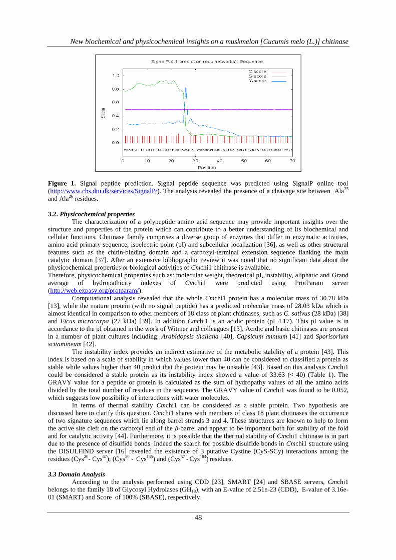
The analysis of Cmchi1 primary structure using SignalP 4.1 Server [14], showed that the 292 amino
acids full length protein contains a 25 amino acid long N-terminal signal peptide which is cleaved off to form
the mature polypeptide (267 amino acid residues). The data suggests the presence of a cleavage site between
Ala25
and Ala26
residues with a mean S score of 0.856 and a discrimination score of 0.857 (Figure 1).
New biochemical and physicochemical insights on a muskmelon [Cucumis melo (L.)] chitinase
48
Figure 1. Signal peptide prediction. Signal peptide sequence was predicted using SignalP online tool
(http://www.cbs.dtu.dk/services/SignalP/). The analysis revealed the presence of a cleavage site between Ala25
and Ala26
residues.
3.2. Physicochemical properties
The characterization of a polypeptide amino acid sequence may provide important insights over the
structure and properties of the protein which can contribute to a better understanding of its biochemical and
cellular functions. Chitinase family comprises a diverse group of enzymes that differ in enzymatic activities,
amino acid primary sequence, isoelectric point (pI) and subcellular localization [36], as well as other structural
features such as the chitin-binding domain and a carboxyl-terminal extension sequence flanking the main
catalytic domain [37]. After an extensive bibliographic review it was noted that no significant data about the
physicochemical properties or biological activities of Cmchi1 chitinase is available.
Therefore, physicochemical properties such as: molecular weight, theoretical pI, instability, aliphatic and Grand
average of hydropathicity indexes of Cmchi1 were predicted using ProtParam server
(http://web.expasy.org/protparam/).
Computational analysis revealed that the whole Cmchi1 protein has a molecular mass of 30.78 kDa
[13], while the mature protein (with no signal peptide) has a predicted molecular mass of 28.03 kDa which is
almost identical in comparison to other members of 18 class of plant chitinases, such as C. sativus (28 kDa) [38]
and Ficus microcarpa (27 kDa) [39]. In addition Cmchi1 is an acidic protein (pI 4.17). This pI value is in
accordance to the pI obtained in the work of Witmer and colleagues [13]. Acidic and basic chitinases are present
in a number of plant cultures including: Arabidopsis thaliana [40], Capsicum annuum [41] and Sporisorium
scitamineum [42].
The instability index provides an indirect estimative of the metabolic stability of a protein [43]. This
index is based on a scale of stability in which values lower than 40 can be considered to classified a protein as
stable while values higher than 40 predict that the protein may be unstable [43]. Based on this analysis Cmchi1
could be considered a stable protein as its instability index showed a value of 33.63 (< 40) (Table 1). The
GRAVY value for a peptide or protein is calculated as the sum of hydropathy values of all the amino acids
divided by the total number of residues in the sequence. The GRAVY value of Cmchi1 was found to be 0.052,
which suggests low possibility of interactions with water molecules.
In terms of thermal stability Cmchi1 can be considered as a stable protein. Two hypothesis are
discussed here to clarify this question. Cmchi1 shares with members of class 18 plant chitinases the occurrence
of two signature sequences which lie along barrel strands 3 and 4. These structures are known to help to form
the active site cleft on the carboxyl end of the β-barrel and appear to be important both for stability of the fold
and for catalytic activity [44]. Furthermore, it is possible that the thermal stability of Cmchi1 chitinase is in part
due to the presence of disulfide bonds. Indeed the search for possible disulfide bonds in Cmchi1 structure using
the DISULFIND server [16] revealed the existence of 3 putative Cystine (CyS-SCy) interactions among the
residues (Cys20
- Cys67
); (Cys50
- Cys
155) and (Cys
57 - Cys
184)
residues.
3.3 Domain Analysis
According to the analysis performed using CDD [23], SMART [24] and SBASE servers, Cmchi1
belongs to the family 18 of Glycosyl Hydrolases (GH18), with an E-value of 2.51e-23 (CDD), E-value of 3.16e-
01 (SMART) and Score of 100% (SBASE), respectively.
New biochemical and physicochemical insights on a muskmelon [Cucumis melo (L.)] chitinase
49
3.3. Secondary structure analysis
The predicted secondary structures of Cmchi1, using CFSSP, GOR 5, HNN, PCI-SS, SopMA and
YASPIN servers (Table 1), showed the predominance of random coils (34.83 – 57.68%) followed by helices
(19.10 – 31.08%) and strands (16.85 – 34.46%). Although these Servers use different algorithms and approaches
to predict secondary structure pattern from primary amino acid sequences, in all the cases the presence of
random coils as secondary structure elements was found to be dominant. Similarly, the class III GH18 family rice
chitinase (DIP3) when analyzed for possible secondary structure elements, i.e., helices, strands and random coils
showed the prevalence of random coils as the main secondary structure element [45], which also supports the
present findings. Random coils are known to be very important to protein folding, translocation and stability
[46].
Table 1: Analysis of secondary structures of Cmchi1 enzyme (Accession GenBank nº AAF64474.1) using
different servers
Cmchi1 Server
name
α-Helix Extended-strand Random coil
Nº of
residues
Percentage
(%)
Nº of
residues
Percentage
(%)
Nº of
residues
Percentage
(%)
CFSSP 82 30,71 92 34,46 93 34,83
GOR 5 51 19,10 62 23,22 154 57,68
HNN 81 30,34 45 16,85 141 52,81
PCI-SS 66 24,72 57 21,35 144 53,93
SopMa 68 25,47 78 29,21 121 45,32
YASPIN 83 31,08 62 23,22 122 45,7
3.4. Model construction, refinement and stereo-chemical evaluation
Computational modeling of protein has been considered more reliable when there is a clear
evolutionary relationship (homology) between a target sequence of the protein on analysis and protein structures
already stored and available in protein structure data banks. The first step in predicting a 3D model for Cmchi1
through comparative molecular modeling consisted in an extensive search for suitable models in the RSCB-PDB
Protein Data Bank. As a result the structures termed: 1HVQ, 1LLO and 2GSJ, all belonging to family GH18 of
plant chitinases, were selected according to parameters like query coverage, E-value, sequence identity and
structural resolution.
The Cmchi1 3D model was constructed using the software Modeller [26] and the quality of the
predicted structure was checked in the MolProbity server [30]. It was observed that the vast majority of ɸ –
dihedral angle pairs were distributed in the most favored and allowed regions of the Ramachandran’s plot
(Figure 2A). On the other hand, an additional optimization step was made using the WinCoot tool [27] in order
to rearrange the Phe32
, Ser251
and Asn256
amino acids in the allowed regions of the Ramachandran’s plot (Figure
2B). The overall structure comprises an 8 -strains and 10 α-helices, which together form a TIM-barrel protein
fold. The eight parallel -strains form the core of the enzyme (Figure 3).
The final model of Cmchi1 was subjected to energy minimization using GalaxyRefine server [28,29].
GalaxyRefine performs the refinement in the overall structure [44]. The method takes into account the general
pattern of the initial protein model and then it performs the refinement based on the backbone structure using a
high-accuracy global distance test (GDT-HA) [47]followed by a side-chain global distance test GDC-SC [48] and
finally, for physical correctness, the software makes use of MolProbity score [30].
The overall model quality (Z-score), overall quality factor and 3D-1D Averaged Score of the Cmchi1
model were measured using ERRAT, VERIFY_3D and ProSA-web, respectively. Using the Protein Structure
Assessment Server (ProSA-web) [33], it was obtained a value of - 9.69 (black dot – Figure 4A). Figure 4B
shows the quality of the model based on the global energy of its structure. According to this result Cmchi1
model can be classified as a good quality model and it is placed on a range of scores typically found for native
conformations of proteins with similar sizes (200 - 300 amino acid residues) in which the three dimensional
structures were solved by X-Ray diffraction [33].

New biochemical and physicochemical insights on a muskmelon [Cucumis melo (L.)] chitinase
50
(2A). Ramachandran’s plot of Cmchi1 protein.
95.1% (252/265) of all residues were in favored
(98%) regions.
98.9% (262/265) of all residues were in allowed
(>99.8%) regions.
There were 3 outliers (phi, psi):
32 PHE (87.4, 106.9)
251 SER (87.0, 121.3)
256 ASN (-54.4, 100,2)
(2B). Ramachandran’s plot of Cmchi1 protein after
being optimized by WinCoot software and energy-
minimized by GalaxyRefine.
95.5% (253/265) of all residues were in favored
(98%) regions.
100.0% (265/265) of all residues were in allowed
(>99.8%) regions.
There were no outliers.
Figure 2. Ramachandran’s plot (MolProbity) showing the dihedral angles ɸ and of amino acid residues from
Cmchi1. The residues located in the most favored regions (95.1%) are shown in light blue curves and the
residues located in the additional allowed regions (98.9%) are in dark blue curves.
Figure 3. Electrostatic potential map shows differences in the distribution of superficial charge along Cmchi1
structure (A). Specular image of Cmchi1: 180º�rotation around their longitudinal axis (B).
(8A) (8B)
(8C)
180º
New biochemical and physicochemical insights on a muskmelon [Cucumis melo (L.)] chitinase
51
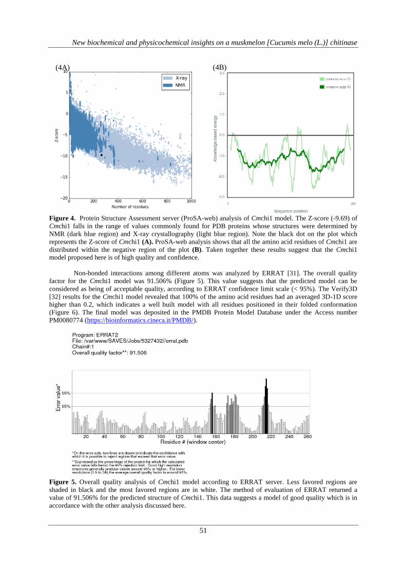
Figure 4. Protein Structure Assessment server (ProSA-web) analysis of Cmchi1 model. The Z-score (-9.69) of
Cmchi1 falls in the range of values commonly found for PDB proteins whose structures were determined by
NMR (dark blue region) and X-ray crystallography (light blue region). Note the black dot on the plot which
represents the Z-score of Cmchi1 (A). ProSA-web analysis shows that all the amino acid residues of Cmchi1 are
distributed within the negative region of the plot (B). Taken together these results suggest that the Cmchi1
model proposed here is of high quality and confidence.
Non-bonded interactions among different atoms was analyzed by ERRAT [31]. The overall quality
factor for the Cmchi1 model was 91.506% (Figure 5). This value suggests that the predicted model can be
considered as being of acceptable quality, according to ERRAT confidence limit scale (< 95%). The Verify3D
[32] results for the Cmchi1 model revealed that 100% of the amino acid residues had an averaged 3D-1D score
higher than 0.2, which indicates a well built model with all residues positioned in their folded conformation
(Figure 6). The final model was deposited in the PMDB Protein Model Database under the Access number
PM0080774 (https://bioinformatics.cineca.it/PMDB/).
Figure 5. Overall quality analysis of Cmchi1 model according to ERRAT server. Less favored regions are
shaded in black and the most favored regions are in white. The method of evaluation of ERRAT returned a
value of 91.506% for the predicted structure of Cmchi1. This data suggests a model of good quality which is in
accordance with the other analysis discussed here.
(4A) (4B)

New biochemical and physicochemical insights on a muskmelon [Cucumis melo (L.)] chitinase
52
Figure 6. Verify 3D plot of Cmchi1 model. Vertical axis represents the averaged 3D-1D profile score for
residues in a 25-residue sliding window. Horizontal axis represents the number of residues in the primary
sequence of the protein. The first and the last 9 residues of the primary sequence are not subjected to analysis.
According to the plot the structure of Cmchi1 shows no error in the distribution of amino acid residues along the
three dimensional space.
3.5. Docking study
A chitinase from muskmelon, Cmchi1, was initially identified in the work of Witmer et al., [13]. A
recombinant version of the enzyme was heterologously expressed in bacteria and purified. Besides to being a
genuine chitinase the recombinant protein was unable to properly hydrolyze glycol chitin, a potential substrate
for this type of enzyme. According to authors this fact (absence of activity) prevented a direct investigation of
the possible protective role of Cmchi1 against phytopathogenic fungi [13]. Based on these information we had
developed a docking study in order to elucidate the molecular properties that possibly justify the absence of
activity of Cmchi1 using the predicted and refined 3D model of the enzyme.
The first step in the docking study consisted in the search for the amino acid residues responsible for
the interaction between Cmchi1 and chitin. Besides the fact that Cmchi1 appears to be unable to properly
interact with chitin or chitin-like ligands [13] there are a sequence of conserved amino acid residues located in
the potential active site of Cmchi1 in which its presence appears to be essential to the catalysis mechanism,
according to CAZypedia database (https://www.cazypedia.org/index.php/Glycoside_Hydrolase_Family_18). In
classical GH18 family chitinases the active site of the enzyme is comprised by the motif, Asp-X-X-Asp-X-Asp-
X-Glu, where “X” stands for any of the other known amino acids residues. In the case of Cmchi1 the conserved
sequence is: Asn120
-X-X-ASP123
-X-ASP125
-X-GLU127
. Surprisingly Cmchi1 presents a mutation in the first
conserved amino acid of the active site, in which a Asn is located in a place where a Asp should be present in
the majority of commonly active chitinases. We suggest that this substitution may explain at least in part the
absence of activity of the enzyme. In addition, when the same region (active site motif) was analyzed in the
sequence of the chitinases used as template, PDB ID: 1HVQ, 1LLO and 2GSJ by means of a multiple sequence
alignment using the software CLC Sequence Viewer v. 7.7.1 (www.clcbio.com) we observed that Cmchi1 was
the only protein that do not conserved the first Asp. Indeed the Asn was present as discussed before (Figure 7).
In addition to the fact that Cmchi1 has a substitution mutation in the first amino acid residue of the
conserved active site motif of chitinases, docking studies revealed that the enzyme cannot perform an optimal
interaction with the substrate. A total of 10 simulations were performed in which the binding of (GlcNac)4 to the
active site of Cmchi1 was examined. The best value of interaction was E-value = – 6.2 kcal.mol-1. This result
demonstrated that the amino acids of the active site of Cmchi1 were prone to interact with (GlcNac)4 molecules
(Figure 8A). However it was observed a distance of ligation of around 12.1 Å from the Asp123
to the ligand
(GlcNac)4, what is completely incompatible with an ideal interaction between enzyme and substrate (Figure 8B).
Taken together these findings: 1) The mutation of Asp120
to Asn120
and 2) The distance of Asp123
to the
ligand may give us a molecular overview that partially explain the possible reason why Cmchi1 is a chitinase
that lacks chitinolytic activity and they are in complete agreement with the work of Witmer et al [13].

New biochemical and physicochemical insights on a muskmelon [Cucumis melo (L.)] chitinase
53
Figure 7. Multiple amino acid sequence alignment of four chitinases from family 18 (GH18) of plant Glycosyl
Hydrolases: 1HVQ, 1LLO, 2GSJ and Cmchi1 using the software CLC Sequence Viewer v. 7.7.1. The active site
motif of the enzymes is indicated by black triangles. The number of residues relative to the N-terminal segment
of the mature proteins is shown on the right side of each sequence.
New biochemical and physicochemical insights on a muskmelon [Cucumis melo (L.)] chitinase
54
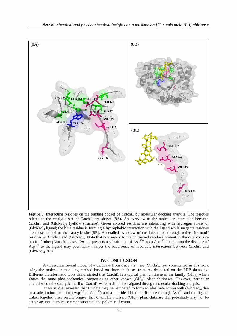
Figure 8. Interacting residues on the binding pocket of Cmchi1 by molecular docking analysis. The residues
related to the catalytic site of Cmchi1 are shown (8A). An overview of the molecular interaction between
Cmchi1 and (GlcNac)4 (yellow structure). Green colored residues are interacting with hydrogen atoms of
(GlcNac)4 ligand; the blue residue is forming a hydrophobic interaction with the ligand while magenta residues
are those related to the catalytic site (8B). A detailed overview of the interaction through active site motif
residues of Cmchi1 and (GlcNac)4. Note that conversely to the conserved residues present in the catalytic site
motif of other plant chitinases Cmchi1 presents a substitution of Asp120
to an Asn120
. In addition the distance of
Asp123
to the ligand may potentially hamper the occurrence of favorable interactions between Cmchi1 and
(GlcNac)4 (8C).
IV. CONCLUSION
A three-dimensional model of a chitinase from Cucumis melo, Cmchi1, was constructed in this work
using the molecular modeling method based on three chitinase structures deposited on the PDB databank.
Different bioinformatic tools demonstrated that Cmchi1 is a typical plant chitinase of the family (GH18) which
shares the same physicochemical properties as other known (GH18) plant chitinases. However, particular
alterations on the catalytic motif of Cmchi1 were in depth investigated through molecular docking analysis.
These studies revealed that Cmchi1 may be hampered to form an ideal interaction with (GlcNac)4 due
to a substitution mutation (Asp120
to Asn120
) and a non ideal binding distance through Asp123
and the ligand.
Taken together these results suggest that Cmchi1is a classic (GH18) plant chitinase that potentially may not be
active against its more common substrate, the polymer of chitin.
(8A) (8B)
(8C)
New biochemical and physicochemical insights on a muskmelon [Cucumis melo (L.)] chitinase
55
V. ACKNOWLEDGEMENTS
The authors would like to thank the educational institutions: Federal University of Ceará and Maurício
de Nassau Faculty which equally contributed to support this work.
REFERENCES [1] F. G. Malinovsky, J. U. Fangel, and W. G. T. Willats, “The role of the cell wall in plant immunity.,”
Front. Plant Sci., vol. 5, no. May, p. 178, 2014.
[2] S. Zeilinger, C. K. M. Tsui, S. Zeilinger, V. K. Gupta, T. E. S. Dahms, R. N. Silva, H. B. Singh, R. S.
Upadhyay, E. V. Gomes, C. K. Tsui, and C. N. S, “Friends or foes? Emerging insights from fungal
interactions with plants.,” FEMS Microbiol. Rev., vol. 40, no. 2, pp. 182–207, 2016.
[3] L. Jiang, J. Wu, S. Fan, W. Li, L. Dong, Q. Cheng, P. Xu, and S. Zhang, “Isolation and Characterization
of a Novel Pathogenesis-Related Protein Gene (GmPRP) with Induced Expression in Soybean (Glycine
max) during Infection with Phytophthora sojae.,” PLoS One, vol. 10, no. 6, p. e0129932, 2015.
[4] J. García-Cristobal, A. García-Villaraco, B. Ramos, J. Gutierrez-Mañero, and J. A. Lucas, “Priming of
pathogenesis related-proteins and enzymes related to oxidative stress by plant growth promoting
rhizobacteria on rice plants upon abiotic and biotic stress challenge.,” J. Plant Physiol., vol. 188, pp. 72–
79, 2015.
[5] G. Senthilraja, T. Anand, J. S. Kennedy, T. Raguchander, and R. Samiyappan, “Plant growth promoting
rhizobacteria (PGPR) and entomopathogenic fungus bioformulation enhance the expression of defense
enzymes and pathogenesis-related proteins in groundnut plants against leafminer insect and collar rot
pathogen.,” Physiol. Mol. Plant Pathol., vol. 82, pp. 10–19, 2013.
[6] B. Chinnasri, T. Borsics, D. A. Christopher, and B. S. Sipes, “Induction of pathogenesis-related gene 1
(PR-1) by acibenzolar-s-methyl application in pineapple and its effect on reniform nematodes
(Rotylenchulus reniformis).,” Agric. Nat. Resour., vol. 1, no. Accepted Manuscript, 2016.
[7] A. S. Rathore and R. D. Gupta, “Chitinases from Bacteria to Human: Properties, Applications, and Future
Perspectives,” Enzyme Res., vol. 2015, pp. 1–8, 2015.
[8] S. Sytwala, F. Günther, and M. F. Melzig, “Lysozyme- and chitinase activity in latex bearing plants of
genus Euphorbia - A contribution to plant defense mechanism,” Plant Physiol. Biochem., vol. 95, pp. 35–
40, 2015.
[9] A. Watanabe, V. HAI NONG, D. Zhang, M. ARAHIRA, N. ASARE YEBOAH, K. UDAKA, and C.
FUKAZAWA, “Molecular Cloning and Ethylene-Inducible Expression of Chi b1 Chitinase from
Soybean (Glycine max (L.) Merr.),” Biosci. Biotechnol. Biochem., vol. 63, no. 2, pp. 251–256, 1999.
[10] V. M. Gomes, A. E. A. Oliveira, and J. Xavier-Filho, “A chitinase and a β-1,3-glucanase isolated from
the seeds of cowpea (Vigna unguiculata L walp) inhibit the growth of fungi and insect pests of the seed.,”
J. Sci. Food Agric., vol. 72, no. 1, pp. 86–90, 1996.
[11] L. Zhe-fu, W. Danqi, L. Annuo, and Z. Weiqin, “Chitinases from Seeds of Zea mays and Coix lachryma-
jobi L. Purification and Some Properties,” Process Biochem., vol. 27, no. 2, pp. 83–88, 1992.
[12] M. Pawełkowicz, K. Zielinski, D. Zielinska, W. Plader, K. Yagi, M. Wojcieszek, E. Siedlecka, G.
Bartoszewski, A. Skarzynska, and Z. Przybecki, “Next generation sequencing and omics in cucumber
(Cucumis sativus L.) breeding directed research,” Plant Sci., vol. 242, pp. 77–88, 2015.
[13] X. Witmer, H. Nonogaki, E. P. Beers, K. J. Bradford, and G. E. Welbaum, “Characterization of chitinase
activity and gene expression in muskmelon seeds.,” Seed Sci. Res., vol. 13, no. 2, pp. 167–178, 2003.
[14] T. N. Petersen, S. Brunak, G. von Heijne, and H. Nielsen, “SignalP 4.0: discriminating signal peptides
from transmembrane regions.,” Nat. Methods, vol. 8, no. 10, pp. 785–6, 2011.
[15] E. Gasteiger, C. Hoogland, A. Gattiker, S. Duvaud, M. R. Wilkins, R. D. Appel, and A. Bairoch, “Protein
Identification and Analysis Tools on the ExPASy Server.,” in The Proteomics Protocols Handbook,
2005, pp. 571–607.
[16] A. Ceroni, A. Passerini, A. Vullo, and P. Frasconi, “Disulfind: A disulfide bonding state and cysteine
connectivity prediction server.,” Nucleic Acids Res., vol. 34, pp. 177–181, 2006.
[17] P. Y. Chou and G. D. Fasman, “Conformational parameters for amino acids in helical, β-sheet, and
random coil regions calculated from proteins.,” Amino Acids, vol. 13, no. 2, pp. 211–222, 1974.
[18] T. Z. Sen, R. L. Jernigan, J. Garnier, and A. Kloczkowski, “GOR V server for protein secondary structure
prediction.,” Bioinformatics, vol. 21, no. 11, pp. 2787–2788, 2005.
[19] Y. Guermeur, C. Geourjon, P. Gallinari, and G. Deléage, “Improved performance in protein secondary
structure prediction by inhomogeneous score combination.,” Bioinformatics, vol. 15, no. 5, pp. 413–421,
1999.
[20] J. R. Green, M. J. Korenberg, and M. O. Aboul-Magd, “PCI-SS: MISO dynamic nonlinear protein
secondary structure prediction.,” BMC Bioinformatics, vol. 10, no. 222, pp. 1–12, 2009.
[21] C. Geourjon and G. Deleage, “SOPMA: Significant improvements in protein secondary structure
New biochemical and physicochemical insights on a muskmelon [Cucumis melo (L.)] chitinase
56
prediction by prediction from multiple alignments.,” Comput. Appl. Biosci., vol. in press, no. 6, pp. 681–
684, 1995.
[22] K. Lin, V. a Simossis, W. R. Taylor, and J. Heringa, “A simple and fast secondary structure prediction
method using hidden neural networks.,” Bioinformatics, vol. 21, no. 2, pp. 152–159, 2005.
[23] A. Marchler-Bauer, M. K. Derbyshire, N. R. Gonzales, S. Lu, F. Chitsaz, L. Y. Geer, R. C. Geer, J. He,
M. Gwadz, D. I. Hurwitz, C. J. Lanczycki, F. Lu, G. H. Marchler, J. S. Song, N. Thanki, Z. Wang, R. A.
Yamashita, D. Zhang, C. Zheng, and S. H. Bryant, “CDD: NCBI’s conserved domain database.,” Nucleic
Acids Res., vol. 43, no. D1, pp. D222–D226, 2015.
[24] I. Letunic, T. Doerks, and P. Bork, “SMART: Recent updates, new developments and status in 2015.,”
Nucleic Acids Res., vol. 43, no. D1, pp. D257–D260, 2015.
[25] H. M. Berman, J. Westbrook, Z. Feng, G. Gilliland, T. N. Bhat, H. Weissig, I. N. Shindyalov, and P. E.
Bourne, “The Protein Data Bank.,” Nucleic Acids Res., vol. 28, no. 1, pp. 235–242, 2000.
[26] N. Eswar, B. Webb, M. A. Marti-Renom, M. S. Madhusudhan, D. Eramian, M.-Y. Shen, U. Pieper, and
A. Sali, Comparative protein structure modeling using Modeller., vol. Chapter 5. 2006.
[27] P. Emsley, B. Lohkamp, W. G. Scott, and K. Cowtan, “Features and development of Coot.,” Acta
Crystallogr. Sect. D Biol. Crystallogr., vol. 66, pp. 486–501, 2010.
[28] J. Ko, H. Park, L. Heo, and C. Seok, “GalaxyWEB server for protein structure prediction and
refinement,” Nucleic Acids Res., vol. 40, no. W1, pp. 294–297, 2012.
[29] W.-H. Shin, G. R. Lee, L. Heo, H. Lee, and C. Seok, “Prediction of Protein Structure and Interaction by
GALAXY Protein Modeling Programs,” Bio Des., vol. 2, pp. 01–11, 2014.
[30] V. B. Chen, W. B. Arendall, J. J. Headd, D. A. Keedy, R. M. Immormino, G. J. Kapral, L. W. Murray, J.
S. Richardson, and D. C. Richardson, “MolProbity: All-atom structure validation for macromolecular
crystallography,” Acta Crystallogr. Sect. D Biol. Crystallogr., vol. 66, no. 1, pp. 12–21, 2010.
[31] C. Colovos and T. O. Yeates, “Verification of protein structures: Patterns of nonbonded atomic
interactions.,” Protein Sci., vol. 2, no. 9, pp. 1511–1519, 1993.
[32] R. Lüthy, J. U. Bowie, and D. Eisenberg, “Assessment of protein models with three-dimensional
profiles.,” Nature, vol. 356, no. 6364, pp. 83–85, 1992.
[33] M. Wiederstein and M. J. Sippl, “ProSA-web: Interactive web service for the recognition of errors in
three-dimensional structures of proteins,” Nucleic Acids Res., vol. 35, no. SUPPL.2, pp. 407–410, 2007.
[34] J. Dundas, Z. Ouyang, J. Tseng, A. Binkowski, Y. Turpaz, and J. Liang, “CASTp: Computed atlas of
surface topography of proteins with structural and topographical mapping of functionally annotated
residues.,” Nucleic Acids Res., vol. 34, pp. 116–118, 2006.
[35] O. Trott and A. J. Olson, “Software news and update AutoDock Vina: Improving the speed and accuracy
of docking with a new scoring function, efficient optimization, and multithreading.,” J. Comput. Chem.,
vol. 31, no. 16, pp. 255–261, 2010.
[36] F. Meins, B. Fritig, H. J. M. Linthorst, J. D. Mikkelsen, J. M. Neuhaus, and J. Ryals, “Plant chitinase
genes,” Plant Mol. Biol. Report., vol. 12, no. 2, pp. 22–28, 1994.
[37] F. Hamel, R. Boivin, C. Tremblay, and G. Bellemare, “Structural and evolutionary relationships among
chitinases of flowering plants,” J. Mol. Evol., vol. 44, no. 6, pp. 614–624, 1997.
[38] J. P. Métraux, L. Streit, and T. Staub, “A pathogenesis-related protein in cucumber is a chitinase.,”
Physiol. Mol. Plant Pathol., vol. 33, no. 1, pp. 1–9, 1988.
[39] T. Taira, A. Ohdomari, N. Nakama, M. Shimoji, and M. Ishihara, “Characterization and antifungal
activity of gazyumaru (Ficus microcarpa) latex chitinases: both the chitin-binding and the antifungal
activities of class I chitinase are reinforced with increasing ionic strength.,” Biosci. Biotechnol. Biochem.,
vol. 69, no. 4, pp. 811–818, 2005.
[40] D. A. Samac, C. M. Hironaka, P. E. Yallaly, and D. M. Shah, “Isolation and characterization of the genes
encoding basic and acidic chitinase in Arabidopsis thaliana.,” Plant Physiol., vol. 93, no. 3, pp. 907–914,
1990.
[41] I. Garmendia, J. Aguirreolea, and N. Goicoechea, “Defence-related enzymes in pepper roots during
interactions with arbuscular mycorrhizal fungi and/or verticillium dahliae.,” BioControl, vol. 51, no. 3,
pp. 293–310, 2006.
[42] Y. Su, L. Xu, S. Wang, Z. Wang, Y. Yang, Y. Chen, and Y. Que, “Identification, phylogeny, and
transcript of chitinase family genes in sugarcane.,” Sci. Rep., vol. 5, p. 10708, 2015.
[43] K. Guruprasad, B. V. B. Reddy, and M. W. Pandit, “Correlation between stability of a protein and its
dipeptide composition: a novel approach for predicting in vivo stability of a protein from its primary
sequence,” "Protein Eng. Des. Sel., vol. 4, no. 2, pp. 155–161, 1990.
[44] A. Terwisscha van Scheltinga, M. Hennig, and B. Dijkstra, “The 1.8 A resolution structure of hevamine,
a plant chitinase/lysozyme, and analysis of the conserved sequence and structure motifs of glycosyl
hydrolase family 18.,” J. Mol. Biol., vol. 262, no. 2, pp. 243–57, 1996.

New biochemical and physicochemical insights on a muskmelon [Cucumis melo (L.)] chitinase
57
[45] X. L. Guo, L. R. Bai, C. Q. Su, L. R. Shi, and D. W. Wang, “Molecular cloning and expression of a class
III chitinase in upland rice.,” Genet. Mol. Res., vol. 3, no. 4, pp. 6860–6870, 2013.
[46] L. J. Smith, K. M. Fiebig, H. Schwalbe, and C. M. Dobson, “The concept of a random coil. Residual
structure in peptides and denatured proteins.,” Fold. Des., vol. 1, no. 5, pp. R95–R106, 1996.
[47] A. Zemla, “LGA: A method for finding 3D similarities in protein structures,” Nucleic Acids Res., vol. 31,
no. 13, pp. 3370–3374, 2003.
[48] D. A. Keedy, C. J. Williams, J. J. Headd, I. W. B. III Arendall, V. B. Chen, G. J. Kapral, R. A. Gillespie,
J. N. Block, A. Zemla, D. C. Richardson, and J. S. Richardson, “The other 90% of the protein:
Assessment beyond the Cαs for CASP8 template-based and high-accuracy models.,” Proteins, vol. 77,
no. 9, pp. 29–49, 2009.
![Physicochemical and Biochemical Reclamation of Soil ... · cultivation leads to reduced soil fertility and increased soil erosion in many areas of tropics [2]. Specific detri- mental](https://static.fdocuments.in/doc/165x107/5edc448cad6a402d6666ddeb/physicochemical-and-biochemical-reclamation-of-soil-cultivation-leads-to-reduced.jpg)