Neutron Scattering in Soft Matter Research: From Biology ...wpage.unina.it/lpaduano/PhD...
56
Neutron Scattering in Soft Matter Research: From Biology to Chemistry Henrich Frielinghaus Jülich Centre for Neutron Science Forschungszentrum Jülich GmbH Lichtenbergstrasse 1 85747 Garching (München) [email protected]
Transcript of Neutron Scattering in Soft Matter Research: From Biology ...wpage.unina.it/lpaduano/PhD...

Neutron Scattering in Soft Matter Research:From Biology to Chemistry
Henrich Frielinghaus
Jülich Centre for Neutron ScienceForschungszentrum Jülich GmbHLichtenbergstrasse 185747 Garching (München)

Locations…

– Instruments
NSE
KWS-1, -2
KWS-3
SPHERESDNS
(TREFF)NOSPEC
MARIATOPAS
Apply for beam-time: www.jcns.info !!!
ORNL: NSEILL : IN12-TAS

Scattering TheoryScattering ModelsConcepts
CytologyModel SystemsConcepts
Materials (Polymers…)Model SystemsConcepts

Scope of the Lectures
• Technical part (what is a neutron? what instruments?)
• Polymer part (what is a polymer? how does it scatter?)
• Soft matter part (what is soft matter? how does it scatter?)
• General part (more general questions)
Links to Biology, Chemistry and materials research.
Mathematics (sometimes rudimentary, sometimes more precise)Fourier transformation (should become plastic) Power laws (should be interpreted directly)

Neutron Sources:
(a)Reactors
235U absobs thermalneutron. Decayto medium heavy nuclei+ 2.5 fast neutrons.Moderation needed (1n).Surplus of 1.5 neutrons.
(b) Spallation Sources
High energy protons hita heavy nucleus (Hg…).Excitation leads to evaporation of 20-25nwith 1 to 100MeV.Decay of nucleus.
The spallation source is pulsed.Peak intensity very high (good for instruments: TOF).The medium flux compares well to reactors(not so well for SANS, NSE…)

New reactor
Old reactor

Reactors
0.8e151990 - 2000FRM-2 Munich
1.5e151970 - 1980HFR Grenoble
2.0e141960 - 1970FRJ-2 Jülich
~1.0e131950 - 1960FRM-1 Munich
Flux [cm-2s-1]PeriodReactor
Spallation Sources
SNS – Oak Ridge, USAJ-PARC – Tokai, JapanECNS – Sweden, Spain…???

Neutron History
1932: Chadwick discovered neutron (2He4 + 4Be96C12 + 0n1). No electric charge =
neutral particle. Standard theory says: 1 up + 2 down quarks (charge - 2 = 0).
1936: Hahn & Meitner observe fission. Mitchell & Powers conduct first neutronscattering experiment.
1942: Fermi built first nuclear reactor (Chicago pile = first controlled chain reaction)
1943: Oak Ridge Graphite Reactor: 3.5MW for production of fissionable material
1945: Shull´s first neutron diffractometer: antiferromagnetic structure of MnO2 was resolved (Nobel prize 1994)

Neutron History
1940s and 1950s : further nuclear reactors were built.
1954: Canadian reactor in Chalk River: Strongest neutron source (3e14 cm-2s-1). Brockhouse developed 3-axis spectrometer for: inelastic scattering of excitations in solid materials (Nobel prize 1994)
Important development: Cold Source. In Harwell, England the cold source ran on liquid hydrogen. (Cold, slower) Neutrons with long wavelengths were accessible.
1960s: High flux reactor in Brookhaven, USA came to operation. Since then thefluxes did not increase dramatically anymore.
1972: High flux reactor in Greoble, France at the Institut Laue Langevin, ILL came to operation. Flux 1.5e15 cm-2s-1 is the highest flux in the world.
Germany:
1955: Munich atom egg.
1960s: Jülich FRJ-2 reactor.
2004: High flux reactor in Munich, Germany at the Technical University of Munich came to operation. Germany´s best reactor.

Instrument Development in Germany
Backscattering Spectrometer ( )Neutron Small Angle Scattering (SANS) diffractometerInstruments for diffuse neutron scattering (DNS)High resolution time-of-flight spectrometers
Spallation Source History
1960s: Pioneer work at the Argonne National Laboratory (Chicago, USA).
Rutherford laboratory, UK: strongest spallation source with a proton beam of 200 kW.
Future: SNS – Oak Ridge, USAJ-PARC – Tokai, JapanECNS – Sweden, Spain…???

Neutron Particle Properties
mn = 1.675 10-27 kg
radioactive particle with =889.1 ± 1.8s
n p+ + e- +ν
practically stable, since v = ~1000m/s and length ~100m.
Spin ½ particle with µn = -1.913 µN (µN is the nuclear magneton)
The kinetic energy is non-relativistic (Newton, de Broglie…). Units are:
1 meV = 1.602 10-22J 9.044 Å (de Broglie wavelength)
437.4 m/s
(E = hν) 0.2418 1012 Hz (used less frequently)
(E = kBT) 11.60 K

nnnnn
Em
h
vm
h
2==λ
Neutron Particle Properties
De Broglie wavelengthwith mn mass
vn velocityEn energy = ½ mn vn
2
Hot: ~2000KThermal: ambient TCold: ~30K (liquid hydrogen / deuterium)

−
Tk
vvv
B
23
2
1exp~)(φ
Velocity distribution in a thermal source

Comparison neutron – photon (x-ray)
vn = (2En/mn)½ = 2187 m/sc = 3.00⋅108 m/sVelocity
λn= h /(2mnEn)½ = 1.81 Åλx= ch/E = 1.24 ÅWavelength
25 meV10 keVTypical energy
µn = –1.91304275(45) µN0Magnetic moment
½1Spin
00Charge
mn = 1.6749286(10) ⋅10–27 kg mphot = 0Mass
neutron= particle wave
X-rays= transversal wave
particle:
Both kinds of radiation used to study materials (soft matter, liquids, …)No charge, but strong interaction of photon with electrons.Magnetic moment allows neutrons to study magnetic structures.Energy given here: λ 1Å. (for SANS λ 6 to 15Å)For this wavelength, the large neutron mass leads to small energies. Relative changes of this energy are easily detectable. These energy changes are suitable for soft-matter research.For x-rays one needs larger efforts to detect changes of the energy.

Typical Scattering Experiment:
source
monochromator
slits
preparation
sample
Θ
analyzer
slits
analysis
detector
fk
fE
vmh
ph
ki
ππ 22 ==
2
2
1mvEi =

Typical Scattering Experiment:
source
monochromator
slits
preparation
sample
Θ
analyzer
slits
analysis
detector
fk
fE
vmh
ph
ki
ππ 22 ==
2
2
1mvEi = In
tens
ityif kkQ
−=
if EEE −=∆

Triple Axis Spectrometer:

Bragg´s Scattering Law:
d
Θ
Θ= sin2dnλ
Θ==
sin
2
d
nk
πλπ
For preparing/analyzing the beam:
``Selective Mirror´´
retardation

Bragg´s Scattering Law:
d
Θ
ik
As a sample:
fk
Q
Θ= sin2dnλ
Θ==
sin
2
d
nk
πλπ
d
nkQ
kkQ if
π2sin2 =Θ=
−=
reciprocal space !!

Bragg´s Scattering Law: (different planes)
d All possible Qof constructive interferenceform also a lattice.
(reciprocal lattice)

The priciples of neutron scattering (Born Approximation…)
neutronsincomingofFlux
inneutronsscatteredofFlux Ω=Ωd
dσΩ
⋅=ΩΣ
d
d
Vd
d σ1
Only 10% of neutrons are scattered (coherently). single scattering event
The nuclei appear as pointlike particles (nuclear physics Fermi pseudo potential)
The priciples of neutron scattering (Born Approximation…)
)exp( jj
j rQibA
=
2A
d
d =Ωσ
= )exp()(3 rQirrdAρ
pointlike particles
continuous density
intensity
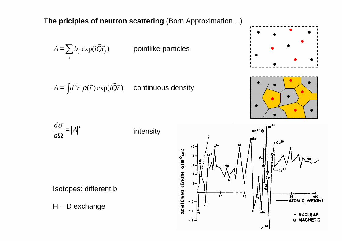
Isotopes: different b
H – D exchange

The priciples of neutron scattering (Born Approximation…)
randomly distributed nuclear spins
n
2)( bb −
b new coherent scattering length
incoherent cross section
structure of a pointlike particle

)()(
)exp()(3
QsQf
rQirrdA
⋅=
= ρ
Problems of x-ray scattering
formfactor of single atoms f(Q)
structure factor of idealpointlike particles s(Q)
At larger angles intensity reduced.
What model of electron distribution ???

Comparison: neutron and x-ray scattering
Chemical modificationsNon-destructive
Thin samples (0.1 – 0.5mm)Penetration: thick samples 1-5mm, and even more (10cm)
(huge effort)Energy transfers comparable to kineticenergy.
(magnetic dichroism)Spin density directly detectable (µn)
Scattering length ~ Z
Resonances prepare certain nuclei
Heavier atoms scatter more ( res.)
Non-systematic scattering lengthIsotope labelling
Each atom can be highlighted
Formfactor of atomHydrogen electron cloud distorted
Pointlike particles (easy modelling)Hydrogen-nucleus easily detectable
X-raysNeutrons

Scales of Plots:
0
1
2
3
4
5
6
7
8
9
10
Linear Scale
length 2
length 2
length 2
length 2
1
10
Logarithmic Scale
factor 2
factor 2
factor 2
factor 28
4
2
positive values only,huge numbers can bedisplayed !!!

1 10 10010-10
10-9
10-8
10-7
10-6
1x10⁻⁵
1x10⁻⁴
10-3
10-2
10-1
100
-4
-2
-1
x-4
x-2
x-1
Scales of Plots: (power laws)

Small Angle Neutron Scattering (SANS)

Small Angle Neutron Scattering (SANS)

Small Angle Neutron Scattering (SANS)
δ
0k
1k Q
δ
λπ /210 == kk
4sin( /2)Q
π δλ
=
0 5 10 15 20 2510-7
10-6
10-5
10-4
10-3
10-2
10-1
100
form factor of sphere with radius R
P(Q
R)
Q R
QQ now
Remark on vectors:
elastic

Q-range of Small Angle Scattering Techniques
2 – 15 Å
~ 6000 Å
Wave Length
10Å – 20µm
0.5 – 20µm
Length Scale
Neutron
Light
Probe
According to D = 2 /Q one can explore particles of size:
10-3 Å-1 < Q < 2·10-5 Å-1Double Crystal Diffractometer
5·10-3 Å-1 < Q < 10-4 Å-1Focusing SANS
0.2Å-1 < Q < 10-3 Å-1Pin - Hole SANS
Three different SANS instrumentsallow to measure a particle size in a range of four orders of magnitude:

∆k/k
∆Ω
∆Ω D
, ∆λ/λ
, ∆λ/λ
∆ΩD
∆Ω,
F
Sample
Θ
Qk1
k0
Θ
2( / )4( )2
Tk kU
T
k kL e
k kπ−Φ ∆=
0 UI L F∆ = ⋅ ⋅ ∆ΩIntensity at sample:
Luminosity of the source:
0( ; ) ( ; )D D
dI Q I D T Q
dδ δΣ∆ = ∆ ⋅ ⋅ ⋅ ⋅ ∆Ω
ΩIntensity at detector:
22
2 T Bk k Tm
=
Intensitiesof a SANS exp.and resolution

Resolution:2
2 2 2 2 2 2 21 1[ ( ) ( ) ( ) ( ) ]
12D E
SD C C D
k d dQ d
L L L L
δλδ δλ
< > = + + + +< >6
Θ2
Optimization: D CL L= SDE d2dd ==
. ( / 3) /opt E CQ k d Lδ = ⋅
and
leads to:
Resolutionof a SANS exp.and intensity
LD as large as possible!One limit: C4γ=∆Ω
.2 4 20 2
( )optU D U D
D
QFI L L L L
L k
δ∆ = ∆Ω ∝
2( / )E Cd L

1 10105
106
107
KWS1: Flux on Sample
λ=7; ∆λ/λ=0.2
Entrance Aperture 3*3 cm2
Reactor Power 20MW
L1.68±0.04
Inte
nsity
[n/
cm2 s]
Collimation [m]
-20 -15 -10 -5 0 5 10 15 20
10-6
10-5
10-4
10-3
10-2
10-1
100
2Γ1/22Γ1/2
No
rmal
ized
Int
ensi
tyPosition [cm]
Primary beam:
Characteristics of SANS
Intensity at sample position:

2 2 2 20 2
(4 ) ( )U D U C DD
F QI L L L L
L k
δγ∆ = ∆Ω ∝
Primary beam:
Focusing SANS

Layout of focusing-mirror SANS
toroidal mirror:•size:20x120 cm2
• 1000Å thick layer of Cu65

Example from focusing-mirror SANS
10-4 10-3102
103
104
105
106
107
108
U-SANS Lightscattering
R=(7140±14)Å
(dΣ/
dΩ)[
cm-1]
Q [Å-1]
Latex Spheres: USANS and light scattering

>−−
≤=
− 1y)y1(1
1y1)y(R
5.02
-3 -2 -1 0 1 2 30.0
0.2
0.4
0.6
0.8
1.0
3x1xR
efle
ktiv
ität
y
-5 -4 -3 -2 -1 0 1 2 3 4 50.0
0.5
1.0
1.5
2.0
t-t mode
s-s mode
Si111; λ=4.48Å
Inte
nsity
δ[∆δ]
2
4 sin 2
WC
B
b e F N λδ
π δ
−
∆ =
exp(-W) Debye-Waller factorF geometrical structure factor
Darwin curve
Double crystal diffractometer (DCD)

Layout of the old “Jülich” instrument
“Agamalian” cut

Resolution of DCD
-90 -60 -30 0 30 60 9010-5
10-4
10-3
10-2
10-1
100
2∆δ(t-t)
∆δ(s-s)=30.5µrad∆δ(t-t)=20.5µrad
t-t mode
Theoretical Resolution: Si111
; λ=4.48Å
s-s mode
Inte
nsity
I(δ)
/I(0)
Scattering Angle δ[µrad]
-90 -60 -30 0 30 60 9010-5
10-4
10-3
10-2
10-1
100
2 ∆δ(t-t)
Experim. Resolution: Si111
; λ=4.48Å
s-s mode
t-t mode
∆δ(s-s)=54.2µrad ∆δ(t-t)=25.8µrad
Inte
sity
I(δ)
/I(0)
Scattering Angle δ [µrad]

0 10 20 30 40 505.5
6.0
6.5
7.0
7.5
8.0
ca.4% Latex in Water
Rg=(0.72±0.03)µm
D=(1.87±0.08)µm
Rg=(0.314±0.004)µm
D=(0.82±0.01)µm
Lnd
Σ* /dΩ
[cm
-1]
Q2 [10-8Å-2]-100 0 100 200 300 400 500
10-6
10-5
10-4
10-3
10-2
10-1
100
2∆δ
4% Latex Dispersion in Water
empty beam latex dispersion
(d=0.796µm)
Nor
mal
ized
Inte
nsity
Scattering Angle δ [µrad]
[ ]D mµ 0.82±0.01
1 3 31 0V c m− 2.89±0.04
21 0− Φ 3.4±0.09
Model System: Latex spheres in solution
Given parameters: D=0.796µm; Φ=3.98 10-2
( ) ( )0 H
I QdQ
d I T A D δ
∗ ∆Σ =Ω ⋅ ⋅ ⋅ ⋅ ∆

Lamella Al-Al 2Cu alloy
Lamella structure shownby optical micrograph
Bragg-Reflectionλ=1.8ÅQm=(45±2.7) 10-5 ÅD=(1.4 ± 0.09)µm

Crystallization of syndio-Polypropylene in d-22 solution -large scale view
(Scale bar 2 µm)
Spherulitic morphologyDKD, Focusing SANS, Pin - Hole SANS
10-5 10-4 10-3 10-2 10-110-3
10-1
101
103
105
107
109
Q-4Q-4
dΣ/d
Ω [c
m-1]
Q [Å-1]
Q-2
0.5% s-PP in d-22room temp.
Rg=3.3µm

Large-scale aggregates formed by the sPP-P(E-co-P) diblocks
Scale bar 2 µm
Multi-level structure:
• small scale: 2-d morphology
• intermediate scale: 1-d morphology
• large scale: mass-fractal features
Columnar morphology
MW=38.4k/106.6k
10-5 10-4 10-3 10-2 10-110-2
100
102
104
106
108
Q-1
Q-3
dΣ/d
Ω [c
m-1]
Q [Å-1]
0.5% sPP-P(E-co-P) in d22room temp.
R=2.75µm

Important design elements of SANS
- Monochromator- Position Sensitive Detector- Neutron Lenses

Velocity selector
0 5 10 15500
1000
1500
2000
2500
3000
λ=7.94Å
f=16000 rpm<λ>=8.01Å
∆λ/λ=0.2
Inte
nsitä
t [a
.u.]
λ [Å]
0.00 0.50 1.00 1.50 2.00 2.500.0
0.5
1.0
1.5
2.0
2.5
3.0
Dornier Selector
λ = 2127 / fTheor
λf=2011+0.456f[f]=1/s
theory
λ*f [
103 Å
/s]
f [104min-1]

Example of sample with oriented anisotropic “particles”:
Isoprene rubber stretched
Position sensitive detector

Gas detector
3 3 1 0.77He n H H MeV+ → + +2D-lattice of wires:
Resolution ~1cmTime per count ~10s

Scintillation detector
6 4 3 4.79Li n He H MeV+ → + +
Ce activated 6Li glass
One neutron gives about 4000 photons of 0.4 m wave length

Lenses for SANS
Focal length=(L1+L2)/4

• Refractive index: n2 = 1 - ξξξξ (n is less than 1, i.e. vacuum is the “optically” more dense region → concave lens is focusing!)
• Parameter: ξξξξ = λλλλ2ρρρρ/ππππ = 1.64⋅ 10-6 λ2 [Å] = 8.05 ⋅ 10-5cm for λ= 7Å neutrons of lenses consisting ofMgF2 with scattering length density: ρ = 5.16 ⋅ 1010 cm-2
• Focal length: f = r / ξξξξ = 248.5m for 7Å neutrons
• Several lenses: fN = f / N = 9.94m for N=25 lenses at 7Å(fine tuning by wave length!)
Typical parameters for “SANS” lenses
-30 -20 -10 0 10 20 30-30
-20
-10
0
10
20
30
s [m
m]
w [mm]
R=20mm
ΦΦΦΦ=52mm

Sample measurement with 7 lenses
4 µm

10-5 10-4 10-3 10-2 10-110-1
100
101
102
103
104
105
106
107
108
dΣ/d
Ω [c
m-1]
Scattering vector Q [Å-1]
Sample measurement with lenses
Jülich measurements:— KWS-2— KWS-3 (focussed)— DKD
JAEA measurements:DKDwith 7 MgF2-lenses(19Å, 10m, small detector)
with 70 MgF2-lenses(6.5Å, 10m, small detector)
with 70 MgF2-lenses(6.5Å, 10m, large detector)
conventional 2m SANS(incoherentbgrnotsubtracted)

Neutron Spin Echo (NSE) Spectrometer
• Small energies detectable (soft matter)• Signal in time domain

Summary of technical part:
if kkQ
−=
if EEE −=∆2
Ad
d =Ωσ
= )exp()(3 rQirrdAρ
10-3 Å-1 < Q < 2·10-5 Å-1Double Crystal Diffractometer
5·10-3 Å-1 < Q < 10-4 Å-1Focusing SANS
0.2Å-1 < Q < 10-3 Å-1Pin - Hole SANS
Length scale given by: D = 2 /Q