Neurotoxicology and Teratology - GeneNetwork and Teratology 33 (2011) 415–421 ⁎ Corresponding...
7
Genetic-based, differential susceptibility to paraquat neurotoxicity in mice Lina Yin a , Lu Lu b,c , Kavita Prasad d , Eric K. Richfield d , Erica L. Unger e , Jialin Xu f , Byron C. Jones a,g, ⁎ a Intercollege Graduate Degree Program in Neuroscience, The Pennsylvania State University, University Park, PA 16802, USA b Department of Anatomy and Neurobiology, University of Tennessee College of Medicine, Memphis, TN 38163, USA c Jiangsu Key Laboratory of Neuroregeneration, Nantong University, Nantong 226001, People's Republic of China d Environmental & Occupational Health Sciences Institute, Robert Wood Johnson Medical School, Rutgers, The State University of New Jersey, Piscataway, NJ 08854, USA e Department of Nutritional Sciences, The Pennsylvania State University, University Park, PA 16802, USA f Department of Statistics, The Pennsylvania State University, University Park, PA 16802, USA g Department of Biobehavioral Health, The Pennsylvania State University, University Park, PA 16802, USA abstract article info Article history: Received 23 November 2010 Received in revised form 21 February 2011 Accepted 23 February 2011 Available online 28 February 2011 Keywords: Paraquat Inbred mice Substantia nigra Gene expression Parkinson's disease Paraquat (PQ) is an herbicide used extensively in agriculture. This agent is also suspected to be a risk factor for Parkinson's disease (PD) by harming nigro-striatal dopamine neurons. There is likely, genetic-based, individual variability in susceptibility to PQ neurotoxicity related PD. In this study, we measured the delivery of PQ to the brain after three weekly injections of PQ at 5 mg kg -1 , PQ-related neural toxicity after three weekly injections of PQ at 1 mg kg -1 or 5 mg kg -1 , PQ-related iron accumulation and PQ-related gene expression in midbrain of DBA/2J (D2) and C57BL/6J (B6) inbred mouse strains after a single injection of PQ at 15 mg kg -1 and 10 mg kg -1 , respectively. Results showed that compared to controls, PQ-treated B6 mice lost greater numbers of dopaminergic neurons in the substantia nigra pars compacta than D2 mice; however, distribution of PQ to the midbrain was equal between the strains. PQ also significantly increased iron concentration in the midbrain of B6 but not D2 mice. Microarray analysis of the ventral midbrain showed greater PQ-induced changes in gene expression in B6 compared to D2 mice. This is the first study to report genetically-based differences in susceptibility to PQ neurotoxicity and to understanding individual differences in vulnerability to PQ neurotoxicity and its relation to PD in humans. © 2011 Elsevier Inc. All rights reserved. 1. Introduction Paraquat (PQ) is an herbicide used world-wide (Bromilow, 2004). Besides its target effect of toxicity to plants in agriculture, PQ is toxic to animals and can cause damage to lungs and brain, among other tissues. Notably, PQ has been reported to increase the risk for sporadic Parkinson's disease (sPD) via its toxicity to dopamine neurons in the substantia nigra. sPD is believed to be the result of environmental risk factors acting on genetically susceptible individuals during aging (Barbeau et al., 1985; Gwinn-Hardy, 2002). Current epidemiologic data show that PQ-associated risk for sPD is an estimated 4–7 times greater than for other environmental factors such as heavy metal exposure or rural residency (Hertzman et al., 1990; Liou et al., 1997; Landrigan et al., 2005). In the laboratory, mice treated with PQ developed neurochemical and behavioral signs consistent with Parkinsonism, including specific dopaminergic (DA) neuron loss and α-synuclein containing protein aggregation in the substantia nigra pars compacta (SNpc) (Manning-Bog et al., 2002; McCormack et al., 2002; Prasad et al., 2007, 2009), specific dopamine depletion in striatum (Shimizu et al., 2003), and decrease of ambulatory activity (Brooks et al., 1999; Fernagut et al., 2007). PQ-induced cell death may be the result of oxidative stress (McCormack et al., 2005; Prasad et al., 2007), and possible intracellular signal pathways involved c-Jun N-terminal protein kinases (JNK) and caspase-3 (Peng et al., 2004). Thus, PQ is considered to be a strong risk factor for sPD, even though the exact biochemical mechanism of PQ neurotoxicity is unknown. Different genetic backgrounds very likely predispose individuals to resistance or to susceptibility to environmental toxicants, such as PQ, that are implicated in sPD. In humans, not every individual exposed to PQ develops sPD. In mice, Smeyne and colleagues have reported strain-dependent susceptibility to another Parkinson's disease related neurotoxin 1-methyl-4-phenyl-1,2,3,6-tetrahydro-pyridine (MPTP), C57BL/6J mice more vulnerable to Swiss–Webster (SW) mice (Hamre et al., 1999). Later they identified a single QTL, Mptp1, for susceptibility to MPTP neurotoxicity in mice (Cook et al., 2003). Based on these data, certain genetic backgrounds likely contribute to differential susceptibility to PQ neurotoxicity. Identifying the specific genes involved may ultimately reveal the underlying molecular mechanisms for PQ neurotoxicity and likely other environmental Neurotoxicology and Teratology 33 (2011) 415–421 ⁎ Corresponding author at: 315 E., Health and Human Development Building, The Pennsylvania State University, University Park, PA 16802, USA. Tel.: +1 814 863 0167; fax: +1 814 863 7525. E-mail address: [email protected] (B.C. Jones). 0892-0362/$ – see front matter © 2011 Elsevier Inc. All rights reserved. doi:10.1016/j.ntt.2011.02.012 Contents lists available at ScienceDirect Neurotoxicology and Teratology journal homepage: www.elsevier.com/locate/neutera
Transcript of Neurotoxicology and Teratology - GeneNetwork and Teratology 33 (2011) 415–421 ⁎ Corresponding...
Neurotoxicology and Teratology 33 (2011) 415–421
Contents lists available at ScienceDirect
Neurotoxicology and Teratology
j ourna l homepage: www.e lsev ie r.com/ locate /neutera
Genetic-based, differential susceptibility to paraquat neurotoxicity in mice
Lina Yin a, Lu Lu b,c, Kavita Prasad d, Eric K. Richfield d, Erica L. Unger e, Jialin Xu f, Byron C. Jones a,g,⁎a Intercollege Graduate Degree Program in Neuroscience, The Pennsylvania State University, University Park, PA 16802, USAb Department of Anatomy and Neurobiology, University of Tennessee College of Medicine, Memphis, TN 38163, USAc Jiangsu Key Laboratory of Neuroregeneration, Nantong University, Nantong 226001, People's Republic of Chinad Environmental & Occupational Health Sciences Institute, Robert Wood Johnson Medical School, Rutgers, The State University of New Jersey, Piscataway, NJ 08854, USAe Department of Nutritional Sciences, The Pennsylvania State University, University Park, PA 16802, USAf Department of Statistics, The Pennsylvania State University, University Park, PA 16802, USAg Department of Biobehavioral Health, The Pennsylvania State University, University Park, PA 16802, USA
⁎ Corresponding author at: 315 E., Health and HumaPennsylvania State University, University Park, PA 1680fax: +1 814 863 7525.
E-mail address: [email protected] (B.C. Jones).
0892-0362/$ – see front matter © 2011 Elsevier Inc. Aldoi:10.1016/j.ntt.2011.02.012
a b s t r a c t
a r t i c l e i n f oArticle history:Received 23 November 2010Received in revised form 21 February 2011Accepted 23 February 2011Available online 28 February 2011
Keywords:ParaquatInbred miceSubstantia nigraGene expressionParkinson's disease
Paraquat (PQ) is an herbicide used extensively in agriculture. This agent is also suspected to be a risk factor forParkinson's disease (PD) by harming nigro-striatal dopamine neurons. There is likely, genetic-based,individual variability in susceptibility to PQ neurotoxicity related PD. In this study, we measured the deliveryof PQ to the brain after three weekly injections of PQ at 5 mg kg−1, PQ-related neural toxicity after threeweekly injections of PQ at 1 mg kg−1or 5 mg kg−1, PQ-related iron accumulation and PQ-related geneexpression in midbrain of DBA/2J (D2) and C57BL/6J (B6) inbred mouse strains after a single injection of PQ at15 mg kg−1 and 10 mg kg−1, respectively. Results showed that compared to controls, PQ-treated B6 mice lostgreater numbers of dopaminergic neurons in the substantia nigra pars compacta than D2 mice; however,distribution of PQ to the midbrain was equal between the strains. PQ also significantly increased ironconcentration in the midbrain of B6 but not D2 mice. Microarray analysis of the ventral midbrain showedgreater PQ-induced changes in gene expression in B6 compared to D2 mice. This is the first study to reportgenetically-based differences in susceptibility to PQ neurotoxicity and to understanding individual differencesin vulnerability to PQ neurotoxicity and its relation to PD in humans.
n Development Building, The2, USA. Tel.: +1 814 863 0167;
l rights reserved.
© 2011 Elsevier Inc. All rights reserved.
1. Introduction
Paraquat (PQ) is an herbicide used world-wide (Bromilow, 2004).Besides its target effect of toxicity to plants in agriculture, PQ is toxicto animals and can cause damage to lungs and brain, among othertissues. Notably, PQ has been reported to increase the risk for sporadicParkinson's disease (sPD) via its toxicity to dopamine neurons in thesubstantia nigra. sPD is believed to be the result of environmental riskfactors acting on genetically susceptible individuals during aging(Barbeau et al., 1985; Gwinn-Hardy, 2002). Current epidemiologicdata show that PQ-associated risk for sPD is an estimated 4–7 timesgreater than for other environmental factors such as heavy metalexposure or rural residency (Hertzman et al., 1990; Liou et al., 1997;Landrigan et al., 2005). In the laboratory, mice treated with PQdeveloped neurochemical and behavioral signs consistent withParkinsonism, including specific dopaminergic (DA) neuron loss andα-synuclein containing protein aggregation in the substantia nigra
pars compacta (SNpc) (Manning-Bog et al., 2002; McCormack et al.,2002; Prasad et al., 2007, 2009), specific dopamine depletion instriatum (Shimizu et al., 2003), and decrease of ambulatory activity(Brooks et al., 1999; Fernagut et al., 2007). PQ-induced cell death maybe the result of oxidative stress (McCormack et al., 2005; Prasad et al.,2007), and possible intracellular signal pathways involved c-JunN-terminal protein kinases (JNK) and caspase-3 (Peng et al., 2004).Thus, PQ is considered to be a strong risk factor for sPD, even thoughthe exact biochemical mechanism of PQ neurotoxicity is unknown.
Different genetic backgrounds very likely predispose individuals toresistance or to susceptibility to environmental toxicants, such as PQ,that are implicated in sPD. In humans, not every individual exposed toPQ develops sPD. In mice, Smeyne and colleagues have reportedstrain-dependent susceptibility to another Parkinson's disease relatedneurotoxin 1-methyl-4-phenyl-1,2,3,6-tetrahydro-pyridine (MPTP),C57BL/6J mice more vulnerable to Swiss–Webster (SW) mice (Hamreet al., 1999). Later they identified a single QTL, Mptp1, forsusceptibility to MPTP neurotoxicity in mice (Cook et al., 2003).Based on these data, certain genetic backgrounds likely contribute todifferential susceptibility to PQ neurotoxicity. Identifying the specificgenes involved may ultimately reveal the underlying molecularmechanisms for PQ neurotoxicity and likely other environmental
416 L. Yin et al. / Neurotoxicology and Teratology 33 (2011) 415–421
risk factors for sPD. It is also likely that PQ-related sPD is a complextrait, influenced by multiple genes acting in response to environmen-tal conditions.
Because it is likely that sPD is a complex trait, modeling theetiology of the disease in animals is best accomplished using a geneticreference population. Inbred strains of rodents can be helpful. Onesuch group of genetically-defined mice is the panel of BXDrecombinant inbred (RI) mice. This panel has proven to be animportant resource for dissecting the molecular architecture ofcomplex traits and the genetic study of gene-environment interaction(Churchill et al., 2004). BXD RI mice were generated by crossingC57BL/6J (B6) and DBA/2J (D2) inbred strains. Currently there are 80BXD RI strains all of which have been genotyped at more than 13,000unique markers. This will enable fine mapping of chromosome andmore precise identification of chromosome loci that contain genesunderlying differential strain susceptibility to this environmentaltoxicant (Darvasi and Soller, 1997; Peirce et al., 2004). Geneticreference populations are useful in identifying genetic variantsassociated with phenotypes, genetic correlations among phenotypesand if available, gene expression data that can be used in describingmechanisms. For example, GeneNetwork (http://www.genenetwork.org) is the online database containing the sequencing information fora number of genetic reference populations, including the BXD RIstrains. It also enables quantitative trait loci (QTL) mapping by usingstrain means of a certain trait/phenotype as the index for associationanalysis to locate the chromosome regions of which the genotypevariations cause the phenotype variation among strains. In this report,we show that between the parental strains of BXD RI mice B6mice aremore sensitive to PQ neurotoxicity than D2 mice, which establishedthe basis for using the larger panel of BXD RI strains of mice tomeasure the phenotype variation in terms of different susceptibility toPQ neurotoxicity, and to pinpoint the underlying genes. This may shedlight to the genetics of sPD.
2. Materials and methods
2.1. Animals
Two inbred strains of mice, C57BL/6J and DBA/2J, obtained fromour vivarium and from the University of Tennessee Health SciencesCenter, were used in this study. The mice were maintained under aconstant light–dark cycle (06:00–18:00, on–off rotation), controlledtemperature (21±2 °C) and humidity (40%). Food (standard Purinarodent diet# 5010) and water were provided ad libitum. Allexperimental protocols were approved by the Penn State InstitutionalAnimal Care and Use Committee and were conducted in accordancewith the National Institutes of Health Guidelines for the Care and Useof Laboratory Animals.
2.2. Experiments
Male and female mice between the ages of 2.5–4 months wereused for three experiments: measurement of brain PQ concentration(experiment I), quantification of dopaminergic neurons in the SNpc(experiment II) and identification of ventral midbrain transcriptome(experiment III). Mice aged 120±10 days were used for measure-
Table 1PQ dosing regimen and sample size.
Exp. Dose mg kg−1 Sample size Mice age Dosing
I 5 5 M and 5 F 2.5–4 months Once wII 1, 5 4–7 M and 4–7 F for each dose 2.5–4 months Once wIII 10 2–4 M 2.5–4 months Single inIV 15 2–4 M and 3–9 F 120±10 days Single in
Abbreviation: VMB: ventral midbrain, CB: cerebellum, DA: dopaminergic neurons, SNpc: su
ment of ventral midbrain iron concentration (experiment IV), asshown in Table 1. To avoid litter specific effects, for every experiment,mice within a litter were assigned to different treatment groups, i.e.,different doses of PQ or saline control.
2.3. PQ treatments
PQ dichloride trihydrate (Sigma Chemicals, St. Louis, MO) solutionwas made fresh in saline and administered by intraperitoneal (i.p.)injection. In experiment I (Table 1), PQ (5 mg kg−1) was injected onceweekly for 3 weeks and brain tissue (ventral midbrain (VMB) andcerebellum(CB)) harvested 24 h after the last injection. 5 male and 5female mice of each strain were used. This is the highest dose used inour toxicology study and required to show equal distribution of PQ tothe VMB. In experiment II, one of two doses of PQ (1 mg kg−1 or5 mg kg−1) vs. saline was injected once weekly for 3 weeks and braintissue was harvested 24 h after the last injection. These doses anddosing regimen were based on the work of McCormack et al. (2002).Sample sizes of 4–7 male and 4–7 female mice of each strain wereused for every treatment. In experiment III, PQ (10 mg kg−1) or salinewas injected once and brain tissue (ventral midbrain) was harvestedat three different time points (24 h, 48 h, and 7 days) after theinjection. The choice of this dose was to maximize possible effects ongene expression and minimize possible acute toxicity. We adminis-tered one dose to capture the acute kinetics of gene expression.Samples consisting of 2–4 male mice of each strain were used forevery time point. In experiment IV, PQ (15 mg kg−1) or saline wasinjected once and the brain tissue (ventral midbrain and caudateputamen) was harvested 24 h later from 2–4 male and 3–9 femalemice of each strain. In this case, the animals were sacrificed within24 h, because we wanted to maximize possible effects on acute ironaccumulation. The mice were killed by CO2 in experiments I and II, orcervical dislocation in experiments III and IV. Brain samples werestored at −80 °C until analysis.
2.4. Brain PQ determinations
Brain PQ was quantified by HPLC MS as described previously(Winnik et al., 2009). Briefly, tissue samples weighing 10–15 mgwereplaced into a 1.5-ml centrifuge tube, mixed with 150 μl 12% acetic acidand sonicated for 20 min. The centrifuge tubes were then placed inhigh-pressure microwaveable Teflon extraction vessels (CEM HP500;CEM Corp., Matthews, NC) and heated in a microwave digestion/extraction system (CEM MARS; CEM Corp.) for 30 min at 50% power.The samples were then centrifuged using a 10-kDa filter (Nanosep10 k Omega Pall Trincor; Omega Pall Corp., Exton, PA) and the filtratewas transferred to HPLC/autosample vials for storage at −30 °C untilanalysis. The PQ separation was carried out on ZORBAX RX-C8,4.6 mm×15 cm, 5-μm column (Agilent Technologies, Santa Clara, CA)with reverse-phase column guard 4.3 mm×1 cm. PQ was eluted atflow rate 0.3 ml/minwith a retention time of 5 minmonitored by bothultraviolet detector and mass spectrometer (Waters 996 photodiodearray detector; Waters Corp., Milford, MA). A 50-μl aliquot wasinjected. Gradient elution was established with three-solvent systemconsisting of 0.1% formic acid in water (A), 0.1% formic acid inmethanol (B), and 0.1% formic acid in acetonitrile (C). Each calibration
regimen (ip) Measurements
eekly ×3 weeks, killed 24 h later PQ tissue (VMB, CB) concentrationeekly ×3 weeks, killed 24 h later The number of DA neurons in the SNpcjection, killed 24 h, 48 h, or 7 days later VMB transcriptomejection, killed 24 h later Iron concentrations in VMB and CP
bstantia nigra pars compacta, CP: caudate putamen, h: hour.

417L. Yin et al. / Neurotoxicology and Teratology 33 (2011) 415–421
curve was prepared separately for each experiment and composed ofPQ standards (Sigma Chemicals) with a concentration range of0–12 ng. As a part of each experiment matrix blanks and spikeswere prepared. Matrix blanks were prepared by adding 150 μl of theextraction solvent to untreated mouse brain tissue. The linear rangewas usually 0–2 ng/μl. Quantification was then carried out using anLCQ ion trap mass spectrometer (Finnigan, San Jose, CA) equippedwith an electrospray ionization source (ESI) and operated usingXcalibur 1.3 software (ThermoFischer Scientific Waltham, MA).Quantification of PQ was then based on integrated peak areas of them/z 186 ion in selective ion monitoring (SIM) mode with isolationwidth m/z.
2.5. Immunohistochemistry
Immediately after sacrifice, mice were perfused with heparanizedsaline (0.15 M, about 15 ml) followed by fresh cold 4% paraformal-dehyde solution (in 0.1 M PB, pH 7.4, about 100 ml). Brains wereremoved and postfixed in the same fixative solution at 4 °C for 24 hand then transferred to increasing concentrations of sucrose (10%, 20%and 30%, in 0.1 M PB, pH 7.4) for 24 h each for cryoprotection. Brainswere then frozen in isopentane cooled by dry ice and stored at−80 °Cuntil sectioning. The frozen fixed brains were cut using a cryostat(Leica CM-3050-S, chamber temperature −20 °C) into 40 μm serialcoronal sections containing the entire SNpc; every fourth section wasfree-floating stained. Staining was performed by washing 3 times inPB (0.1 M, pH7.4). Non-specific binding was blocked by immersionin10% normal goat serum (NGS) for 1.5 h in carrier solution (1% NGS,0.3% triton X-100 and 1%BSA in 0.1 M PB) at room temperature. Nextwas a 48 h incubation with rabbit anti-Tyrosine Hydroxylase (TH)primary antibody (Chemicon AB152, 1:4000) at 4 °C, followed bywashing 3 times with carrier solution, and finally, incubation withbiotinylated goat anti-rabbit IgG (H+L) antibody (Vector BA-1000,1:400) for 24 h at 4 °C. The color was developed by using avidin–biotinylated enzyme complex (Vector PK-6100, Burlingame, CA).After drying overnight, the sections were counterstained using Cresylviolet.
2.6. Stereology
The number of TH positive (TH+) and TH negative (TH−) neuronsin the SNpc were counted using the optical fractionator (StereoInvestigator, MBF bioscience, Williston, VT, USA), with a countingframe measuring 61 μm(x)×61 μm (y), grid size measuring 200 μm(x)×100 μm (y), and with 0.5 μm top and bottom guard zones.Images captured at low power (40×) were used to evaluate thestaining and tissue quality, reorder the brain sections from rostral tocaudal and to trace the region of interest (SNpc). Images captured athigh power (1000×) were used for cell counting. Cell counting wasperformed on one side of each section (showing the best tissuequality) and by counting every other section. In total, four sectionswere subjected for each brain. Cell counting followed the criteriondescribed by Thiruchelvam et al. (2004). Within one counting frame,TH+ neurons counted must show both brown TH staining in the cellbody and blue nissl staining in the nucleus, and the nucleus does nottouch or cross the red avoidance lines of the counting frame; TH−neurons counted must show blue nissl staining without cytoplasmicbrown TH staining. Plus, the TH− neurons must have round to ovalshape with smooth nuclear membrane, one macronucleolus withmore than one small nucleoli, equal size with the nuclei of TH+neurons, and the nucleus does not touch or cross the red avoidancelines of the counting frame. The total numbers of TH+ and TH−neurons in the SNpc of one brain hemisphere were estimated by thesoftware.
2.7. Microarray analysis
The VMB was dissected and placed in RNAlater (Ambion AM7020,Foster City, CA) immediately after cervical dislocation. Total RNA wasisolated using RNeasy Lipid Tissue MiniKit (Qiagen 74804, Valencia,CA). The quantity and purity of RNAwere evaluated using a NanodropND-1000 spectrophotometer ((NanoDrop Technologies Inc, Wilming-ton, USA). Only samples with an OD 260/280 ratio of 1.8 to 2 and anOD 260/230 ratio of 1.8 or greater were used for microarray analysis.RNA integrity was assessed using an Agilent Bioanalyzer 2100(Agilent Technologies, USA). The RNA from each sample wasprocessed using the Illumina TotalPrep RNA amplification kit(Ambion, Austin, TX, USA) to generate biotinylated, amplified cRNA.The biotin-labeled cRNA was then evaluated using a NanoDrop ND-1000. Those samples with OD 260/280 ratios between 2.0 and 2.3were immediately hybridized to an Illumina Sentrix Mouse WG-6V1oligomer BeadChip slide (Illumina, San Diego, CA, USA) followingstandard Illumina protocols. A total of 24 ventral midbrain sampleswere processed using 4 Illumina slides. Each slide contained both PQ-treated and saline treated samples and from both strains, assigned atrandom. The array consists of 46,632 unique probe sequences, each 50nucleotides in length. This particular data set was processed using theIllumina Rank Invariant method. Values were log2 transformed, andvariance of each array was stabilized to 4 units (SD of 2 units) and re-centered to a mean of 8.
2.8. Brain iron analysis
Eachmouse brain was dissected into ventral midbrain (VMB) andcaudate putamen (CP) immediately after death as described byBoone et al. (1997). Total iron concentrations in VMB and CP weredetermined according to the modified procedures of Erikson et al.(1997). Briefly, brain tissue was weighted and combined withultrapure nitric acid (VMB=400 μl, CP=200 μl) (JT Baker 9598–00,Phillipsburg, NJ) in a 1.5 ml polypropylene centrifuge tube. Alltissues were digested for at least 24 h at 60 °C and then re-suspended with an equivalent amount of nanopure water. Eachsample was further diluted 1:50 with 0.2% ultrapure nitric acid andimmediately analyzed for iron by atomic absorption spectropho-tometry (Perkin Elmer AAnalyst 600, Perkin Elmer, Norwalk, CT).Standards were prepared by diluting a Perkin Elmer iron standard(PE# N9300126) in 0.2% ultrapure nitric acid and blanks preparedwith digesting and diluting reagents to control for possiblecontamination.
2.9. Data analysis
Brain PQ and iron concentrations were compared by strain and bysex using ANOVA for a between-subjects variables experiment. Forthe numbers of neurons (TH+ and TH−), the main effects of strainand sex were compared using ANOVA. Post-hoc comparisonsbetween treatment and control means for each strain were madeusing one-tailed Dunnett's t-tests, and strain differences for treat-ment effects were analyzed using Contrast (Kutner et al., 2004).Microarray data were analyzed using Nexus Expression (BioDiscov-ery Inc, El Segundo CA).
3. Results
3.1. PQ concentration in ventral midbrain and cerebellum
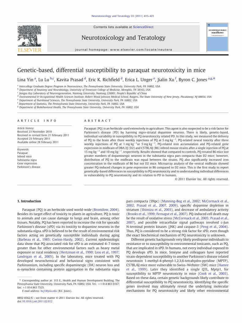
There were no strain, sex, or brain region differences in PQconcentrations in the VMB or CB (Fig. 1).
Ventral Midbrain
B6-F B6-M D2-F D2-M B6-F B6-M D2-F D2-M
PQ
Co
nce
ntr
atio
n(n
g/m
gT
issu
e)
PQ
Co
nce
ntr
atio
n(n
g/m
gT
issu
e)
0.0
0.1
0.2
0.3
0.4
0.5 Cerebellum
0.0
0.1
0.2
0.3
0.4
0.5
Fig. 1. PQ concentration in ventral midbrain and cerebellum. Black bars represent strain C57BL/6J females andmales (B6-F and B6-M); gray bars represent strain DBA/2J females andmales (D2-F and D2-M). Tissue PQ concentrations are equal between the two strains, two brain regions, and sexes, after 3 weekly ip injections at 5 mg kg−1. Data are presented asmeans±SEM.
418 L. Yin et al. / Neurotoxicology and Teratology 33 (2011) 415–421
3.2. Effects of PQ on TH+ neurons in the SNpc
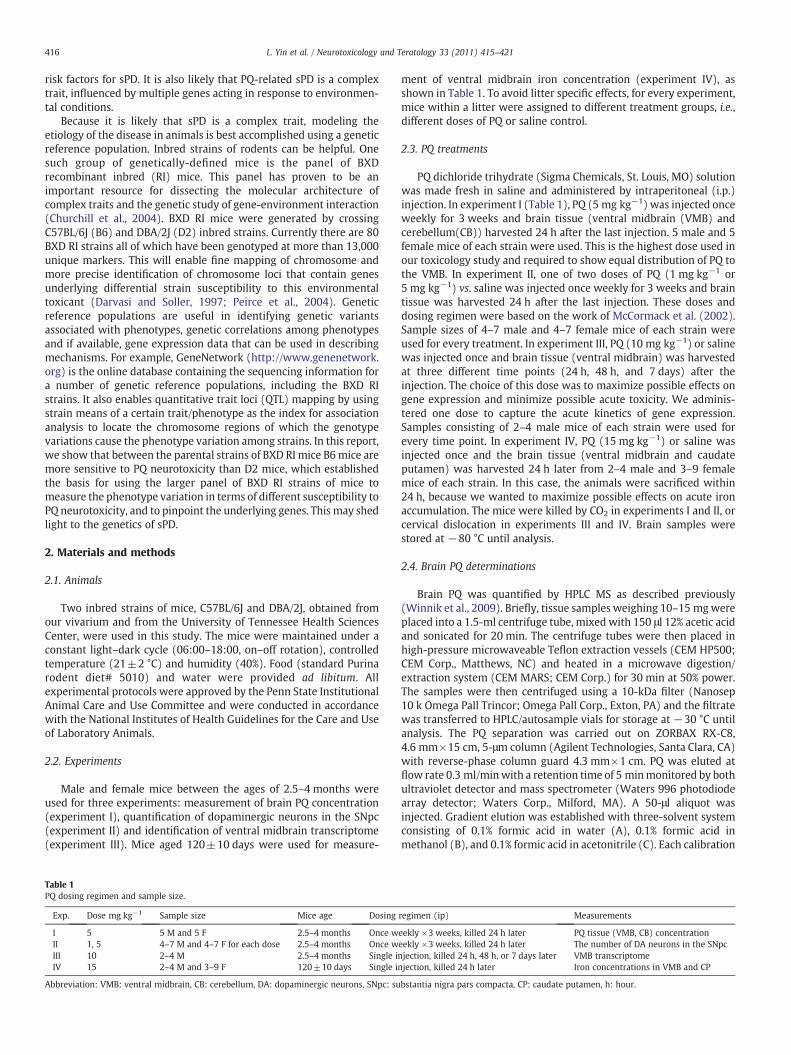
Themain effects for strain and sex were not significant (F1,51b1 forboth). The main effect of treatment was significant (F2,51=38.58,pb0.001) and accounted for 54% of total variance by est.ω2. Therewasalso a significant strain by treatment interaction (F2,51=3.25,pb0.05). At 1 mg kg−1, PQ produced significant TH+ neuron loss inB6 (pb0.05) but not in D2 mice (Fig. 2). 5 mg kg−1 producedsignificant TH+ neuron loss in D2 mice (17.6%, pb0.001) and in B6mice (31.4%, pb0.0001). Moreover the TH+ neuron loss at 5 mg kg−1
in B6 was significantly greater than in D2 mice (pb0.01). When weanalyzed data of each sex separately, in males, PQ induced TH+neuron loss showed the same pattern as pooled sexes in both strains.
Fig. 2. Stereology of cell counting. A–D, micrographs taken at 40× magnification. The four secthe whole SNpc. Brown color indicates tyrosine hydroxylase (TH) positive staining, and bluemagnification. F, differential strain susceptibility to PQ neurotoxicity. In both strains, 5 mg kgmore TH+ than DBA/2J. Data are presented as means±SEM. G, micrographs captured at 40bottom is from a PQ treated male C57BL/6J mouse. The red arrows point to the SNpc.
In females however, only 5 mg kg−1 produced significant TH+ loss inD2 (pb0.05) and in B6 (pb0.001), and there was no strain differenceat this dose (data not shown). We also quantified the number of THnegative (TH−) neurons in the SNpc (data not shown). In D2 mice,significant TH− neuron loss was observed at 1 mg kg−1 and at5 mg kg−1 (pb0.05). In B6 mice, no significant TH− neuron loss wasobserved at either dose.
3.3. Effect of PQ on iron concentration in VMB
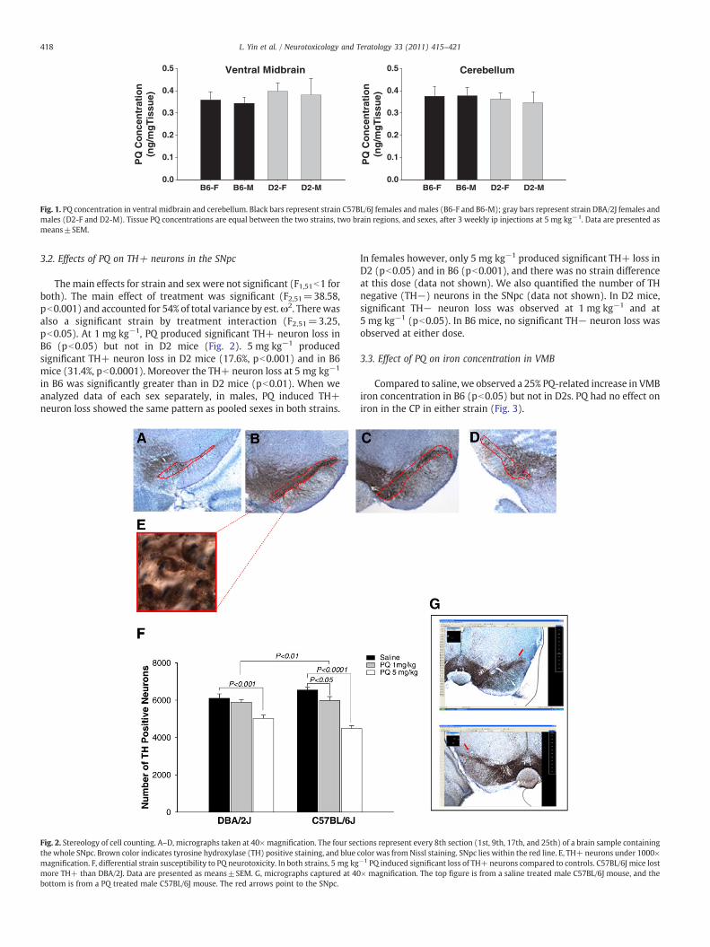
Compared to saline, we observed a 25% PQ-related increase in VMBiron concentration in B6 (pb0.05) but not in D2s. PQ had no effect oniron in the CP in either strain (Fig. 3).
tions represent every 8th section (1st, 9th, 17th, and 25th) of a brain sample containingcolor was fromNissl staining. SNpc lies within the red line. E, TH+ neurons under 1000×−1 PQ induced significant loss of TH+ neurons compared to controls. C57BL/6J mice lost× magnification. The top figure is from a saline treated male C57BL/6J mouse, and the

DBA/2J
VM
B F
e C
on
cen
trat
ion
(ug
/g t
issu
e)
0
2
4
6
8
10
12
14
16
18Saline15mg/kg PQ
P<0.05
DBA/2J
CP
Fe
Co
nce
ntr
atio
n (
ug
/g t
issu
e)
0
5
10
15
20CTL15mg/kg PQ
C57BL/6J
C57BL/6J
Fig. 3. PQ enhances ventral midbrain iron concentration. A 25% increase in ventralmidbrain iron concentration was observed in C57BL/6J mice injected with a single ipinjection of 15 mg kg−1 PQ. No such change was observed in the DBA/2J mice. Data arepresented as means±SEM.
419L. Yin et al. / Neurotoxicology and Teratology 33 (2011) 415–421
3.4. Effect of PQ on gene expression in the VMB
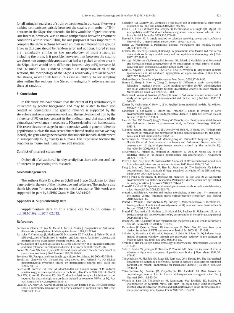
Genes showingmore thana1.5-fold change (increase or decrease) inresponse to PQ and p-value less than 0.05 in paired-t-test (PQ vs. saline)were defined as differentially expressed genes. B6mice evinced greaternumbers of differentially expressed genes than D2 mice at all timepoints (Fig. 4). 24 h after PQ injection, the numbers of the differentiallyexpressed genes in B6 andD2were 5 (4−decreased, 1+ increased) and1+, respectively. 48 h after PQ injection, the numbers of differentiallyexpressed genes in B6 and D2were 204 (82−, 122+) and 12 (5−, 7+),respectively. 7 days after PQ injection, the numbers of differentiallyexpressed genes in B6 and D2were 358 (225−, 133+) and 7 (2−, 5+),respectively, as shown in Fig. 4B. In summary, the numbers ofdifferentially expressed genes kept increasing from 24 h to 48 h to7 days after PQ treatment in B6 mice (numbers of differentiallyexpressed genes=5, 204 and 358, respectively) while the pattern wasdifferent in strainD2mice, peakingat 48 h and thendecreasingat7 days(numbers of differentially expressed genes=1, 12 and 7).
We performed gene enrichment analysis using Nexus Expressionto group differentially expressed genes by molecular function, usinggene ontology terms. As shown in Table 2 in the Supplementary data,the top three most significantly over represented groups at 48 h in B6mice in order were iron ion binding, aromatic-L-amino-acid decar-boxylase activity which is involved in dopamine synthesis, and thiol–disulfide exchange intermediate activity (1 gene) which is related toredox homeostasis. At 7 days, the top three were iron ion binding,selenium binding, and the NADH dehydrogenase (ubiquinone)activity. The molecular functions of the differential expressed genesin other treatment groups are presented in Table 2c of theSupplementary data.
4. Discussion
4.1. Strain differences in PQ neurotoxicity
Our primary hypothesis that B6 and D2 mice evince differential,genetically-based susceptibility to PQ neurotoxicity was confirmed.Compared to D2 mice, B6 mice lost greater numbers of dopaminergicneurons in the SNpc, gained more VMB iron, and showed greaternumbers of differentially expressed genes after PQ treatments (vs. salinecontrol). Moreover, we also found no change in the number of non-dopaminergic (tyrosinehydroxylasenegative, TH−) neurons in the SNpcin B6 mice after the PQ treatment as well, which confirmed the specifictoxic effect of PQ on striatal dopaminergic system. However, D2mice didshow significant TH− neuron loss in the SNpc after the same treatment.Thismight indicate strain specific biological change, or a challenge to theTH and Nissl double staining strategy. A recent paper reported theadvantage of usingneuron specific nuclear antigen (NeuN) to specificallyrecognize neuronal nuclei, overcoming Nissl staining which universallystains all neuronal RNA (Prasad and Richfield, 2010). By using thisstaining technique in future studies,wemight be able to answerwhetherthe loss of TH− neurons shown in D2mice was due to PQ toxicity or thestaining technique. PQhasbeen reported todecrease striatal dopamine inmice (Shimizu et al., 2003). PQ in combined with Maneb has beenreported to reduce tyrosine hydroxylase (TH) immunoreactivity in thedorsal striatum in mice, however, PQ alone did not show this effect(Thiruchelvam et al., 2000). In our study, the significant loss of THpositive neurons in both strains was not accompanied by a reciprocalincrease in TH negative neurons which indicated that TH per sewas notdownregulated (Prasad and Richfield, 2008).
4.2. PQ neurotoxicity is not related to distribution
Under our dosing regimen, we also found that the differential strainsusceptibility to PQneurotoxicitywasnot related todistributionof PQ tothe brain. Indeed, the seemingly equal distribution between ventralmidbrain and cerebellum might indicate that unlike MPTP whoseneurotoxic effects rely on being transported into DA neurons bydopamine transporter (DAT), PQ neurotoxicity may not depend onsimilar transport. We cannot be sure on this point at this time becausewe tested the entire ventral midbrain. The mechanism underlying whyDAneurons aremore vulnerable to PQneurotoxicity than other types ofneurons is not clear. One possibility is the interaction between iron anddopamine. Dopamine metabolism yields reactive oxygen species (ROS)whichmay accumulate inDAneuronswith aging andmake the neuronsmore sensitive than other types of neurons to neurotoxins such as PQ.Moreover, iron per se can produce ROS.
4.3. Role of iron in PQ neurotoxicity
Iron plays a central role in oxidative stress (Dauer and Przedborski,2003) and is reported to induce the formation of α-synucleinprotofibrils in brain (Golts et al., 2002; Rhodes and Ritz, 2008). Ironhas been shown to exacerbate the neurotoxic effect of PQ as well(Peng et al., 2007). In this study, we found, for the first time, that PQcan increase iron in VMB, an effect that is strain-dependent.Furthermore, gene expression and enrichment analysis revealedthat iron ion binding was the most overrepresented molecularfunction among the differentially expressed genes after PQ treatmentin B6 mice. The pattern of numbers of DA neuron loss, elevated VMBiron, and differentially expressed iron-related genes shown in B6 (vs.D2) mice supports the involvement of iron in PQ neurotoxicity, andimplies that the differential strain capabilities to regulate VMB ironhomeostasis may well contribute to the differential strain suscepti-bility to PQ neurotoxicity. In addition, we found that caudate putameniron concentration was refractory to PQ treatment. These support thehypothesis of specificity of PQ toxicity in DA neurons in the VMB

-1.5 -1.0 -0.5 0.0 0.5 1.0 1.5
0.0
0.5
1.0
1.5
2.0
2.5
3.0
24h DBA2 PQ VS CTL
log_2 Fold Change-0.5 0.0 0.5 1.0 1.5
0.0
0.5
1.0
1.5
2.0
2.5
3.0
48h DBA2 PQ VS CTL
log_2 Fold Change-0.5 0.0 0.5
0.0
0.5
1.0
1.5
2.0
2.5
3.0
7d DBA2 PQ VS CTL
log_2 Fold Change
-0.5 0.0 0.5 1.0 1.5 2.0 2.5
01
23
24h B6 PQ VS CTL
log_2 fold change-1 0 1 2
01
23
4
48h B6 PQ VS CTL
log_2 fold change-1 0 1 2 3 4
01
23
4
7d B6 PQ VS CTL
log_2 fold change
-lo
g_1
0 p
-val
ue,
Fo
ld C
han
ge
-lo
g_1
0 p
-val
ue,
Fo
ld C
han
ge
-lo
g_1
0 p
-val
ue,
Fo
ld C
han
ge
-lo
g_1
0 p
-val
ue,
Fo
ld C
han
ge
-lo
g_1
0 p
-val
ue,
Fo
ld C
han
ge
-lo
g_1
0 p
-val
ue,
Fo
ld C
han
ge
A
B Numbers of differentially expressed genes
24 h 48 h 7 dChange
B6 D2 B6 D2 B6 D2
Increased 1 1 122 7 133 5
Decreased 4 0 82 5 225 2
Total 5 1 204 12 358 7
Fig. 4. Gene expression change after paraquat treatment. A, Volcano plot of differentially expressed genes (blue colored) between experiment (PQ) and saline control groups, in eachstrain and at each time point. The Y-axis displays the –log10 of p values from paired t-tests, the x-axis displays the log2 of the fold change of gene expression level (experiment/control). The dark red lines correspond to p-value of 0.05 and fold changes of (±) 1.5. Genes with pb0.05, and fold change more than 1.5, were identified as differentially expressedand are highlighted in blue. B, the numbers of differentially expressed genes (pb0.05, 1.5b fold change b–1.5).
420 L. Yin et al. / Neurotoxicology and Teratology 33 (2011) 415–421
(including SNpc) andmay implicate local regulation of ironhomeostasisin VMBDA neurons. In humans, Iron accumulation in the VMB has beenreported in Parkinson's disease patients, and the more accumulatedVMB iron themore severe the disease (Bartzokis et al., 1999; Berg et al.,2001; Sofic et al., 1991). Thus, genes regulating VMB iron homeostasismight be novel therapeutic targets to prevent PQ induced sPD.
4.4. Gene expression
Ourmost salient finding in gene expressionwas PQ-related changein iron ion binding protein genes in the B6 strain. Alternatively, otherthan DOPA decarboxylase, no genes directly related to dopaminechanged expression in response to PQ. Exactly why this is the case isunclear; however the former finding may help strengthen ourunderstanding of the relationship between iron and dopamine.Mitochondrial dysfunction has been intensively studied as the basisfor PQ neurotoxicity. The results are mixed as to whether PQ causesmitochondrial oxidative damage, and whether mitochondrial com-plex I is the major site of PQ-induced ROS production in the brain(Castello et al., 2007; Choi et al., 2008; Cochemé and Murphy, 2008;Richardson et al., 2005). Our study does suggest mitochondrialinvolvement in PQ neurotoxicity. Mitochondrial genes (Ndufs7,Ndufc2, Ndufv2, Grx2, and Sdhd) were differentially expressedbased on the transcriptome data. The first 4 of these play important
roles in maintaining mitochondrial complex I activity and theirexpression was increased after PQ treatment. Sdhd is a subunit ofmitochondrial complex II and its expression was increased after PQtreatment as well. Real-time PCR verification of these gene expressionchanges will help to elucidate their biological role in response to PQ.
In the D2 strain, no iron or dopamine related genes wereresponsive to PQ. Most of the genes that did respond were relatedto intermediate metabolism and reactive oxygen species (ROS).
Other than iron homeostasis and mitochondrial response, severalother biological processes were affected by PQ, based on our transcrip-tome data. These include the ubiquitin proteosome system, lipidmetabolism, catecholamine biosynthesis, and inflammatory response.This shows that PQ neurotoxicity is a highly complex trait. Thus, webelieve using systematic genetic analysis will lead to better understand-ing themechanism of PQ neurotoxicity and its role in the etiology of sPD.
4.5. Stereology of cell counting
On a methodological note, Schmitz and Hof (2005) reviewed theliterature concerning stereological methods and sources of bias inselecting sections. In their view, when comparing between regions oranimals (strain and sex), the sections to be counted for featuresshould be picked at random in order to avoid bias. In the presentsituation, we chosewhich sections to count a priori and to be the same
421L. Yin et al. / Neurotoxicology and Teratology 33 (2011) 415–421
for all animals regardless of strain or treatment. In our case, if weweremaking comparisons strictly between the strains on number of TH+neurons in the SNpc, the potential for bias would be of great concern.Our interest, however, was to make comparisons between treatmentconditions within strain. Thus, for our purposes it was important tocompare the same sections between animals in different dose groups.Error in this case should be random error and not bias. Inbred strainsare remarkably similar in the morphology of most structures,including the brain. Is it possible, however, that between the strains,we chose non comparable areas so that had we picked another area inthe SNpc, there would be no difference in sensitivity to PQ between B6and D2 mice? This is indeed possible. On inspection of all of thesections, the morphology of the SNpc is remarkably similar betweenthe strains, so we think bias in this case is unlikely. As for samplingsites within the sections, the Stereo Investigator™ software assignsthese at random.
5. Conclusion
In this work, we have shown that the extent of PQ neurotoxicity isinfluenced by genetic background and may be related to brain ironcontent or homeostasis. The genetic influence is supported by thestereology and gene expressionwork and the involvement of iron by theinfluence of PQ on iron content in the midbrain and that many of thegenes that showchange in response toPQare related to ironhomeostasis.This research sets the stage formore extensivework in genetic referencepopulations, such as the BXD recombinant inbred strains so that wemayidentify the genes andgenenetworks that underlie individual differencesin susceptibility to PQ toxicity in humans. This is possible because thegenomes in mouse and humans are 90% syntenic.
Conflict of interest statement
On behalf of all authors, I hereby certify that there exist no conflictsof interest in presenting this research.
Acknowledgements
The authors thank Drs. Steven Schiff and Bruce Gluckman for theirgenerosity in the use of the microscope and software. The authors alsothank Ms. Ann Tomaszewicz for technical assistance. This work wassupported in part by USPHS Grant # U01AA014425.
Appendix A. Supplementary data
Supplementary data to this article can be found online atdoi:10.1016/j.ntt.2011.02.012.
References
Barbeau A, Cloutier T, Roy M, Plasse L, Paris S, Poirier J. Ecogenetics of Parkinson'sdisease: 4-hydroxylation of debrisoquine. Lancet 1985;2:1213–6.
Bartzokis G, Cummings JL, Markham CH, Marmarelis PZ, Treciokas LJ, Tishler TA, et al.MRI evaluation of brain iron in earlier- and later-onset Parkinson's disease andnormal subjects. Magn Reson Imaging 1999;17:213–22.
Berg D, GerlachM, YoudimMB, Double KL, Zecca L, Riederer P, et al. Brain iron pathwaysand their relevance to Parkinson's disease. J Neurochem 2001;79:225–36.
Boone EM, Cook MN, Hou X, Jones BC. Sex and strain influence the effect of ethanol oncentral monoamines. J Stud Alcohol 1997;58:590–9.
Bromilow RH. Paraquat and sustainable agriculture. Pest Manag Sci 2004;60:340–9.Brooks AI, Chadwick CA, Gelbard HA, Cory-Slechta DA, Federoff HJ. PQ elicited
neurobehavioral syndrome caused by dopaminergic neuron loss. Brain Res1999;27:1-10.
Castello PR, Drechsel DA, Patel M. Mitochondria are a major source of PQ-inducedreactive oxygen species production in the brain. J Biol Chem 2007;282:14186–93.
Choi WS, Kruse SE, Palmiter RD, Xia Z. Mitochondrial complex I inhibition is notrequired for dopaminergic neuron death induced by rotenone, MPP+, or PQ. ProcNatl Acad Sci U S A 2008;105:15136–41.
Churchill GA, Airey DC, Allayee H, Angel JM, Attie AD, Beatty J, et al. The CollaborativeCross, a community resource for the genetic analysis of complex traits. Nat Genet2004;36:1133–7.
Cochemé HM, Murphy MP. Complex I is the major site of mitochondrial superoxideproduction by PQ. J Biol Chem 2008;283:1786–98.
Cook R, Lu L, Gu J, Williams RW, Smeyne RJ. Identification of a single QTL, Mptp1, forsusceptibility to MPTP-induced substantia nigra pars compacta neuron loss inmice.Brain Res Mol Brain Res 2003;110:279–88.
Darvasi A, Soller M. A simple method to calculate resolving power and confidenceinterval of QTL map location. Behav Genet 1997;27:125–32.
Dauer W, Przedborski S. Parkinson's disease: mechanisms and models. Neuron2003;39:889–909.
Erikson KM, Pinero DJ, Connor JR, Beard JL. Regional brain iron, ferritin and transferrinconcentrations during iron deficiency and iron repletion in developing rats. J Nutr1997;127:2030–8.
Fernagut PO, Hutson CB, Fleming SM, Tetreaut NA, Salcedo J, Masliah E, et al. Behavioraland histopathological consequences of PQ intoxication in mice: effects of alpha-synuclein over-expression. Synapse 2007;61:991-1001.
Golts N, Snyder H, Frasier M, Theisler C, Choi P, Wolozin B. Magnesium inhibitsspontaneous and iron-induced aggregation of alpha-synuclein. J Biol Chem2002;277:16116–23.
Gwinn-Hardy K. Genetics of parkinsonism. Mov Disord 2002;17:645–56.Hamre K, Tharp R, Poon K, Xiong X, Smeyne RJ. Differential strain susceptibility
following 1-methyl-4-phenyl-1,2,3,6-tetrahydropyridine (MPTP; administrationacts in an autosomal dominant fashion: quantitative analysis in seven strains ofMus musculus. Brain Res 1999;15:91-103.
Hertzman C,WiensM, Bowering D, Snow B, Calne D. Parkinson's disease: a case–controlstudy of occupational and environmental risk factors. Am J Ind Med 1990;17:349–55.
Kutner M, Nachtsheim C, Neter J, Li W. Applied linear statistical models. 5th edition.McGraw-Hill; 2004.
Landrigan PJ, Sonawane B, Butler RN, Trasande L, Callan R, Droller D. Earlyenvironmental origins of neurodegenerative disease in later life. Environ HealthPerspect 2005;113:1230–3.
Liou HH, Tsai MC, Chen CJ, Jeng JS, Chang YC, Chen SY, et al. Environmental risk factorsand Parkinson's disease: a case–control study in Taiwan. Neurology 1997;48:1583–8.
Manning-Bog AB, McCormack AL, Li J, Uversky VN, Fink AL, Di Monte DA. The herbicidePQ causes up-regulation and aggregation of alpha-synuclein in mice: PQ and alpha-synuclein. J Biol Chem 2002;277:1641–4.
McCormack AL, Thiruchelvam M, Manning-Bog AB, Thiffault C, Langston JW, Cory-Slechta DA, et al. Environmental risk factors and Parkinson's disease: selectivedegeneration of nigral dopaminergic neurons caused by the herbicide PQ.Neurobiol Dis 2002;10:119–27.
McCormack AL, Atienza JG, Johnston LC, Andersen JK, Vu S, Di Monte DA. Role ofoxidative stress in PQ-induced dopaminergic cell degeneration. J Neurochem2005;93:1030–7.
Peirce JL, Lu L, Gu J, Silve LM, Williams RW. A new set of BXD recombinant inbred linesfrom advanced intercross populations in mice. BMC Genet 2004;29:5–7.
Peng J, Mao XO, Stevenson FF, Hsu M, Andersen JK. The herbicide PQ inducesdopaminergic nigral apoptosis through sustained activation of the JNK pathway.J Biol Chem 2004;279:32626–32.
Peng J, Peng L, Stevenson FF, Doctrow SR, Andersen JK. Iron and PQ as synergisticenvironmental risk factors in sporadic Parkinson's disease accelerate age-relatedneurodegeneration. J Neurosci 2007;27:6914–22.
Prasad K, Richfield EK. Sporadic midbrain dopamine neuron abnormalities in laboratorymice. Neurobiol Dis 2008;32:262–72.
Prasad K, Richfield EK. Number and nuclear morphology of TH+ and TH− neurons inthe mouse ventral midbrain using epifluorescence stereology. Exp Neurol2010;225:328–40.
Prasad K, Winnik B, Thiruchelvam MJ, Buckley B, Mirochnitchenko O, Richfield EK.Prolonged toxicokinetics and toxicodynamics of PQ in mouse brain. Environ HealthPerspect 2007;115:1448–53.
Prasad K, Tarasewicz E, Mathew J, Strickland PA, Buckley B, Richardson JR, et al.Toxicokinetics and toxicodynamics of PQ accumulation in mouse brain. Exp Neurol2009;215:358–67.
Rhodes SL, Ritz B. Genetics of iron regulation and the possible role of iron in Parkinson'sdisease. Neurobiol Dis 2008;32:183–95.
Richardson JR, Quan Y, Sherer TB, Greenamyre JT, Miller GW. PQ neurotoxicity isdistinct from that of MPTP and rotenone. Toxicol Sci 2005;88:193–201.
Shimizu K, Matsubara K, Ohtaki K, Fujimaru S, Saito O, Shiono H. PQ induces long-lasting dopamine overflow through the excitotoxic pathway in the striatum offreely moving rats. Brain Res 2003;976:243–52.
Schmitz C, Hof PR. Design-based stereology in neuroscience. Neuroscience 2005;130:813–31.
Sofic E, Paulus W, Jellinger K, Riederer P, Youdim MB. Selective increase of iron insubstantia nigra zona compacta of parkinsonian brains. J Neurochem 1991;56:978–82.
Thiruchelvam M, Richfield EK, Baggs RB, Tank AW, Cory-Slechta DA. The nigrostriataldopaminergic system as a preferential target of repeated exposures to combinedparaquat and maneb: implications for Parkinson's disease. J Neurosci 2000;20:9208–14.
Thiruchelvam MJ, Powers JM, Cory-Slechta DA, Richfield EK. Risk factors fordopaminergic neuron loss in human alpha-synuclein transgenic mice. Eur JNeurosci 2004;19:845–54.
Winnik B, Barr DB, Thiruchelvam M, Montesano MA, Richfield EK, Buckley B.Quantification of paraquat, MPTP, and MPP+ in brain tissue using microwaveassisted solvent extraction (MASE) and high performance liquid chromatography-mass spectrometry. Anal Bioanal Chem 2009;395:195–201.