Neurology Peripheral Neurology - nuhem.com fellowship... · Bells Palsy . Peripheral VII nerve...
42
Neurology Peripheral Neurology Alison Ruiz PA-C
Transcript of Neurology Peripheral Neurology - nuhem.com fellowship... · Bells Palsy . Peripheral VII nerve...

Neurology Peripheral Neurology
Alison Ruiz PA-C

Case Presentation 50 y/o M presents with c/o rt facial droop. Woke this
morning and his wife noticed it. Pt denies other symptoms. No slurred speech, no weakness, no dizziness. Pt states his upper lip feels heavy and he can’t close his rt eye very well.
No previous episode
No F/C/N/V/D. But states had a fever and cough approx 2 weeks ago
No other complaints

History
• HTN • Hyperlipidemia
PMH
• None
PSH
• Stroke (mother 76 y/o) • MI (father 79 y/o)
FH
• Nonsmoker • 2 drinks per week
SH

PE Vitals: T99.0, HR 86, BP 140/80, R 14, Pulse ox 99%
HEENT: PERRL, EOMi, rt sided facial droop, asymmetry in smile. Loss of nasolabial fold Unable to lift rt eyebrow
Neck supple full rom. No carotid bruits
Lungs: CTA bilat
CV RRR S1S2
Abd: soft NT ND NABS
Ext: no C/C/E

PE cont Neuro Speech clear. No focal deficits. Muscle strength 5/5 and equal bilat throughout- UE and
LE. Blow out cheeks test: Buccinator function is disrupted Unable to raise eyebrow. Loss of nasolabial fold and
nasal flaring Negative for expressive or receptive aphasia Patellar tendon, brachioradialis, triceps, achilles tendon
2+ bilat. Pronator Drift WNL, Rhomberg WNL, finger to nose WNL
Presenter
Presentation Notes
Bucinator muscle: Motor innervation is from the buccal branch of the facial nerve (cranial nerve VII)

Diagnostic Tests ?????
Presenter
Presentation Notes
None…this is bells palsy…clinical diagnosis If you thought it was something central then do CT/ MRI brain

Motor Neurons
Presenter
Presentation Notes
Bilateral innervation of the upper half of the face results in sparing of the forehead muscles in an upper motor neurone lesion

Bell’s Palsy The pathophysiology is not clear Theories Inflammation and edema of the nerve due to infectious
processes, leading to nerve compression Ischemic mononeuropathy due to disturbance in the
circulation in the vasa nervorum (the arterial branches supplying the nerve), leading to edema from the subsequent ischemic neuritis
The most common cause is idiopathic

What causes Bell’s and Who is at risk?
Common triggers include
Stress, trauma, fever, tooth extraction and “chilling” episode from exposure to the drafts and cold
Common diseases associated with the Bell’s
DM, HTN, HIV, Sarcoidosis, Sjogren’s, parotid nerve tumors, eclampsia, amyloidosis and recipients of intranasal influenza vaccine
Presenter
Presentation Notes
Inflammatory and autoimmune processes

Bells Palsy Peripheral VII nerve palsy (Facial Nerve) • Symptoms include • Ipsilateral tongue numbness, • Loss of taste, • Ear pain, • Overt paralysis preceded by a sensation of numbness or
weakness on affected side, Tinnitus, Drooling, Inability to keep liquids in the mouth, Occipital headache
Physical findings • Loss of ability to wrinkle nose • raise eyebrow, blow out cheeks • unable to purse lips allowing food, liquids and air to
escape • loss of nasolabial fold and nasal flaring

Differential Diagnosis Stroke
Shingles
Presenter
Presentation Notes
Look for central findings: expressive or receptive aphasia (sparing of forehead) Lesions are present: think Ramsay hunt- vesicles on TM or oropharynx

Algorithm Acute Facial Weakness
Central or Peripheral?
Peripheral Herpes zoster oticus
Bells Palsy
Ramsay Hunt Syndrome
Central
MRI Evaluate ischemia,
inflammatory, infectious disease
Consider
CSF
ESR
Serologic studies Syphilis, HIV, Vasculitis Admit patient

Treatment for Bell’s Palsy
Prednisone • Start within 2-14 days of onset • 1mg/kg/day • Usually 7 days
Valacyclovir 1000mg bid for 7 days or Famvir 750mg tid for 7
days
Wear an eyelid patch at night to prevent drying out the
cornea

Trigeminal Nerve (V)
3 Branches • Ophthalmic nerve V1 • Maxillary nerve V2 • Mandibular nerve V3
Mixed motor and sensory nerve. • Motor innervation for the muscles of
mastication • Sensation from the face, scalp, conjunctiva,
globe, mucous membranes of the sinuses, tongue, teeth and part of the external TM

Trigeminal Nerve Dysfunction Presentation Pain, paraesthesias, dysesthesias, and anesthesias
Motor dysfunction typically presents as difficulty chewing or difficulty swallowing
Peripheral Lesions
Cause loss of sensation or pain in only one division
Central Lesions
Consider if positive findings in 2 or more divisions
Presenter
Presentation Notes
Dysesthesias- abnromal sensations

Trigeminal Neuralgia
Tic Douloureaux • Slight predilection in women • Increased incidence >60 years of age
Mechanism • Compression of the trigeminal nerve root
within millimeters of the entry into the pons

Presentation
Sudden onset of pain • Paroxysms of severe unilateral pain
in the distribution of the trigeminal nerve
Last only seconds, with normal findings on neurologic exam • There is no pain between paroxysms

Causes Vascular compression by artery or vein
Saccular aneurysm
AV malformation
Vestibular Schwannomas
Meningioma
Epidermoid cyst
Tumor
Primary demyelinating disorders •MS •Charcot-Marie Tooth (rare)
Trigeminal Amyloidoma
Small infarct or angioma in the brainstem
Famillial

Diagnosing
Consider in all patients with unilateral facial pain
Look for “red flags” of other diseases

Abnormal Neuro exam • Think MS, Tumor,
hemmorhage, stroke
Abnormal oral, dental, or ear exam
Age < 40 yrs • unusual Bilateral SXs
Dizziness or vertigo • BPV, Stroke, Dehydration,
Labrinythitis

Hearing loss Numbness
Pain lasting > 2 minutes
Pain outside of trigeminal distribution
Visual changes

Treatment for Trigeminal Neuralgia
Carbamazepine 100mg PO bid and then increased in dosage as needed Treatment is very effective If fails, then the pt is unlikely to have trigeminal neuralgia
Presenter
Presentation Notes
Carbamazepine-Anticonvulsant- decreases nerve impulses

Case Presentation 30 y/o M presents to the ER with weakness in his lower
extremities at the ankle area bilat. States started 3 days ago but he is having trouble walking now. Today developed tingling in thighs bilat. He was recently diagnosed with mononucleosis and has been home in bed for 2 weeks.
Fevers 102.0 last week, but resolved in the past 3 days. No N/V/D.
Denies cough, sob, chest pain or abdominal pain

History PMH Mononucleosis
PSH Hernia repair 10 years ago
SH Nonsmoker Drinks alcohol mostly beer 4-6 drinks per week
FH HTN both parents

Physical Exam Vitals: T 99.5 po, R 18, Pulse 89, Pulse ox 100%, BP
120/68
HEENT: PERRL EOMi
Neck supple full rom. No nuccal rigidity
Lungs: CTA bilat. NO W/R/R
CV RRR S1S2
Abd: soft slight RUQ tenderness
Ext: no C/C/E

Neuro Physical exam Neuro Motor weakness bilat LE 3/5 plantar and dorsiflexion, flexion
and extension. Motor weakness bilat UE 4/5 with hand grasp, flexion and
extension of biceps bilat. Otherwise normal 5/5 strength Difficulty getting out of chair. Gait is limited to standing, unable to step without
assistance. Sensory with light and sharp touch equal and intact bilat LE and
UE. CN II-XII grossly intact Finger to nose WNL Rhomberg difficult to assess because patient has trouble
standing

Differential Diagnosis ?
Presenter
Presentation Notes
Gillain Barre MS Transverse Myelitis Viral Chronic inflammatory demyelinating polyneuropathy Conversion disorder/hysterical paralysis Human immunodeficiency virus (HIV) peripheral neuropathy Neurotoxic fish or shellfish poisoning Paraneoplastic neuropathy Poliomyelitis Porphyria polyneuropathy Spinal cord compression Spinal cord syndromes, particularly postinfection Tick paralysis[89] Toxic neuropathies (eg, arsenic, thallium, organophosphates, lead) Vasculitic neuropathies Vitamin deficiency (eg, vitamin B-12, folate, thiamine) West Nile encephalitis Bilateral strokes Acute cerebellar ataxia syndromes Posterior fossa structural lesion

Diagnostic Testing in ER CBC
14,000 WBC Otherwise normal
BMG WNL
LFTs Elevated Alk phos
ESR and CPK Sed Rate elevated Normal CPK
Urinalysis- WNL
LP Protein 500

Guillan Barre Syndrome Demyelinating disease
primarily affecting the peripheral nervous system.
Slight male predominance.
Affects the elderly and young adults most commonly.
Presenter
Presentation Notes
Lower extremity weakness usually begins first and ascends symmetrically and progressively over the first several days. Upper extremity, trunk, facial, and oropharyngeal weakness is observed to a variable extent. Marked asymmetrical weakness calls the diagnosis of GBS into question. Patients may be unable to stand or walk despite reasonable strength, especially when ophthalmoparesis or impaired proprioception is present. Despite frequent complaints of paresthesias, objective sensory changes are minimal. A well-demarcated sensory level should not be observed in patients with GBS; such a finding calls the diagnosis of GBS into question. Reflexes are absent or reduced early in the disease course. Hyporeflexia or areflexia of involved areas represents a major clinical finding on examination of the patient with GBS. Pathologic reflexes, such as the Babinski sign, are absent. Hypotonia can be observed with significant weakness.
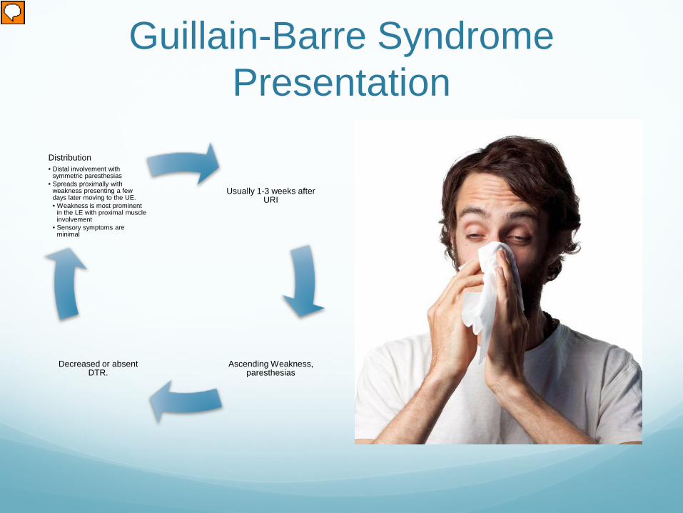
Guillain-Barre Syndrome Presentation
Usually 1-3 weeks after URI
Ascending Weakness, paresthesias
Decreased or absent DTR.
Distribution • Distal involvement with
symmetric paresthesias • Spreads proximally with
weakness presenting a few days later moving to the UE. • Weakness is most prominent
in the LE with proximal muscle involvement
• Sensory symptoms are minimal
Presenter
Presentation Notes
May initially present with difficulty rising from a chair Weakness can be profound and involve the face and respiratory muscles. Starts with the lower extremities Symmetric Does not usually cause sensory loss

Disease Progression Progressive
Phase • Lasts few
days to 3-4 weeks
Plateau Phase
• Days to weeks
Recovery Phase
• Weeks to months

Work up LP will show elevated protein levels >400 Normal protein levels do not rule out the GBS
ESR and CPK may be elevated with myopathies
LFTs are elevated in some patient with demyelinating disease
Nerve Conduction studies are common
MRI is sensitive, but nonspecific, for diagnosis. However, it can reveal nerve root enhancement and may be an effective diagnostic adjunct

GB Management Largely Supportive
Should have a respiratory evaluation of FVC
Admitted to the ICU if the FVC < 20mL/kg or intubated if it is less than 15mL/kg.
Plasmapheresis and IV immunoglobin can reduce recovery time by 50%

A Few Points about Transverse Myelitis
May present by itself Or as symptomatology of
Multiple Sclerosis
Paraparesis, which is initially flaccid and then spastic Preceded by back pain
The thoracic cord is most often affected
Presenter
Presentation Notes
TM is seen as a feature of relapsing and remitting MS Loss of sensation with a sensory level on the trunk (starts distally and ascends) and bowel and bladder dysfunction Paraparesis-a slight paralysis or weakness of both legs

Transverse Myelitis Resembles Guillain-Barre,
But it is asymmetric involvement
Has definite sensory level Complete lack of UE
involvement Urinary incontinence
symptoms CSF pleocystosis make the
dx of GB less likely
Presenter
Presentation Notes
Pleocytosis-icrease in wbc count in CSF

Transverse Myelitis Diagnostics
The spinal cord MRI will almost always confirm the presence of a lesion within the spinal cord, whereas the brain MRI may provide clues to other underlying causes, especially MS.
Presenter
Presentation Notes
Brain MRI may help to identify the existence of areas of demyelination in other regions of the central nervous system other than the spinal cord. CSF studies may be helpful to determine if antibodies are being synthesized within the central nervous system (e.g., oligoclonal bands), a finding that may support a diagnosis of MS.
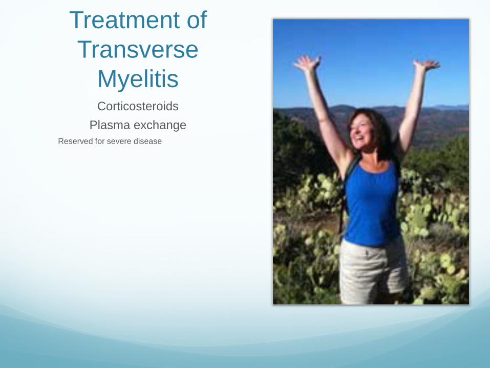
Treatment of Transverse
Myelitis Corticosteroids
Plasma exchange Reserved for severe disease

Myasthenia Gravis Pathophysiology • Bimodal occurring first in
20s and 30s in women and then in the 6th and 7th decades of life in men
• Many etiologies • All lead to formation of
autoantibiodies directed against nicotinic acetylcholine receptors (AchRs) at the neuromuscular junction

Myasthenia Gravis Muscular weakness and
fatigability are the hallmarks
Ocular muscle weakness is one of the first signs In up to 40% of patients Ptosis, diplopia and blurred vision
If bulbar muscle involvement then may have dysarthria and dysphagia
Acute crisis occurs in 15-20% of patients and presents as respiratory failure. Usually occurs in the 1st two years of the disease.

Diagnostic Testing Clinical findings and serologic testing
Endrophonium (Tensilon) test
Electromyography
Receptor antibody testing is positive
At this point, you have called neurology!!!!!
Presenter
Presentation Notes
Endrophonuim test- done at bedside. Measure the distance between the upper and lower eyelids in the most affected eye, then measuring again following IV administration of edrophonium, a short acting acetylcholinesterage blocking agent.

Treatment Cholinesterase inhibitors
Pyridostigmine and neostigmine Used for outpt chronic
therapy
Immunosuppressant drugs Used for chronic control
Thymectomy
Immunomodulatory Therapy Plasmapheresis and IVIg are
used for severe exacerbation
Consult!!!!

References Tintanilli 1118
Adams 995-997, 1043-1050
Medscape http://emedicine.medscape.com/article/315632-overview