Christine M. Betzold MSN NP IBCLC UCI Assistant Clinical ...
Nervous System
Syahruramdhani, MSN., M.Sc
School of NursingMedical Faculty and Health Sciences
Universitas Muhammadiyah Yogyakarta


#FACT
• If you pick up something hot, for
example, you may find that the
grasping muscles in your hand relax and
you drop it even before the sensation
of extreme heat or pain reaches your
conscious perception.

Describe the gross and microscopic anatomy of the spinal cord
• The spinal cord is a cylinder of nervous tissue that arises from the brainstem at
the foramen magnum of the skull.
• In adults, it averages about 1.8 cm thick and 45 cm long.
• The spinal cord is divided into cervical, thoracic, lumbar, and sacral regions.


Describe the gross and microscopic anatomy of the spinal cord
The meninges are three protective, connective tissue coverings that encircle the
spinal cord and brain. From superficial to deep they are the:
• (1) Dura mater (The most superficial of the three spinal meninges is a thick strong layer)
• (2) Arachnoid mater (This layer, the middle of the meningeal membranes); and
• (3) Pia mater (This innermost meninx is a thin transparent connective tissue layer).


Describe the gross and microscopic anatomy of the spinal cord
• The spinal cord, like the brain,
consists of two kinds of
nervous tissue called gray and
white matter.
Describe the gross and microscopic anatomy of the spinal cord
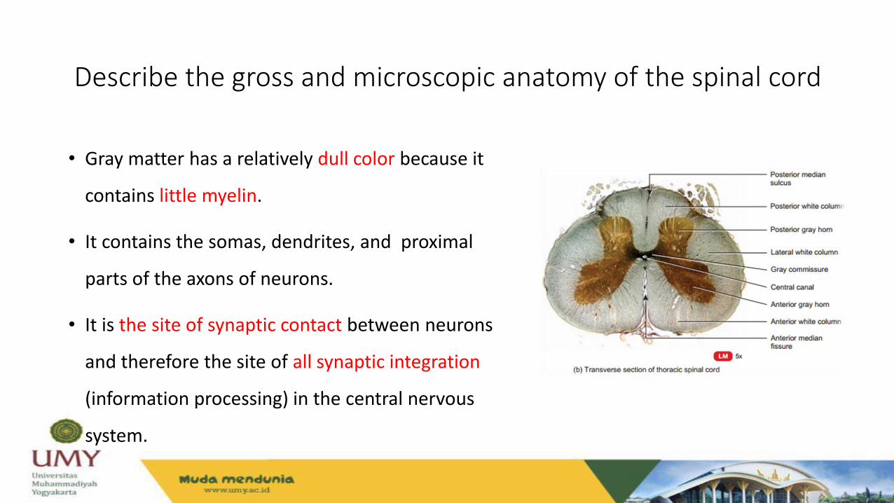
• Gray matter has a relatively dull color because it
contains little myelin.
• It contains the somas, dendrites, and proximal
parts of the axons of neurons.
• It is the site of synaptic contact between neurons
and therefore the site of all synaptic integration
(information processing) in the central nervous
system.

Describe the gross and microscopic anatomy of the spinal cord
• White matter contains an abundance of
myelinated axons, which give it a bright, pearly
white appearance.
• It is composed of bundles of axons, called
tracts, that carry signals from one part of the
CNS to another.

Name the major conduction pathways of the spinal cord and state their functions;
• The spinal cord serves three principal functions
1. Conduction
2. Locomotive
3. Reflexes

Name the major conduction pathways of the spinal cord and state their functions;
• Knowledge of the locations and functions of the spinal tracts is essential in
diagnosing and managing spinal cord injuries.
• Ascending tracts carry sensory information up the cord and descending Tracts
conduct motor impulses down.
• All nerve fibers in a given tract have a similar origin, destination, and function.
Many of these Fibers have their origin or destination in a region called the
brainstem.


Nature of Reflex
• Reflexes are quick, involuntary, stereotyped reactions of glands
or muscles to stimulation.
• This definition sums up four important properties of a reflex:
1. Require stimulation
2. Quick
3. Involuntary
4. Stereotyped

Reflexes
• Somatic reflexes/spinal reflexes.Example: the quick withdrawal of your hand from a hot stove or the lifting of your foot when you step on something sharp.
Many somatic reflexes are initiated by proprioceptors, organs that monitor the position and movements of body parts.
• Visceral reflexesExample: high blood pressure activates a visceral baroreflex.

Somatic Reflexes• A somatic reflex employs a reflex arc, in which signals travel along the
following pathway:
1. Somatic receptors in the skin,
2. Afferent nerve fibers,
3. Interneurons,
4. Efferent nerve fibers,
5. Skeletal muscles

Muscle Spindle
• Muscle spindles are proprioceptors embedded in the skeletal muscles
that respond to stretching of the muscle.
• A spindle contains 3 to 12 modified muscle fibers and a few nerve
fibers, all wrapped in a fibrous capsule.
• The muscle fibers within a spindle are called intrafusal fibers, while
those of the rest of the muscle are called extrafusal fibers.


Muscle Spindle
• Muscle spindles have three types of nerve fibers:
• 1. Primary afferent fibers,
• 2. Secondary afferent fibers, and
• 3. Gamma motor neurons.

The strestch reflex
• The tendency of a muscle to contract when it is stretched.
• For example, if your head starts to tip forward, it stretches muscles such as the
semispinalis and splenius capitis of the nuchal region (back of your neck).
• Reciprocal inhibition, a reflex phenomenon that prevents muscles from working
against each other by inhibiting antagonists.

The golgi tendon reflex
• Golgi tendon organs are proprioceptors located in a tendon near its
junction with a muscle.
• The Golgi tendon reflex is a response to excessive tension on the tendon.
• The Golgi tendon reflex also functions when some parts of a muscle
contract more than others.


The flexor (withdrawal) reflex
• A flexor reflex is the quick contraction of flexor muscles resulting in the
withdrawal of a limb from an injurious stimulus.
• For example, suppose you are wading in a lake and step on a broken bottle with
your right foot.
• The protective function of this reflex requires more than a quick jerk like a tendon
reflex, so it involves more complex neural pathways.

The crossed extensor reflex
• The crossed extensor reflex is the contraction of extensor muscles in
the limb opposite from the one that is withdrawn.
• To produce this reflex, branches of the afferent nerve fibers cross from
the stimulated side of the body to the contralateral side of the spinal
cord.


References
• Saladin, K., 2003. Integration and Control. Anatomy and Physiology:
The Unity of Form and Function, 3rd Edition.United States of
America: The McGraw-Hill Companies, Inc. Chapter 13, Pages: 481-
513.
• Tortora, GJ, Derrickson, B., 2012. Principles of Anatomy & Physiology,
13th Edition. United States of America: John Wiley & Sons, Inc.
Chapter 13, Pages: 492-526.

Alhamdulillah THANK YOU