Mutations in sphingosine-1-phosphate lyase cause nephrosis ...
date post
21-Dec-2015Category
view
225download
1
Nephrotic Syndrome
Dr. Raid Jastania

Causes
• Minimal Change disease (lipoid nephrosis)
• Membranous glomerulonephritis
• Focal segmental glomerulosclerosis
• Membranoproliferative glomerulonephritis

Minimal Change GN
Focal Segmental
GN
Membranous GN
Membrano-proliferative GN
Good Prognosis
Bad Prognosis

Minimal Change Disease
• Clinical presentation

Minimal Change Disease
• Most common cause of nephrotic syndrome in children 2-3 years

Minimal Change Disease
• Pathogenesis

Minimal Change Disease
• Pathogenesis:
• Disorder of T-cells
• Suspected mediators of epithelial cell injury:– IL-8, TNF
• Nephrin gene mutation

Minimal Change Disease
• Light Microscopy

Minimal Change Disease
• Light Microscopy
–Normal
–Lipoid Nephrosis
Minimal Change Disease
• Immuno Fluorescence:

Minimal Change Disease
• Immuno Fluorescence:
–Negative
Minimal Change Disease
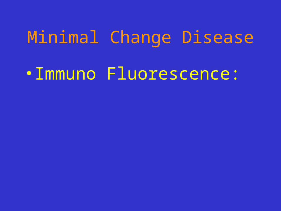
• Electron Microscopy:

Minimal Change Disease
• Electron Microscopy:
–Loss of Foot Processes of epithelial cells
–Uniform and Diffuse

Minimal Change Disease
• Prognosis

Minimal Change Disease
• The changes are reversible
• 90% respond to steroid
• May recur
• 5% progress to CRF

Membranous Glomerulonephritis
• Clinical Presentation

Membranous Glomerulonephritis
• Nephrotic syndrome
• Non-selective proteinuria
• 30-50 years of age

Membranous Glomerulonephritis
• Primary or Secondary

Membranous Glomerulonephritis
• Primary or Secondary– 85% 15%
» 1. Infections (HepB, malaria)» 2. Cancer (Lung ca, colon ca)» 3. SLE» 4. Gold, mercury» 5. Drugs (penicillamine, capropril,
NSAID’s)

Membranous Glomerulonephritis
• Pathogenesis

Membranous Glomerulonephritis
• Pathogenesis
• Immune complex
–Mostly: In-situ
–Few: Circulating
• Complement activation (MAC)

Membranous Glomerulonephritis
• Light microscopy:

Membranous Glomerulonephritis
• Light microscopy:
• Thick GBM (diffuse)
• Spikes

Membranous Glomerulonephritis• Electron microscopy:

Membranous Glomerulonephritis
• Electron microscopy:
• Sub-epithelial deposits

Membranous Glomerulonephritis
• Immuno Fluorescence:

Membranous Glomerulonephritis
• Immuno Fluorescence:
• Ig, comp. deposits

Membranous Glomerulonephritis
• Prognosis

Membranous Glomerulonephritis
• Less response to steroid
• 40% progress to CRF

Minimal Change GN
Focal Segmental
GN
Membranous GN
Membrano-proliferative GN
Good Prognosis
Bad Prognosis

Focal Segmental Glomerulosclerosis
• Primary or Secondary

Focal Segmental Glomerulosclerosis
• Primary or Secondary
»1. Infections (HIV)
»2. Drugs/Toxins (Heroin)
»3. Inherited
»4. Progress from other GN

Focal Segmental Glomerulosclerosis
• Pathogenesis

Focal Segmental Glomerulosclerosis
• Pathogenesis:
• Relation to Minimal change GN
• Progression from other GN
• Ablation nephropathy

Focal Segmental Glomerulosclerosis
• Light microscopy

Focal Segmental Glomerulosclerosis
• Light microscopy:
• Hyalinosis/Sclerosis

Focal Segmental Glomerulosclerosis
• Immuno Fluorescence:

Focal Segmental Glomerulosclerosis
• Immuno Fluorescence:
• IgM, Comp.

Focal Segmental Glomerulosclerosis
• Electron Microscopy:

Focal Segmental Glomerulosclerosis
• Electron Microscopy:
• Not specific
• Deposits in areas of hyalinosis
• Epithelial cell detachment

Focal Segmental Glomerulosclerosis
• Prognosis

Focal Segmental Glomerulosclerosis
• 50% progress to CRF
• Recurrence in transplant
• Collapsing FSGS

Membranoproliferative Glomerulonephritis
• Clinical presentation

Membranoproliferative Glomerulonephritis
• Nephrotic, Nephritic-Nephrotic
Membranoproliferative Glomerulonephritis
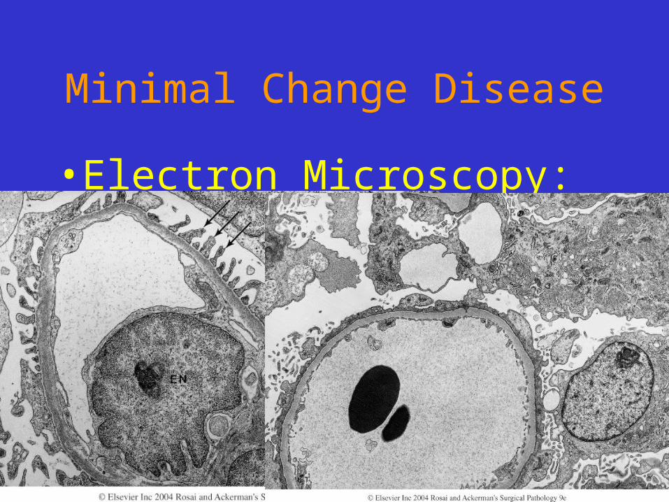
• Light Microscopy:

Membranoproliferative Glomerulonephritis
• Light Microscopy:
• Thick GBM
• Tram-Track
• Large Glomeruli
• Mesangial proliferation

Membranoproliferative Glomerulonephritis
• Electron Microscopy:

Membranoproliferative Glomerulonephritis
• Electron Microscopy:
• Type I: sub-endothelial deposits
• Type II: dense-deposit disease (within GBM)

Membranoproliferative Glomerulonephritis
• Immuno Fluorescence:
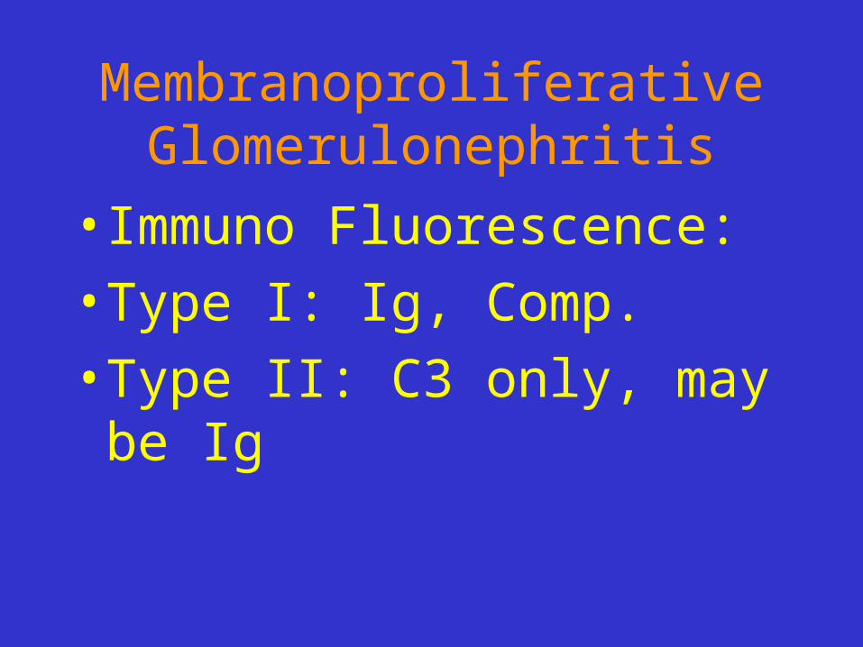
Membranoproliferative Glomerulonephritis
• Immuno Fluorescence:
• Type I: Ig, Comp.
• Type II: C3 only, may be Ig

Membranoproliferative Glomerulonephritis
• Prognosis:
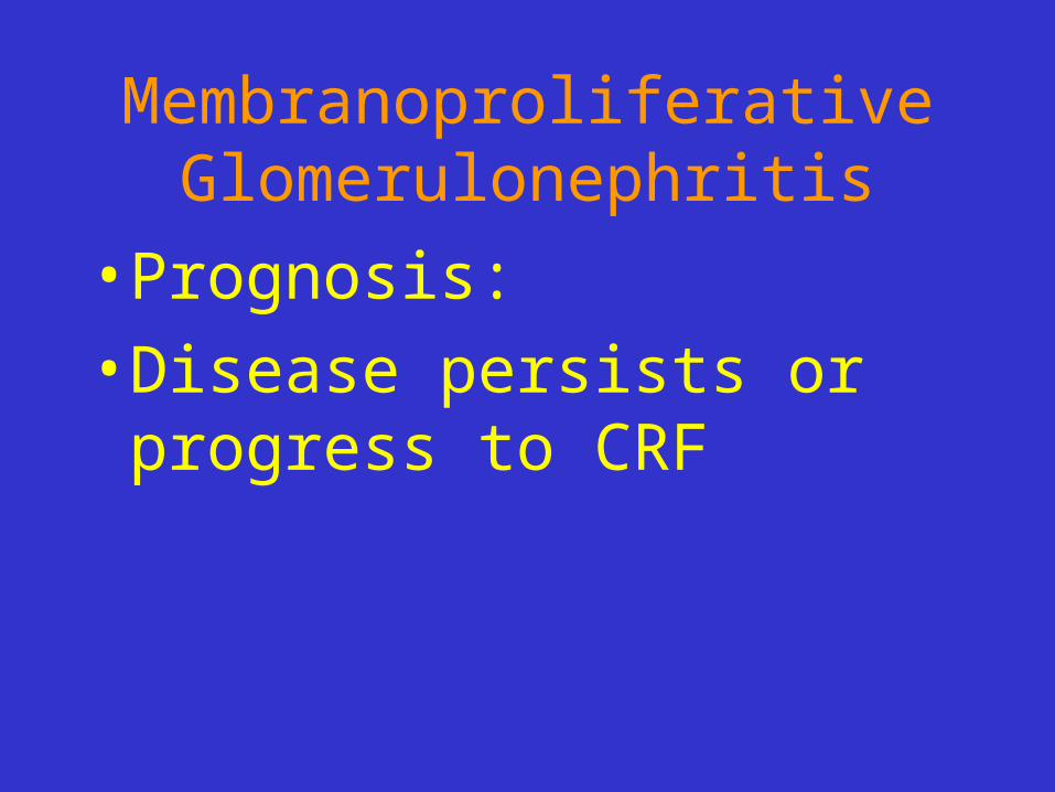
Membranoproliferative Glomerulonephritis
• Prognosis:
• Disease persists or progress to CRF

Minimal Change GN
Focal Segmental
GN
Membranous GN
Membrano-proliferative GN
Good Prognosis
Bad Prognosis