Neonatal pain facial expression: Evaluating the primal ...
12
Neonatal pain facial expression: Evaluating the primal face of pain Martin Schiavenato a, * , Jacquie F. Byers b , Paul Scovanner c , James M. McMahon a , Yinglin Xia d , Naiji Lu e , Hua He e a University of Rochester, School of Nursing, 601 Elmwood Ave., Box SON, Rochester, NY 14642-8404, USA b College of Nursing, University of Central Florida, Orlando, FL 32816, USA c School of Electrical Engineering and Computer Science, University of Central Florida, Orlando, FL 32816, USA d Department of Medicine, University of Rochester, Rochester NY 14642, USA e Department of Biostatistics and Computational Biology, University of Rochester, Rochester NY 14642, USA Received 29 November 2007; received in revised form 1 July 2008; accepted 9 July 2008 Abstract The primal face of pain (PFP) is postulated to be a common and universal facial expression to pain, hardwired and present at birth. We evaluated its presence by applying a computer-based methodology consisting of ‘‘point-pair” comparisons captured from video to measure facial movement in the pain expression by way of change across two images: one image before and one image after a painful stimulus (heel-stick). Similarity of facial expression was analyzed in a sample of 57 neonates representing both sexes and 3 ethnic backgrounds (African American, Caucasian and Hispanic/Latino) while controlling for these extraneous and potentially modulating factors: feeding type (bottle, breast, or both), behavioral state (awake or asleep), and use of epidural and/or other peri- natal anesthesia. The PFP is consistent with previous reports of expression of pain in neonates and is characterized by opening of the mouth, drawing in of the brows, and closing of the eyes. Although facial expression was not identical across or among groups, our analyses showed no particular clustering or unique display by sex, or ethnicity. The clinical significance of this commonality of pain display, and of the origin of its potential individual variation begs further evaluation. Ó 2008 International Association for the Study of Pain. Published by Elsevier B.V. All rights reserved. Keywords: Neonatal pain; Primal face of pain; Facial expression; Pain assessment; PFP 1. Introduction The ability to express pain is thought to be present at birth as an adaptation to species survival [34]. An evolu- tionary view proposes that facial expression of emotions in general is a hardwired ability that is modulated through learned behavior [7,11]. Specifically to pain, Craig and colleagues’ [5] Sociocommunication Model of Infant Pain proposes that the facial expression of pain in infants is a product of both biological and social fac- tors. That is, it can be theorized that infants are equipped with a ‘‘primal face of pain” (PFP), an inborn or biologically determined ability to display distress like pain, which is censored or modulated through various developmental and sociocultural factors [30]. Support- ing this is evidence that facial expressions of pain are more consistent in infants than in adults [6]. However, although a consistent display of pain has been observed even across various stimuli [24], evidence supporting a universal display of pain in infants has not been conclu- sive and remains poorly understood. Regarding commonality of pain display, much has been written about the role of culture and race/ethnicity in the expression and overall assessment and treatment of pain [13,25,28]. However, it is not clear to what extent 0304-3959/$34.00 Ó 2008 International Association for the Study of Pain. Published by Elsevier B.V. All rights reserved. doi:10.1016/j.pain.2008.07.009 * Corresponding author. Tel.: +1 585 276 4037; fax: +1 585 273 1270. E-mail address: [email protected] (M. Schiavenato). www.elsevier.com/locate/pain Pain 138 (2008) 460–471
Transcript of Neonatal pain facial expression: Evaluating the primal ...
www.elsevier.com/locate/pain
Pain 138 (2008) 460–471
Neonatal pain facial expression: Evaluating the primal face of pain
Martin Schiavenato a,*, Jacquie F. Byers b, Paul Scovanner c, James M. McMahon a,Yinglin Xia d, Naiji Lu e, Hua He e
a University of Rochester, School of Nursing, 601 Elmwood Ave., Box SON, Rochester, NY 14642-8404, USAb College of Nursing, University of Central Florida, Orlando, FL 32816, USA
c School of Electrical Engineering and Computer Science, University of Central Florida, Orlando, FL 32816, USAd Department of Medicine, University of Rochester, Rochester NY 14642, USA
e Department of Biostatistics and Computational Biology, University of Rochester, Rochester NY 14642, USA
Received 29 November 2007; received in revised form 1 July 2008; accepted 9 July 2008
Abstract
The primal face of pain (PFP) is postulated to be a common and universal facial expression to pain, hardwired and present atbirth. We evaluated its presence by applying a computer-based methodology consisting of ‘‘point-pair” comparisons captured fromvideo to measure facial movement in the pain expression by way of change across two images: one image before and one image aftera painful stimulus (heel-stick). Similarity of facial expression was analyzed in a sample of 57 neonates representing both sexes and 3ethnic backgrounds (African American, Caucasian and Hispanic/Latino) while controlling for these extraneous and potentiallymodulating factors: feeding type (bottle, breast, or both), behavioral state (awake or asleep), and use of epidural and/or other peri-natal anesthesia. The PFP is consistent with previous reports of expression of pain in neonates and is characterized by opening of themouth, drawing in of the brows, and closing of the eyes. Although facial expression was not identical across or among groups, ouranalyses showed no particular clustering or unique display by sex, or ethnicity. The clinical significance of this commonality of paindisplay, and of the origin of its potential individual variation begs further evaluation.� 2008 International Association for the Study of Pain. Published by Elsevier B.V. All rights reserved.
Keywords: Neonatal pain; Primal face of pain; Facial expression; Pain assessment; PFP
1. Introduction
The ability to express pain is thought to be present atbirth as an adaptation to species survival [34]. An evolu-tionary view proposes that facial expression of emotionsin general is a hardwired ability that is modulatedthrough learned behavior [7,11]. Specifically to pain,Craig and colleagues’ [5] Sociocommunication Model of
Infant Pain proposes that the facial expression of painin infants is a product of both biological and social fac-
0304-3959/$34.00 � 2008 International Association for the Study of Pain. P
doi:10.1016/j.pain.2008.07.009
* Corresponding author. Tel.: +1 585 276 4037; fax: +1 585 2731270.
E-mail address: [email protected](M. Schiavenato).
tors. That is, it can be theorized that infants areequipped with a ‘‘primal face of pain” (PFP), an inbornor biologically determined ability to display distress likepain, which is censored or modulated through variousdevelopmental and sociocultural factors [30]. Support-ing this is evidence that facial expressions of pain aremore consistent in infants than in adults [6]. However,although a consistent display of pain has been observedeven across various stimuli [24], evidence supporting auniversal display of pain in infants has not been conclu-sive and remains poorly understood.
Regarding commonality of pain display, much hasbeen written about the role of culture and race/ethnicityin the expression and overall assessment and treatmentof pain [13,25,28]. However, it is not clear to what extent
ublished by Elsevier B.V. All rights reserved.
M. Schiavenato et al. / Pain 138 (2008) 460–471 461
these are environmental factors such as cultural normson communication and expression, versus possibleinborn genetic differences that govern facial display.Although consistently tracked in medical research, thelabels of ‘‘race” and ‘‘ethnicity” are obscure in meaningat best [36,37]. Similarly, gender/sex differences arereported in pain research and while some can be attrib-uted to social norms [23,27] others, as is the case of stud-ies in newborns, cannot. Guinsburg and colleagues [17]found more facial expression on term and pre-termfemales, while Holditch-Davis and colleagues [18] foundthat pre-term males displayed more facial expression.On the other hand, Fuller [12] found no differencesbetween the sexes in facial expression at 2 weeks to 6months of age; while Grunau and Craig [15] found nosex differences in newborn expression but did find differ-ences in the speed of response with males demonstratinga quicker facial display than females.
Facial expressions form the cornerstone of clinicaland research pediatric pain instruments [30]; thus, theevaluation of a common and inherited facial expressionfor pain, the PFP that an evolutionary view would pur-port, is critically in question. The purpose of this studywas to compare the facial expression of term newbornsto a painful stimulus, across sex and ethnic back-grounds, while controlling for possible extraneous con-founding effects. Computer methods were applied inan attempt to refine the measurement of facialexpression.
2. Methods
2.1. Study design and sample
This prospective observational study used a quotasampling technique to recruit ‘‘well” neonates at thenewborn nursery of a large metropolitan hospital whowere (a) term (37–43 weeks gestation), without gesta-tional or delivery complications, without history ofmaternal drug use (see Table 1 for inclusion/exclusioncriteria), and (b) represented a specific sex and ethnicorigin: males and females ascribed as Asian, or Afri-can-American, or Caucasian, or Hispanic/Latino. Wedefine ethnicity here as a sociocultural, subjectivelyassigned and non-genetic attribute. The hospital’s con-vention of assigning the infant’s ‘‘race” according tothe mother’s self-ascribed ‘‘race” (i.e., that which coin-
Table 1Inclusion/exclusion criteria
1. Vaginal delivery2. Term pregnancy (37–43 week gestation)3. No history of pregnancy complications4. No history/evidence of genetic or congenital disorders5. No history of substance abuse during pregnancy6. Apgar scores at 1 and 5 min >6
cides with our definition and categories for ‘‘ethnicity”)at the time of admission was applied. Admittedly, thismethod does not identify infants of multi-ethnic,multi-racial background. To avoid potential confound-ing effects of cesarean anesthesia, only vaginal birthswere included. Metabolic screening (e.g. phenylketon-uria test) is State-mandated and the heel-stick requiredfor blood collection was chosen as the common painstimulus in the study. Hospital protocol required theprocedure to take place 24 h after beginning oral feedsand before discharge. The heel-stick was carried outby hospital staff per normal protocol using a standardlancet device. The neonate’s head and chest wererecorded using a tripod digital video camera (KodakZ740). The picture was focused on the neonate’s face;sound was recorded and the staff member was instructedto state ‘‘stick” immediately prior to engaging the lancet.Recorded events consisted of a brief baseline prior to theevent, the heel-stick, and the immediate reaction follow-ing it. Total analyzed video time did not exceed 30 s pernewborn.
2.2. Data collection
The study was approved by both university and hos-pital Institutional Review Boards, and parental consentwas obtained in advance. A chart review was performedto derive demographic and medical history data includ-ing sex, race/ethnicity, gestational age, postnatal age,birth measurements, Apgar scores, and evidence of preg-nancy or delivery complications. Use of epidural and orother perinatal anesthesia was noted for potentiallyinteractive neurobehavioral effects [3,26]. Similarly,feeding type, bottle, breast or both, was tracked to con-trol for potential differences in bonding and/or maternalcontact and interaction [35]. Behavioral state is impli-cated in pain expression with infants that are awakeexpressing more pain response [1,15,20,32]. Behavioralstate was scored at baseline as either asleep, character-ized by closed eyes; or awake, characterized by openeyes. Soothing techniques were utilized on fussing/whimpering infants to ensure a non-crying baseline.These infants were coded awake with eyes open orclosed.
2.3. Video analysis
Our examination of facial expression led us to theNeonatal Facial Coding System, NFCS [14], a com-monly used tool in this population and specificallydesigned for coding pain action. Facial action codedwith the NFCS and consistently associated with painin term neonates includes brow bulging, eyes squeezedshut, deepening of the nasolabial furrow, open mouth,and taut tongue [4,15,16,31]. These actions are generallycoded for presence/absence in video analyses and point
462 M. Schiavenato et al. / Pain 138 (2008) 460–471
to anatomical engagement (e.g. area of facial move-ment). A scheme was developed that allowed us to mea-sure (a) gross movement and cry (taut tongue, chin quiverand cry), and (b) fine facial activity (other implicatedfacial action based on the NFCS as listed above).‘‘Gross movement and cry” were evaluated by the prin-cipal investigator (a trained and certified NFCS rater)by viewing digital video recordings played back in realtime on a computer with frame by frame manipulationsoftware and noting the presence/absence of chin quiver,taut tongue and/or cry. These actions were looked atindependently because of their previous stated involve-ment in pain expression and because they were not con-ducive to our computerized image measurementmethods.
2.4. Baseline and reaction images
Baseline image was defined as a ‘‘neutral” or the non-crying/non-grimacing image immediately before theheel-stick. Reaction image was defined as the still picturedisplaying the initial moment of maximal expression fol-lowing the heel-stick. Baseline instantly preceded heel-stick, and reaction occurred almost immediately afterheel-stick. Specifically, for reaction, we were careful tocapture any facial action after the heel-stick but beforeany potentially intervening act such as squeezing of heelfor blood. Rad Video Tools (Rad Game Tools, Kirk-land, WA) was used to convert the digital video file intoa series of still images (jpegs) from which to choose base-line and reaction pictures. IrfanView (Irfan Skiljan,www.irfanview.net) was used to examine and select thetwo images.
2.5. Computerized image measurement
‘‘Fine facial activity” as tracked by this methodologyis an innovative attempt at measuring intensity of NFCSassociated pain behavior by means of digital image anal-ysis. Recent advances in computer vision are exploringsystems with the potential for automatic recognition offacial actions and identification of expressed emotion[2,33]. These computer systems use the Facial ActionCoding System (FACS) developed by Ekman and Frie-sen [10], the oldest and the most widely used tool forcoding facial expression in general. The NFCS was
Table 2NFCS facial action and point-pair comparison
NFCS facial actiona Corresponding point-pair
1.Brow bulge 1. Point-pair 1: between the2. Eye squeeze 2. Point-pair 2 (right side),3. Nasolabial furrow, cheek raise (proxy) 3. Point-pair 4 (right side),4. Horizontal mouth/lip movement 4. Point-pair 6: between lip5. Vertical mouth/lip movement 5. Point-pair 7: between me
a [4].
derived from the FACS [4,15]. Along this line ofresearch, a technique proposed by Pantic and Patras[21] was attempted to track facial action and measuremovement. They proposed using particle filtering totrack 20 facial points in a video image assuming a sta-tionary head or small head rotations. In earlier phasesof this research, we attempted similar affine transforma-tions to account for minor changes in head movement;however, these affine transformations were prone toerror due to the lack of a sufficient number of rigid reg-istration points on the face and to considerable headmovement by the infants following the heel-stick. Thus,we devised a ‘‘point-pair method” that used facial pointsbut rather than base the measurement on moving video,we used the two captured still images (baseline and reac-tion) for comparison to help overcome the issue of headmovement. This method accounts for rigid transforma-tions in a two dimensional image plane. Thus, manualimage selection performed by the principal investigatorattempted to minimize movement and approximateposition while capturing the fullness of the expression.Head movement, which was common across neonates,was evaluated and considered to introduce randomerror. This methodology does not completely precludethe subjectiveness associated with current methods inselecting or judging an image. However, it offers a higherlevel of measurement and precision in terms ofmovement as compared to current methods.
2.6. Point-pair method
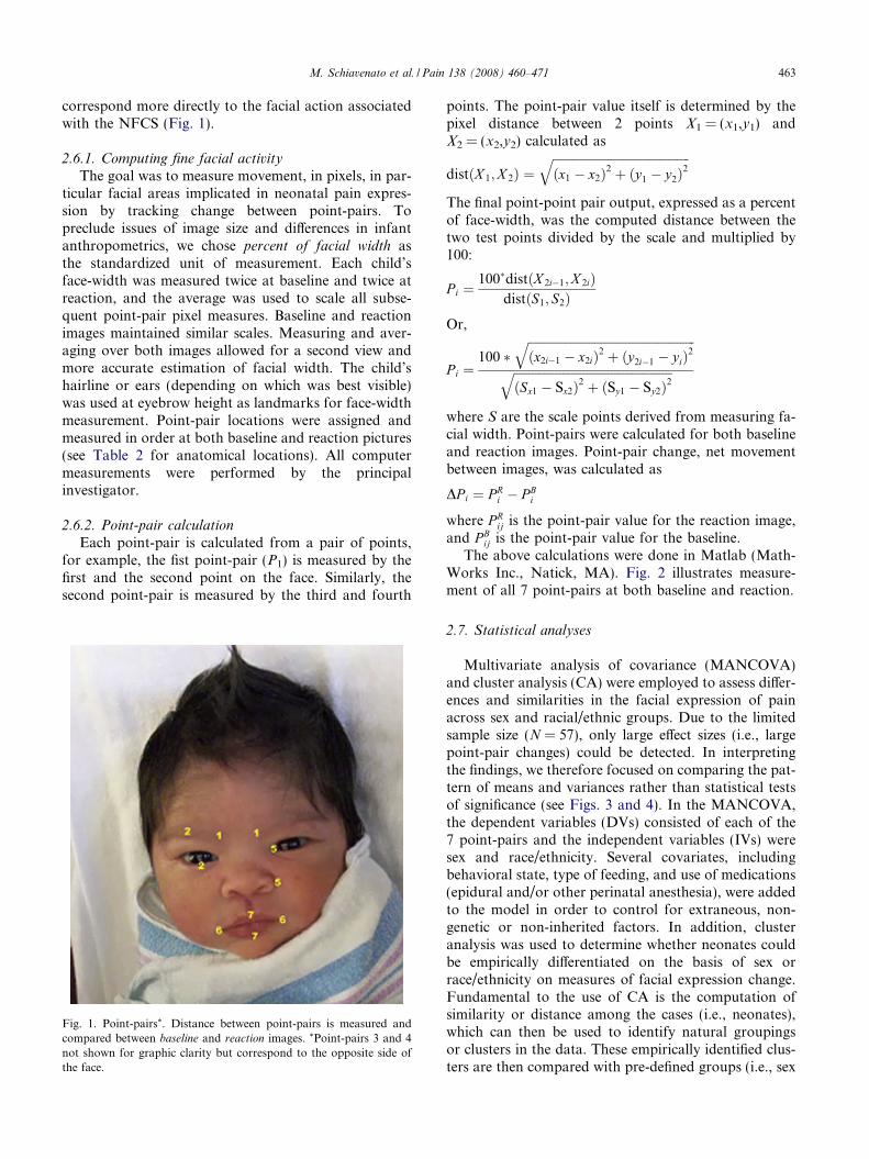
We devised 7 pairs of points (point-pairs) that coin-cide anatomically with the pain facial actions associatedwith the NFCS (Table 2). Because nasolabial furrowingor deepening of a portion of the face is difficult to mea-sure in a flat two-dimensional image, a substitute mea-sure was developed (point-pairs 4 and 5). Based onfacial action common to both the NFCS and FACS[4], ‘‘cheek raise” was used as a proxy for nasolabial fur-rowing. A point on the infraorbital triangle itself is dif-ficult to obtain because of the relative lack of anatomicallandmarks in the area. Therefore, the alar-facial groovewas chosen as a readily identifiable landmark, and itsmovement was anchored at the ipsilateral medial can-thus which is an assumed reference point with relativestability in facial expression. The remaining point-pairs
medial borders of the eyebrows; to track horizontal brow movementpoint-pair 3 (left side): from mid eyebrow to mid lower eyelidpoint-pair 5 (left side): from medial canthi to alar-facial groovecornersdial upper and lower lip vermilion border
M. Schiavenato et al. / Pain 138 (2008) 460–471 463
correspond more directly to the facial action associatedwith the NFCS (Fig. 1).
2.6.1. Computing fine facial activity
The goal was to measure movement, in pixels, in par-ticular facial areas implicated in neonatal pain expres-sion by tracking change between point-pairs. Topreclude issues of image size and differences in infantanthropometrics, we chose percent of facial width asthe standardized unit of measurement. Each child’sface-width was measured twice at baseline and twice atreaction, and the average was used to scale all subse-quent point-pair pixel measures. Baseline and reactionimages maintained similar scales. Measuring and aver-aging over both images allowed for a second view andmore accurate estimation of facial width. The child’shairline or ears (depending on which was best visible)was used at eyebrow height as landmarks for face-widthmeasurement. Point-pair locations were assigned andmeasured in order at both baseline and reaction pictures(see Table 2 for anatomical locations). All computermeasurements were performed by the principalinvestigator.
2.6.2. Point-pair calculation
Each point-pair is calculated from a pair of points,for example, the fist point-pair (P1) is measured by thefirst and the second point on the face. Similarly, thesecond point-pair is measured by the third and fourth
Fig. 1. Point-pairs*. Distance between point-pairs is measured andcompared between baseline and reaction images. *Point-pairs 3 and 4not shown for graphic clarity but correspond to the opposite side ofthe face.
points. The point-pair value itself is determined by thepixel distance between 2 points X1 = (x1,y1) andX2 = (x2,y2) calculated as
distðX 1;X 2Þ ¼ffiffiffiffiffiffiffiffiffiffiffiffiffiffiffiffiffiffiffiffiffiffiffiffiffiffiffiffiffiffiffiffiffiffiffiffiffiffiffiffiffiffiffiffiffiðx1 � x2Þ2 þ ðy1 � y2Þ
2q
The final point-point pair output, expressed as a percentof face-width, was the computed distance between thetwo test points divided by the scale and multiplied by100:
P i ¼100�distðX 2i�1;X 2iÞ
distðS1; S2ÞOr,
P i ¼100 �
ffiffiffiffiffiffiffiffiffiffiffiffiffiffiffiffiffiffiffiffiffiffiffiffiffiffiffiffiffiffiffiffiffiffiffiffiffiffiffiffiffiffiffiffiffiffiffiffiffiffiffiffiffiffiffiðx2i�1 � x2iÞ2 þ ðy2i�1 � yiÞ
2qffiffiffiffiffiffiffiffiffiffiffiffiffiffiffiffiffiffiffiffiffiffiffiffiffiffiffiffiffiffiffiffiffiffiffiffiffiffiffiffiffiffiffiffiffiffiffiffiffiffiffiffiðSx1 � Sx2Þ2 þ ðSy1 � Sy2Þ2
q
where S are the scale points derived from measuring fa-cial width. Point-pairs were calculated for both baselineand reaction images. Point-pair change, net movementbetween images, was calculated as
DP i ¼ P Ri � P B
i
where P Rij is the point-pair value for the reaction image,
and P Bij is the point-pair value for the baseline.
The above calculations were done in Matlab (Math-Works Inc., Natick, MA). Fig. 2 illustrates measure-ment of all 7 point-pairs at both baseline and reaction.
2.7. Statistical analyses
Multivariate analysis of covariance (MANCOVA)and cluster analysis (CA) were employed to assess differ-ences and similarities in the facial expression of painacross sex and racial/ethnic groups. Due to the limitedsample size (N = 57), only large effect sizes (i.e., largepoint-pair changes) could be detected. In interpretingthe findings, we therefore focused on comparing the pat-tern of means and variances rather than statistical testsof significance (see Figs. 3 and 4). In the MANCOVA,the dependent variables (DVs) consisted of each of the7 point-pairs and the independent variables (IVs) weresex and race/ethnicity. Several covariates, includingbehavioral state, type of feeding, and use of medications(epidural and/or other perinatal anesthesia), were addedto the model in order to control for extraneous, non-genetic or non-inherited factors. In addition, clusteranalysis was used to determine whether neonates couldbe empirically differentiated on the basis of sex orrace/ethnicity on measures of facial expression change.Fundamental to the use of CA is the computation ofsimilarity or distance among the cases (i.e., neonates),which can then be used to identify natural groupingsor clusters in the data. These empirically identified clus-ters are then compared with pre-defined groups (i.e., sex
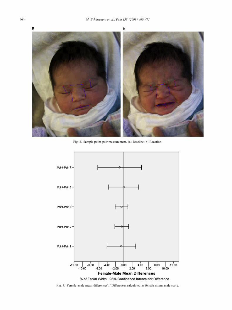
Fig. 2. Sample point-pair measurement. (a) Baseline (b) Reaction.
Fig. 3. Female–male mean differences*. *Differences calculated as female minus male score.
464 M. Schiavenato et al. / Pain 138 (2008) 460–471
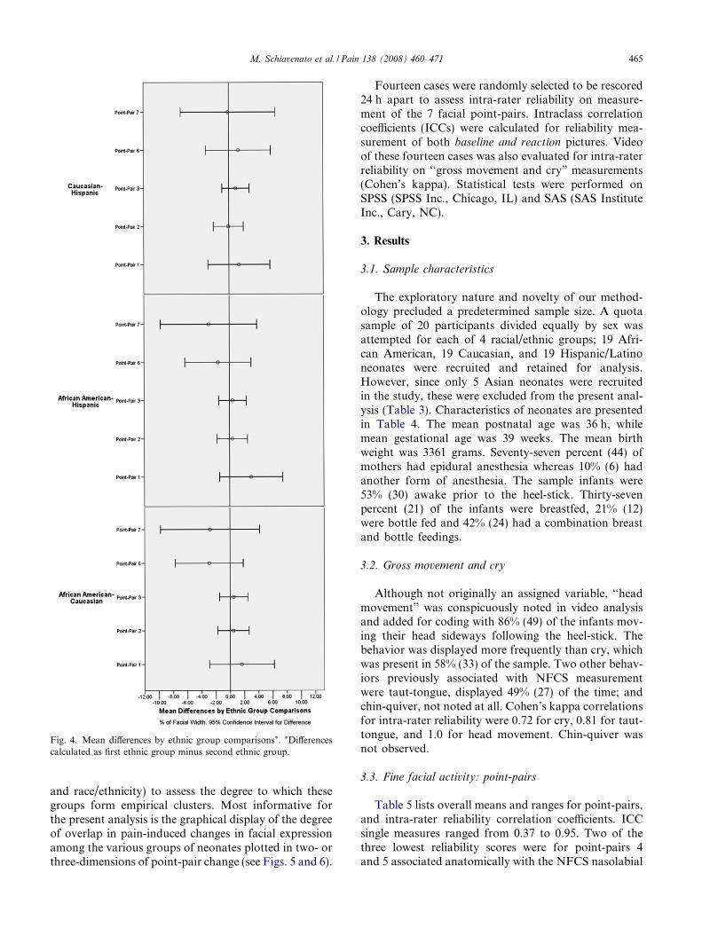
Fig. 4. Mean differences by ethnic group comparisons*. *Differencescalculated as first ethnic group minus second ethnic group.
M. Schiavenato et al. / Pain 138 (2008) 460–471 465
and race/ethnicity) to assess the degree to which thesegroups form empirical clusters. Most informative forthe present analysis is the graphical display of the degreeof overlap in pain-induced changes in facial expressionamong the various groups of neonates plotted in two- orthree-dimensions of point-pair change (see Figs. 5 and 6).
Fourteen cases were randomly selected to be rescored24 h apart to assess intra-rater reliability on measure-ment of the 7 facial point-pairs. Intraclass correlationcoefficients (ICCs) were calculated for reliability mea-surement of both baseline and reaction pictures. Videoof these fourteen cases was also evaluated for intra-raterreliability on ‘‘gross movement and cry” measurements(Cohen’s kappa). Statistical tests were performed onSPSS (SPSS Inc., Chicago, IL) and SAS (SAS InstituteInc., Cary, NC).
3. Results
3.1. Sample characteristics
The exploratory nature and novelty of our method-ology precluded a predetermined sample size. A quotasample of 20 participants divided equally by sex wasattempted for each of 4 racial/ethnic groups; 19 Afri-can American, 19 Caucasian, and 19 Hispanic/Latinoneonates were recruited and retained for analysis.However, since only 5 Asian neonates were recruitedin the study, these were excluded from the present anal-ysis (Table 3). Characteristics of neonates are presentedin Table 4. The mean postnatal age was 36 h, whilemean gestational age was 39 weeks. The mean birthweight was 3361 grams. Seventy-seven percent (44) ofmothers had epidural anesthesia whereas 10% (6) hadanother form of anesthesia. The sample infants were53% (30) awake prior to the heel-stick. Thirty-sevenpercent (21) of the infants were breastfed, 21% (12)were bottle fed and 42% (24) had a combination breastand bottle feedings.
3.2. Gross movement and cry
Although not originally an assigned variable, ‘‘headmovement” was conspicuously noted in video analysisand added for coding with 86% (49) of the infants mov-ing their head sideways following the heel-stick. Thebehavior was displayed more frequently than cry, whichwas present in 58% (33) of the sample. Two other behav-iors previously associated with NFCS measurementwere taut-tongue, displayed 49% (27) of the time; andchin-quiver, not noted at all. Cohen’s kappa correlationsfor intra-rater reliability were 0.72 for cry, 0.81 for taut-tongue, and 1.0 for head movement. Chin-quiver wasnot observed.
3.3. Fine facial activity: point-pairs
Table 5 lists overall means and ranges for point-pairs,and intra-rater reliability correlation coefficients. ICCsingle measures ranged from 0.37 to 0.95. Two of thethree lowest reliability scores were for point-pairs 4and 5 associated anatomically with the NFCS nasolabial
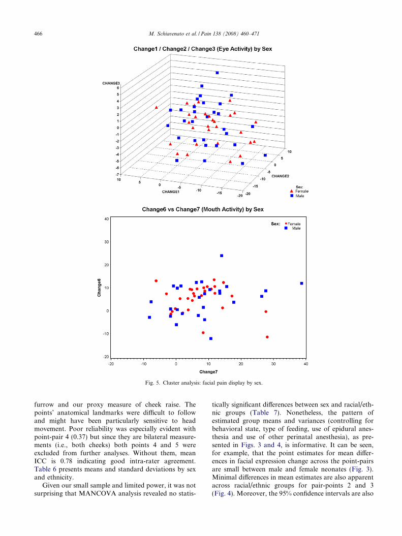
Fig. 5. Cluster analysis: facial pain display by sex.
466 M. Schiavenato et al. / Pain 138 (2008) 460–471
furrow and our proxy measure of cheek raise. Thepoints’ anatomical landmarks were difficult to followand might have been particularly sensitive to headmovement. Poor reliability was especially evident withpoint-pair 4 (0.37) but since they are bilateral measure-ments (i.e., both cheeks) both points 4 and 5 wereexcluded from further analyses. Without them, meanICC is 0.78 indicating good intra-rater agreement.Table 6 presents means and standard deviations by sexand ethnicity.
Given our small sample and limited power, it was notsurprising that MANCOVA analysis revealed no statis-
tically significant differences between sex and racial/eth-nic groups (Table 7). Nonetheless, the pattern ofestimated group means and variances (controlling forbehavioral state, type of feeding, use of epidural anes-thesia and use of other perinatal anesthesia), as pre-sented in Figs. 3 and 4, is informative. It can be seen,for example, that the point estimates for mean differ-ences in facial expression change across the point-pairsare small between male and female neonates (Fig. 3).Minimal differences in mean estimates are also apparentacross racial/ethnic groups for pair-points 2 and 3(Fig. 4). Moreover, the 95% confidence intervals are also
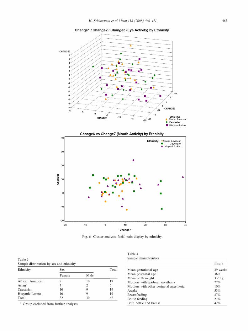
Fig. 6. Cluster analysis: facial pain display by ethnicity.
Table 3Sample distribution by sex and ethnicity
Ethnicity Sex Total
Female Male
African American 9 10 19Asiana 3 2 5Caucasian 10 9 19Hispanic Latino 10 9 19Total 32 30 62
a Group excluded from further analyses.
Table 4Sample characteristics
Result
Mean gestational age 39 weeksMean postnatal age 36 hMean birth weight 3361 gMothers with epidural anesthesia 77%Mothers with other perinatal anesthesia 10%Awake 53%Breastfeeding 37%Bottle feeding 21%Both bottle and breast 42%
M. Schiavenato et al. / Pain 138 (2008) 460–471 467
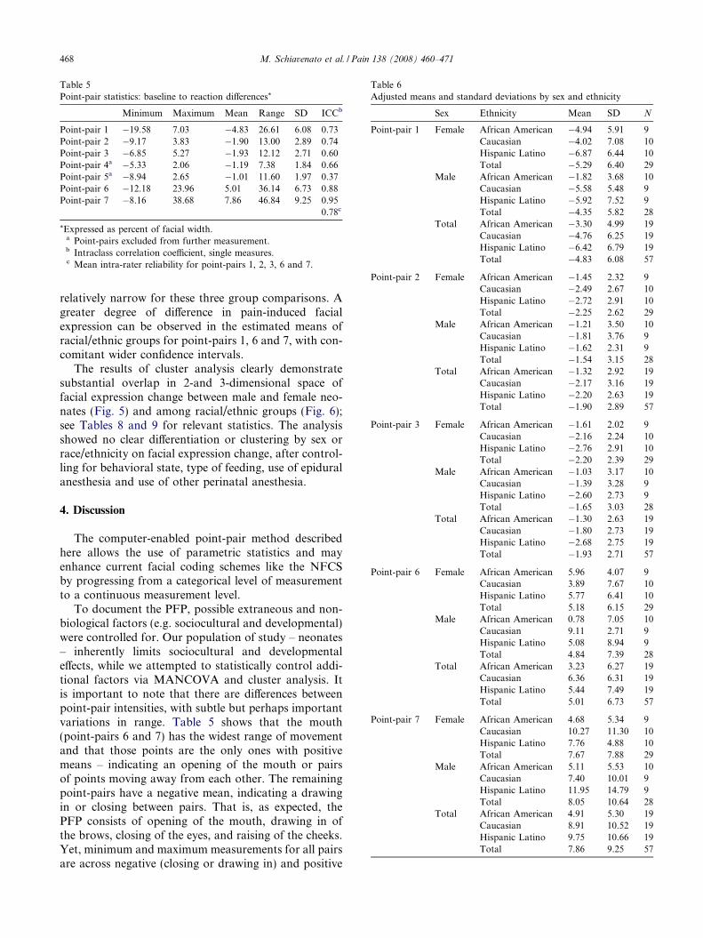
Table 5Point-pair statistics: baseline to reaction differences*
Minimum Maximum Mean Range SD ICCb
Point-pair 1 �19.58 7.03 �4.83 26.61 6.08 0.73Point-pair 2 �9.17 3.83 �1.90 13.00 2.89 0.74Point-pair 3 �6.85 5.27 �1.93 12.12 2.71 0.60Point-pair 4a �5.33 2.06 �1.19 7.38 1.84 0.66Point-pair 5a �8.94 2.65 �1.01 11.60 1.97 0.37Point-pair 6 �12.18 23.96 5.01 36.14 6.73 0.88Point-pair 7 �8.16 38.68 7.86 46.84 9.25 0.95
0.78c
*Expressed as percent of facial width.a Point-pairs excluded from further measurement.b Intraclass correlation coefficient, single measures.c Mean intra-rater reliability for point-pairs 1, 2, 3, 6 and 7.
Table 6Adjusted means and standard deviations by sex and ethnicity
Sex Ethnicity Mean SD N
Point-pair 1 Female African American �4.94 5.91 9Caucasian �4.02 7.08 10Hispanic Latino �6.87 6.44 10Total �5.29 6.40 29
Male African American �1.82 3.68 10Caucasian �5.58 5.48 9Hispanic Latino �5.92 7.52 9Total �4.35 5.82 28
Total African American �3.30 4.99 19Caucasian �4.76 6.25 19Hispanic Latino �6.42 6.79 19Total �4.83 6.08 57
Point-pair 2 Female African American �1.45 2.32 9Caucasian �2.49 2.67 10Hispanic Latino �2.72 2.91 10Total �2.25 2.62 29
Male African American �1.21 3.50 10Caucasian �1.81 3.76 9Hispanic Latino �1.62 2.31 9Total �1.54 3.15 28
Total African American �1.32 2.92 19Caucasian �2.17 3.16 19Hispanic Latino �2.20 2.63 19Total �1.90 2.89 57
Point-pair 3 Female African American �1.61 2.02 9Caucasian �2.16 2.24 10Hispanic Latino �2.76 2.91 10Total �2.20 2.39 29
Male African American �1.03 3.17 10Caucasian �1.39 3.28 9Hispanic Latino �2.60 2.73 9Total �1.65 3.03 28
Total African American �1.30 2.63 19Caucasian �1.80 2.73 19Hispanic Latino �2.68 2.75 19Total �1.93 2.71 57
Point-pair 6 Female African American 5.96 4.07 9Caucasian 3.89 7.67 10Hispanic Latino 5.77 6.41 10Total 5.18 6.15 29
Male African American 0.78 7.05 10Caucasian 9.11 2.71 9Hispanic Latino 5.08 8.94 9Total 4.84 7.39 28
Total African American 3.23 6.27 19Caucasian 6.36 6.31 19Hispanic Latino 5.44 7.49 19Total 5.01 6.73 57
Point-pair 7 Female African American 4.68 5.34 9Caucasian 10.27 11.30 10Hispanic Latino 7.76 4.88 10Total 7.67 7.88 29
Male African American 5.11 5.53 10Caucasian 7.40 10.01 9Hispanic Latino 11.95 14.79 9Total 8.05 10.64 28
Total African American 4.91 5.30 19Caucasian 8.91 10.52 19Hispanic Latino 9.75 10.66 19Total 7.86 9.25 57
468 M. Schiavenato et al. / Pain 138 (2008) 460–471
relatively narrow for these three group comparisons. Agreater degree of difference in pain-induced facialexpression can be observed in the estimated means ofracial/ethnic groups for point-pairs 1, 6 and 7, with con-comitant wider confidence intervals.
The results of cluster analysis clearly demonstratesubstantial overlap in 2-and 3-dimensional space offacial expression change between male and female neo-nates (Fig. 5) and among racial/ethnic groups (Fig. 6);see Tables 8 and 9 for relevant statistics. The analysisshowed no clear differentiation or clustering by sex orrace/ethnicity on facial expression change, after control-ling for behavioral state, type of feeding, use of epiduralanesthesia and use of other perinatal anesthesia.
4. Discussion
The computer-enabled point-pair method describedhere allows the use of parametric statistics and mayenhance current facial coding schemes like the NFCSby progressing from a categorical level of measurementto a continuous measurement level.
To document the PFP, possible extraneous and non-biological factors (e.g. sociocultural and developmental)were controlled for. Our population of study – neonates– inherently limits sociocultural and developmentaleffects, while we attempted to statistically control addi-tional factors via MANCOVA and cluster analysis. Itis important to note that there are differences betweenpoint-pair intensities, with subtle but perhaps importantvariations in range. Table 5 shows that the mouth(point-pairs 6 and 7) has the widest range of movementand that those points are the only ones with positivemeans – indicating an opening of the mouth or pairsof points moving away from each other. The remainingpoint-pairs have a negative mean, indicating a drawingin or closing between pairs. That is, as expected, thePFP consists of opening of the mouth, drawing in ofthe brows, closing of the eyes, and raising of the cheeks.Yet, minimum and maximum measurements for all pairsare across negative (closing or drawing in) and positive
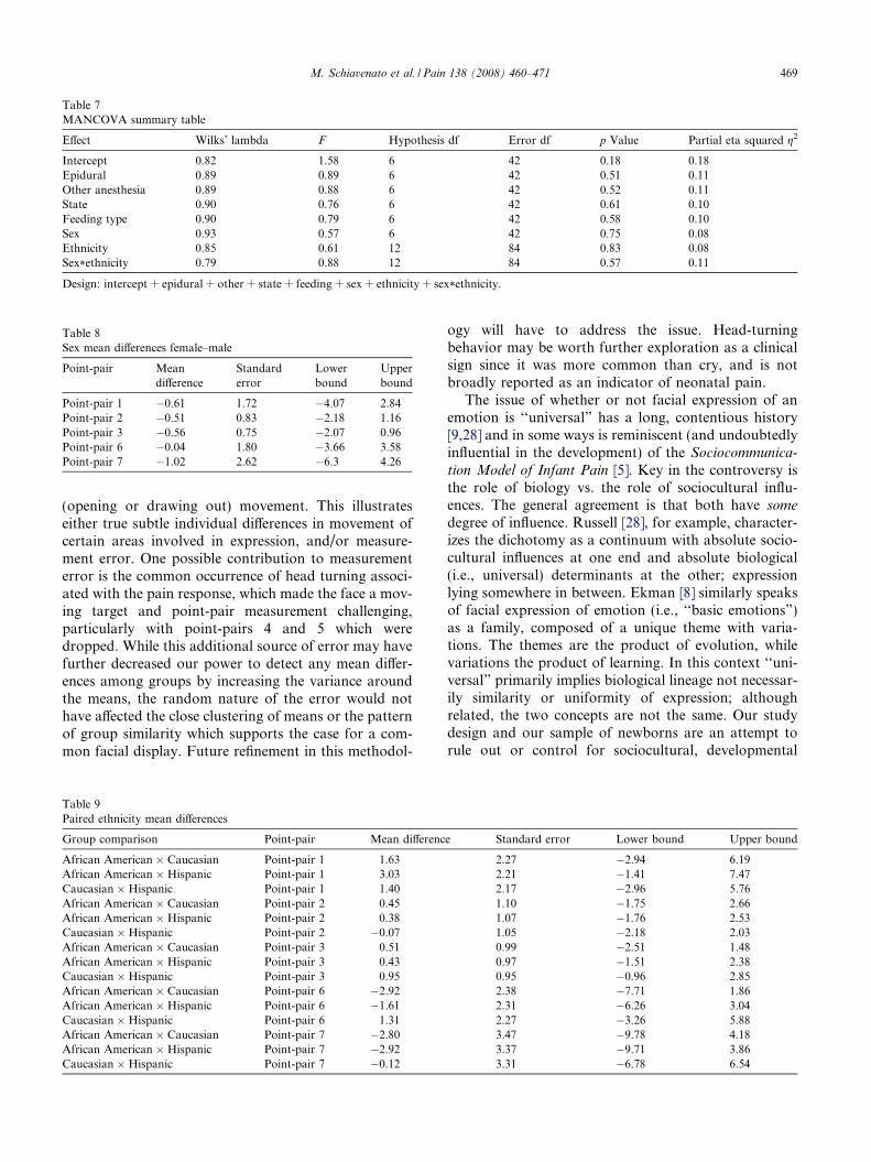
Table 7MANCOVA summary table
Effect Wilks’ lambda F Hypothesis df Error df p Value Partial eta squared g2
Intercept 0.82 1.58 6 42 0.18 0.18Epidural 0.89 0.89 6 42 0.51 0.11Other anesthesia 0.89 0.88 6 42 0.52 0.11State 0.90 0.76 6 42 0.61 0.10Feeding type 0.90 0.79 6 42 0.58 0.10Sex 0.93 0.57 6 42 0.75 0.08Ethnicity 0.85 0.61 12 84 0.83 0.08Sex*ethnicity 0.79 0.88 12 84 0.57 0.11
Design: intercept + epidural + other + state + feeding + sex + ethnicity + sex*ethnicity.
Table 8Sex mean differences female–male
Point-pair Meandifference
Standarderror
Lowerbound
Upperbound
Point-pair 1 �0.61 1.72 �4.07 2.84Point-pair 2 �0.51 0.83 �2.18 1.16Point-pair 3 �0.56 0.75 �2.07 0.96Point-pair 6 �0.04 1.80 �3.66 3.58Point-pair 7 �1.02 2.62 �6.3 4.26
M. Schiavenato et al. / Pain 138 (2008) 460–471 469
(opening or drawing out) movement. This illustrateseither true subtle individual differences in movement ofcertain areas involved in expression, and/or measure-ment error. One possible contribution to measurementerror is the common occurrence of head turning associ-ated with the pain response, which made the face a mov-ing target and point-pair measurement challenging,particularly with point-pairs 4 and 5 which weredropped. While this additional source of error may havefurther decreased our power to detect any mean differ-ences among groups by increasing the variance aroundthe means, the random nature of the error would nothave affected the close clustering of means or the patternof group similarity which supports the case for a com-mon facial display. Future refinement in this methodol-
Table 9Paired ethnicity mean differences
Group comparison Point-pair Mean differenc
African American � Caucasian Point-pair 1 1.63African American � Hispanic Point-pair 1 3.03Caucasian � Hispanic Point-pair 1 1.40African American � Caucasian Point-pair 2 0.45African American � Hispanic Point-pair 2 0.38Caucasian � Hispanic Point-pair 2 �0.07African American � Caucasian Point-pair 3 0.51African American � Hispanic Point-pair 3 0.43Caucasian � Hispanic Point-pair 3 0.95African American � Caucasian Point-pair 6 �2.92African American � Hispanic Point-pair 6 �1.61Caucasian � Hispanic Point-pair 6 1.31African American � Caucasian Point-pair 7 �2.80African American � Hispanic Point-pair 7 �2.92Caucasian � Hispanic Point-pair 7 �0.12
ogy will have to address the issue. Head-turningbehavior may be worth further exploration as a clinicalsign since it was more common than cry, and is notbroadly reported as an indicator of neonatal pain.
The issue of whether or not facial expression of anemotion is ‘‘universal” has a long, contentious history[9,28] and in some ways is reminiscent (and undoubtedlyinfluential in the development) of the Sociocommunica-
tion Model of Infant Pain [5]. Key in the controversy isthe role of biology vs. the role of sociocultural influ-ences. The general agreement is that both have some
degree of influence. Russell [28], for example, character-izes the dichotomy as a continuum with absolute socio-cultural influences at one end and absolute biological(i.e., universal) determinants at the other; expressionlying somewhere in between. Ekman [8] similarly speaksof facial expression of emotion (i.e., ‘‘basic emotions”)as a family, composed of a unique theme with varia-tions. The themes are the product of evolution, whilevariations the product of learning. In this context ‘‘uni-versal” primarily implies biological lineage not necessar-ily similarity or uniformity of expression; althoughrelated, the two concepts are not the same. Our studydesign and our sample of newborns are an attempt torule out or control for sociocultural, developmental
e Standard error Lower bound Upper bound
2.27 �2.94 6.192.21 �1.41 7.472.17 �2.96 5.761.10 �1.75 2.661.07 �1.76 2.531.05 �2.18 2.030.99 �2.51 1.480.97 �1.51 2.380.95 �0.96 2.852.38 �7.71 1.862.31 �6.26 3.042.27 �3.26 5.883.47 �9.78 4.183.37 �9.71 3.863.31 �6.78 6.54

470 M. Schiavenato et al. / Pain 138 (2008) 460–471
and other non-biological influences on the expression ofpain. In other words, the PFP documented here is anattempt to isolate and capture the universal, inborn,inherited expression associated with a painful stimulus.Based on our preliminary analyses, such a facial displayappears to be similar across sex and ethnicity. Or, saiddifferently, no systematic variations in facial pain dis-play were noted between males and females, or betweenthe 3 ethnic groups. This suggests that individual varia-tion in neonatal expression in response to pain may begenetically influenced. The role of genes in determiningnot just the presence of the display at birth but alsopotential variations within it deserves further examina-tion [21].
Discernment of what constitutes pain expression isof utmost importance in its assessment and manage-ment. The presence of a universal human facial paindisplay has theoretical and practical consequences inmeasurement tools like facial pain scales, which arebased on a common understanding of facial expres-sion in the evaluation of pain [30]. An importantproduct of this study is the establishment, in termsof our metric, of the effect of a heel-stick on facialexpression in our population. Questions that arisefrom this include, how does expression vary by stimu-lus intensity? Further, the mechanics of the expressionbeyond that which is presented here need further eval-uation. For example, how does the expression occuror how is it displayed across time? Finally, document-ing the nature of the PFP as a baseline expressionwith increasing precision as shown here may help toquantify the role of possible developmental and socio-cultural contributions in sculpting the expression ofpain throughout life.
Acknowledgements
Dr. Schiavenato thank Drs. Ken Craig and Xin Tufor their mentoring, encouragement and influence inthe development of this work. The authors also thankthe hospital which the study was conducted and thereviewers for their keen and lucid comments andsuggestions.
The authors have no financial or personal relation-ships or affiliations to disclose that could influence orbias our work on this manuscript.
References
[1] Ahn Y. The relationship between behavioral states and painresponses to various NICU procedures in premature infants. JTrop Pediatr 2006;52:201–5.
[2] Bartlett MS, Littlewort G, Frank MG, Lainscsek C, Fasel IR,Movellan J. Automatic recognition of facial actions in spontane-ous expressions. J Multimedia 2006;1:22–35.
[3] Beilin Y, Bodian CA, Weiser J, Hossain S, Arnold I, FeiermanDE, et al. Effect of labor epidural analgesia with and withoutfentanyl on infant breast-feeding: a prospective, randomized,double-blind study. Anesthesiology 2005;106:1211–7.
[4] Craig KD, Hadjistavropoulos HD, Grunau RV, Whitfield MF. Acomparison of two measures of facial activity during pain in thenewborn child. J Pediatr Psychol 1994;19:305–18.
[5] Craig KD, Korol CT, Pillai RR. Challenges of judging pain invulnerable infants. Clin Perinatol 2002;29:445–57.
[6] Craig KD, Prkachin KM, Grunau RE. The facial expression ofpain. In: Turk DC, Melzack R, editors. Handbook of painassessment. New York: The Guilford Press; 2001. p. 53–169.
[7] Ekman P. Biological and cultural contributions to body and facialmovements. In: Blacking J, editor. The anthropology of thebody. London: Academic Press; 1977. p. 34–84.
[8] Ekman P. Basic emotions. In: Dalgleish T, Power M, editors.Handbook of cognition and emotion. New York: John Wiley &Sons; 1999.
[9] Ekman P. Facial expressions. In: Dalgleish T, Power M, editors.Handbook of cognition and emotion. New York: John Wiley &Sons; 1999.
[10] Ekman P, Friesen WV. Facial action coding system. PaloAlto: Consulting Psychologists Press; 1978.
[11] Fridlund AJ. Human facial expression: an evolutionary view. SanDiego: Academic Press; 1994.
[12] Fuller BF. Infant gender differences regarding acute establishedpain. Clin Nurs Res 2002;11:190–203.
[13] Green CR, Anderson KO, Baker TA, Campbell LC, Decker S,Fillingim RB, et al. The unequal burden of pain: confrontingracial and ethnic disparities in pain. Pain Med 2003;4:277–94.
[14] Grunau RE, Oberlander T, Holsti L, Whitfield MF. Bedsideapplication of the neonatal facial coding system in pain assess-ment of premature neonates. Pain 1998;76:277–86.
[15] Grunau RE, Craig KD. Pain expression in neonates: facial actionand cry. Pain 1987;28:395–410.
[16] Grunau RE, Johnston CC, Craig KD. Neonatal facial and cryresponses to invasive and non-invasive procedures. Pain1990;42:295–305.
[17] Guinsburg RE, de Araujo Peres C, Branco de Almeida MF, deCassia Xavier Balda R, Cassia Berenguel R, Tonelotto J, et al.Differences in pain expression between male and female newborninfants. Pain 2000;85:127–33.
[18] Holditch-Davis D, Brandon DH, Schwartz T. Development ofbehaviors in preterm infants: relation to sleeping and waking.Nurs Res 2003;52:307–17.
[19] Johnston CC, Stevens BJ, Franck LS, Jack A, Stremler R, Platt R.Factors explaining lack of response to heel stick in pretermnewborns. J Obstet Gynecol Neonatal Nurs 1999;28:587–94.
[20] Pantic M, Patras I. Detecting facial actions and their temporalsegments in nearly frontal-view face image sequences. Presented atIEEE International Conference on Systems, Man and Cybernet-ics, Waikoloa, HI. October 10-12, 2005.
[21] Peleg G. Hereditary family signature of facial expression. ProcNatl Acad Sci USA 2006;103:15921.
[22] Pool GJ, Schwegler AF, Theodore BR, Fuchs PN. Role of gendernorms and group identification on hypothetical and experimentalpain tolerance. Pain 2007;129:122–9.
[23] Prkachin KM. The consistency of facial expressions of pain: acomparison across modalities. Pain 1992;51:297–306.
[24] Rahim-Williams FB, Riley III JL, Herrera D, Campbell CM,Hastie BA, Fillingim RB. Ethnic identity predicts experimentalpain sensitivity in African Americans and Hispanics. Pain2007;129:177–84.
[25] Ransjo-Arvidson AB, Matthiesen AS, Lilja G, Nissen E, Wid-strom AM, Uvnas-Moberg K. Maternal analgesia during labordisturbs newborn behavior: effects on breastfeeding, temperature,and crying. Birth 2001;28:5–12.

M. Schiavenato et al. / Pain 138 (2008) 460–471 471
[26] Robinson ME, Wise EA, Gagnon C, Fillingim RB, Price DD.Influences of gender role and anxiety on sex differences intemporal summation of pain. J Pain 2004;5:77–82.
[27] Rosmus C, Johnston CC, Chan-Yip A, Yang F. Pain response inChinese and non-Chinese Canadian infants: is there a difference?Soc Sci Med 2000;51:175.
[28] Russell JA. Facial expressions of emotion: what lies beyondminimal universality? Psycho Bull 1995;118:379.
[29] Schiavenato M. Facial expression and pain assessment in thepediatric patient: the primal face of pain. J Spec Pediatr Nurs2008;13.
[30] Stevens B, McGrath P, Gibbins S, Beyene J, Breau L, Camfield C,et al. Determining behavioural and physiological responses topain in infants at risk for neurological impairment. Pain2007;127:94–102.
[31] Stevens B, Johnston CC. Physiological responses of prematureinfants to a painful stimulus. Nurs Res 1994;43:226–31.
[32] Susskind JM, Littlewort G, Bartlett MS, Movellan J, AndersonAK. Human and computer recognition of facial expressions ofemotion. Neuropsychologia 2007;45:152–62.
[33] Williams AC. Facial expression of pain: an evolutionary account.Behav Brain Sci 2002;25:439–88.
[34] Winberg J. Mother and newborn baby: mutual regulation ofphysiology and behavior – a selective review. Dev Psychobiol2005;47:217–29.
[35] Winker MA. Measuring race and ethnicity: why and how? JAMA2004;292:1612–4.
[36] Winker MA. Race and ethnicity in medical research: requirementsmeet reality. J Law Med Ethics 2006;34:520–5. 480.