CLINICAL PHARMACY IN CARDIOLOGY CLINICAL PHARMACY IN CARDIOLOGY.

Neonatal Cardiology Review Nicole Bowie, NNP-/BC, PNP Neonatal Nurse Practitioner Jackson Memorial Hospital, Miami, FL
The speaker has signed a disclosure form and indicated she has no significant financial interest or relationship with the companies or the manufacturer(s) of any commercial product and/or service that will be discussed as part of this presentation.
Session Summary This session provides an overview of cyanotic, acyanotic, obstructive, and other congenital heart defects. There will also be a brief discussion regarding tachyarrhythmias, brady arrhythmias, and pulseless arrests, as well as compensated, decompensated, and irreversible shock.
Session Objectives Upon completion of this presentation, the participant will be able to:
understand fetal circulation;
understand assessment of the cardiac system;
discuss tachy and brady arrhythmias;
- recognize congenital heart disease, including - acyanotic lesions - obstructive lesions - cyanotic lesions
Test Questions 1. Infants with Tetralogy of Fallot who experience “hypoxic tet spells” are placed in knee chest
position in order to:
a. Increase the left to right shunting b. Increase the systemic vascular resistance c. Decrease the systemic vascular resistance
2. A 3-month-old with Down syndrome exhibits poor weight gain, tachypnea and grade 2/6
murmur. CX reveals cardiomegaly. Of the following, which is the MOST likely diagnosis?
a. Coarctation of the aorta b. Complete atrioventricular septal defect c. Perimembranous VSD
3. A pan systolic murmur is noted on exam and the infant also has bilateral ventricular dilatation
on ECHO and increased pulmonary vascularity on CXR. The likely etiology is:
a. Large PDA b. Large VSD c. Pulmonary stenosis
B12 FANNP 27th National NNP Symposium: Clinical Update and Review
B12: Neonatal Cardiology Review Page 1 of 12
4. A 28-week old infant on DOL 5 has a symptomatic PDA, he may experience which of the following symptoms:
a. Oliguria b. Hypertension c. Weak radial pulses
5. Pulmonary vascularity is decreased in all of the below except:
a. Tetralogy of Fallot b. TAPVR c. Tricuspid atresia
References Gomella, T. L., Cunningham ,D., & Eyal, F. G. (2009). Neonatology: Management, procedures, on-call problems,
diseases, and drugs (6th ed.). New York, NY: McGraw Hill Medical.
Kenner, C. & Lott, J. W. (2007). Comprehensive neonatal care: An interdisciplinary approach (4th ed.). St Louis, MO: Saunders Elsevier.
Verklan, M. T. & Walden, M. (2010). Core curriculum for neonatal intensive care nursing (4th ed.). St. Louis, MO: Saunders Elsevier.
FANNP 27th National NNP Symposium: Clinical Update and Review
B12: Neonatal Cardiology Review Page 2 of 12

CardiologyReviewNicole Thompson‐Bowie, NNP‐BC, PNP
Embryology• Begins developing between the 3rd to 7th week gestation with completion at 10 weeks
• 1st organ to function in utero
• Fetal heartbeat can be heard at 6wks
• Starts as long structure with 2 tubes
Embryology• Elongates & Twists to Right
• Separates the Atria and Ventricles
• Formation of Valves
• Mitral & Tricuspid
• Great Vessel Formation
• Aorta, Pulmonary Arteries and Veins
TheHeart
• The heart consists of four chambers
• Valves that open and close to allow blood to enter and leave these vessels and chambers.
• S1= Closing of TV and MV (AV valves)
• S2= Closing of AV and PV(semilunar valves)
Vena Cava
Right Ventricle
Right Atrium
Pulmonary Veins
Pulmonary Artery
Lungs
LeftVentricle
Left Atrium
Systemic CirculationRenal Vascular Bed(Volume Regulator))
Aorta
FANNP 27th National NNP Symposium: Clinical Update and Review
B12: Neonatal Cardiology Review Page 3 of 12
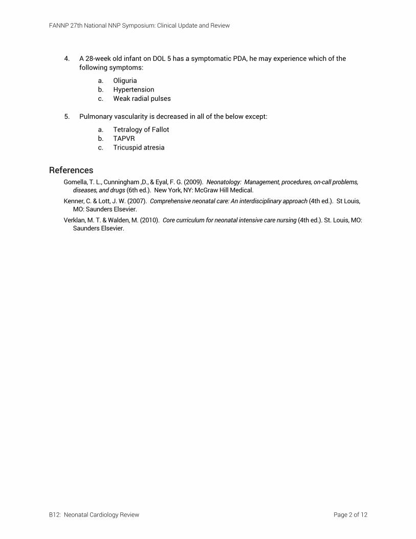
FetalCirculation
Things unique to Fetal Circulation
• Foramen Ovale
• Ductus Arteriosus
• Ductus Venosus
• Placenta
• Umbilical Vessels
• Dominant Right Heart – pumping 2/3 of combined ventricular output
High pulmonary vascular resistance
Low resistance placenta
Parallel Circulation
CVO
RV LV
2/3 1/3
Systemic circulation
ExtrauterineCardiovascularChanges
• Pulmonary Vascular resistance (PVR)
• Systemic Vascular Resistance (SVR)
• Ductus Arteriosus closes• Oxygen and PGE2 lessens
• Ductus Venosus closes
• Foramen Ovale closes
CardiacOutput
Cardiac Output ( CO )
• The volume of blood ejected by the heart in 1 minute
• CO = stroke volume x heart rate
• 200 ml/kg/min
• Neonates increase HR in response to low CO
Stroke Volume (SV) is the difference between the ventricular end diastolic volume and the end systolic volume (1.5ml/kg )
• SV is affected by preload, contractility and afterload
Contractility (inotropy)The speed of ventricular contractionContractility is affected byCatecholamine‐ increase contractilityAcidosis, hypoxia… decrease contractility
ConductionSystem
FANNP 27th National NNP Symposium: Clinical Update and Review
B12: Neonatal Cardiology Review Page 4 of 12

Bloodpressure• Measurement of the pressure on the walls of the vessels as blood is pumped
• Determined by • Peripheral vascular resistance • Cardiac output
• Systolic: end of each heart contraction• Diastolic: immediately before each contraction.• Pulse pressure
• Widened= PDA (blood runs off into pulmonary artery during diastole
• Narrow= pericardial tamponade, intravascular depletion and ECMO pt
Shock• State of inadequate circulatory blood volume
• Results in decreased perfusion and oxygenation to tissues lactic acidosis heart failure• Hypovolemic: loss of volume
• Acute blood loss, pleural effusion, skin disruption
• Cardiogenic• Heart fails due to tamponade, tension pneumothorax, CHD
• Distributive‐ sepsis, body release toxins
Compensated Uncompensated
BP still normal Hypotensive
AssessmentofCardiac• Physical Assessment & History
• Observation
• Auscultation
• Palpation : PMI
• Diagnostics
• EKG
• Chest XRAY
• Hyperoxia Test
• Pre and Post Ductal Saturations
• Echocardiogram
Sounds• S1: closure of MV/TV
• S2: closure of Ao/pulmonic valve. Should be split!
• S3: extra sound may be normal in newborn related to ventricle filling.
• S4‐rare, myocardial disease
S1
S2
Murmur• Turbulent blood flow • Innocent versus pathologic murmurs
• FT infant may have murmur @24‐48hr due to PDA closing benign
• Location• Intensity (1‐6)• Radiation• Timing
• Continuous: pathologic• Systole: usually benign• Diastole : PATHOLOGIC
MurmurandHeartSounds
VSD Harsh pansystolic LLSB
PDA Continuous machinery
Truncus Arteriosus
Harsh systolic, single S2
Valvular Stenosis Loud ejection click
PPS Radiates to axilla and back `
FANNP 27th National NNP Symposium: Clinical Update and Review
B12: Neonatal Cardiology Review Page 5 of 12

CommonElectrolyteDisturbances
• Hyperkalemia= peaked T waves, widened QRS
• Hypokalemia‐ prominent U waves
• Hypercalcemia= short Qt interval
• Hypocalcemia= prolonged Qt interval
Dysrhythmias
• Brady arrhythmias
• Sinus Bradycardia
• Heart Block
• Tachyarrhythmia
• Sinus tachycardia
• Supraventricular Tachycardia
SVT
Supraventricular tachycardia (SVT)• Heart rate sustained at > 220bpm
• Dual AV node pathway
• Treatment
• Ice
• Vagal maneuver
• Adenosine: rapid infusion 1‐2 sec followed by NS
• Cardioversion may be needed
CongenitalHeartDisease• <1% of all newborns,
• Prenatal Dx in about 50‐80% of the time
• 30% of patients with chromosomal anomalies have CHD
• Multifactorial causes (90% of cases )
• Biggest risk factor = Family History of CHD
IncidenceofDefectsTruncus
TAPVR
AV Canal
HPLH
Coarctation 5-8%
PDA 4-10% (in FT)
TGA 5%
ASD 5-10%
Pulmonary Stenosis (5-8%)
Tetralogy of Fallot (8-10%)
VSD 20-25%
Acyanotic Heart Disease Obstructive Lesions Cyanotic Heart Disease
Atrial septal defect (ASD) Aortic stenosis (AS) Transposition of the great arteries(TGA)
Ventricular septal defect (VSD)
Pulmonary stenosis (PS) Tetralogy of fallot (TOF)
Patent ductus arteriosus (PDA)
Coarctation of the aorta (CoA)
Total anomalous pulmonary venous return (TAPVR)
Atrioventricular Canal Truncus arterious (TA)
Tricuspid atresia
Pulmonary atresia
Hypoplastic left heart (HLHS)
Ebstein’s anomaly
FANNP 27th National NNP Symposium: Clinical Update and Review
B12: Neonatal Cardiology Review Page 6 of 12

I.AcyanoticHeartDefects
• Left to Right shunt
• Cardiomegaly
• Increased pulmonary vascular markings
• CHF when PVR drops
• Pulmonary over circulation
PatentDuctusArteriosus• Stealing effect from systemic circulation & the increased pulmonary blood flow………
Hypotension Oliguria
Peripheral vasoconstriction Metabolic acidosis
Hyper dynamic precordium Widened pulse pressure
Pulmonary edema; CHF Respiratory distress
Continuous Loud machinery murmur
ManagementofPDA:
• Term Infant
• Coiling closure at 3 months
• Preterm infant
• Conservative: Fluid restriction & Diuretics
• Hemodynamically significant PDA • Indomethacin/Ibuprofen
• Surgical Ligation
ASDHemodynamics • Oxygenated blood from the left atrium is shunted to the right atrium then into the right ventricle and back to the lungs
• Rarely get CHF• Systolic ejection murmur• The increased volume and work of the RV leads to RV hypertrophy
Management• Treat CHF• Intractable CHF: surgical repair is necessary
VSD• Most common CHD
• FLOW: L‐>R shunting via ventricular septum causing increased pulmonary blood flow
• Harsh pan systolic (holosystolic) murmur
• Urgency depends on size of VSD• Small: usually resolves by itself
• Large: causes CHF in 6‐8 weeks
ManagementofVSD
Mild VSD
Fluid restriction, Diuretics, Digoxin
Moderate to severe VSD
– pulmonary banding, suturing or patching the of the defect
FANNP 27th National NNP Symposium: Clinical Update and Review
B12: Neonatal Cardiology Review Page 7 of 12

AtrioventricularCanal• Abnormal development of the endocardial cushion
• Common in Down syndrome
• Treatment• PA Banding
• AD , VSD closure and reconstruction of valve
ASD
VSDFloppy tricuspid valve
Floppy mitral valve
Complete AV Canal Partial AV canal
1 valve Mitral regurgitation
CongestiveHeartFailure• The heart no longer able to pump adequate amount of blood to meet the needs of the body
• Results in systemic and venous congestion
• Can be caused by such things as CHD, infection, severe anemia, birth asphyxia and dysrhythmias
• Tachycardia, tachypnea,• sudden weight gain or poor weight gain
• Poor feeding • Hepatomegaly
• Arrhythmias
• Cardiomegaly
Eisenmenger’s Syndrome
II.LesionsObstructingBloodFlow
• Pulmonary Stenosis (PS)
• Aortic Stenosis (AS)
• Coarctation of the Aorta (CoAo)
PulmonaryStenosis
• Obstruction of blood flow to pulmonary bed
• May be valvular (90%), subvalvular, or supravalvular
• Usually associated with large VSD or ASD
• Sudden death is possible in more severe PS (Critical PS)
• Harsh Systolic ejection murmur
PulmonaryStenosis
• Usually normal heart size.
• Severe pulmonary stenosis will result in decreased pulmonary blood flow
AorticStenosis
• Obstruction of Blood flow to body
Types:• Valvular
• Supravalvular: usually associated with William’s Syndrome
• Subvalvular
• Peripheral pulses are weak and thready
• Narrow pulse pressure is present in severe AS
FANNP 27th National NNP Symposium: Clinical Update and Review
B12: Neonatal Cardiology Review Page 8 of 12
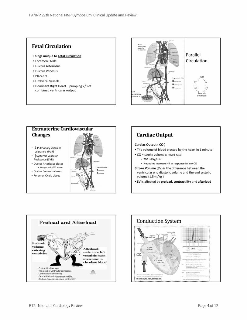
CoarctationoftheAorta• Strong pulses in upper extremities compared to lower extremities
• Severe cases may have
• LV pressure overload
• Loud S3 gallop is usually present
Mild headaches
Mod CHF
severe Shock
Management • Treat the heart failure (digoxin & Lasix )
• Prostin
• Surgical Intervention • Anastomosis
• Grafting
• Balloon angioplasty
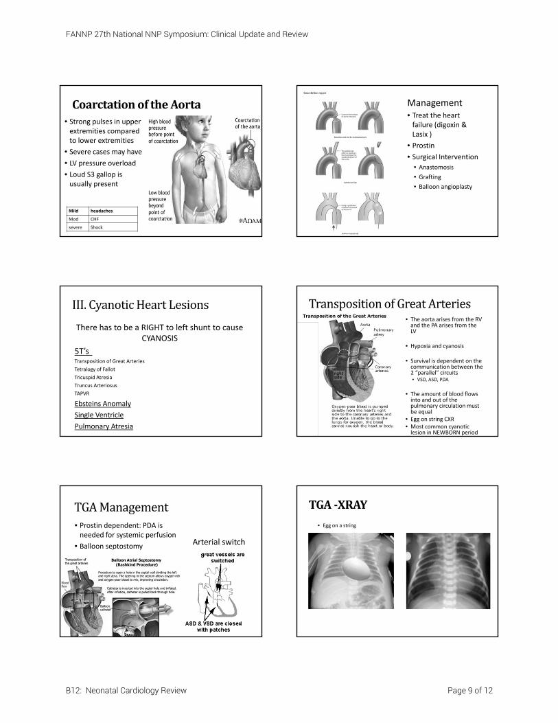
III.CyanoticHeartLesions
There has to be a RIGHT to left shunt to cause CYANOSIS
5T’sTransposition of Great Arteries
Tetralogy of Fallot
Tricuspid Atresia
Truncus Arteriosus
TAPVR
Ebsteins Anomaly
Single Ventricle
Pulmonary Atresia
TranspositionofGreatArteries• The aorta arises from the RV and the PA arises from the LV
• Hypoxia and cyanosis
• Survival is dependent on the communication between the 2 “parallel” circuits • VSD, ASD, PDA
• The amount of blood flows into and out of the pulmonary circulation must be equal
• Egg on string CXR• Most common cyanotic lesion in NEWBORN period
TGAManagement• Prostin dependent: PDA is needed for systemic perfusion
• Balloon septostomy Arterial switch
TGA‐XRAY
• Egg on a string
FANNP 27th National NNP Symposium: Clinical Update and Review
B12: Neonatal Cardiology Review Page 9 of 12

TetralogyofFallot• MOST COMMON CYANOTICHEART DISEASE
• Includes 4 abnormalities:
1) RVOT obstruction
2) RVH
3) VSD
4) overriding of the aorta
Severity depends on pulmonary
stenosis degree
Cyanosis Sats 75‐85%
Boot shaped heart
Tet (Hypoxic )Spells Knee chest, morphine, O2, beta blocker
Murmur SEM
ManagementofTOF
• Treat CHF
• Prostin
• Surgical
• Blalock‐Taussig Shunt: subclavian to pulmonary artery
• Total surgical correction 3‐6 months
TOF‐XRAY
Boot shape heart on X‐RAY Excuse me, what atrium was I suppose to connect to????
Total Anomalous Pulmonary Venous Return
The pulmonary veins drain oxygenated blood directly or indirectly into the right atrium instead of the left atrium
TAPVR• Obstructive cyanosis due to R‐> L mixing at ASD level
• Nonobstructive CHF
• XRAY: Snowman Heart
• Surgical Correction: The pulmonary veins are reconnected to the left atrium and the ASD is closed. Performed within the first weeks after the child’s birth
TAPVRXRAY
• Snowman
FANNP 27th National NNP Symposium: Clinical Update and Review
B12: Neonatal Cardiology Review Page 10 of 12

TruncusArteriosus
• Only a single arterial trunk leaves the heart – supplies pulmonary, systemic and coronary circulation
• Large VSD is always present
• Cyanosis varies and depends on the amount of Pulmonary blood flow
• Associated with DiGeorge syndrome
TruncusArteriosusManagement
• Rastelli Operation
• Conduit is placed from the Right Ventricle to the Pulmonary Artery
• Tricuspid valve is absent, RV and PA are Hypoplastic with decreased PBF
• 1‐2% of all CHD• ASD, VSD, or PDA are necessary for survival
• Single S2
TricuspidAtresia ManagementofTricuspidAtresiaB‐T Shunt Glenn Fontan
Need to get blood into pulmonary artery circulation
PulmonaryAtresia• Communication at the atrial level is necessary for life
• These patients are duct dependent
• Single S2
Ebstein’sAnomaly
• Extremely large heart
• Abnormal development of the tricuspid valve
• Weak TV PVR Cyanosis
FANNP 27th National NNP Symposium: Clinical Update and Review
B12: Neonatal Cardiology Review Page 11 of 12

Ebstein’sAnomaly
• Treatment
• Prostin
• Treat heart failure
• Pulmonary artery banding
• Surgery
HypoplasticLeftHeartSyndrome
• 1 – 2% of all CHD
• Must have PFO/ASD – allow LA to receive oxygenated blood
• PDA dependent to ensure systemic circulation
Left Heart Underdeveloped
1. LV Hypoplastic
2. Aortic Valve atresia or stenosis
3. Mitral valve atresia or stenosis
4. Aortic arch Hypoplastic
HLHS
Presentation‐ HLHSCyanosis Signs an symptoms of CHFPoor perfusion : pulmonary over‐circulationSever metabolic acidosis
HypoplasticLeftHeartSyndrome
Medical Management:• Compassionate Care
• PGE1 infusion
• Must balance circuit of pulmonary and systemic circulation
• Keep sats 75 to 85%
• Avoid excessive pulmonary vasodilation PBF CHF
Surgical Management:
• Norwood: rebuild the tiny ascending aorta
• Stage II: Glenn Operation
• State III: Fontan procedure
• Cardiac Transplant
Ruleof4’sinCardiacPatient
• pH= should be 7.40• Acidosis= lactic acid build up= muscle fatigue= bad cardiac contractility and function
• CO2= in the 40’s• respiratory acidosis
• Hematocrit= at least 40• Need higher Oxygen carrying capacity
• Potassium= level in the 4 range• Na/K pump regulates influx of electrical impulses to regulate heart muscle contraction.
• Hyperkalemia can create lethal arrhythmias
Maternal DiabetesHypertrophic cardiomyopathy, TGA, VSD
Maternal Lupus Heart Block
Maternal Alcohol Abuse TOF
Maternal Rubella PDA, PPS
Down's syndrome40% have CHD,AVC, VSD most common
Turner syndrome Coarctation of the aorta
DiGeorge Syndrome Truncus arteriosus
FANNP 27th National NNP Symposium: Clinical Update and Review
B12: Neonatal Cardiology Review Page 12 of 12