CONGENITAL CARDIOLOGY TODAY · pediatric electrophysiology program is ... patient is managed by the...
20
Introduction Extremely Low Birth Weight (ELBW) infants (weight <1kg at birth) frequently present with a large, hemodynamically significant Patent Ductus Arteriosus (PDA). 1 The PDA in ELBW infants can contribute to worse outcomes. 2-4 However, there are no management algorithms that exist for ELBW infants with a hemodynamically significant PDA. Established treatment options include the administration of cyclooxygenase (COX) inhibitors and surgical ligation of the PDA (SLP) via a thoracotomy. COX-inhibitors are not always effective. 5-7 SLP has not been shown to positively impact survival. 5,7-9 Procedural complications and long-term sequelae 7-9 have made this option less attractive. There is also a general belief that treatment of the PDA beyond the first few weeks of life in the ELBW infants may not significantly alter outcomes. 1,3,5-6 This has led to a practice of a trial of medical therapy in the first 2-3 weeks of life, and if unsuccessful, no further intervention is sought. 2-3, 6-7 Many neonates continue to languish with a large, hemodynamically significant PDA on a ventilator with evolving Chronic Lung Disease (CLD) and Pulmonary Hypertension (PHT). Transcatheter PDA closure (TCPC) is the established mode of treatment for PDAs in children >5 kg.10. TCPC in ELBW infants is slowly being implemented by many centers. 11-13 Advantages of TCPC in ELBW newborns include immediate PDA closure compared to several days to weeks until closure with medications, 11-12 and that TCPC is less invasive than SLP. 11-12 Despite these perceived advantages, TCPC does not feature in the current treatment algorithm for PDA in the ELBW newborn. The challenges that prevent this therapy from becoming readily available to this population of patients include: a general concern for any procedural adverse events in extremely small babies, need for miniaturizing catheters and devices required for TCPC, concern of transporting the patient to a different environment for the procedure, use of radiation and contrast to perform TCPC, and lack of awareness of the availability of TCPC as a treatment option for ELBW newborns, among others. The aim of this report is to describe how we established a robust TCPC program for ELBW infants. The TCPC Program for ELBW Infant Currently, we have a one-of-a-kind infants comprehensive PDA program unlike anything in the United States which has enabled us to provide exceptional care for these infants both during their hospitalization and throughout their childhood. This has been accomplished through several years of extremely collegial and collaborative work between the neonatologists, cardiologists, congenital cardiac surgeons, and pulmonologists, among others, along with support from hospital administration. Generally, any ELBW newborn with a large, hemodynamically-significant PDA would have a course of medical therapy to close the PDA. If the therapy fails (the PDA is still large and hemodynamically significant based on echocardiographic and clinical assessment), Establishing a Robust Transcatheter PDA Closure Program for Extremely Low Birth Weight Infants March 2018; Volume 16; Issue 3 North American Edition IN THIS ISSUE Establishing a Robust Transcatheter PDA Closure Program for Extremely Low Birth Weight Infants By Shyam Sathanandam, MD; Leah Apalodimas, MSN, APN, CCRN, CPNP; Mark Weems, MD; B. Rush Waller, MD; Ranjit Philip, MD ~Page 1 Potts Shunt for Children with Severe Pulmonary Hypertension By Pirooz Eghtesady, MD, PhD; R. Mark Grady, MD ~Page 12 Medical News, Products & Information ~Page 15 CONGENITAL CARDIOLOGY TODAY Editorial and Subscription Offices 16 Cove Rd, Ste. 200 Westerly, RI 02891 USA www.CongenitalCardiologyToday.com Follow on Twitter @CCardiology Recruitment ads: pages 2, 5, 8, 13, 14, 16, 18, 19 CONGENITAL CARDIOLOGY TODAY Timely News and Information for BC/BE Congenital/Structural Cardiologists and Surgeons By Shyam Sathanandam, MD; Leah Apalodimas, MSN, APN, CCRN, CPNP; Mark Weems, MD; B. Rush Waller, MD; Ranjit Philip, MD MEDICAL MEETINGS Echocardiography of CHD Apr. 15-17, 2018; Philadelphia, PA USA https://chop.cloud-cme.com/ aph.aspx?P=5&EID=602# EPIC-SEC (Emory Practical Intervention Course Southeastern Consortium) Apr. 19-21, 2018; Atlanta, GA USA www.epicsec.org SCAI 2018 Apr. 25-27, 2018: San Diego, CA USA www.scai.org/SCAI2018/ 52 nd Annual Meeting of the AEPC May 9-12, 2018; Athens, Greece http://www.aepc2018.org International PDA Symposium May 18-19, 2018; Memphis, TN USA Call 901.516.8933 CONGENITAL CARDIOLOGY TODAY CALL FOR CASES AND OTHER ORIGINAL ARTICLES Do you have interesting research results, observations, human interest stories, reports of meetings, etc. to share? Submit your manuscript to: [email protected]
Transcript of CONGENITAL CARDIOLOGY TODAY · pediatric electrophysiology program is ... patient is managed by the...
Introduction
Extremely Low Birth Weight (ELBW) infants (weight <1kg at birth) frequently present with a large, hemodynamically significant Patent Ductus Arteriosus (PDA).1 The PDA in ELBW infants can contribute to worse outcomes.2-4 However, there are no management algorithms tha t ex i s t f o r ELBW in fan ts w i t h a hemodynamically significant PDA. Established treatment options include the administration of cyclooxygenase (COX) inhibitors and surgical ligation of the PDA (SLP) via a thoracotomy. COX-inhibitors are not always effective.5-7 SLP has not been shown to positively impact survival.5,7-9 Procedural complications and long-term sequelae7-9 have made this option less attractive. There is also a general belief that treatment of the PDA beyond the first few weeks of life in the ELBW infants may not significantly alter outcomes.1,3,5-6 This has led to a practice of a trial of medical therapy in the first 2-3 weeks of life, and if unsuccessful, no further intervention is sought.2-3, 6-7 Many neonates continue to languish with a large, hemodynamically significant PDA on a ventilator with evolving Chronic Lung Disease (CLD) and Pulmonary Hypertension (PHT).
Transcatheter PDA closure (TCPC) is the established mode of treatment for PDAs in children >5 kg.10. TCPC in ELBW infants is slowly being implemented by many centers.11-13 Advantages of TCPC in ELBW newborns include
immediate PDA closure compared to several days to weeks until closure with medications,11-12 and that TCPC is less invasive than SLP.11-12 Despite these perceived advantages, TCPC does not feature in the current treatment algorithm for PDA in the ELBW newborn. The challenges that prevent this therapy from becoming readily available to this population of patients include: a general concern for any procedural adverse events in extremely small babies, need for miniaturizing catheters and devices required for TCPC, concern of transporting the patient to a different environment for the procedure, use of radiation and contrast to perform TCPC, and lack of awareness of the availability of TCPC as a treatment option for ELBW newborns, among others. The aim of this report is to describe how we established a robust TCPC program for ELBW infants.
The TCPC Program for ELBW Infant
Currently, we have a one-of-a-kind infants comprehensive PDA program unlike anything in the United States which has enabled us to provide exceptional care for these infants both during their hospitalization and throughout their childhood. This has been accomplished through several years of extremely collegial and collaborative work between the neonatologists, cardiologists, congenital cardiac surgeons, and pulmonologists, among others, along with support from hospital administration. Generally, a n y E L B W n e w b o r n w i t h a l a r g e , hemodynamically-significant PDA would have a course of medical therapy to close the PDA. If the therapy fails (the PDA is still large and hemodynamically signif icant based on echocardiographic and clinical assessment),
Establishing a Robust Transcatheter PDA Closure Program for Extremely Low Birth Weight Infants
March 2018; Volume 16; Issue 3North American Edition
IN THIS ISSUE
Establishing a Robust Transcatheter PDA Closure Program for Extremely Low Birth Weight InfantsBy Shyam Sathanandam, MD; Leah Apalodimas, MSN, APN, CCRN, CPNP; Mark Weems, MD; B. Rush Waller, MD; Ranjit Philip, MD~Page 1
Potts Shunt for Children with Severe Pulmonary HypertensionBy Pirooz Eghtesady, MD, PhD; R. Mark Grady, MD~Page 12
Medical News, Products & Information ~Page 15
CONGENITAL CARDIOLOGY TODAYEditorial and Subscription Offices16 Cove Rd, Ste. 200Westerly, RI 02891 USAwww.CongenitalCardiologyToday.comFollow on Twitter @CCardiology
Recruitment ads: pages 2, 5, 8, 13, 14, 16, 18, 19
CONGENITAL CARDIOLOGY TODAYTimely News and Information for BC/BE Congenital/Structural Cardiologists and Surgeons
By Shyam Sathanandam, MD; Leah Apalodimas, MSN, APN, CCRN, CPNP; Mark Weems, MD; B. Rush Waller, MD; Ranjit Philip, MD
MEDICAL MEETINGS
Echocardiography of CHDApr. 15-17, 2018; Philadelphia, PA USAhttps://chop.cloud-cme.com/aph.aspx?P=5&EID=602#
EPIC-SEC (Emory Practical Intervention Course Southeastern Consortium)Apr. 19-21, 2018; Atlanta, GA USAwww.epicsec.org
SCAI 2018Apr. 25-27, 2018: San Diego, CA USAwww.scai.org/SCAI2018/
52nd Annual Meeting of the AEPCMay 9-12, 2018; Athens, Greecehttp://www.aepc2018.org
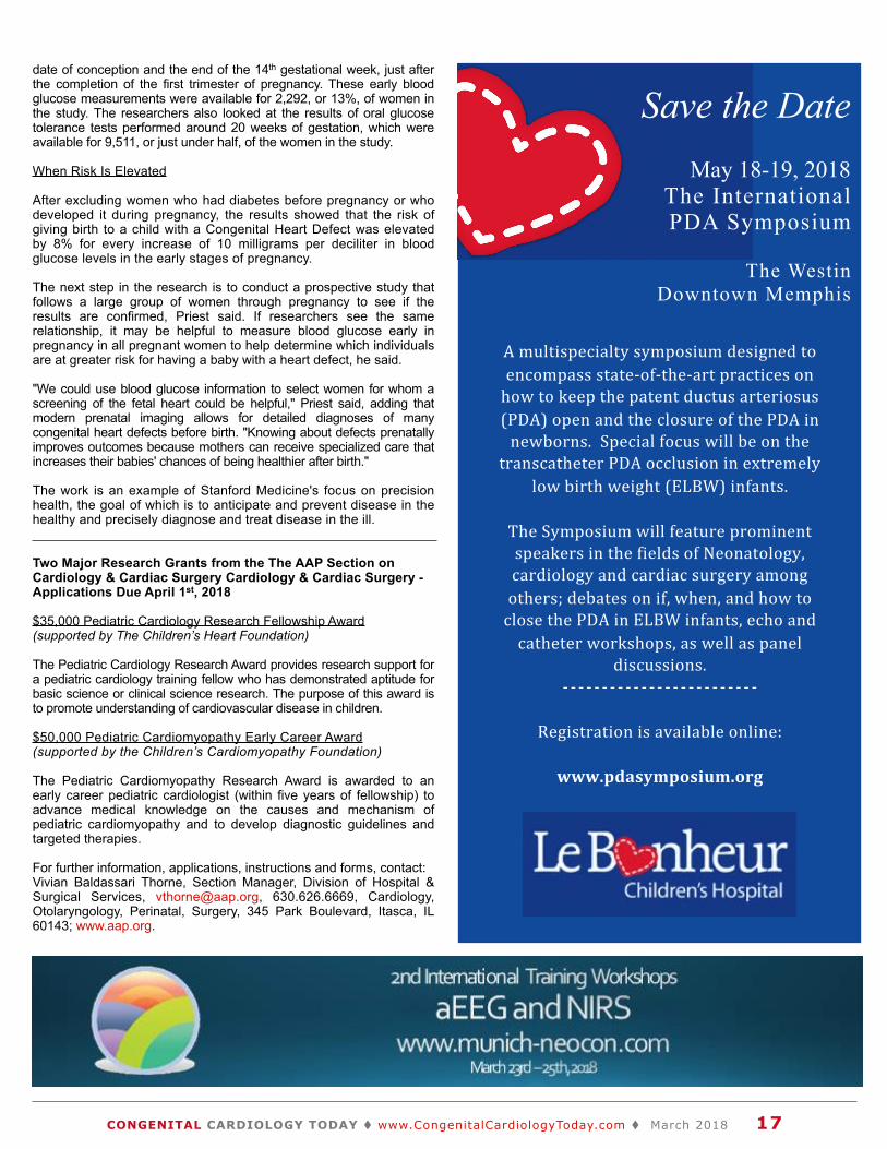
International PDA Symposium May 18-19, 2018; Memphis, TN USACall 901.516.8933 CONGENITAL CARDIOLOGY TODAY
CALL FOR CASES AND OTHER ORIGINAL ARTICLES
Do you have interesting research results, observations, human interest stories, reports of meetings, etc. to share? Submit your manuscript to: [email protected]
The Heart Institute at theCHILDREN’S HOSPITAL OF PITTSBURGH OF UPMC
Is EXPANDING!
With a strategic plan for growth and expansion, the Division of Cardiology within the Heart Institute of the Children’s Hospital of Pittsburgh of UPMC / University of Pittsburgh School of Medicine is recruiting additional faculty positions.
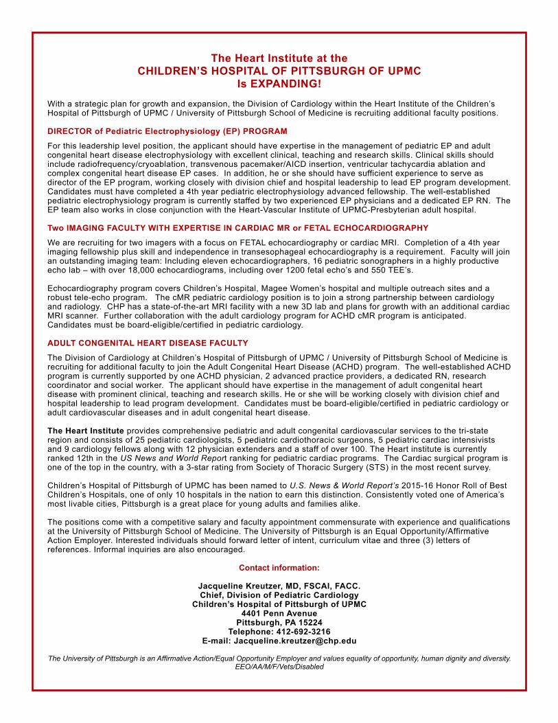
DIRECTOR of Pediatric Electrophysiology (EP) PROGRAM
For this leadership level position, the applicant should have expertise in the management of pediatric EP and adult congenital heart disease electrophysiology with excellent clinical, teaching and research skills. Clinical skills should include radiofrequency/cryoablation, transvenous pacemaker/AICD insertion, ventricular tachycardia ablation and complex congenital heart disease EP cases. In addition, he or she should have sufficient experience to serve as director of the EP program, working closely with division chief and hospital leadership to lead EP program development. Candidates must have completed a 4th year pediatric electrophysiology advanced fellowship. The well-established pediatric electrophysiology program is currently staffed by two experienced EP physicians and a dedicated EP RN. The EP team also works in close conjunction with the Heart-Vascular Institute of UPMC-Presbyterian adult hospital.
Two IMAGING FACULTY WITH EXPERTISE IN CARDIAC MR or FETAL ECHOCARDIOGRAPHY
We are recruiting for two imagers with a focus on FETAL echocardiography or cardiac MRI. Completion of a 4th year imaging fellowship plus skill and independence in transesophageal echocardiography is a requirement. Faculty will join an outstanding imaging team: Including eleven echocardiographers, 16 pediatric sonographers in a highly productive echo lab – with over 18,000 echocardiograms, including over 1200 fetal echo’s and 550 TEE’s.
Echocardiography program covers Children’s Hospital, Magee Women’s hospital and multiple outreach sites and a robust tele-echo program. The cMR pediatric cardiology position is to join a strong partnership between cardiology and radiology. CHP has a state-of-the-art MRI facility with a new 3D lab and plans for growth with an additional cardiac MRI scanner. Further collaboration with the adult cardiology program for ACHD cMR program is anticipated. Candidates must be board-eligible/certified in pediatric cardiology.
ADULT CONGENITAL HEART DISEASE FACULTY
The Division of Cardiology at Children’s Hospital of Pittsburgh of UPMC / University of Pittsburgh School of Medicine is recruiting for additional faculty to join the Adult Congenital Heart Disease (ACHD) program. The well-established ACHD program is currently supported by one ACHD physician, 2 advanced practice providers, a dedicated RN, research coordinator and social worker. The applicant should have expertise in the management of adult congenital heart disease with prominent clinical, teaching and research skills. He or she will be working closely with division chief and hospital leadership to lead program development. Candidates must be board-eligible/certified in pediatric cardiology or adult cardiovascular diseases and in adult congenital heart disease.
The Heart Institute provides comprehensive pediatric and adult congenital cardiovascular services to the tri-state region and consists of 25 pediatric cardiologists, 5 pediatric cardiothoracic surgeons, 5 pediatric cardiac intensivists and 9 cardiology fellows along with 12 physician extenders and a staff of over 100. The Heart institute is currently ranked 12th in the US News and World Report ranking for pediatric cardiac programs. The Cardiac surgical program is one of the top in the country, with a 3-star rating from Society of Thoracic Surgery (STS) in the most recent survey.
Children’s Hospital of Pittsburgh of UPMC has been named to U.S. News & World Report’s 2015-16 Honor Roll of Best Children’s Hospitals, one of only 10 hospitals in the nation to earn this distinction. Consistently voted one of America’s most livable cities, Pittsburgh is a great place for young adults and families alike.
The positions come with a competitive salary and faculty appointment commensurate with experience and qualifications at the University of Pittsburgh School of Medicine. The University of Pittsburgh is an Equal Opportunity/Affirmative Action Employer. Interested individuals should forward letter of intent, curriculum vitae and three (3) letters of references. Informal inquiries are also encouraged.
Contact information:
Jacqueline Kreutzer, MD, FSCAI, FACC. Chief, Division of Pediatric Cardiology
Children’s Hospital of Pittsburgh of UPMC4401 Penn Avenue
Pittsburgh, PA 15224Telephone: 412-692-3216
E-mail: [email protected]
The University of Pittsburgh is an Affirmative Action/Equal Opportunity Employer and values equality of opportunity, human dignity and diversity. EEO/AA/M/F/Vets/Disabled

then the patient is referred for TCPC after a multi-disciplinary discussion between the primary neonatologist, consulting cardiologist and cardiac surgeon. To date, we have performed TCPC on 80 ELBW infants that weighed <2 kg at the time of the procedure, 37 of whom were <1 kg with the smallest weighing 640 grams at the time of TCPC. The median gestation age for these patients is 25 weeks (range 22-27 weeks). The patient is managed by the neonatologists in the Neonatal Intensive Care Unit (NICU) with the cardiologist assessing the child once a week till
hospital discharge. Recently, we have started an out-patient PDA clinic for follow-up of these patients. The PDA clinic encompasses a multi-specialty team that is invested in discovering the long-term outcomes ELBW infants who continue to have PDAs and those who have undergone PDA closures. Cardiology, pulmonology, developmental pediatrics, nutrition, and nursing are all integral members of this multi-specialty team. Infants that are seen by pulmonology for bronchopulmonary dysplasia and also have a PDA (open or closed) are seen in this clinic by both pulmonology and cardiology teams with expertise in pulmonary hypertension. Growth and development are vital areas of tracking, most especially in our formerly, ELBW, premature patients. Nutrition and feeding abilities are regularly assessed at these visits as well.
TCPC Technique
All cases are performed in the cardiac catheterization lab (Figure 1). The interventional cardiologist rounds with the NICU staff for at least two days prior to the procedure in order to understand how to best manage the patient during the procedure. Our specialized technique included making modifications to how we transport the patient and how the staff cares for them in the catheterization laboratory. Prior to transport, the entire team including the cath lab staff, the interventional cardiologist, and anesthesia team receive bedside hand-off in the NICU.
We utilize a Giraffe Shuttle which is a mobile isolate that provides electrical power for ventilator support and thermoregulation. Intubated patients remain ventilated with the Neopuff (versus manual bag ventilation) in order to decrease risk of extubation, maintain lung volumes, and decrease risk of causing oxygenation-ventilation problems in these fragile infants. Our patients are returned to the NICU post-procedure in the same fashion with another bedside hand-off with the NICU team. In the catheterization lab, the temperature is increased to 75 degrees Fahrenheit and a heat lamp is placed over the patient to support thermoregulation. A temperature probe is placed in the esophagus to monitor core temperature throughout the procedure. The patient is kept warm using heat lamps and a heating blanket with continuous temperature monitoring via an esophageal probe. The blood pressure (BP) cuff is placed around the left lower extremity and is cycled every 5-minutes. A Trans-Thoracic Echo (TTE) is performed initially by the non-invasive cardiologist standing at the head end of the table to determine ideal windows for scanning during the procedure. Measurement of the PDA length, and diameters at the Pulmonary Artery (PA) end, and aortic end are obtained from various windows. The patient is prepped and draped in the standard sterile fashion. The patient is draped with a clear drape so that the entire body can be visualized at all times during the procedure. A 4-French, 7cm, introducer sheath is placed in the right femoral vein using ultrasound-guidance. Intravenous heparin bolus is not administered. Procedural heparinized saline flushes are sufficient to maintain the activated clotting times between 200-250 seconds. Prophylactic antibiotic is administered.
Under fluoroscopy, a 4-French Glide catheter (Terumo, Japan) and a 0.035”, Wholey wire (Medtronic, Minneapolis, MN) are used to access the PDA antegrade and to cross into the aorta. Hand injection of contrast through this catheter is used to obtain an aortogram in straight lateral projection for measurement of the PDA (Figure-2). An appropriate size Microvascular plug (MVP; Medtronic, Minneapolis, MN) is advanced through the same catheter and deployed in position across the PDA (Figures 2). If an Amplatzer device (Abbott Structural Heart, Abbott Park, IL) is chosen for occlusion, the femoral venous sheath is exchanged for a longer delivery sheath/catheter over a wire placed in the descending aorta through the PDA. The device is delivered through this sheath using standard technique. Presence of residual shunting and obstruction to the aorta or the left pulmonary artery (LPA) by the device are checked using TTE with the device repositioned as necessary (Figure 2). The esophageal temperature probe on fluoroscopy usually corresponds to the aortic end of the PDA and is helpful in device positioning.
CONGENITAL CARDIOLOGY TODAY t www.CongenitalCardiologyToday.com t March 2018 3
Figure 1. Photograph of the procedure performed in the cath lab using echocardiographic and fluoroscopic guidance.
Figure 2. PDA occlusion using the Microvascular plug (MVP) using transthoracic echocardiography (TTE) and fluoroscopic guidance.A. Color Doppler TTE image showing a large PDA.B. Color Doppler image post PDA closure using the MVP demonstrating no PDA and stenosis of the left pulmonary artery (LPA).C. Angiogram performed in straight lateral projection demonstrating a large tubular PDA.D. Angiogram post-occlusion of PDA with MVP shows no LPA stenosis. The white arrow demonstrates the relationship of the distal end of the device to the esophageal temperature probe.

A hand injection of contrast is performed through a Y-adapter connected to the end of the catheter in a straight lateral projection to assess device positioning and stenosis of the proximal LPA (Figure 2). TTE, in conjunction with palpation of the femoral arterial pulsations and non-invasive BP measurement in the left lower extremity prior to and post-device occlusion, helps rule out stenosis of the descending aorta. Once the position of the device is found to be acceptable, it is detached from the delivery wire, the catheter and sheath are removed, and patient is transported back to the NICU. Follow-up TTE and chest radiographs are obtained 6-hours post-procedure and as clinically indicated thereafter. An earlier practice of obtaining a lower extremity venous and arterial Doppler following the procedure is not followed anymore as there have been no access vessel complications.
Lessons Learned
As with any procedure, TCPC in ELBW infants has a learning curve. We have achieved a level of comfort that the median procedure time (physician scrub in to scrub out time) is less than 30 minutes; the fluoroscopy time is less than 5 minutes (median radiation exposure < 4 mGy), the median contrast volume used is <2mL/kg among the most recent 50 ELBW patients referred for TCPC. Figure 3 illustrates the gradual decline in the procedure age and weight of the patients in whom TCPC was performed. More recently, with referrals coming from other centers, we have seen a slight increase in the procedure weight of the ELBW infants undergoing TCPC. Similarly, there is a gradual decline in the procedure time and fluoroscopy time with experience.
The learning curve started nearly 6 years ago, when we noticed that formerly premature infants who were now much older were being referred for pulmonary hypertension studies to the catheterization laboratory. These older infants had severe bronchopulmonary dysplasia, sometimes with ventilator dependency, and large PDAs with some already developing irreversible pulmonary hypertension. Most of these children had undergone medical therapy to attempt PDA closure earlier in life. We began to wonder, if there was an intention to close the PDA with medical therapy that failed, then why were they not referred sooner for another form of PDA closure? We, therefore, decided to embark on altering the traditional TCPC technique to be suitable for ELBW infants.
The first step was to understand the morphology of the PDA in ELBW infants. What we discovered was that the morphology of these PDAs resemble their fetal counterpart and are large and long with a slightly tortuous connection to the PA giving a “hockey stick” appearance (Figure 4).14 We defined this morphology as Type F PDAs, as they are “fetal type” PDAs and do not fit the conventional classification system proposed by Krichenko.15 Having an understanding of the distinctive morphology of the PDAs in premature infants was an important step in the process of
CONGENITAL CARDIOLOGY TODAY t www.CongenitalCardiologyToday.com t March 2018 4
Figure 4. A collage of angiograms performed in children <1kg, demonstrating similar PDA morphology. The median PDA diameter at the pulmonary artery (PA) end was 3.4 mm and the median length of the PDA was 10.6 mm. These “fetal type”, or Type-F PDAs have a “hockey-stick” appearance - Tubular and long with a slight angulation as it connects to the PA from the aorta.
Figure 3. Graphical illustrations of the learning curve associated with TCPC performed in 100 consecutive ELBW infants weighing <3kg represented in sequence.A. Gradual decline in the procedure weight of patients undergoing TCPC. We were consistently
performing procedures in children <1 kg after 40 cases. There is a gradual rise in the procedure weights afterwards as other centers started referring slightly older and bigger patients for TCPC.
B. There is a similar trend towards performing TCPC at a younger age after the first 40 cases and a slight increase in patient age more recently with new referring centers.
C. Downward trend in procedure times despite TCPC being performed in smaller and younger patients. This coincided with the exclusive use of the MVP as there is no sheath exchange, and almost one size device used for all, making the procedure simpler.
D. Downward trend in fluoroscopy times for similar reasons.
Seeking Pediatric Cardiologist to Join Established Practice in Highly Desirable Raleigh, NC
WakeMed Physician Practices is a multi-specialty group with over 300 physicians in a variety of specialty areas. Our organization places a high value on quality, caring and compassionate physicians and is committed to providing state-of-the-art health care to our patients. Serving the community since 1961, WakeMed Health & Hospitals is a nationally recognized, private, not-for-profit health care organization founded and based in Raleigh, N.C. WakeMed is a leader in cardiac and vascular care, women’s and children’s services, emergency medicine and trauma care, physical rehabilitation, orthopaedics and neurosciences.
We are currently seeking physicians with an interest and training in Pediatric Cardiology to join our expanding group in Raleigh, NC. Our team provides comprehensive care and management of cardiac conditions from pre-natal diagnosis through early adulthood. Covering conditions from murmurs, syncope, high blood pressure, high cholesterol, chest pain, palpitations, arrhythmias, viral infections and cardiomyopathies to name a few. The ideal candidate will have experience and interest in outpatient medicine, fetal echocardiography and exercise testing. This expansion supports WakeMed’s growing pediatric subspecialties division which includes endocrine, weight management, surgical, gastroenterology, neonatology & special infant care, orthopaedics, urology and ENT. WakeMed Children’s hospital has a 48 bed level IV NICU, 10 bed Pediatric ICU, and children’s ED with over 40,000 visits per year. In addition, WakeMed Women’s has strong division of maternal fetal medicine with over 8000 deliveries per year and the WakeMed Health system is part of an accountable care organization.
As an employer of choice, we offer a full range of benefits and attractive compensation package with a competitive base salary commensurate with experience plus incentives. Comprehensive benefit package includes paid malpractice, CME, association dues and membership, vacation, health, life, and dental insurance, matching 403b retirement plan and other options, relocation assistance and/or signing bonus. Eligible candidates should be BE/BC in pediatric cardiology with 3-5 years post fellowship experience preferred. Visa sponsorship is not available at this time.
Experienced physicians interested in learning more are encouraged to visit our website at www.wakemed.org or submit a C.V. to our applicant system for review.
You are also welcome to inquire in confidence to Jessica Kilduff,
Director of Provider Recruitment at [email protected].

improving our technique and appropriate device selection.
Traditionally, TCPC involves femoral arterial access to perform an aortogram. However, the prevalence of arterial access complications in children <2 kg is very high, with a real risk of limb loss.16 Therefore, similar to what was described by other operators,11,13,17 we only use femoral venous access for these interventions. TTE guidance eliminates the need for arterial access and aortograms. Ultrasound is used to guide needle access into the common femoral vein and to prevent inadvertent femoral arterial puncture. Additionally, ultrasound provides visualization of the needle entering the vein and care is taken not to puncture the back wall of the vein. All operators at our site routinely use ultrasound-guided vascular access for patients of all ages. Therefore, the expertise in this technique is very high. In fact, it only takes one needle puncture to access the right femoral vein, and we have seldom needed to access the vein more than once. The left femoral vein has only been used twice, secondary to the presence of a right lower extremity PICC line. The idea of TCPC performed at the patient’s bedside in the NICU without fluoroscopy relying entirely on TTE is attractive.17-18 However, the complete avoidance of fluoroscopy would limit the ability to manage device complications such as malposition or embolization.
Our goal is to perform TCPC limiting the occurrence of any adverse event. Every catheter maneuver that is made within the patient must be intentional and precise with care taken to not impose any superfluous
movements. Precision of movements will help decrease procedure time and prevent complications. We prefer the use of an angled glide catheter, which is one of the most flexible catheters, along with the 0.035” Wholey wire with an atraumatic tip. This combination prevents catheter-wire size mismatch and is less likely to cause any vessel injury. Initially, like many other operators, we used the Amplatzer Vascular Plug–II (AVP-II) device for TCPC.11-12,17,19 However, there are several pitfalls when using this device. As stated earlier, the PDA size is very consistent at least in the smaller (<1.5 kg) neonates. In our series, the median PDA diameter at the PA end was 3.4 mm, and the median length of the PDA was 10.6 mm (Figure 4). It was frustrating that the 4mm AVP-II was a bit too small for the PDA, and the 6mm AVP-II was a bit too large, and a 5mm AVP-II was not manufactured. Therefore, the MVP-5Q device is practically a one-size-fits-all for PDAs for patients <1.5 kg.20 This was the most commonly used device in our series with its use in 58 of the 80 children <2 kg that underwent TCPC. Secondly, the disks of the AVP-II have the potential to cause stenosis of either the LPA or the aorta.17,19 We had one LPA stenosis caused by the use of a 6 mm AVP-II in a 1 kg neonate. This complication was identified during the procedure itself and the device was retrieved easily. The PDA in that particular patient went into spasm and did not reopen leading to a “deviceless” closure of the PDA. The MVP device being a “diskless” device is less likely to cause vessel stenosis.20 Third, extremely small patients do not tolerate stiff wires and delivery cables to cross the tricuspid and pulmonary valves that are required for device deployment. The delivery cable of the MVP is less stiff compared to other devices and, consequentially, easier to maneuver through the heart in neonates weighing as small as 600 grams.20 In the past two years, we have almost exclusively used the MVP, especially in neonates ≤1.2 kg because of the less stiff delivery cable. The MVP has the advantage of being delivered through the same catheter that is used to cross the PDA,20 avoiding the need for a sheath exchange unlike the AVP-II.11-12,17,19
We had one mortality relatively early in our experience during the sheath exchange needed to implant an AVP-II in an 840-grams neonate. The sheath exchange led to an inadvertent laceration of the inferior vena cava that was recognized only at the end of the procedure, and the infant could not be resuscitated. Besides this one mortality and the LPA stenosis that was immediately “cured” by removing the device, there was one other procedure-related complication. The third complication was in a 900-grams child that had a small pericardial effusion before the procedure that increased in size 24-hours post-procedure. Though there were no signs of tamponade in this patient, it was decided to electively drain the effusion percuataneously. The effusion was hemorrhagic in nature and therefore attributed as a procedural complication. In fact, all three adverse events happened relatively early in our experience when the AVP-II was exclusively utilized. There have been no TCPC procedure related adverse events in the most recent 60 patients and none with the use of the MVP. We, therefore, believe that if TCPC is performed carefully, it is a safe procedure for most ELBW infants and the MVP may be the most appropriate device to use in the smallest of patients. With the ADO-II AS US IDE trial (Amplatzer Duct Occluder II Additional Sizes, United States Investigation Device Exemption Clinical Trial, IDE#G160190, Identifier #NCT03055858 on clinicaltrials.gov) recently completing patient enrollments, we could soon have an FDA-approved device available for TCPC in ELBW infants.21
As we progressed to performing TCPC earlier in life, we found a distinct benefit in early TCPC vs. delayed TCPC. Early TCPC (within the first 4 weeks of life) seems to afford faster weaning off ventilator and oxygen support and lower incidence in development of Pulmonary Hypertension (PHT). In fact, ELBW neonates undergoing TCPC early in life seem to have no PHT compared to their older counterparts, who almost invariably seem to have PHT associated with their large PDA. It becomes challenging to treat the PHT in these patients with pulmonary vasodilators in the presence of large left-to-right shunt across the PDA. We have not encountered the dreaded “Post-PDA Ligation Syndrome” that sometimes occurs after surgical PDA ligation.22 Anecdotally, when we have performed TCPC early, we have observed that these patients have not gone on to develop IVH or NEC post-TCPC. It is hard to prove
CONGENITAL CARDIOLOGY TODAY t www.CongenitalCardiologyToday.com t March 2018 6
Figure 5. Graph showing the increase in the number of patients referred and the number of referring centers for TCPC for ELBW infants over the past 3 years. Each color bar represents a different center.
Figure 6. Graph showing the increase in the number of ELBW infants undergoing TCPC along with a decline in the number of surgical PDA ligations from 2014-2017.
WED. SEPT. 5, 2O18
W W W . P I C S Y M P O S I U M . C O M
SAVE THE DATE
PICS-AICSMGM GRAND LAS VEGAS
SEPT. 5-8, 2O18
PICS-AICSPediatric and Adult Interventional Cardiac Symposium
m
W W W . P I C S Y M P O S I U M . C O M
if PDA closure early in life prevents the development of IVH and NEC. A randomized control trial comparing outcomes following early TCPC for large, hemodynamically-significant PDA vs. no PDA closure may be necessary to answer the question of whether early TCPC is beneficial with improved patient outcomes. In our fairly large experience with TCPC in ELBW infants, there seems to be a trend towards possible benefit to closing the PDA earlier in life.
Expansion of the TCPC Program
This experience has sincerely convinced us that TCPC is the “correct” treatment for ELBW infants with a large, hemodynamically significant PDA. This conviction has genuinely made us resolve that TCPC should be in the management algorithm for ELBW infants with a large PDA. We are absolutely dedicated to the care and welfare of these ELBW infants, and are determined to expand the availability of this procedure to all at-risk ELBW infants. Our series of 80 TCPC in children <2 kg, with 37 being performed in children <1 kg to date, represents the largest series in ELBW infants. The neonate that underwent TCPC at 640 grams is the smallest patient on record to undergo this procedure. This has led to a spike in the referrals for this procedure. Figure 5 demonstrates the increasing number of centers referring ELBW infants for TCPC and Figure 6 demonstrates the change in practice at our site from SLP to TCPC for failure of pharmacotherapy. Our greatest success is reflected by the increasing referrals from our neonatology colleagues in the smaller outlying communities of rural Arkansas, Mississippi, and Tennessee after receiving informative brochures detailing our experience. As we continue to successfully perform this procedure on infants <1 kg, we anticipate that the number of TCPCs performed at our site will increase exponentially. We even had one patient referred for TCPC from a center that is 6 hours away from us. This 700-gram neonate was transferred to our facility and back after the procedure via air transportation. This patient has subsequently been discharged home and is without any major sequelae. This is a huge accomplishment and represents the true success of our program.
Conclusions
In summary, TCPC is feasible and is safe in most ELBW infants. TCPC is less invasive than surgical PDA ligation and will definitively close the PDA, unlike medical therapy. The MVP and the ADO-II AS may be the devices ideally suited for these patients. We established a robust TCPC program which is all inclusive from management of ELBW newborns that failed medical therapy to close the PDA to long-term follow-up in a dedicated, multi-specialty PDA clinic. The success of the program at our center should be easy to replicate throughout the country. With the increasing use of this procedure, TCPC is likely to find its way into the management algorithm of ELBW infants with a large, hemodynamically-significant PDA.
If you want to learn more about TCPC in ELBW infants, you can consider attending the 2018 International PDA Symposium. This inaugural symposium will be held on May 18th-19th, 2018 at The Westin in downtown Memphis, Tennessee. This is an exciting and creative approach to foster multi-specialty dialogue among neonatologists, pediatric cardiologists, and pediatric cardiovascular surgeons, as well as other sub-specialists. The symposium will feature informative talks, debates, hands-on sessions, and a live TCPC case. Experts from around the world in each of these specialties will be in Memphis to promote lively discourse. A panel discussion at the end of the meeting will attempt to come up with a consensus statement.
References
1. Hamrick SE, Hansmann G. Patent ductus arteriosus of the preterm infant. Pediatrics. 2010;125(5):1020-30.
2. Noori S, McCoy M, Friedlich P, Bright B, Gottipati V, Seri I, Sekar K. Failure of ductus arteriosus closure is associated with increased mortality in preterm infants. Pediatrics. 2009 Jan;123(1):e138-44.
3. Chorne N, Leonard C, Piecuch R, Clyman R. Patent ductus arteriosus and its treatment as risk factors for neonatal and neurodevelopemental morbidity. Pediatrics. 2007. 119:1165.
4. Jim WT, Chiu NC, Chen MR, et al. Cerebral hemodynamic change and intraventricular hemorrhage in very low birth weight infants with patent ductus arteriosus. Ultrasound Med Biol 2005;31:197–202.
5. Koehne PS, Bein G, Alexi-Meskhishvili V, Weng Y, Bührer C, Obladen M. Patent ductus arteriosus in very low birthweight
CONGENITAL CARDIOLOGY TODAY t www.CongenitalCardiologyToday.com t March 2018 8
PEDIATRIC CARDIOLOGY YALE UNIVERSITY SCHOOL OF
MEDICINE The Section of Pediatric Cardiology at the Yale University School of Medicine and Yale New Haven Children's Hospital is recruiting a BE/BC pediatric cardiologist with advanced training and expertise in cardiac catheterization and intervention at the Assistant Professor level. This individual will join a team of pediatric cardiologists and advanced nursing practi t ioners to provide comprehensive congenital heart care and cardiac intervention to patients throughout the state and region. The Section has an active research program with numerous opportunities for participation in basic, translational, and clinical research. The successful candidates will receive a faculty appointment in the Yale Department of Pediatrics at the academic level commensurate with experience and qualifications. Yale University and the Department of Pediatrics offer an excellent benefits package. The greater New Haven and Connecticut Shoreline area offers an excellent quality of life with immense cultural and recreational opportunities. Interested applicants should submit Curriculum Vitae, Cover Letter and 3 references electronically to:
apply.interfolio.com/47204 Yale University is an equal opportunity, affirmative action employer. Women, minorities, persons with disabilities and protected veterans are encouraged to apply. Review of applications will begin immediately and continue until the position is filled.
PEDIATRIC CARDIOLOGY YALE UNIVERSITY SCHOOL OF MEDICINE
The Section of Pediatric Cardiology at the Yale University School of Medicine and Yale New Haven Children's Hospital is recruiting a BE/BC pediatric cardiologist with advanced training and expertise in cardiac catheterization and intervention at the Assistant Professor level. This individual will join a team of pediatric cardiologists and advanced nursing practitioners to provide comprehensive congenital heart care and cardiac intervention to patients throughout the state and region. The Section has an active research program with numerous opportunities for participation in basic, translational, and clinical research. The successful candidates will receive a faculty appointment in the Yale Department of Pediatrics at the academic level commensurate with experience and qualifications. Yale University and the Department of Pediatrics offer an excellent benefits package. The greater New Haven and Connecticut Shoreline area offers an excellent quality of life with immense cultural and recreational opportunities. Interested applicants should submit Curriculum Vitae, Cover Letter and 3 references electronically to:
apply.interfolio.com/47204 Yale University is an equal opportunity, affirmative action employer. Women, minorities, persons with disabilities and protected veterans are encouraged to apply. Review of applications will begin immediately and continue until the position is filled.

CONGENITAL CARDIOLOGY TODAY t www.CongenitalCardiologyToday.com t March 2014 9
infants: complications of pharmacological and surgical treatment. J Perinat Med. 2001;29(4):327-34.
6. Liebowitz M, Clyman RI. Prophylactic Indomethacin Compared with Delayed Conservative Management of the Patent Ductus Arteriosus in Extremely Preterm Infants: Effects on Neonatal Outcomes. J Pediatr. 2017 Aug;187:119-126.e1.
7. Kabra NS, Schmidt B, Roberts RS, Doyle LW, Pable L, Fanaroff A. Neurosensory impairment after surgical closure of patent ductus arteriosus in extremely low birth weight infants: Results from the trial of indomethacin prophylaxis in preterms. J Pediatr, 2007;150(3):229-234.
8. Zbar RI, Chen AH, Behrendt DM, Bell EF, Smith RJ. Incidence of vocal fold paralysis in infants undergoing ligation of patent ductus arteriosus. Ann Thorac Surg. 1996 Mar;61(3):814-6.
9. Seghaye MC, Grabitz R, Alzen G, Trommer F, Hörnchen H, Messmer BJ, von Bernuth G. Thoracic sequelae after surgical closure of the patent ductus arteriosus in premature infants. Acta Paediatr. 1997 Feb;86(2):213-6.
10. El-Said HG, Bratincsak A, Foerster SR, Murphy JJ, Vincent J, Holzer R, PorrasD, Moore J, Bergersen LJ. Safety of percutaneous patent ductus arteriosus closure: an unselected multicenter population experience. Am Heart Assoc. 2013 Nov 27;2(6):e000424.
11. Zahn EM, Nevin P, Simmons C, Garg R. A novel technique for transcatheter patent ductus arteriosus closure in extremely preterm infants using commercially available technology. Catheter Cardiovasc Interv. 2015 Feb 1;85(2):240-8.
12. Abu Hazeem AA, Gillespie MJ, Thun H, Munson D, Schwartz MC, Dori Y, Rome JJ, Glatz AC. Percutaneous closure of patent ductus arteriosus in small infants with significant lung disease may offer faster recovery of respiratory function when compared to surgical ligation. Catheter Cardiovasc Interv. 2013;82(4):526-33.
13. Narin N, Pamukcu O, Baykan A, Sunkak S, Ulgey A, Uzum K. Percutaneous PDA Closure in Extremely Low Birth Weight Babies. J Interv Cardiol. 2016 Dec;29(6):654-660.
14. Philip R, Rush Waller B, Agrawal V, Wright D, Arevalo A, Zurakowski D, Sathanandam S. Morphologic characterization of the patent ductus arteriosus in the premature infant and the choice of transcatheter occlusion device. Catheter Cardiovasc Interv. 2016 Feb 1;87(2):310-7.
15. Krichenko A, Benson LN, Burrows P, Moes CA, McLaughlin P, Freedom RM. Angiographic classification of the isolated persistently patent ductus arteriousus and implication for percutaneous catheter occlusion. Am J Cardiol. 1989;63:877–80.
16. Alexander J, Yohannan T, Abutineh I, Agrawal V, Lloyd H, Zurakowski D, Wal ler BR, Sathanandam S. Ultrasound-guided Femoral Arterial A c c e s s i n P e d i a t r i c C a r d i a c Catheter iza t ions : A Prospect ive Evaluation of the Prevalence, Risk Factors and Mechanism for Acute Loss of Arterial Pulse. Catheter Cardiovasc Interv. Catheter Cardiovasc Interv. 2016 Dec;88(7):1098-1107.
17. Zahn EM, Peck D, Phillips A, Nevin P, Basaker K, Simmons C, McRae ME, Early T, Garg R. Transcatheter Closure of Patent Ductus Arteriosus in Extremely Premature Newborns: Early Results and Midterm Follow-Up. JACC Cardiovasc Interv. 2016 Dec 12;9(23):2429-2437.
18. Bentham J, Meur S, Hudsmith L, Archer N, Wilson N. Echocardiographically guided catheter closure of arterial ducts in small preterm infants on the neonatal intensive care unit. Catheter Cardiovasc Interv. 2011 Feb 15;77(3):409-15.
19. Backes CH, Cheatham SL, Deyo GM, Leopold S, Ball MK, Smith CV, Garg V, Holzer RJ, Cheatham JP, Berman DP. Percutaneous Patent Ductus Arteriosus (PDA) Closure in Very Preterm Infants: Feasibility and Complications. J Am Heart Assoc.2016; 5: e002923.
20. Sathanandam S, Justino H, Waller BR, Radtke W, Qureshi AM. Initial clinical experience with the Medtronic Micro Vascular Plug™ in transcatheter occlusion of PDAs in extremely premature infants. Catheter Cardiovasc Interv. 2017 May;89(6):1051-1058.
21. ht tps: / /c l in ical t r ia ls.gov/ct2/show/NCT03055858.
22. El-Khuffash AF, Jain A, Weisz D, Mertens L, McNamara PJ. Assessment and treatment of post patent ductus ar ter iosus l iga t ion syndrome. J Pediatr. 2014 Jul;165(1):46-52.e1.
CCT
Leah Apalodimas, MSN, APN, CCRN, CPNPNurse Practitioner of Cardiac Catheterization LabLeBonheur Children HospitalUniversity of Tennessee Health Science CenterMemphis, TN USA
Corresponding Author
Shyam K. Sathanandam, MD, FSCAIMedical Director of the Invasive Cardiac Imaging and Interventional Catheterization LaboratoryLeBonheur Children HospitalUniversity of Tennessee Health Science CenterMemphis, TN USATel: 901.287.6380; Fax: 901.287.5915 [email protected]
Mark Weems, MDAssistant ProfessorDivision of NeonatologyUniversity of Tennessee Health Science CenterMemphis, TN USAAssociate Editor, Pediatrics in Review
Ranjit Philip, MDCardiac Imaging and Interventional Catheterization LaboratoryLeBonheur Children HospitalUniversity of Tennessee Health Science CenterMemphis, TN USA
B. Rush Waller, MDCardiac Imaging and Interventional Catheterization LaboratoryLeBonheur Children HospitalUniversity of Tennessee Health Science CenterMemphis, TN USA
https://www.ncbi.nlm.nih.gov/pubmed/?term=Yohannan%20T%5BAuthor%5D&cauthor=true&cauthor_uid=27535615
https://www.ncbi.nlm.nih.gov/pubmed/?term=Yohannan%20T%5BAuthor%5D&cauthor=true&cauthor_uid=27535615
https://www.ncbi.nlm.nih.gov/pubmed/?term=Abutineh%20I%5BAuthor%5D&cauthor=true&cauthor_uid=27535615
https://www.ncbi.nlm.nih.gov/pubmed/?term=Abutineh%20I%5BAuthor%5D&cauthor=true&cauthor_uid=27535615
https://www.ncbi.nlm.nih.gov/pubmed/?term=Abutineh%20I%5BAuthor%5D&cauthor=true&cauthor_uid=27535615
https://www.ncbi.nlm.nih.gov/pubmed/?term=Abutineh%20I%5BAuthor%5D&cauthor=true&cauthor_uid=27535615
https://www.ncbi.nlm.nih.gov/pubmed/?term=Phillips%20A%5BAuthor%5D&cauthor=true&cauthor_uid=27931595
https://www.ncbi.nlm.nih.gov/pubmed/?term=Phillips%20A%5BAuthor%5D&cauthor=true&cauthor_uid=27931595
https://www.ncbi.nlm.nih.gov/pubmed/?term=Qureshi%20AM%5BAuthor%5D&cauthor=true&cauthor_uid=27888552

RIGHTCHOICE.
The only transcatheter pulmonary valve specifi cally designed for RVOT conduits and bioprosthetic valves. The longest studied, with the largest body of clinical evidence at 7 years post-implant.* Over 11 years of implants, more than 12,000 patients’ lives have been changed.
Melody TPV — The Right Choicefor Your Patients
Melody™
Transcatheter Pulmonary Valve (TPV) System
Restoring lives for
11 years and counting.
* Melody Transcatheter Pulmonary Valve Study: Post Approval Study of the Original IDE Cohort.
©2018 Medtronic. All rights reserved. UC201809495 EN 02/2018
Not intended to constitute medical advice or in any way replace the independent medical judgment of a trained and licensed physician with respect to any patient needs or circumstances. Melody TPV is not suitable for all patients and ease of use, outcomes, and performance may vary. See the Instructions for Use for indications, contraindications, precautions, warnings, and adverse events.
201809495EN Melody Right Choice CCT Ad.indd 1 2/21/18 1:17 PM
Melody™ Transcatheter Pulmonary Valve, Ensemble™ II Transcatheter Valve Delivery System
Important Labeling Information for the United States
Indications: The Melody TPV is indicated for use in the management of pediatric and adult patients who have a clinical indication for intervention on a dysfunctional right ventricular outflow tract (RVOT) conduit or surgical bioprosthetic pulmonary valve that has ≥ moderate regurgitation, and/or a mean RVOT gradient ≥35 mm Hg.
Contraindications: None known.
Warnings/Precautions/Side Effects: DO NOT implant in the aortic or mitral position. Pre-clinical bench testing of the Melody valve suggests that valve function and durability will be extremely limited when used in these locations.
DO NOT use if patient’s anatomy precludes introduction of the valve, if the venous anatomy cannot accommodate a 22 Fr size introducer, or if there is significant obstruction of the central veins.
DO NOT use if there are clinical or biological signs of infection including active endocarditis. Standard medical and surgical care should be strongly considered in these circumstances.
Assessment of the coronary artery anatomy for the risk of coronary artery compression should be performed in all patients prior to deployment of the TPV.
To minimize the risk of conduit rupture, do not use a balloon with a diameter greater than 110% of the nominal diameter (original implant size) of the conduit for pre-dilation of the intended site of deployment, or for deployment of the TPV.
The potential for stent fracture should be considered in all patients who undergo TPV placement. Radiographic assessment of the stent with chest radiography or fluoroscopy should be included in the routine postoperative evaluation of patients who receive a TPV.
If a stent fracture is detected, continued monitoring of the stent should be performed in conjunction with clinically appropriate hemodynamic assessment. In patients with stent fracture and significant associated RVOT obstruction or regurgitation, reintervention should be considered in accordance with usual clinical practice.
Potential procedural complications that may result from implantation of the Melody device include the following: rupture of the RVOT conduit, compression of a coronary artery, perforation of a major blood vessel, embolization or migration of the device, perforation of a heart chamber, arrhythmias, allergic reaction to contrast media, cerebrovascular events (TIA, CVA), infection/sepsis, fever, hematoma, radiation-induced erythema, blistering, or peeling of skin, pain, swelling, or bruising at the catheterization site.
Potential device-related adverse events that may occur following device implantation include the following: stent fracture*, stent fracture resulting in recurrent obstruction, endocarditis, embolization or migration of the device, valvular dysfunction (stenosis or regurgitation), paravalvular leak, valvular thrombosis, pulmonary thromboembolism, hemolysis.
* The term “stent fracture” refers to the fracturing of the Melody TPV. However, in subjects with multiple stents in the RVOT it is difficult to definitively attribute stent fractures to the Melody frame versus another stent.
For additional information, please refer to the Instructions for Use provided with the product or available on http://manuals.medtronic.com.
CAUTION: Federal law (USA) restricts this device to sale by or on the order of a physician.
©2018 Medtronic. All rights reserved. Medtronic, Medtronic logo and Further, Together are trademarks of Medtronic. All other brands are trademarks of a Medtronic company.
UC201809495 EN 02/2018
Important Labeling Information for Geographies Outside of the United States
Indications: The Melody™ TPV is indicated for use in patients with the following clinical conditions: Patients with regurgitant prosthetic right ventricular outflow tract (RVOT) conduits or bioprostheses with a clinical indication for invasive or surgical intervention, OR
Patients with stenotic prosthetic RVOT conduits or bioprostheses where the risk of worsening regurgitation is a relative contraindication to balloon dilatation or stenting
Contraindications: Venous anatomy unable to accommodate a 22 Fr size introducer sheath
Implantation of the TPV in the left heart
RVOT unfavorable for good stent anchorage
Severe RVOT obstruction, which cannot be dilated by balloon
Obstruction of the central veins
Clinical or biological signs of infection
Active endocarditis
Known allergy to aspirin or heparin
Pregnancy
Potential Complications/Adverse Events: Potential procedural complications that may result from implantation of the Melody device include the following: rupture of the RVOT conduit, compression of a coronary artery, perforation of a major blood vessel, embolization or migration of the device, perforation of a heart chamber, arrhythmias, allergic reaction to contrast media, cerebrovascular events (TIA, CVA), infection/sepsis, fever, hematoma, radiation-induced erythema, pain, swelling or bruising at the catheterization site.
Potential device-related adverse events that may occur following device implantation include the following: stent fracture*, stent fracture resulting in recurrent obstruction, endocarditis, embolization or migration of the device, valvular dysfunction (stenosis or regurgitation), paravalvular leak, valvular thrombosis, pulmonary thromboembolism, hemolysis.
* The term “stent fracture” refers to the fracturing of the Melody TPV. However, in subjects with multiple stents in the RVOT it is difficult to definitively attribute stent fractures to the Melody frame versus another stent.
For additional information, please refer to the Instructions for Use provided with the product or available on http://manuals.medtronic.com.
The Melody Transcatheter Pulmonary Valve and Ensemble II Transcatheter Delivery System has received CE Mark approval and is available for distribution in Europe.
medtronic.com710 Medtronic Parkway Minneapolis, MN 55432-5604 USA Tel: (763) 514-4000 Fax: (763) 514-4879 Toll-free: (800) 328-2518
LifeLine CardioVascular Technical Support Tel: (877) 526-7890 Tel: (763) 526-7890 Fax: (763) 526-7888 [email protected]
201809495EN Melody Right Choice CCT Ad.indd 2 2/21/18 1:18 PM
Introduction
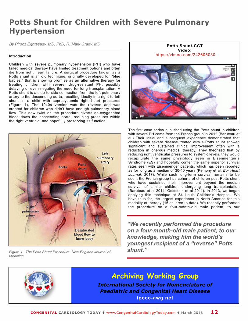
Children with severe pulmonary hypertension (PH) who have failed medical therapy have limited treatment options and often die from right heart failure. A surgical procedure known as a Potts shunt is an old technique, originally developed for "blue babies," that is showing promise as an alternative therapy for treating children with severe, drug-resistant PH, possibly delaying or even negating the need for lung transplantation. A Potts shunt is a side-to-side connection from the left pulmonary artery to the descending aorta, resulting ideally in a right-to-left shunt in a child with suprasystemic right heart pressures (Figure 1). The 1940s version was the reverse and was created for children who didn’t have enough pulmonary blood flow. This new twist on the procedure diverts de-oxygenated blood down the descending aorta, reducing pressures within the right ventricle, and hopefully preserving its function.
The first case series published using the Potts shunt in children with severe PH came from the French group in 2012 (Baruteau et al.) Their initial and subsequent experience demonstrated that children with severe disease treated with a Potts shunt showed significant and sustained clinical improvement often with a reduction in onerous medical therapy. They theorized that by reducing right ventricular pressures to systemic levels, they would recapitulate the same physiology seen in Eisenmenger’s Syndrome (ES) and hopefully confer the same superior survival rates seen with Eisenmenger patients, which has been reported as for long as a median of 30-40 years (Kempny et al. Eur Heart Journal, 2017). While such long-term survival remains to be seen, the French group has cohorts of children post-Potts shunt who have sustained their improvement beyond the median survival of similar children undergoing lung transplantation (Baruteau et al 2014; Goldstein et al 2011). In 2013, we began applying this technique at St. Louis Children’s Hospital. We have thus far, the largest experience in North America for this modality of therapy (15 children to date). We recently performed the procedure on a four-month-old male patient, to our
Potts Shunt for Children with Severe Pulmonary HypertensionBy Pirooz Eghtesady, MD, PhD; R. Mark Grady, MD
Archiving Working GroupInternational Society for Nomenclature of Paediatric and Congenital Heart Disease
ipccc-awg.net
CONGENITAL CARDIOLOGY TODAY t www.CongenitalCardiologyToday.com t March 2018 12
Figure 1. The Potts Shunt Procedure. New England Journal of Medicine.
Potts Shunt-CCTVideo:
https://vimeo.com/242605030
“We recently performed the procedure on a four-month-old male patient, to our knowledge, making him the world’s youngest recipient of a “reverse” Potts shunt.”

CONGENITAL CARDIOLOGY TODAY t www.CongenitalCardiologyToday.com t March 2018 13
knowledge, making him the world’s youngest recipient of a “reverse” Potts shunt.
The Case
In March 2017, a term infant presented at 19 Days of Life with tachypnea and cyanosis to a local hospital in New York state, where he quickly decompensated, became profoundly hypoxemic, resulting in emergent intubation. The initial echocardiogram demonstrated severe right ventricular hypertension, a patent foramen ovale with right to left shunting, and an otherwise structurally normal heart. Continued struggles with oxygenation led eventually to the child being placed on veno-arterial extracorporeal membrane oxygenation (ECMO). With subsequent aggressive medical therapy, including use of PH specific medications, the child was successfully decannulated from ECMO although he still required substantial ICU care. Despite decannulation, his PH remained severe, with suprasystemic right heart pressures. Pulmonary angiogram demonstrated markedly abnormal pulmonary arteries (diffusely hypoplastic with truncated branching patterns) suggesting that the child’s PH was due to developmental abnormalities of the pulmonary bed. With little improvement in his PH, the child was transferred to our institution for possible Potts shunt or lung transplantation.
The patient came to St. Louis Children’s Hospital on full mechanical ventilation via tracheostomy, FiO2 60%, inhaled nitric o x i d e , e p o p r o s t e n o l , m i l r i n o n e a n d s i l d e n a f i l . O u r echocardiogram confirmed the presence of suprasystemic right ventricular pressures with a BNP of 2697 pg/ml (normal <39 pg/ml). After evaluation and discussion, he underwent an uncomplicated Potts shunt with direct anastomosis between the descending aorta and the Left Pulmonary Artery (LPA). His post-operative course was uncomplicated, and he was transferred back home on Post-operative Day 13. At the time of transfer, he was no longer on IV or inhaled vasoactive medications. He was on CPAP pressure support while awake with upper extremity saturations >90% on FiO2 40%. BNP was 52 pg/ml. The child was eventually discharged home, and is now a year out from his Potts shunt. His clinical course has been stable with weight gain on home ventilator management, as well as sildenafil, bosentan and SQ treprostinil.
Discussion
Pulmonary hypertension in children can be a devastating diagnosis with very poor outcomes. Until recently, for children with severe PH who have failed medical therapy, lung transplantation has been the only option. Now, however, treating these children with a Potts shunt has proven to be a successful strategy in palliating their disease and providing an attractive alternative. Success has led to a significant improvement in quality of life and other diagnostic markers of disease, often with a concomitant decrease in medication burden. Sustained benefit has been seen extending beyond 8-10 years post-shunt (Barteau et al 2014). The ages of successfully treated children ranges from the infant to the teenager with a variety of different etiologies for their PH. At our institution, we do not consider a Potts shunt a contraindication to future lung transplant. In fact, we have successfully transplanted a child who failed to improve
The congenital heart professionals network exists to facilitate communications between congenital heart professionals locally, regionally, and globally.
JOIN TODAY www.ch ipnetwork .o rg
Pediatric Cardiologist at the level of Associate or Full Professor
The University of Kentucky College of Medicine and Kentucky Children’s Hospital (KCH) is recruiting a Pediatric Cardiologist at the level of Associate or Full Professor.
The successful candidate will require established clinical excellence in pediatric cardiology, and will oversee and ensure a high standard of mentoring younger faculty, teaching, quality clinical performance, and scholarly activity within the Division. This position further supports our expansion under the "one program, two sites" partnership for adult and pediatric congenital heart disease between UK Healthcare (KCH) and Cincinnati Children's Hospital Medical Center (CCHMC). The UK team currently includes 10 pediatric cardiologists, and program services at the UK campus include cardiac surgery, interventional cardiology, invasive electrophysiology, fetal echocardiography, advanced cardiac imaging, and adult congenital heart disease. Leadership opportunities exist in this exciting program. The ambulatory practice includes multiple outreach sites in central and eastern Kentucky region.
UK Healthcare, the clinical enterprise of the University of Kentucky, encompasses multiple medical campuses as well as the clinical activities of all six health care colleges. KCH serves as a subspecialty regional referral and pediatric care center for the state of Kentucky as well as parts of West Virginia, Tennessee, and Ohio. KCH is the region’s level I pediatric trauma center including a state of the art pediatric emergency department, newly planned entrance, outpatient areas, and 70 bed NICU opening in May 2018. Additionally, in 2017, UK embarked on a partnership with Shriner’s Children’s Hospital, a premier pediatric orthopedic organization. This venture includes the Shriner’s newly built clinic space on the UK Healthcare campus and all inpatient care of these patients be performed in KCH.
As the horse capital of the world, Lexington offers southern hospitality in a world class atmosphere with the Kentucky Horse Park, home of the 2010 World Equestrian Games, and Keeneland, host of the 2015 Breeders Cup. It is a family-friendly town with a wide range of neighborhood communities. The surrounding counties have excellent schools, both private and public. The city offers a rich cultural and food scene. For those who love sports, the University of Kentucky Wildcats athletics boast highly competitive SEC men’s and women’s teams, including the outstanding men’s basketball team. Lexington is also close to an impressive array of outdoor activities given the proximity to lakes, rivers and mountains, punctuated by standout destinations such as the Red River Gorge, Natural Bridge, and Mammoth Cave National Park.
To Apply, Please Contact:
Douglas Schneider, MDChief, Division of Pediatric Cardiology
Department of Pediatrics, University of KentuckyMedical Director, Kentucky Children’s Hospital Congenital Heart Program
Director, Pediatric Catheterization LabEmail: [email protected]

CONGENITAL CARDIOLOGY TODAY t www.CongenitalCardiologyToday.com t March 2018 14
from his Potts shunt due to progressive right ventricular failure. While long term benefits remain to be seen, we feel the Potts shunt holds great promise in treating a child with severe PH and should be a part of every pediatric PH caregiver’s armamentarium. To that end, we, along with the French group, have initiated an international Potts shunt registry to help gather worldwide data on how this relatively simple procedure can be utilized for treating these very compromised children.
Conclusion
Potts shunt has the potential to offer c h i l d r e n w i t h s e v e r e p u l m o n a r y hypertension the possibility of extended survival and improved quality of life. The Potts shunt should be considered as the f i r s t i n t e r v e n t i o n a l s t e p i n t h e management of children with severe pulmonary hypertension who are being c o n s i d e r e d f o r p o s s i b l e l u n g transplantation.
References
Figure 1: The Potts Shunt Procedure. New England Journal of Medicine.Video: https://vimeo.com/242605030
CCT
About the Authors
Pirooz Eghtesady, MD, PhD
Dr. Eghtesady is Emerson Chair of Pediatric Cardiothoracic Surgery at Washington University School of Medicine in St. Louis, and Cardiothoracic Surgeon-in-Chief at St. Louis Children’s Hospital. He is also Co-Director of the St. Louis Children’s and Washington University Heart Center.
R. Mark Grady, MD
Dr. Grady is a Pediatric Cardiologist at St. Louis Children’s and Washington University Heart Center. He is also an Associate Professor of Pediatrics, Cardiology at Washington University School of Medicine in St. Louis.
R. Mark Grady, MD St. Louis Children’s and Washington University Heart Center One Children’s Place St. Louis, MO 63110 USA Phone: 314-454-6165 Fax: 314-454-2381 [email protected]
www.stlouischildrens.org
Follow
CONGENITAL CARDIOLOGY TODAY
on Twitter:@CCardiology
Chief of Pediatric Cardiology
The Department of Pediatrics, UT Health San Antonio Joe R & Teresa Lozano Long School of Medicine together with the Heart Center at University Children’s Health is recruiting a Division Chief of Pediatric Cardiology at the level of Associate or Ful l Professor. The successful candidate wi l l require established clinical excellence, experience in leadership, as well as academic recognition. The applicant must be board certified in pediatric cardiology and either possess or be able to easily obtain an unrestricted Texas medical license.
The candidate will join an established academic clinical practice with 5 pediatric cardiologists and 2 congenital heart surgeons. Inpatient services are provided at University Hospital with a dedicated, variable acuity Pediatric and Congenital Cardiac Unit. The Program serves the county and much of South and West Texas. The Chief of Pediatric Cardiology will help guide the Heart Center as it enters an exciting phase of development. The Joe R. & Teresa Lozano Long School of Medicine has 230 medical students at each level. Cardiology faculty is engaged in the training of these medical students and 35 pediatric residents. The candidate will oversee and ensure a high standard of teaching, quality clinical performance and scholarly activity within the Division.
All faculty appointments are designated as security sensitive positions. UT Health San Antonio is an equal employment opportunity/affirmative action employer including protected veterans and persons with disabilities.
UT Health offers a competitive salary, comprehensive insurance package, and a generous retirement plan.
Interested individuals should apply online at
https://uthscsa.edu/hr/employment.asp
Principal Author
Pirooz Eghtesady, MD, PhD St. Louis Children’s and Washington University Heart Center One Children’s Place St. Louis, MO 63110 USA Phone: 314-454-6165 Fax: 314-454-2381 [email protected]
www.stlouischildrens.org

AI That Sees the Invisible: AliveCor and Mayo Clinic Announce Collaboration to Develop Groundbreaking AI Technology to Help Prevent Sudden Cardiac Death
AliveCor, a leader in FDA-cleared personal electrocardiogram (ECG) technology, today announced a collaboration with Mayo Clinic to develop tools for medical and non-medical personnel to easily screen for Long QT Syndrome (LQTS) early by combining AliveCor’s AI technology with Mayo’s patented algorithms. LQTS is both a congenital and acquired disorder; the inherited form leaves 160,000 people in the U.S. vulnerable to this deadly disease and causes 3,000 to 4,000 sudden deaths in children and young adults annually in the U.S. The acquired form of LQTS is often caused by medications, such as antibiotics and antidepressants, prescribed to many millions of patients every year.
Through this collaboration, new methods and techniques to detect LQTS will be developed for AliveCor’s Kardia Mobile device, a device that is both portable and affordable. The collaboration envisions a tool that will enable people to practice preventive medicine on an unprecedented scale and provide instantaneous results previously unavailable unless patients visited a doctor’s office.
“To prevent this type of sudden death, increased awareness and screening is critical. AliveCor’s patented artificial intelligence technology, algorithms and millions of ECGs, paired with Mayo Clinic’s extensive data and world-leading clinical expertise will mean enhanced safety and decreased risk for many,” said Vic Gundotra, CEO, AliveCor. “This new technology could one day allow pharmacists, coaches and others to actively screen for and prevent sudden cardiac deaths.”
AliveCor provides the first consumer-ready, clinically validated and FDA-cleared ECG to give patients a more complete view of their heart health, improve proactive monitoring and create a new standard of cardiac care. The ECG holds a vast amount of information about a person’s overall health and applies machine learning to millions of ECG recordings as an important enhancement to the traditional ECG analysis.
“This agreement makes our vision of universal screening for the early detection of Long QT Syndrome – a potentially lethal, yet highly treatable condition – one step closer to reality. The electrical heart cycle is emerging as the next vital sign. With very few exceptions, we now know that a prolonged cycle – whether caused by genetics, drugs, electrolyte disturbances or by other diseases – indicates increased risk for early death,” said Michael J. Ackerman, MD, PhD, Director of Mayo Clinic’s Windland Smith Rice Sudden Death Genomics Laboratory. ̊ “Any of these deaths could be averted with simple preventive and/or counteractive measures.”
CONGENITAL CARDIOLOGY TODAY t www.CongenitalCardiologyToday.com t March 2018 15
Medical News, Products & InformationCompiled by Kate Baldwin, Special Projects Editor
PediatricCardiologySPECIALTY REVIEW IN
American Academy of Pediatrics Section on Cardiology & Cardiac Surgeryin collaboration with the Society of Pediatric Cardiology Training Program Directors
August 13-17, 2018 | Chicago PediatricCardiologyCourse.com

The investment from Mayo adds to the recent infusion of $30 million of capital AliveCor received in spring of 2017 and will accelerate innovations in heart health and continue the rapid expansion of the business.
Mayo Clinic and Dr. Michael J. Ackerman have a financial interest in the technology referenced in this news release. Mayo Clinic will use any revenue it receives to support its not-for-profit mission in patient care, education and research.
The FDA-cleared Kardia Mobile is the most clinically validated mobile ECG solution on the market and is recommended by leading cardiologists and used by people worldwide for accurate ECG recordings. Kardia Pro is the first AI-enabled platform for doctors to monitor patients for the early detection of AF. For more information, visit alivecor.com.
Higher Blood Sugar Early in Pregnancy Raises the Baby's Risk of a Congenital Heart Defect - Published online Dec. 15, 2017 in The Journal of Pediatrics.
For many years, physicians have known that women with diabetes face an increased risk of giving birth to babies with heart defects. Some studies have also suggested a link between non-diabetic mothers' blood sugar levels and babies' heart defect risk. However, the new study, led by researchers at the Stanford University School of Medicine, is the first to examine this question in the earliest part of pregnancy, when the fetal heart is forming.
"Most women who have a child with Congenital Heart Disease are not diabetic," said the study's senior author, James Priest, MD, Assistant Professor of Pediatric Cardiology. "We found that in women who don't already have diabetes or develop diabetes during pregnancy, we can still measure risk for having a child with Congenital Heart Disease (CHD) by looking at their glucose values during the first trimester of pregnancy."
The study's lead author is Emmi Helle, MD, PhD, an affiliate in Pediatric Cardiology and former postdoctoral scholar.
One challenge associated with conducting the research was the fact that maternal blood glucose is not routinely measured in non-diabetic pregnant women. Instead, women typically receive an oral glucose tolerance test halfway through pregnancy to determine whether they have gestational diabetes, but this test is performed well after the fetal heart has formed.
The research team studied medical records from 19,107 pairs of mothers and their babies born between 2009 and 2015. The records included details of the mothers' prenatal care, including blood test results and any cardiac diagnoses made for the babies during pregnancy or after birth. Infants with certain genetic diseases, those born from multiple pregnancies and those whose mothers had extremely low or high body-mass-index measures were not included in the study. Of the infants in the study, 811 were diagnosed with congenital heart disease, and the remaining 18,296 were not.
The scientists analyzed blood glucose levels from any blood sample collected from the mothers between four weeks prior to the estimated
CONGENITAL CARDIOLOGY TODAY t www.CongenitalCardiologyToday.com t March 2018 16
Stanford Pulmonary Artery Reconstruction and Right Ventricle Rehabilitation Symposium
Li Ka Shing Center for Learning and Knowledge Stanford, CA
https://med.stanford.edu/cme/courses/2018/pa_full.html
APR 14 - APR 15
2018SAT - SUN
Your Recruitment Advertising Includes:
• Full color Recruitment ad in the issue(s)• Your recruitment listing in the email blast
for the issue(s) with a hot link • 3-Step Special Recruitment Opportunity
Website Section in three (3) areas of the website
• We can create your recruitment ad at no extra charge!
For more Information Contact: Tony Carlson
+1.301.279.2005 or [email protected]
CONGENITAL CARDIOLOGY TODAY
We Can Help You Recruit:* • Pediatric Cardiologists• Pediatric Interventional
Cardiologist• Adult Cardiologist
focused on CHD• Congenital/Structural
Heart Surgeons• Echocardiographers • EPs• Pediatric Transplant
Cardiologist
*Reach over 6,000 BC/BE Cardiologists focused on CHD worldwide
date of conception and the end of the 14th gestational week, just after the completion of the first trimester of pregnancy. These early blood glucose measurements were available for 2,292, or 13%, of women in the study. The researchers also looked at the results of oral glucose tolerance tests performed around 20 weeks of gestation, which were available for 9,511, or just under half, of the women in the study.
When Risk Is Elevated
After excluding women who had diabetes before pregnancy or who developed it during pregnancy, the results showed that the risk of giving birth to a child with a Congenital Heart Defect was elevated by 8% for every increase of 10 milligrams per deciliter in blood glucose levels in the early stages of pregnancy.
The next step in the research is to conduct a prospective study that follows a large group of women through pregnancy to see if the results are confirmed, Priest said. If researchers see the same relationship, it may be helpful to measure blood glucose early in pregnancy in all pregnant women to help determine which individuals are at greater risk for having a baby with a heart defect, he said.
"We could use blood glucose information to select women for whom a screening of the fetal heart could be helpful," Priest said, adding that modern prenatal imaging allows for detailed diagnoses of many congenital heart defects before birth. "Knowing about defects prenatally improves outcomes because mothers can receive specialized care that increases their babies' chances of being healthier after birth."
The work is an example of Stanford Medicine's focus on precision health, the goal of which is to anticipate and prevent disease in the healthy and precisely diagnose and treat disease in the ill.
Two Major Research Grants from the The AAP Section on Cardiology & Cardiac Surgery Cardiology & Cardiac Surgery - Applications Due April 1st, 2018
$35,000 Pediatric Cardiology Research Fellowship Award(supported by The Children’s Heart Foundation)
The Pediatric Cardiology Research Award provides research support for a pediatric cardiology training fellow who has demonstrated aptitude for basic science or clinical science research. The purpose of this award is to promote understanding of cardiovascular disease in children.
$50,000 Pediatric Cardiomyopathy Early Career Award(supported by the Children’s Cardiomyopathy Foundation)
The Pediatric Cardiomyopathy Research Award is awarded to an early career pediatric cardiologist (within five years of fellowship) to advance medical knowledge on the causes and mechanism of pediatric cardiomyopathy and to develop diagnostic guidelines and targeted therapies.
For further information, applications, instructions and forms, contact:Vivian Baldassari Thorne, Section Manager, Division of Hospital & Surgical Services, [email protected], 630.626.6669, Cardiology, Otolaryngology, Perinatal, Surgery, 345 Park Boulevard, Itasca, IL 60143; www.aap.org.
CONGENITAL CARDIOLOGY TODAY t www.CongenitalCardiologyToday.com t March 2018 17
Save the DateMay 18-19, 2018
The InternationalPDA Symposium
The WestinDowntown Memphis
Amultispecialtysymposiumdesignedtoencompassstate-of-the-artpracticesonhowtokeepthepatentductusarteriosus(PDA)openandtheclosureofthePDAinnewborns.SpecialfocuswillbeonthetranscatheterPDAocclusioninextremely
lowbirthweight(ELBW)infants.
TheSymposiumwillfeatureprominentspeakersinthefieldsofNeonatology,cardiologyandcardiacsurgeryamongothers;debatesonif,when,andhowtoclosethePDAinELBWinfants,echoandcatheterworkshops,aswellaspanel
discussions.- - - - - - - - - - - - - - - - - - - - - - - - -
Registrationisavailableonline:
www.pdasymposium.org

Call for Europe-Wide Screening of Babies for Heart Defects
Newswire. All babies across Europe should be routinely screened for Critical Congenital Heart Defects (CCHD) within 24 hours of their birth, say a group of experts led by a University of Birmingham Professor and Honorary Consultant Neonatologist at Birmingham Women's Hospital.
The European Pulse Oximetry Screening Workgroup (EPOSW), a group of neonatologists and paediatric cardiologists, including presidents of leading European Neonatal Scientific Societies, has published a consensus statement recommending screening with pulse oximetry for all babies across Europe.
CCHD occur in around two in every 1,000 newborn babies, and are a leading cause of infant death. Timely diagnosis is crucial for the best outcome for these babies, but current screening methods may miss up to 50% of affected newborn infants, and those sent home before diagnosis frequently die or suffer major morbidity. However, babies with CCHD often have low blood oxygen levels which can be detected quickly and non-invasively by pulse oximetry screening (POS), using a simple sensor placed on newborn infants' hand and foot.
This medical device monitors the oxygen saturation of a patient's blood through their skin, as opposed to measuring oxygen levels directly through a blood sample.
EPOSW's statement, published in Lancet Child Adolescent Health, is a culmination of almost a decade's work and calls for POS in all European countries for newborn babies after six hours of life or before discharge - preferably within 24 hours of birth.
The recommendations follow the PulseOx study led by a team from University of Birmingham and Birmingham Women's Hospital in 2011, which screened over 20,000 newborn babies for critical heart defects using POS. This study, and an important meta-analysis of the test published by the same team in 2012, has led to POS being used by an increasing number of hospitals in the UK and Europe. However, to date, only a few countries such as Poland, Ireland and Switzerland have issued national guidelines recommending universal screening with pulse oximetry.
Senior author Professor Andrew Ewer, of the Institute of Metabolism and Systems Research at the University of Birmingham, said, "These recommendations are the culmination of almost a decade's work driven by one focus; to prevent as many babies as possible from dying as a result of undetected heart defects. "Surgical and catheter interventions for CCHD now lead to excellent outcomes for most cases of CCHD, but timely detection is essential. POS improves early detection of CCHD in newborn babies by identifying those with low oxygen saturations. "POS has been shown to be simple, quick, painless, consistent and cost-effective and acceptable to both staff and parents. We have tried to create common, shared, flexible, and evidence-based recommendations for use and standardisation of POS for early detection of CCHD across Europe. These recommendations should be considered at a national level as an approach to better identify CCHD, and other life-threatening conditions, in newborn babies."
The UK National Screening Committee is currently considering the evidence for POS and whether it should be introduced to the UK national screening programme.
The European Pulse Oximetry Screening Workgroup comprises of Paolo Manzoni, Gerard R Martin, Luc JI Zimmermann, Anne de-Wahl Granelli, Avroy A Fanaroff, Ilona C. Narayen, Daniele De Luca, Julie Thomas-Chabaneix, Thorsten Orlikowsky, Umberto Simeoni, Irana G. Soldatova, Julije Mestrovic, Barry S Rodgers-Gray, Virgilio P Carnielli, Antonio Del Vecchio, Sergio Picardo, Robert M Tulloh, Maximo Vento, Manuel Sanchez Luna, and Andrew Ewer.
The authors represent the following institutions and organisations: • Neonatology and Neonatal Intensive Care Unit, S.Anna Hospital,
Turin, Italy.• Children's National Health System, Washington, DC, USA• Neonatology Division, Complutense University of Madrid, Madrid,
Spain.• Pediatric Intensive Care Unit, University Hospital of Split, Split,
Croatia.• Division of Pediatrics, Université de Lausanne, Lausanne,
Switzerland• Neonatology Division, Maastricht UMC+, Maastricht, the
Netherlands.• Institute of Metabolism and Systems Research, University of
Birmingham, UK.• Birmingham Women's Hospital, Birmingham Women's and
Children's NHS Foundation Trust, UK.
A pulse oximeter is a machine which can measure, non-invasively, the amount of oxygen being carried around the body by the red blood cells called oxygen saturations. Oxygen from the lungs is bound to hemoglobin in the red-blood cells forming oxyhemoglobin. If oxygen is not bound then this forms de-oxyhemoglobin. In health, almost all haemoglobin is oxyhemoglobin and so the oxygen saturations (i.e. the percentage of haemoglobin which has bound oxygen) is close to 100%. The pulse oximeter measures this by passing light through the peripheral blood vessels (e.g. a finger-tip in an adult or a hand or foot in a baby). Oxyhaemoglobin and de-oxyhaemoglobin absorb this light in different way and the proportion of light absorbed can be analysed by software within the oximeter which then calculates the percentage of haemoglobin saturated with oxygen.
To read the PulseOx Study led by the University of Birmingham in 2011 v i s i t : h t tp : / /www. the lance t . com/ jou rna ls / l ance t /a r t i c l e /PIIS0140-6736(11)60753-8/abstract and for the meta-analysis visit http://www.thelancet.com/journals/lancet/article/PIIS0140-6736(12)60107-X/fulltext
Birmingham Women's Hospital, part of Birmingham Women's and Children's NHS Foundation Trust, is a centre of excellence, providing a range of specialist health care services to over 50,000 women and their families every year from Birmingham, the West Midlands and beyond. As well as delivering over 8,000 babies a year, it offers a full range of gynaecological, maternity and neonatal care, as well as a comprehensive genetics service, which serves men and women. Its fertility centre is one of the best in the country, while the fetal medicine centre receives regional and national referrals. The hospital is also an international centre for education, research and development with a research budget of over £3 million per year. In April 2016, it was announced as the national miscarriage research centre - the first of its kind in the UK - in partnership with Tommy's Baby Charity (https://www.tommys.org).
CONGENITAL CARDIOLOGY TODAY t www.CongenitalCardiologyToday.com t March 2018 18
Save the DateMay 18-19, 2018
The InternationalPDA Symposium
The WestinDowntown Memphis
w w w. m e t h o d i s t m d . o r g / c m e or call 901.516.8933 for the program.

CONGENITAL CARDIOLOGY TODAY t www.CongenitalCardiologyToday.com t March 2018 19
CONGENITAL CARDIOLOGY TODAY
© 2018 by Congenital Cardiology Today (ISSN 1554-7787-print; ISSN 1554-0499-online). Published monthly. All rights reserved. www.CongenitalCardiologyToday.com
Publication Company Address:11502 Elk Horn Dr. Ste. 201Clarksburg, MD 20871 USATel: +1.301.279.2005
Publishing Management:• Tony Carlson, Founder, President & Sr.
Editor - [email protected]• Richard Koulbanis, Group Publisher &
Editor-in-Chief - [email protected]• John W. Moore, MD, MPH, Group
Medical Editor - [email protected] • Kate Baldwin, Sr. Editor - Special
Projects - [email protected]
Editorial Board: Teiji Akagi, MD; Zohair Al Halees, MD; Mazeni Alwi, MD; Felix Berger, MD; Fadi Bitar, MD; Jacek Bialkowski, MD; Mario Carminati, MD; Anthony C. Chang, MD, MBA; John P. Cheatham, MD; Bharat Dalvi, MD, MBBS, DM; Horacio Faella, MD; Yun-Ching Fu, MD; Felipe Heusser, MD; Ziyad M. Hijazi, MD, MPH; Ralf Holzer, MD; Marshall Jacobs, MD; R. Krishna Kumar, MD, DM, MBBS; John Lamberti, MD; Gerald Ross Marx, MD; Tarek S. Momenah, MBBS, DCH; Toshio Nakanishi, MD, PhD; Carlos A. C. Pedra, MD; Daniel Penny, MD, PhD; James C. Perry, MD; P. Syamasundar Rao, MD; Shakeel A. Qureshi, MD; Andrew Redington, MD; Carlos E. Ruiz, MD, PhD; Girish S. Shirali, MD; Horst Sievert, MD; Hideshi Tomita, MD; Gil Wernovsky, MD; Zhuoming Xu, MD, PhD; William C. L. Yip, MD; Carlos Zabal, MD
Free Subscription to Qualified Professionals: Send your name, title(s), hospital or practice name, work address and url, phone, fax and email to: [email protected].
Statements or opinions expressed in Congenital Cardiology Today reflect the views of the authors and sponsors, and are not necessarily the views of Congenital Cardiology Today.
Official publication of the CHiP Network
Outstanding Opportunity for Medical Director of Pediatric Heart Failure and Transplant Program in Miami
Nicklaus Children's Hospital (formerly Miami Children’s Hospital), a 289-bed freestanding children's hospital and Level III trauma center, and Pediatric Specialists of America (PSA), the physician-led group practice of Nicklaus Children's Health System, are seeking an exceptional candidate to join its esteemed Heart Program to help develop and lead a new Heart Failure and Heart Transplant Program.
This exciting and unique opportunity will allow an individual to help develop all aspects of pre- and post-transplant care, including writing new protocols, developing treatment paradigms, and determining programmatic staffing requirements in a cardiac program with a track record of clinical and academic excellence and innovation exceeding 20 years. Experience with all aspects of transplant care is required, including the ability to manage patients with acute and chronic heart failure and failing congenital heart physiology. The candidate will need to meet all OPTN qualifications as a Primary Heart Transplant Physician. Submission of a transplant log is strongly suggested.
The Medical Director of Heart Failure and Heart Transplantation will work as part of a team of 14 pediatric cardiologists, 7 cardiac intensivists, 3 cardiac surgeons and more than a dozen advanced nurse practitioners. There is an active ACGME accredited pediatric cardiology fellowship, a 20-year history of training cardiac ICU fellows and advanced training programs in electrophysiology and cardiology hospitalist practice. Cardiology inpatients are cared for in our brand new, state of the art, 190-bed Advanced Pediatric Care Pavilion. The cardiology section consists of 34 beds and utilizes an acuity adjusted type model for clinical care, with each bed space capable of providing critical care services.
The Heart Program at Nicklaus Children’s, a world leader in pediatric cardiology and cardiovascular surgery for the care of children with congenital heart disease, serves as a beacon to families confronting the reality of a child or newborn with a heart defect. It offers the most innovative and least invasive approaches to the treatment of congenital heart disease, including many first-in-the-world procedures that were pioneered right here by The Heart Program’s own internationally renowned cardiologists and cardiovascular surgeons. It remains the only cardiovascular surgical program to offer real-time outcomes reporting (http://www.pediatricheartsurgery.com/realtimeoutcomes/cvperformance.aspx).
Founded in 1950, the rebranded Nicklaus Children’s Hospital is renowned for excellence in all aspects of pediatric medicine and has numerous subspecialty programs that are routinely ranked among the best in the nation. It is also home to the largest pediatric teaching program in the southeastern U.S. Many of the physicians on our staff have trained or worked at other leading medical institutions. Join a phenomenal team that brings lifelong health and hope to children and their families through innovative and compassionate care.
Nicklaus Children’s Hospital is located in Miami, Florida, and offers all of the advantages of a tropical, diverse, metropolitan community. Competitive compensation and benefits package. DFW
Qualified candidates please contact:Joyce Berger, Physician Recruiter
[email protected] or 786-624-3510pediatricspecialistsofamerica.org
Looking for Career Opportunities in Congenital/Structural Heart Disease (CHD)?
Go to: www.CongenitalCardiologyToday.com/Recruit/Recruit.html
CONGENITAL CARDIOLOGY
TODAY

*Reach over 6,000 BC/BE Cardiologists focused on CHD worldwide
CONGENITAL
CARDIOLOGY TODAY
We Can Help You Recruit:* • Pediatric Cardiologists• Pediatric Interventional Cardiologist• Adult Cardiologist focused on CHD• Congenital/Structural Heart Surgeons
• Echocardiographers • EPs• Pediatric Transplant Cardiologist
Your Recruitment Advertising Includes:
• Full color Recruitment ad in the issue(s)• Your recruitment listing in the email blast
for the issue(s) with a hot link. • 3-Step Special Recruitment Opportunity
Website Section in three (3) areas of the website.
• We can create your recruitment ad at no extra charge!
For more Information Contact: Tony Carlson
+1.301.279.2005 or [email protected]

NIT-OCCLUD® Coil System for PDA Closure
Designed For the Safe and Atraumatic Occlusion of the Congenital Heart Defect PDA (Patent Ductus Arteriosus)
Distributed by:B. Braun Interventional Systems Inc.
824 Twelfth Avenue | Bethlehem, PA 18018 | USA Tel 877 836 2228 | Fax 610 849 1334 | www.bisusa.org
Repositionable and Retrievable Prior to Release
Tight and Compact Windings Ensure Efficient Occlusion
Designed to Match Individual Morphologies and Sizes
INDICATIONS FOR USE: The Nit-Occlud® PDA coil is a permanently implanted prosthesis indicated for percutaneous, transcatheter closure
of small to moderate size patent ductus arteriosus with a minimum angiographic diameter less than 4mm.
NIT-OCCLUD BRIEF STATEMENT: Do not implant the Nit-Occlud PDA into patients who have endocarditis, endarteritis, active infection, pulmonary hypertension (calculated PVR greater
than 5 Wood Units), thrombus in a blood vessel through which access to the PDA must be obtained, thrombus in the vicinity of the implantation site at the time of the implantation or patients with a body weight < 11 lbs. (5kg). An angiogram must be performed prior to implantation for measuring
length and diameter of the PDA. Only the pfm medical implantation delivery catheter should be used to implant the device. Administration of 50 units of heparin per kg bodyweight should be injected after femoral sheaths are placed. Antibiotics should be given before (1 dose) and after implantation
(2 doses) to prevent infection during the implant procedure. Do not implant the Nit-Occlud PDA in an MR environment. Do not pull the Nit-Occlud coil through heart valves or ventricular chambers. Contrast media should not be injected through the implantation catheter. The catheter must not be connected to high pressure injectors. Patients may have an allergic response to this device due to small amounts of nickel that has been shown to be released from the device in very small amounts. If the patient experiences allergic symptoms, such as difficulty in breathing or swelling of the face or
throat, he/she should be instructed to seek medical assistance immediately. Antibiotic prophylaxis should be performed to prevent infective endocarditis during first 6 months after coil implantation. Potential Adverse Events: Air embolism, Allergic reaction to drug/contrast, Apnea, Arrhythmia requiring
medical treatment or pacing, Arteriovenous fistula, Bacterial endocarditis, Blood loss requiring transfusion, Chest pain, Damage to the tricuspid or pulmonary valves, Death, Embolization of the occluder, requiring percutaneous or surgical intervention, Endarteritis, False aneurysm of the femoral
artery, Fever, Headache/ Migraine, Heart failure, Hemolysis after implantation of the occluder, Hypertension, Hypotension or shock, Infection, Myocardial infarction, Occluder fracture or damage, Perforation of the heart or blood vessels, Stenosis of the left pulmonary artery or descending thoracic aorta,
Stroke/TIA, Thromboembolism (cerebral or pulmonary), Valvular Regurgitation, Vessel damage at the site of groin puncture (loss of pulse, hematoma etc.). Nit-Occlud is a registered trademark of pfm medical Inc.
Rx only CV9064 - 5/17 ©2017 B. Braun Interventional Systems Inc.