Neck Imaging Reporting and Data System€¦ · Neck Imaging Reporting and Data System: ......
41
Bethany Cavazuti Patricia Hudgins Tanya Rath Char Branstetter Kristen Baugnon Amanda Corey Ashley Aiken Neck Imaging Reporting and Data System: An Atlas of NI-RADS Categories for Head and Neck Cancer
Transcript of Neck Imaging Reporting and Data System€¦ · Neck Imaging Reporting and Data System: ......

Bethany Cavazuti
Patricia Hudgins
Tanya Rath
Char Branstetter
Kristen Baugnon
Amanda Corey
Ashley Aiken
Neck Imaging Reporting and Data System: An Atlas of NI-RADS Categories for Head and Neck Cancer

Disclosures
The authors have no relevant disclosures.

Introduction to NIRADS • Developed for surveillance imaging in patients with treated
H&N cancer
• In accordance with ACR’s charge to deliver patient
centered, data driven, outcomes based care
• Modeled after BI-RADS system
• Aims to:
• Provide numerical levels of suspicion to guide patient care
• Standardize approach with linked management
recommendations
• Generate data-mineable reports to further optimize
surveillance algorithms, accuracy, inter-observer variability
• Highlight radiologists’ added value in patient care

Introduction to NIRADS • Surveillance: CECT with concurrent PET for initial follow up 12
wks after H&N cancer treatment
• These categories are easily adapted to MR
• Limited management options:
• Keep patient on routine surveillance if imaging is negative
• Recommend directed inspection or shorter term follow up
• Proceed to additional imaging: PET, MR, etc
• Biopsy
• Therefore, simple suspicion categories were established in
accordance with input from ENT, radiation oncology,
hematology/oncology, and pathology colleagues to guide care

NIRADS Categories
Category 1 – No evidence of recurrence
Category 2 – Low suspicion of recurrence Ill-defined, only mild or moderate FDG uptake
Category 3 – High suspicion of recurrence Discrete, new or enlarging, intense FDG uptake
Category 4 – Definitive recurrence Path proven, clinical or radiographic progression
ACR-NIRADS

PET + CECT neck
CECT neck + chest
CECT neck
CECT neck + chest
Surveillance Algorithm here At our centers, initial follow up is 8-12 wks
after surgery or completion of CRT
If negative
8-12 mos
If negative
6 months
If negative
6 months

NIRADS Recommendations
NIRADS 1
NIRADS 4
NIRADS 3
NIRADS 2
Routine f/u (6 mo)
Short f/u (3 mo), PET, or
direct inspection
Biopsy
Clinical care of recurrence
Category Linked Recommendation
ACR-NIRADS

NIRADS category 2 is defined as questionable recurrence
Divided into two subcategories for the primary site:
a) Superficial (mucosal surface) – recommend direct
inspection
b) Deep, ill-defined soft tissue – recommend short
interval f/u or PET
NIRADS category 2 should be considered when CECT and
PET findings are DISCORDANT:
Robust enhancement without associated FDG uptake
Focal FDG uptake without anatomical correlate
NIRADS 2 Subcategories
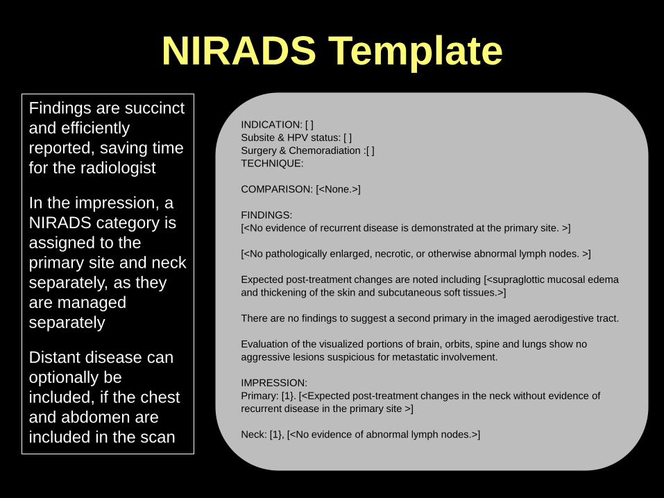
NIRADS Template
INDICATION: [ ]
Subsite & HPV status: [ ]
Surgery & Chemoradiation :[ ]
TECHNIQUE:
COMPARISON: [<None.>]
FINDINGS:
[<No evidence of recurrent disease is demonstrated at the primary site. >]
[<No pathologically enlarged, necrotic, or otherwise abnormal lymph nodes. >]
Expected post-treatment changes are noted including [<supraglottic mucosal edema
and thickening of the skin and subcutaneous soft tissues.>]
There are no findings to suggest a second primary in the imaged aerodigestive tract.
Evaluation of the visualized portions of brain, orbits, spine and lungs show no
aggressive lesions suspicious for metastatic involvement.
IMPRESSION:
Primary: [1}. [<Expected post-treatment changes in the neck without evidence of
recurrent disease in the primary site >]
Neck: [1}, [<No evidence of abnormal lymph nodes.>]
Findings are succinct
and efficiently
reported, saving time
for the radiologist
In the impression, a
NIRADS category is
assigned to the
primary site and neck
separately, as they
are managed
separately
Distant disease can
optionally be
included, if the chest
and abdomen are
included in the scan

NIRADS Template Legend
CECT Surveillance Legend:
Primary
1: No evidence of recurrence: routine surveillance
2: Low suspicion
a) Superficial abnormality (skin, mucosal surface): direct visual
inspection
b) Ill-defined deep abnormality: short interval follow-up*or PET
3: High suspicion (new or enlarging discrete nodule/ mass): biopsy
4: Definitive recurrence (path proven or clinical progression): no biopsy needed
Nodes
1: No evidence of recurrence: routine surveillance
2: Low suspicion (ill- defined): short interval follow-up or PET
3: High suspicion (new or enlarging lymph node): biopsy if clinically needed
4: Definitive recurrence (path proven or clinical progression): no biopsy needed
*short interval follow- up: 3 months at our institution
A legend is included
at the bottom of
every NIRADS report
Allows interpretation
by any clinician
viewing the report
with direct guidance
based on category
making NIRADS
accessible to any
physician

NIRADS Lexicon
Non-mass like soft tissue
• Hypo-enhancing distortion of soft tissue and fat planes (1)
Masses
• Morphology: Ill-defined (2) versus discrete (3)
• Enhancement: Mild (2) versus robust (3)
• FDG uptake relative to background: Mild (2) versus
intense (3)
Mucosal abnormality
• Mucoid density (1)
• Diffuse linear enhancement (benign radiation mucositis) (1)
• Focal mucosal enhancement of FDG uptake (2a)

NIRADS Lexicon
Lymph nodes
• Residual nodal tissue with:
• No FDG uptake relative to backgroun(1)
• Mild FDG uptake relative to background(2)
• Intense FDG uptake relative to background (3)
• Growing lymph node:
• Along expected nodal drainage without definite
abnormal morphology (2)
• With abnormal morphology (3)
• With intense FDG (3 or 4)

3 mo post CRT Staging scan
T1 N2c BOT SCCA
NIRADS 1 Primary/Neck
Primary: 1 Neck: 1
Routine surveillance, 6 mo CECT

Primary: 1 Neck: 1 Routine surveillance, 6 mo CECT
NIRADS 1 imaging findings: • No abnl soft tissue
• Non-mass like distortion of
soft tissues
• “Mucoid” density mucosal
edema
• Diffuse linear mucosal
enhancement after radiation
• No abnl FDG uptake
NIRADS 1 Primary/Neck
3 mo post-CRT No abnl soft tissue
No abnl FDG uptake Approximately 4% are positive
for disease

3 mo post CRT
Primary: 2a
Direct inspection: radiation
injury, f/u PET neg
Staging scan
T4a BOT SCCA
NIRADS 2a Primary

Primary: 2a
NIRDAS 2 primary site
imaging findings: • Focal superficial mucosal
enhancement
• Focal mucosal FDG uptake
NIRADS 2a Primary
3 mo post CRT • No abnl soft
tissue
enhancement
• Moderate FDG
uptake
NIRADS 2 is most useful when CECT
and PET findings are DISCORDANT:
• Abnl enhancement with no FDG
uptake (i.e. scar or granulation
tissue)
• Ulceration with avid FDG uptake
(i.e. radiation effect)

Primary: 2a
Direct inspection &
endoscopic biopsy: SCCA
T2N0 glottic SCCA s/p CRT 2011
Poor follow-up, new hoarseness
NIRADS 2a Primary
• No abnl soft tissue
enhancement
• Moderate FDG uptake
• Most NIRADS 2a are
false positive:
• Only 17% are
positive for disease
• Goal of NIRADS 2a is
to direct clinical
inspection and biopsy
if necessary

2 mo post treatment
Neck: 2 Recommend
PET
NIRADS 2 neck imaging
findings: • Questionable nodal
recurrence or residual
nodal disease with mild or
intermediate FDG uptake
NIRADS 2 Neck
• Enlarged right level Iia
lymph node
• Central necrosis
Follow up: Salvage ND was negative
Note: This pt was imaged at OSH with
CECT only (no PET), and surgeon
elected to proceed with salvage ND
Current practice would dictate obtaining
a PET prior to proceeding to ND

NIRADS 3 Primary
Primary: 3
4 mo post resection and CRT
CT biopsy: persistent SCCA
Staging MRI
Maxillary SCCA

NIRADS 3 Primary
NIRADS 3 imaging findings: • New or definitely enlarging
mass
• Discrete nodule/mass with
robust enhancement
• Intense focal FDG uptake
Primary: 3
Recommend biopsy
(image guided or clinical)
4 mo post resection and CRT
• Focal abnormal soft tissue
with bony erosion
• Intense focal FDG uptake Approximately 59% are
positive for disease

NIRADS 3 Primary
Primary: 3
Endoscopic
biopsy: recurrent
SCCA
T4aN0 laryngeal SCC s/p TL, B ND, and CRT
• Focal abnormal
soft tissue
enhancement
• Intense focal
FDG uptake
To differentiate NIRADS 2 from 3: work backwards!
Do you want to biopsy this lesion?
Is there a discrete target?
If no, NIRADS 2

NIRADS 3 Neck
Revision neck dissection positive for recurrence
Neck: 3
3 mo post tx
T2N2b oral tongue SCCA
9 mo post resection,
ND and CRT

NIRADS 3 Neck
Revision neck dissection positive for recurrence
Neck: 3
9 mo post resection,
ND and CRT
NIRADS 3 neck imaging
findings: • New or definite
enlarging lymph nodes
• Intense focal FDG
uptake
• Enlarging abnl LN
• Intense focal FDG
uptake

pT4aN2cM0 laryngeal cancer s/p
TL and bilateral ND
Exam concerning for recurrence
at R neck/stoma
NIRADS 4 imaging
findings: • Pathologically proven
recurrence
• Definite radiologic or
clinical progression
• Definitive recurrence on
a single study
NIRADS 4
Neck: 4
To differentiate NIRADS 3 from 4: work
backwards!
Does this lesion need a biopsy? Is there
anything else it could be? If no, NIRADS 4

Test your skills!
Review the following cases
Assign NI-RADS level
Unknown Cases

Staging scan
pT1N2cM0 SCC L GTS
Unknown Case 1
5 mo post TORS and L ND
Primary: 2a
NIRADS 1 NIRADS 4 NIRADS 3 NIRADS 2
• Ulceration
• Focal mucosal
FDG uptake
• DISCORDANT!
Clinicians noted
ulceration without
evidence of
recurrence
Recommend direct
inspection

Staging scan
T4aN2bM0 L lat
oral tongue SCC
Unknown Case 2
4 mo post resection
• No abnormal
enhancement or
nodularity along
the flap
• No abnormal
FDG uptake
NIRADS 1 NIRADS 4 NIRADS 3 NIRADS 2
Primary: 1
Continue routine
surveillance

Unknown Case 3
CT guided biopsy
positive for
recurrent disease
Staging scan
L nasal cavity alveolar
rhabdomyosarcoma
Long term f/u after multiple
rounds of chemotherapy
Neck: 3
• New mass
• Intense focal FDG
uptake
NIRADS 1 NIRADS 4 NIRADS 3 NIRADS 2
Recommend tissue
sampling

Unknown Case 4
Staging scan
T1N2c L BOT SCC
6 mo after CRT
Primary: 1
Neck: 1
• No abnormal
mucosal
enhancement
• No abnormal FDG
uptake
NIRADS 1 NIRADS 4 NIRADS 3 NIRADS 2
Continue routine
surveillance

• Discrete masslike soft tissue with
differential enhancement
• Intense focal FDG uptake
Unknown Case 5
rypT4bN0 L soft palate SCC
S/p extensive resection and
reconstruction
Patient lost to f/u
Biopsy not performed
Returns 4 mo after resection
CT guided biopsy
Definite progression on imaging:
• Increased size
• Increased FDG uptake
Neck: 3 Neck: 4
NIRADS 1 NIRADS 4 NIRADS 3 NIRADS 2
Positive for recurrent
disease
Recommend tissue
sampling

Unknown Case 6
Staging scan
pT1N2aM0 L tonsil SCC
4 mo s/p TORS and
ND, completed CRT
Neck: 2
Now 7 mo post tx
• Nodular ill-defined
enhancement
adjacent to
surgical clips in L
submandibular
region
• Mild associated
FDG uptake
Neck: 1
NIRADS 1 NIRADS 4 NIRADS 3 NIRADS 2
No change in size, decr’d FDG activity
Likely residual submandibular gland
Recommend short
interval follow up
During TORS, surgeons may leave
a portion of the SMG
Additionally, would be rare for an
oropharyngeal H&N ca to go to a
level Ib node
Note: Primary 2a

Unknown Case 7
Recurrent L BOT SCC s/p
TL w pec flap and R ND
PET/CT done 1 wk later
Neck: 2 Neck: 3
CT guided biopsy
positive for SCC
NIRADS 1 NIRADS 4 NIRADS 3 NIRADS 2
Abnormal soft tissue
in the region of a left
level IV LN adjacent
to L CCA
Intense focal FDG
uptake
Recommend
PET
Recommend tissue
sampling

Unknown Case 8
T4N0M0 myoepithelial cancer R
maxillary sinus s/p composite
resection
CT guided biopsy
Primary: 3
Short term f/u 4
mo after biopsy
Primary: 2a • Discrete soft
tissue
abnormality
• Focal FDG
uptake
What now?
NIRADS 1 NIRADS 4 NIRADS 3 NIRADS 2
No malignant cells in
multiple passes in three
different areas
Interval improvement
in that area
Changes were likely
post treatment related
Recommend
tissue sampling

• Discrete
lesion with
differential
enhancement
• Intense focal
FDG uptake
Unknown Case 9
Staging scan
Supraglottic SCC with transglottic spread
4 mo after TL
Primary: 3
Patient went on to clinical
biopsy and proven recurrence
NIRADS 1 NIRADS 4 NIRADS 3 NIRADS 2
Recommend
tissue sampling

Unknown Case 10
Staging scan
Nasopharyngeal cancer
4 mo after CRT
Primary: 1 • Non-mass like
distortion of soft tissues
• No abnl FDG uptake
NIRADS 1 NIRADS 4 NIRADS 3 NIRADS 2
Continue routine
surveillance

Unknown Case 11
Staging scan
R oropharyngeal SCC
3 mo after CRT
Primary: 2a
6 mo after CRT
Primary: 3
NIRADS 1 NIRADS 4 NIRADS 3 NIRADS 2
• Focal mucosal
enhancement
• Mild focal
mucosal FDG
uptake
• Definitely enlarging mass
• Intense focal FDG uptake
Recommend tissue
sampling
Recommend direct
inspection
If no abnormality, short
interval follow up
Clinical biopsy positive
for recurrence

Unknown Case 12
Staging scan
HPV + Tonsil SCC w
B metastatic LNs
3 mo after CRT
Neck: 2
6 mo after CRT
Neck: 1
NIRADS 1 NIRADS 4 NIRADS 3 NIRADS 2
• Abnormal
enhancing nodular
soft tissue
• Mild FDG uptake
No abnl FDG uptake
Recommend short
interval follow up
Continue routine
surveillance

Unknown Case 13
6 mos p XRT
Staging scan
R tonsil SCC
3 mo after CRT
Primary: 2a
Primary: 1
NIRADS 1 NIRADS 4 NIRADS 3 NIRADS 2
• Focal mucosal
enhancement
• Mild focal FDG
uptake
No abnl FDG uptake
Recommend direct
inspection
If no abnormality,
short interval follow
up
Continue routine
surveillance

Unknown Case 14
Staging scan
R BOT SCC with B
metastatic nodes
3 mo after CRT
Primary: 2a
Neck: 2
Neck: 2
6 mo after CRT
Primary: 1
Pt underwent TL 8 mo post CRT for nonfxning
larynx and salvage R ND: residual SCC in 2 LNs
NIRADS 1 NIRADS 4 NIRADS 3 NIRADS 2
Recommend direct
inspection for
pirmary and short
interval follow up for
the neck
• Focal mucosal
enhancement
• Mild focal
mucosal FDG
uptake
• Persistent
enlarged node
• No focal FDG
uptake

Conclusions
NIRADS was developed to assist in evaluating and
reporting on patients with treated H&N cancer
Allows for succinct, efficient reports which effectively
communicate results with linked recommendations to
guide care
Several specific ways to influence patient care,
including recommendations for routine surveillance,
direct inspection, shorter interval follow up or
additional modality, or biopsy

References Aiken, AH, Farley, A, Baugnon, KL, et al. Implementation of a Novel Surveillance Template for Head and Neck
Cancer: Neck Imaging Reporting and Data System (NI-RADS). JACR. 2016; 13: 743-746.
Krieger, DA, Hudgins, PA, Nayak, GK, et al. Initial Performance of NI-RADS Template to Predict Recurrence
of Head and Neck Squamous Cell Carcinoma. Presented at ASNR 2016, accepted for publication in AJNR
2017.