navigator | MM-107 2015/10/21 We lead the way in Digital...
7
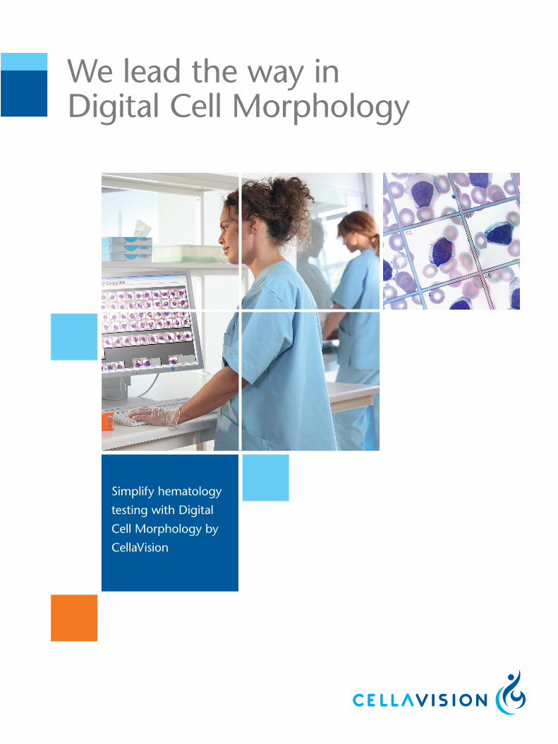
We lead the way in Digital Cell Morphology Simplify hematology testing with Digital Cell Morphology by CellaVision
Transcript of navigator | MM-107 2015/10/21 We lead the way in Digital...
We lead the way in Digital Cell Morphology
Simplify hematology
testing with Digital
Cell Morphology by
CellaVision
www.cellavision.com
We lead the way in Digital Cell MorphologyCellaVision is the world-leading provider of digital solutions for medical microscopy in the fi eld of hematology. We offer a proven technology that replaces or complements the microscope to create an automated digital cell morphology workfl ow. Our method has been adopted by forward-thinking laboratories all over the world and is proven to reduce turnaround time, enhance the degree of standardization and improve quality.
Want to learn more?
Visit www.cellavision.com to learn more about how Digital Cell Morphology can transform your hematology workfl ow, and to fi nd out how to contact us or one of our distributors.
nav
igat
or
| M
M-1
07 2
015/
10/2
1
A proven, multi-benefi t technology
CellaVision offers your hematology lab four principal benefi ts; the improved EFFICIENCY that comes with automating manual pro-cesses, enhanced QUALITY of results by promoting consistency and standardization; improved CONNECTIVITY that facilitates collaboration within and between labs; and a general advance-ment of your staff’s PROFICIENCY in performing cell differentials.
EFFICIENCY QUALITY
PROFICIENCYCONNECTIVITY
DIGITAL CELLMORPHOLOGY
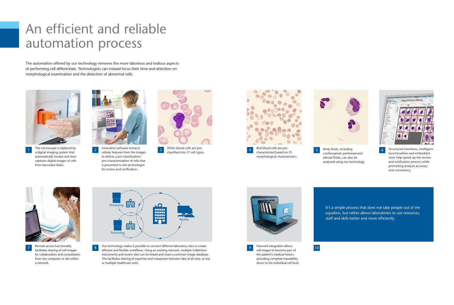
An effi cient and reliable automation processThe automation offered by our technology removes the more laborious and tedious aspects of performing cell differentials. Technologists can instead focus their time and attention on morphological examination and the detection of abnormal cells.
Innovative software extracts cellular features from the images to deliver a pre-classifi cation/pre-characterization of cells that is presented to the technologist for review and verifi cation.
21
7
The microscope is replaced by a digital imaging system that automatically locates and then captures digital images of cells from barcoded slides.
Remote access functionality facilitates sharing of cell images for collaboration and consultation, from any computer or site within a network.
8 Our technology makes it possible to connect different laboratory sites to create effi cient and fl exible workfl ows. Using an existing network, multiple CellaVision instruments and review sites can be linked and share a common image database. This facilitates sharing of expertise and manpower between labs of all sizes, at one or multiple healthcare units.
White blood cells are pre-classifi ed into 17 cell types.
3
Review
Processing
Processing
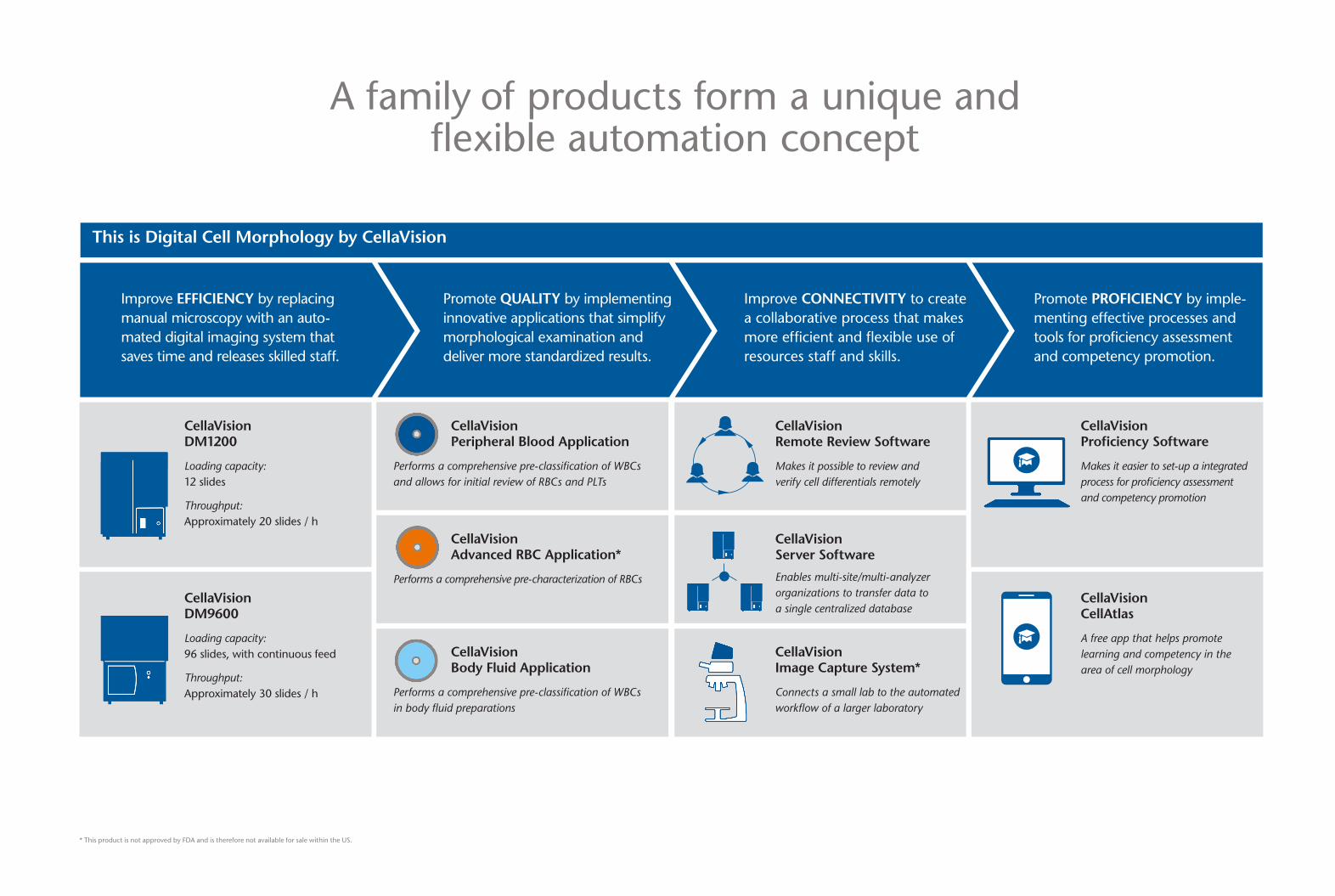
Improve CONNECTIVITY to create a collaborative process that makes more effi cient and fl exible use of resources staff and skills.
Promote PROFICIENCY by imple-menting effective processes and tools for profi ciency assessment and competency promotion.
Improve EFFICIENCY by replacing manual microscopy with an auto-mated digital imaging system that saves time and releases skilled staff.
* This product is not approved by FDA and is therefore not available for sale within the US.
Promote QUALITY by implementing innovative applications that simplify morphological examination and deliver more standardized results.
This is Digital Cell Morphology by CellaVision
CellaVision DM1200
Loading capacity: 12 slides
Throughput: Approximately 20 slides / h
CellaVision DM9600
Loading capacity: 96 slides, with continuous feed
Throughput: Approximately 30 slides / h
CellaVision Profi ciency Software
Makes it easier to set-up a integrated process for profi ciency assessment and competency promotion
CellaVision CellAtlas
A free app that helps promote learning and competency in the area of cell morphology
A family of products form a unique and fl exible automation concept
CellaVision Image Capture System*
Connects a small lab to the automated workfl ow of a larger laboratory
CellaVision Server Software
Enables multi-site/multi-analyzer organizations to transfer data to a single centralized database
CellaVision Remote Review Software
Makes it possible to review andverify cell differentials remotely
CellaVision Peripheral Blood Application
Performs a comprehensive pre-classifi cation of WBCs and allows for initial review of RBCs and PLTs
CellaVision Advanced RBC Application*
Performs a comprehensive pre-characterization of RBCs
CellaVision Body Fluid Application
Performs a comprehensive pre-classifi cation of WBCs in body fl uid preparations
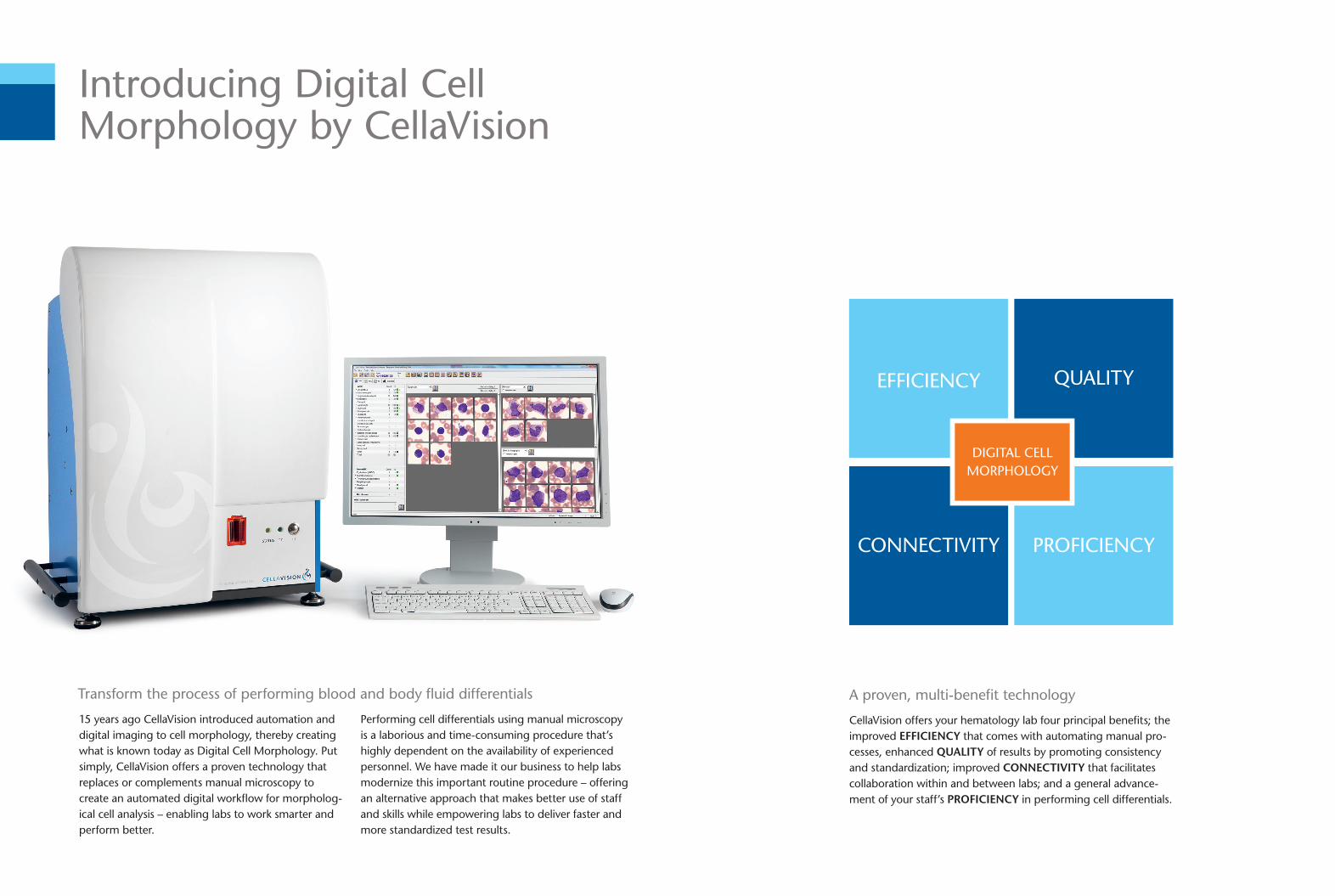
Introducing Digital Cell Morphology by CellaVision
15 years ago CellaVision introduced automation and digital imaging to cell morphology, thereby creating what is known today as Digital Cell Morphology. Put simply, CellaVision offers a proven technology that replaces or complements manual microscopy to create an automated digital workfl ow for morpholog-ical cell analysis – enabling labs to work smarter and perform better.
Performing cell differentials using manual microscopy is a laborious and time-consuming procedure that’s highly dependent on the availability of experienced personnel. We have made it our business to help labs modernize this important routine procedure – offeringan alternative approach that makes better use of staff and skills while empowering labs to deliver faster and more standardized test results.
Transform the process of performing blood and body fl uid differentials
54 Red blood cells are pre-characterized based on 21 morphological characteristics.
Structured interfaces, intelligent functionalities and embedded tools help speed up the review and verifi cation process while promoting analysis accuracy and consistency.
6Body fl uids, including cerebrospinal, peritoneal and pleural fl uids, can also be analysed using our technology.
9 10Network integration allows cell images to become part of the patient’s medical history providing complete traceability down to the individual cell level.
It’s a simple process that does not take people out of the equation, but rather allows laboratories to use resources, staff and skills better and more effi ciently.
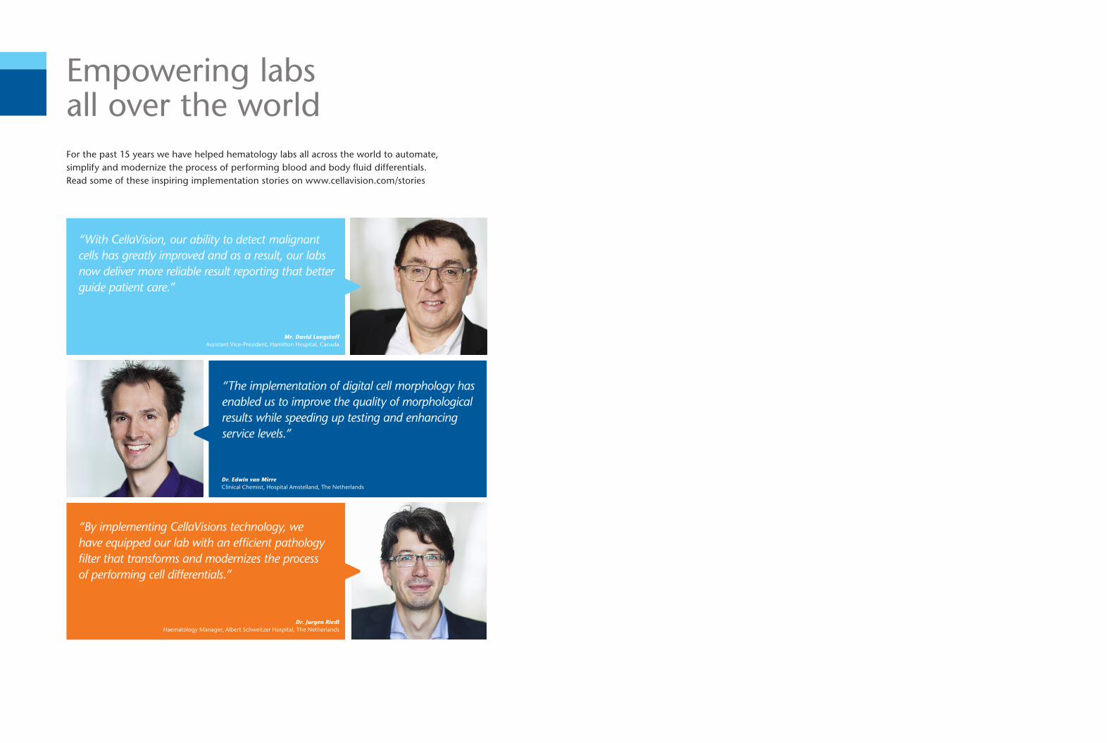
Empowering labs all over the worldFor the past 15 years we have helped hematology labs all across the world to automate, simplify and modernize the process of performing blood and body fl uid differentials. Read some of these inspiring implementation stories on www.cellavision.com/stories
Dr. Jurgen RiedlHaematology Manager, Albert Schweitzer Hospital, The Netherlands
“With CellaVision, our ability to detect malignant cells has greatly improved and as a result, our labs now deliver more reliable result reporting that better guide patient care.”
Dr. Edwin van Mirre Clinical Chemist, Hospital Amstelland, The Netherlands
“The implementation of digital cell morphology has enabled us to improve the quality of morphological results while speeding up testing and enhancing service levels.”
“By implementing CellaVisions technology, we have equipped our lab with an effi cient pathology fi lter that transforms and modernizes the process of performing cell differentials.”
Mr. David LangstaffAssistant Vice-President, Hamilton Hospital, Canada
EFFICIENCY QUALITY
PROFICIENCYCONNECTIVITY
DIGITAL CELLMORPHOLOGY
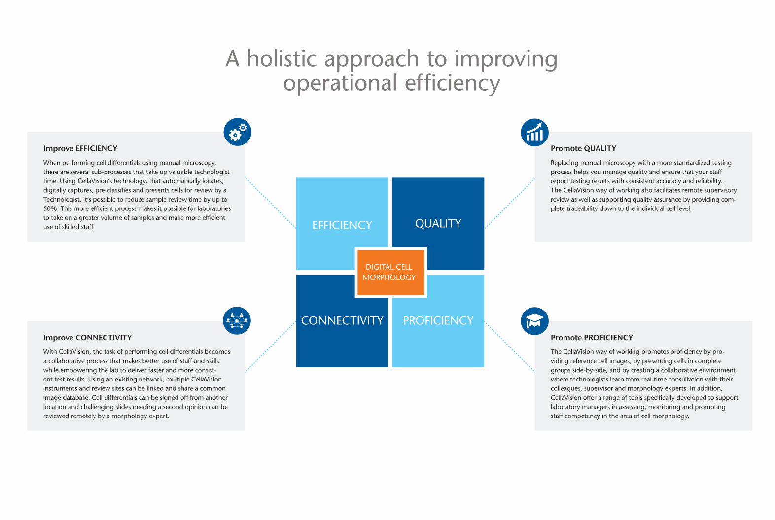
Improve EFFICIENCY When performing cell differentials using manual microscopy, there are several sub-processes that take up valuable technologist time. Using CellaVision’s technology, that automatically locates, digitally captures, pre-classifi es and presents cells for review by a Technologist, it’s possible to reduce sample review time by up to 50%. This more effi cient process makes it possible for laboratories to take on a greater volume of samples and make more effi cient use of skilled staff.
Improve CONNECTIVITY With CellaVision, the task of performing cell differentials becomes a collaborative process that makes better use of staff and skills while empowering the lab to deliver faster and more consist-ent test results. Using an existing network, multiple CellaVision instruments and review sites can be linked and share a common image database. Cell differentials can be signed off from another location and challenging slides needing a second opinion can be reviewed remotely by a morphology expert.
Promote PROFICIENCY The CellaVision way of working promotes profi ciency by pro-viding reference cell images, by presenting cells in complete groups side-by-side, and by creating a collaborative environment where technologists learn from real-time consultation with their colleagues, supervisor and morphology experts. In addition, CellaVision offer a range of tools specifi cally developed to support laboratory managers in assessing, monitoring and promoting staff competency in the area of cell morphology.
A holistic approach to improving operational effi ciency
Promote QUALITY Replacing manual microscopy with a more standardized testing process helps you manage quality and ensure that your staff report testing results with consistent accuracy and reliability. The CellaVision way of working also facilitates remote supervisory review as well as supporting quality assurance by providing com-plete traceability down to the individual cell level.
54 Red blood cells are pre-characterized based on 21 morphological characteristics.
Structured interfaces, intelligent functionalities and embedded tools help speed up the review and verifi cation process while promoting analysis accuracy and consistency.
6Body fl uids, including cerebrospinal, peritoneal and pleural fl uids, can also be analysed using our technology.
9 10Network integration allows cell images to become part of the patient’s medical history providing complete traceability down to the individual cell level.
It’s a simple process that does not take people out of the equation, but rather allows laboratories to use resources, staff and skills better and more effi ciently.
Empowering labs all over the worldFor the past 15 years we have helped hematology labs all across the world to automate, simplify and modernize the process of performing blood and body fl uid differentials. Read some of these inspiring implementation stories on www.cellavision.com/stories
Dr. Jurgen RiedlHaematology Manager, Albert Schweitzer Hospital, The Netherlands
“With CellaVision, our ability to detect malignant cells has greatly improved and as a result, our labs now deliver more reliable result reporting that better guide patient care.”
Dr. Edwin van Mirre Clinical Chemist, Hospital Amstelland, The Netherlands
“The implementation of digital cell morphology has enabled us to improve the quality of morphological results while speeding up testing and enhancing service levels.”
“By implementing CellaVisions technology, we have equipped our lab with an effi cient pathology fi lter that transforms and modernizes the process of performing cell differentials.”
Mr. David LangstaffAssistant Vice-President, Hamilton Hospital, Canada
EFFICIENCY QUALITY
PROFICIENCYCONNECTIVITY
DIGITAL CELLMORPHOLOGY
Improve EFFICIENCY When performing cell differentials using manual microscopy, there are several sub-processes that take up valuable technologist time. Using CellaVision’s technology, that automatically locates, digitally captures, pre-classifi es and presents cells for review by a Technologist, it’s possible to reduce sample review time by up to 50%. This more effi cient process makes it possible for laboratories to take on a greater volume of samples and make more effi cient use of skilled staff.
Improve CONNECTIVITY With CellaVision, the task of performing cell differentials becomes a collaborative process that makes better use of staff and skills while empowering the lab to deliver faster and more consist-ent test results. Using an existing network, multiple CellaVision instruments and review sites can be linked and share a common image database. Cell differentials can be signed off from another location and challenging slides needing a second opinion can be reviewed remotely by a morphology expert.
Promote PROFICIENCY The CellaVision way of working promotes profi ciency by pro-viding reference cell images, by presenting cells in complete groups side-by-side, and by creating a collaborative environment where technologists learn from real-time consultation with their colleagues, supervisor and morphology experts. In addition, CellaVision offer a range of tools specifi cally developed to support laboratory managers in assessing, monitoring and promoting staff competency in the area of cell morphology.
A holistic approach to improving operational effi ciency
Promote QUALITY Replacing manual microscopy with a more standardized testing process helps you manage quality and ensure that your staff report testing results with consistent accuracy and reliability. The CellaVision way of working also facilitates remote supervisory review as well as supporting quality assurance by providing com-plete traceability down to the individual cell level.
A proven, multi-benefi t technology
CellaVision offers your hematology lab four principal benefi ts; the improved EFFICIENCY that comes with automating manual pro-cesses, enhanced QUALITY of results by promoting consistency and standardization; improved CONNECTIVITY that facilitates collaboration within and between labs; and a general advance-ment of your staff’s PROFICIENCY in performing cell differentials.
EFFICIENCY QUALITY
PROFICIENCYCONNECTIVITY
DIGITAL CELLMORPHOLOGY
An effi cient and reliable automation processThe automation offered by our technology removes the more laborious and tedious aspects of performing cell differentials. Technologists can instead focus their time and attention on morphological examination and the detection of abnormal cells.
Innovative software extracts cellular features from the images to deliver a pre-classifi cation/pre-characterization of cells that is presented to the technologist for review and verifi cation.
21
7
The microscope is replaced by a digital imaging system that automatically locates and then captures digital images of cells from barcoded slides.
Remote access functionality facilitates sharing of cell images for collaboration and consultation, from any computer or site within a network.
8 Our technology makes it possible to connect different laboratory sites to create effi cient and fl exible workfl ows. Using an existing network, multiple CellaVision instruments and review sites can be linked and share a common image database. This facilitates sharing of expertise and manpower between labs of all sizes, at one or multiple healthcare units.
White blood cells are pre-classifi ed into 17 cell types.
3
Review
Processing
Processing
Improve CONNECTIVITY to create a collaborative process that makes more effi cient and fl exible use of resources staff and skills.
Promote PROFICIENCY by imple-menting effective processes and tools for profi ciency assessment and competency promotion.
Improve EFFICIENCY by replacing manual microscopy with an auto-mated digital imaging system that saves time and releases skilled staff.
* This product is not approved by FDA and is therefore not available for sale within the US.
Promote QUALITY by implementing innovative applications that simplify morphological examination and deliver more standardized results.
This is Digital Cell Morphology by CellaVision
CellaVision DM1200
Loading capacity: 12 slides
Throughput: Approximately 20 slides / h
CellaVision DM9600
Loading capacity: 96 slides, with continuous feed
Throughput: Approximately 30 slides / h
CellaVision Profi ciency Software
Makes it easier to set-up a integrated process for profi ciency assessment and competency promotion
CellaVision CellAtlas
A free app that helps promote learning and competency in the area of cell morphology
A family of products form a unique and fl exible automation concept
CellaVision Image Capture System*
Connects a small lab to the automated workfl ow of a larger laboratory
CellaVision Server Software
Enables multi-site/multi-analyzer organizations to transfer data to a single centralized database
CellaVision Remote Review Software
Makes it possible to review andverify cell differentials remotely
CellaVision Peripheral Blood Application
Performs a comprehensive pre-classifi cation of WBCs and allows for initial review of RBCs and PLTs
CellaVision Advanced RBC Application*
Performs a comprehensive pre-characterization of RBCs
CellaVision Body Fluid Application
Performs a comprehensive pre-classifi cation of WBCs in body fl uid preparations
54 Red blood cells are pre-characterized based on 21 morphological characteristics.
Structured interfaces, intelligent functionalities and embedded tools help speed up the review and verifi cation process while promoting analysis accuracy and consistency.
6Body fl uids, including cerebrospinal, peritoneal and pleural fl uids, can also be analysed using our technology.
9 10Network integration allows cell images to become part of the patient’s medical history providing complete traceability down to the individual cell level.
It’s a simple process that does not take people out of the equation, but rather allows laboratories to use resources, staff and skills better and more effi ciently.
Empowering labs all over the worldFor the past 15 years we have helped hematology labs all across the world to automate, simplify and modernize the process of performing blood and body fl uid differentials. Read some of these inspiring implementation stories on www.cellavision.com/stories
Dr. Jurgen RiedlHaematology Manager, Albert Schweitzer Hospital, The Netherlands
“With CellaVision, our ability to detect malignant cells has greatly improved and as a result, our labs now deliver more reliable result reporting that better guide patient care.”
Dr. Edwin van Mirre Clinical Chemist, Hospital Amstelland, The Netherlands
“The implementation of digital cell morphology has enabled us to improve the quality of morphological results while speeding up testing and enhancing service levels.”
“By implementing CellaVisions technology, we have equipped our lab with an effi cient pathology fi lter that transforms and modernizes the process of performing cell differentials.”
Mr. David LangstaffAssistant Vice-President, Hamilton Hospital, Canada
EFFICIENCY QUALITY
PROFICIENCYCONNECTIVITY
DIGITAL CELLMORPHOLOGY
Improve EFFICIENCY When performing cell differentials using manual microscopy, there are several sub-processes that take up valuable technologist time. Using CellaVision’s technology, that automatically locates, digitally captures, pre-classifi es and presents cells for review by a Technologist, it’s possible to reduce sample review time by up to 50%. This more effi cient process makes it possible for laboratories to take on a greater volume of samples and make more effi cient use of skilled staff.
Improve CONNECTIVITY With CellaVision, the task of performing cell differentials becomes a collaborative process that makes better use of staff and skills while empowering the lab to deliver faster and more consist-ent test results. Using an existing network, multiple CellaVision instruments and review sites can be linked and share a common image database. Cell differentials can be signed off from another location and challenging slides needing a second opinion can be reviewed remotely by a morphology expert.
Promote PROFICIENCY The CellaVision way of working promotes profi ciency by pro-viding reference cell images, by presenting cells in complete groups side-by-side, and by creating a collaborative environment where technologists learn from real-time consultation with their colleagues, supervisor and morphology experts. In addition, CellaVision offer a range of tools specifi cally developed to support laboratory managers in assessing, monitoring and promoting staff competency in the area of cell morphology.
A holistic approach to improving operational effi ciency
Promote QUALITY Replacing manual microscopy with a more standardized testing process helps you manage quality and ensure that your staff report testing results with consistent accuracy and reliability. The CellaVision way of working also facilitates remote supervisory review as well as supporting quality assurance by providing com-plete traceability down to the individual cell level.
We lead the way in Digital Cell Morphology
Simplify hematology
testing with Digital
Cell Morphology by
CellaVision
www.cellavision.com
We lead the way in Digital Cell MorphologyCellaVision is the world-leading provider of digital solutions for medical microscopy in the fi eld of hematology. We offer a proven technology that replaces or complements the microscope to create an automated digital cell morphology workfl ow. Our method has been adopted by forward-thinking laboratories all over the world and is proven to reduce turnaround time, enhance the degree of standardization and improve quality.
Want to learn more?
Visit www.cellavision.com to learn more about how Digital Cell Morphology can transform your hematology workfl ow, and to fi nd out how to contact us or one of our distributors.
nav
igat
or
| M
M-1
07 2
015/
10/2
1