Natural Durability of Citharexylum spinosum and Morus alba ... · (2015). “Wood and mold...
15
PEER-REVIEWED ARTICLE bioresources.com Mansour et al. (2015). “Wood and mold fungi,” BioResources 10(3), 5330-5344. 5330 Natural Durability of Citharexylum spinosum and Morus alba Woods Against Three Mold Fungi Maisa M. A. Mansour, a Mohamed Z. M. Salem, b,* Mohamed H. Khamis, c and Hayssam M. Ali c,d The natural durability of wood to mold fungi was tested under laboratory conditions with locally sourced Citharexylum spinosum and Morus alba woods. The mold fungi were Penicillium selerotigenum, Paecilomyces variotii, and Aspergillus niger. Changes in surface elemental composition were evaluated with energy dispersive X-ray spectroscopy (EDX) and the biodeterioration of wood surfaces by scanning electron microscope (SEM). The C peak element of C. spinosum wood was affected significantly (P = 0.0004) and decreased from 49.91% in the control specimens to 47%, 40.1%, and 40% with P. selerotigenum, A. niger, and P. variotii, respectively. Also, the C peak element of M. alba heartwood significantly decreased (P < 0.0001) from 51.33% in the control specimens to 41.49%, 45.66%, and 43.66% in wood inoculated with A. niger, P. variotii, and P. selerotigenum, respectively. The elements Al and Cu were observed in high percentages with M. alba heartwood inoculated by P. variotii. The methanol extract from M. alba heartwood showed good inhibition against the growth of A. niger at a concentration of 32 μg/mL, and the methanol extract from C. spinosum wood showed remarkable inhibition against the growth of P. variotii at a concentration of 8 μg/mL. The results of this study clearly showed the changes that occur in wood samples as a result of fungal infestation. Keywords: Morus alba; Citharexylum spinosum; Mold fungi; EDX; SEM; Elemental composition; Methanol extract Contact information: a: Conservation Department, Faculty of Archaeology, Cairo University, Giza, 12613, Egypt; b: Forestry and Wood Technology Department, Faculty of Agriculture (EL-Shatby), Alexandria University, Alexandria, Egypt; c: Timber Trees Research Department, Sabahia Horticulture Research Station, Horticulture Research Institute, Agriculture Research Center; Alexandria, Egypt; d: Botany and Microbiology Department, College of Science, King Saud University, P.O. Box 2455, Riyadh 11451, Saudi Arabia; *Corresponding author: [email protected] INTRODUCTION Wood is a natural organic material that consists of hemicellulose, cellulose, lignin, and minor amounts of extraneous materials, mostly in the form of organic extractives and inorganic minerals such as ash (Pettersen 1984). Generally, wood has an elemental composition of about 50% carbon, 6% hydrogen, 43% oxygen, and trace amounts of nitrogen and several metal ions. While in storage, wood may deteriorate, discolor, stain, or be left with other devaluing damage as a result of different types of mold that either penetrate or live only on the surface of the wood (Sirmah et al. 2009). Mildews or mold fungi are not capable of degrading the structural components of wood, i.e., cellulose, lignin, and hemicellulose; therefore, they do not cause a decrease in wood strength (Viitanen and Ritschkoff 1991; Hukka and Viitanen, 1999; Daniel 2003; Ghosh et al. 2008). Biodeteriogenic fungi have
Transcript of Natural Durability of Citharexylum spinosum and Morus alba ... · (2015). “Wood and mold...

PEER-REVIEWED ARTICLE bioresources.com
Mansour et al. (2015). “Wood and mold fungi,” BioResources 10(3), 5330-5344. 5330
Natural Durability of Citharexylum spinosum and Morus alba Woods Against Three Mold Fungi
Maisa M. A. Mansour,a Mohamed Z. M. Salem,b,* Mohamed H. Khamis,c and
Hayssam M. Ali c,d
The natural durability of wood to mold fungi was tested under laboratory conditions with locally sourced Citharexylum spinosum and Morus alba woods. The mold fungi were Penicillium selerotigenum, Paecilomyces variotii, and Aspergillus niger. Changes in surface elemental composition were evaluated with energy dispersive X-ray spectroscopy (EDX) and the biodeterioration of wood surfaces by scanning electron microscope (SEM). The C peak element of C. spinosum wood was affected significantly (P = 0.0004) and decreased from 49.91% in the control specimens to 47%, 40.1%, and 40% with P. selerotigenum, A. niger, and P. variotii, respectively. Also, the C peak element of M. alba heartwood significantly decreased (P < 0.0001) from 51.33% in the control specimens to 41.49%, 45.66%, and 43.66% in wood inoculated with A. niger, P. variotii, and P. selerotigenum, respectively. The elements Al and Cu were observed in high percentages with M. alba heartwood inoculated by P. variotii. The methanol extract from M. alba heartwood showed good inhibition against the growth of A. niger at a concentration of 32 μg/mL, and the methanol extract from C. spinosum wood showed remarkable inhibition against the growth of P. variotii at a concentration of 8 μg/mL. The results of this study clearly showed the changes that occur in wood samples as a result of fungal infestation.
Keywords: Morus alba; Citharexylum spinosum; Mold fungi; EDX; SEM; Elemental composition;
Methanol extract
Contact information: a: Conservation Department, Faculty of Archaeology, Cairo University, Giza,
12613, Egypt; b: Forestry and Wood Technology Department, Faculty of Agriculture (EL-Shatby),
Alexandria University, Alexandria, Egypt; c: Timber Trees Research Department, Sabahia Horticulture
Research Station, Horticulture Research Institute, Agriculture Research Center; Alexandria, Egypt; d:
Botany and Microbiology Department, College of Science, King Saud University, P.O. Box 2455, Riyadh
11451, Saudi Arabia; *Corresponding author: [email protected]
INTRODUCTION
Wood is a natural organic material that consists of hemicellulose, cellulose,
lignin, and minor amounts of extraneous materials, mostly in the form of organic
extractives and inorganic minerals such as ash (Pettersen 1984). Generally, wood has an
elemental composition of about 50% carbon, 6% hydrogen, 43% oxygen, and trace
amounts of nitrogen and several metal ions.
While in storage, wood may deteriorate, discolor, stain, or be left with other
devaluing damage as a result of different types of mold that either penetrate or live only
on the surface of the wood (Sirmah et al. 2009). Mildews or mold fungi are not capable
of degrading the structural components of wood, i.e., cellulose, lignin, and hemicellulose;
therefore, they do not cause a decrease in wood strength (Viitanen and Ritschkoff 1991;
Hukka and Viitanen, 1999; Daniel 2003; Ghosh et al. 2008). Biodeteriogenic fungi have

PEER-REVIEWED ARTICLE bioresources.com
Mansour et al. (2015). “Wood and mold fungi,” BioResources 10(3), 5330-5344. 5331
the ability to use substrates such as the simple sugars and starches that are present in ray
cells and axial cell lumens to sustain their growth and reproduction, thereby altering
wood and wood products (Sequeira et al. 2012; Rosado et al. 2013). A significantly large
growth of mold fungi, however, may act as a medium for other possible means of decay
(Scheerer et al. 2009; Sterflinger and Piñar 2013).
The natural durability or resistance of wood against decay or mold fungi is
primarily dependent on the chemical composition of the wood (Philip et al. 1995).
Jeloková and Šindler (1997) found that the durability of beech and spruce wood against
Trametes versicolor and Serpula lacrymans was increased by tannins. On the other hand,
the sugar contents have a negative effect on the wood durability of beech wood (Jeloková
and Šindler 2001). Fungal mycelium utilizes the sapwood in all wood species; some
wood species like oak, elm, walnut, and mulberry, which have high natural extractives in
heartwoods, are resistant to fungal attack (Kazemi 2007). Several woods, especially the
heartwood, contain extractives that are toxic for fungi (Neya et al. 2004; Arango et al.
2006; Taylor et al. 2006; Maranhão et al. 2013).
White mulberry (Morus alba L.) is native to northern China and has been widely
cultivated and naturalized abroad. It is a wild plant available throughout the year, in large
numbers in Beni-Suef, Egypt (Hussein et al. 2010). The wood has been reported to be
durable (Se Golpayegani et al. 2010; Se Golpayegani 2011). Different wood, bark, and
leaves extracts of M. alba have shown antibacterial, antifungal, antioxidant, antiviral, and
anti-inflammatory properties (Chung et al. 2003; El-Beshbishy et al. 2006; Lokegaonkar
and Nabar 2011; Salem et al. 2013). Many biological compounds have been identified
from different extracts of M. alba, such as tannins, phytosterols, sitosterols, saponins,
triterpenes, flavanoids, benzofuran derivatives, morusimic acid, anthocyanins,
anthroquinones, glycosides, stilbenes, 2-arylbenzofurans, and oleanolic acid (Chen et al.
2005; Yogisha and Raveesha 2009). Resorcinol, which corresponded to the highest
proportion of heartwood extracts (Salem et al. 2013; Se Golpayegani et al. 2014), has
been reported to have preservative effects against basidiomycete fungi (Adikaram et al.
2010) and against Coptotermes formosanus termites (Yamaguchi et al. 2002).
Citharexylum spinosum, also known as C. quadrangulare or C. fruticosum,
belongs to the family Verbenaceae, which includes common plants known as
Fiddlewood, is a species of flowering plant native to southern Florida in the United
States, the Caribbean, Guyana, Suriname, and Venezuela (Huxley et al. 1997; Wagner et
al. 1999; Mar and Pripdeevech 2014). Iridoid glucoside, the 7-ß-O-acetate of lamiide,
along with the iridoid glucosides lamiide, lamiidoside, duranterectoside C, 8-epiloganin,
and the lignan glucoside (þ)-lyonirenisol-3a-O-ß-D-glucopyranoside, have been isolated
from the aerial parts of C. spinosum (Khalifa et al. 2002; Balázs et al. 2006).
Additionally, the extracts and essential oils from the flowers of C. spinosum have been
shown to exhibit antibacterial and antioxidant qualities (Wei and Shibamoto 2007; Mar
and Pripdeevech 2014).
The aim of the present study was to evaluate the natural durability of Morus alba
and Citharexylum spinosum woods against the mold fungi Penicillium selerotigenum,
Paecilomyces variotii, and Aspergillus niger. These three mold fungi are common molds
growing on wood surfaces, where, the wood used in humidified conditions such as in
bathrooms or sauna rooms.The evaluation was carried out by elemental analysis using
energy dispersive X-ray spectroscopy (EDX) as well as studying the fungal growth on the
surface of the wood using a scanning electron microscope (SEM).

PEER-REVIEWED ARTICLE bioresources.com
Mansour et al. (2015). “Wood and mold fungi,” BioResources 10(3), 5330-5344. 5332
EXPERIMENTAL
Chemicals All of the chemicals used in the present study were of high analytical grade from
Fluka and Sigma-Aldrich Co. (USA).
Preparation of Wood Samples In the present study, locally sourced samples of white mulberry (Morus alba L.,
family: Moraceae) heartwood and Florida fiddlewood (Citharexylum spinosum L.,
family: Verbenaceae) were used. The samples were collected from various locations
around Alexandria city, Egypt, during August 2014. Wood samples measuring 20 × 20 ×
20 mm were prepared at the Faculty of Agriculture, Alexandria University sawmill
workshop and air-dried at room temperature for about three months.
Fungal Inoculation The mold fungi Penicillium selerotigenum, Paecilomyces variotii, and Aspergillus
niger were used to inoculate the wood samples at the Conservation Department, Faculty
of Archaeology, Cairo University, Giza. Spore suspensions were prepared separately by
adding 10 mL of sterilized distilled water to each plated, 15-day-old PDA culture of each
fungus and freeing spores through the use of a camel brush (Darwish et al. 2013). Spore
suspensions were individually strained through muslin and standardized to contain
1.2×10⁶ spores/mL using a haemocytometer slide. Wood blocks (20 × 20 × 20 mm) of
each wood type were oven-dried at 105 °C for 24 h, then autoclaved at 121 °C for 20
min. After cooling, three blocks of each wood type were used for each fungus. The wood
blocks were sprayed until they were covered with a spore suspension of each fungus and
incubated for eight weeks at 25±1 °C and 65% relative humidity (RH). Non-inoculated
wood blocks were used as controls. An SEM (FEI Quanta 200 SEM FEG, USA) was
used to study the fungal colonization of the wood surfaces by examining the changes in
the surface morphology and particle size of the deteriorated samples. The microbial
deterioration of both the inoculated and non-inoculated wood samples was studied by
measuring the elemental distribution by energy dispersive X-ray spectroscopy (EDX) as
well as the microbial growth on wood surface using the SEM (Danilatos and Robinson
1979). EDX analysis was used to measure the changes in the chemical composition of the
un-inoculated and inoculated wood surfaces.
Wood Extracts The methanol extracts of 50-g, air-dried, powdered samples of M. alba heartwood
and C. spinosum wood were used to determine the antifungal activity of extracts by
measuring the minimum inhibitory concentration (MIC) against the three tested mold
fungi. The MIC values of the extracts were determined according to Eloff (1998), with
slight modification. The concentrations of the methanol extracts ranged from 8 μg/mL to
1000 μg/mL.
Statistical Analysis Analysis of variance (ANOVA) procedure (SAS version 8.2, 2001, USA) in a
completely randomized design (CRD) was used to study the statistical differences
between the two wood species in terms of changes in surface elemental analysis as
affected by the mold fungi P. selerotigenum, P. variotii, and A. niger. Fisher's least

PEER-REVIEWED ARTICLE bioresources.com
Mansour et al. (2015). “Wood and mold fungi,” BioResources 10(3), 5330-5344. 5333
significant difference (LSD) at a 5% level of probability was used to measure the
differences among the means of the elemental composition of the wood surfaces.
RESULTS AND DISCUSSION
SEM and EDX Analyses of Wood Inoculated with A. niger, P. variotii, and P. selerotigenum
The surface analysis of C. spinosum wood and M. alba heartwood samples
inoculated with A. niger, P. variotii, and P. selerotigenum was performed by EDX and
SEM. Results are shown in Tables 1 and 2 and Figs. 1-6.
Table 1 presents the changes in elemental composition of the surface of C.
spinosum wood samples as affected by the three mold fungi by comparison with the un-
inoculated control samples. The C peak element was significantly affected (P = 0.0004)
and decreased from the 49.91% seen in the control to 47%, 40.1%, and 40% as a result of
the wood samples being inoculated with P. selerotigenum, A. niger, and P. variotii,
respectively. The results showed that A. niger and P. variotii consumed a high amount of
C in comparison with the control. The change in the Oelement peak of the wood samples
was not significantly affected by inoculation with P. selerotigenum (34.55%), A. niger
(34.67%), and P. variotii (35.15%) in comparison with the control (35.76%). The amount
of N observed in control samples was 4.85%, and this was completely consumed by the
three mold fungi tested. Si element peak in the wood showed significant effects after
inoculation with A. niger (3.14%) in comparison with the control treatment (1.45%). The
P element peak decreased from 3.38% (control) to 1.32% (A. niger) and 0.23% (P.
selerotigenum) but increased by 5.25% in surface wood inoculated by P. variotii. Al
increased significantly (P < 0.0001) from 0.47% in the control to 6.09% as a result of
inoculation with P. selerotigenum.
Table 1. Elemental Analysis (Atomic %) of C. spinosum Wood Inoculated with Three Mold Fungi
Element Control
Wood + A. niger
Wood + P. variotii
Wood + P. selerotigenum
LSD0.05 P value
C 49.91* 40.10 40.00 47 4.10 0.0004 O 35.76 34.67 35.15 34.55 ns 0.256 N 4.85 0.00 0.00 0.00 1.74 0.0004 P 3.38 1.32 5.25 0.23 2.34 0.005 Al 0.47 0.75 1.84 6.09 1.35 <0.0001 Si 1.45 3.14 0.00 1.78 0.29 <0.0001 S 0.00 1.98 4.13 0.84 0.46 <0.0001 K 0.67 1.59 0.66 0.56 0.63 0.017
Ca 0.89 4.51 0.57 0.56 1.41 0.0005 Zn 0.00 3.44 0.93 2.67 0.82 <0.0001 Mg 1.38 0.00 4.15 0.00 0.10 <0.0001 Cl 0.00 1.40 2.42 0.55 0.16 <0.0001 Mn 0.55 0.00 0.29 0.00 0.39 0.0341 Fe 1.56 0.74 3.76 0.48 0.98 0.0002 Cu 0.00 6.36 0.84 4.67 0.78 <0.0001
*: Values are the mean of three replicates. ns: Not significant at 0.05 level of probability according to Fisher's least significant difference.

PEER-REVIEWED ARTICLE bioresources.com
Mansour et al. (2015). “Wood and mold fungi,” BioResources 10(3), 5330-5344. 5334
The highest amount of S element observed in the inoculated wood samples was in
those samples inoculated by P. variotii (4.13%), and the highest amount of Zn element
was observed in the wood inoculated with A. niger (3.44%). S and Zn elements were not
detected in the control samples. Ca element peak was significantly increased (P = 0.0005)
in the surface of C. spinosum wood, from 0.89% in the control to 4.51% in wood
inoculated by A. niger. The highest amount of Mg element peak was observed in wood
inoculated with P. variotii (4.15%) and was not observed at all in the wood samples
inoculated with A. niger and P. selerotigenum in comparison with the 1.38% of the
control. Furthermore, a significant amount of Cl element was found in the wood samples
inoculated by P. variotii (2.42%) but was not observed in the control. Cu element was not
observed in un-inoculated C. spinosum wood samples but was observed in significant (P
< 0.0001) amounts in wood treated by A. niger and P. selerotigenum at 6.36% and
4.67%, respectively. The highest amount of Fe element, 3.76%, was observed in wood
samples treated with P. variotii in comparison with the control amount of 0.69%.
Table 2 shows the changes in elemental composition of the M. alba heartwood
samples as a result of inoculation by the three mold fungi A. niger, P. variotii, and P.
selerotigenum in comparison with the un-inoculated control samples. The presence of C
was decreased significantly from 51.33% in the control to 41.49%, 45.66%, and 43.66%
in the wood samples inoculated with A. niger, P. variotii, and P. selerotigenum,
respectively. The O presence on the surface of the wood samples was not significantly
affected in wood inoculated with A. niger (36.91%), P. variotii (33.73%), and P.
selerotigenum (34.91%) in comparison with the control (36.24%). N was almost entirely
consumed by the three mold fungi, where a small amount was observed in the surfaces of
wood samples inoculated by A. niger (0.007%) in comparison with the control (1.07%).
The presence of P decreased from 2.01% in the control to 1.07% in samples inoculated
by A. niger and to 0.30% with wood inoculated by P. variotii and increased to 3.46% in
samples inoculated by P. selerotigenum.
Table 2. Elemental Analysis (Atomic %) of Inoculated M. Alba Heartwood with Three Mold Fungi
Element Control Wood + A. niger
Wood + P. variotii
Wood + P. selerotigenum
LSD0.05 P value
C 51.33* 43.66 45.66 41.49 2.44 0.0006 O 36.24 36.91 33.73 34.91 ns 0.0510 N 1.07 0.007 0.00 0.00 0.015 <0.0001 P 2.01 1.07 0.30 3.46 1.22 0.0462 Al 1.29 0.36 7.81 4.57 2.41 <0.0001 Si 0.00 2.15 2.76 2.26 1.09 <0.0001 S 0.00 1.24 0.92 3.51 0.45 <0.0001 K 2.18 2.84 0.58 0.61 0.47 0.0109 Ca 0.68 5.53 0.65 0.41 4.24 0.0004 Zn 0.00 1.7 0.41 1.29 0.71 <0.0001 Mg 2.1 0.00 0.00 3.12 1.22 <0.0001 Cl 0.00 0.58 0.80 2.13 0.44 <0.0001 Mn 1.53 0.17 0.14 0.00 0.79 0.0242 Fe 0.69 0.34 0.87 0.39 ns 0.6117 Cu 0.00 3.44 5.31 1.84 1.34 <0.0001
*: Values are the mean of three replicates. ns: Not significant at 0.05 level of probability according to Fisher's least significant difference.

PEER-REVIEWED ARTICLE bioresources.com
Mansour et al. (2015). “Wood and mold fungi,” BioResources 10(3), 5330-5344. 5335
Compared to the 1.29% of the control, the presence of Al was increased
significantly (<0.0001) to 7.81% in wood inoculated with P. variotii and 4.57% in wood
samples inoculated with P. selerotigenum but decreased to 0.36% in samples inoculated
with A. niger. The presence of Ca on the surface of M. alba heartwood was increased
significantly from 0.68% in the control to 5.53% in samples inoculated by A. niger. Mg
was observed in wood inoculated with P. selerotigenum at 3.12% but was not observed in
the wood samples inoculated with A. niger and P. variotii in comparison with the 2.1% of
the control. Si was observed at 2.76%, 2.15% and 2.26% on surface of M. alba
heartwood inoculated by P. variotii, A. niger and P. selerotigenum, respectively, but it
was not observed in the control samples. S, Zn, Cl, and Cu were not observed in the
elemental composition of the control wood but were observed with different percentages
in the wood samples inoculated with the three mold fungi. For example, Cu was observed
at 5.31% and 3.44% in heartwood surface of M. alba inoculated with P. variotii, and A.
niger, respectively, but was not observed in the control.
As was reported by Ljaljević-Grbić et al. (2013), the mold fungi used in this
experiment utilize available sugars, hemicellulose, proteins, and amino acids as the
primary means of deteriorating wood materials. The distribution and growth of the fungal
hyphae of the three tested fungi over the surface of C. spinosum wood, as observed by
SEM, are shown in Figs. 1, 2, and 3. The hyphae and spores of A. niger (Fig. 1) and P.
variotii (Fig. 2) inundated the surface of the C. spinosum wood, while the anatomical
structure of the P. selerotigenum wood was partially visible and not completely covered
with spores and hyphae (Fig. 3).
A- Control treatment; B, C- Inoculated samples
Fig. 1. SEM images of the wood surface of C. spinosum inoculated with A. niger
A- Control treatment; B, C- Inoculated samples
Fig. 2. SEM images of the surface of C. spinosum wood inoculated with P. variotii
PEER-REVIEWED ARTICLE bioresources.com
Mansour et al. (2015). “Wood and mold fungi,” BioResources 10(3), 5330-5344. 5336
A- Control treatment; B, C- Inoculated samples
Fig. 3. SEM images of the wood surface of C. spinosum inoculated with P. selerotigenum
The observed conidiophores of the tested A. niger were smooth-walled, hyaline,
and turning dark towards the vesicle. The conidial heads were biseriate, and the phialidic
conidia were globular and black (Figs. 1 and 4). The fruiting bodies and ascospore
formations of P. variotii can be clearly seen in Fig. 2C. The mycelium of P.
selerotigenum typically consists of highly branched, septate, and usually colorless hyphae
(Lamsal et al. 2013). The sexual reproduction of perfect stage Penicillium resulting in the
production of cleistothecium ascocarp can be seen in Figs. 3C and 6C.
Figures 4 to 6 show that the surface of M. alba heartwood was totally covered by
the spores and hyphae of the three tested mold fungi. It has previously been reported that
M. alba wood from Australia and Asia is nonresistant to decay (Tewari 1978), but two
stilbene-type compounds, oxyresveratrol trans-2,3',4,5’-tetrahydroxy-stilbene 1, a
component of heartwood, and its 4'-prenyl derivative, have been isolated from the xylem
tissue of the M. alba samples inoculated with Fusarium solani f. sp. mori (Takasugi et al.
1978; Takahashi and Shirata 1982). These compounds had previously been extracted
from Osage orange wood and shown good antifungal activity (Barnes and Gerber 1955).
A- Control treatment; B, C- Inoculated samples Fig. 4. SEM images of the surface of M. alba wood inoculated with A. niger
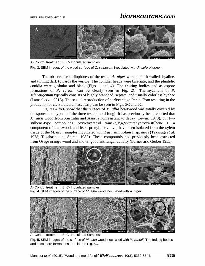
A- Control treatment; B, C- Inoculated samples
Fig. 5. SEM images of the surface of M. alba wood inoculated with P. variotii. The fruiting bodies and ascospore formations are clear in Fig. 5C.

PEER-REVIEWED ARTICLE bioresources.com
Mansour et al. (2015). “Wood and mold fungi,” BioResources 10(3), 5330-5344. 5337
A- Control treatment; B,C- Inoculated samples
Fig. 6. SEM images of the wood surface of M. alba inoculated with P. selerotigenum
P. variotii, a soft-rot fungus, is called a green mold because of the color of its
spores and mycelium (Yoon et al. 2007) and may be recognized by its dense brush-like
spore-bearing structures (Pitt 1979; Domsch et al. 1980). It produces a yellow
discoloration in oak wood during storage and drying (Bauch et al. 1991). P. variotii
produces ample amounts of xylanase (Schmidt et al. 1979) and excretes glucuronidase.
Additionally, the fungus P. variotii may be used to obtain tannins that are suitable for
adhesives (Schmidt et al. 1984; Schmidt and Weißmann 1986). Also, the sugars found in
spent sulfite liquor can be converted by P. variotii for use as animal feed (Forss et al.
1986).
Species of Penicillium are known for their ability to produce extracellular
enzymes including cellulase (Krogh et al. 2004). Additionally, some Penicillum species
can degrade pectin, while a greater number of species are able to degrade xylan (Yoon et
al. 2007). The production of xylanase enzymes by Aspergillus spp. has been studied by
de Vries and Visser (2001); the hydrolytic enzymes involved include endo-1,4-β-
glucanases; exo-1,4-β-glucanase, and 1,4-β-glucosidases for cellulose hydrolysis; and
endo-1,4-β-xylanases and β-xylosidases for hemicelluloses hydrolysis (Biely and
Tenkanen 1998).
By growing mold fungi over the surfaces of wood, there were large fruiting
structures that release vast numbers of spores in nature, as shown in Figs. 1 through 6
(Mansour and Salem 2015; Mansour et al. 2015).
In our previously works (Mansour and Salem 2015; Mansour et al. 2015), we
started to find the changes in the full elemental composition of woods as affected by
mold fungi using the EDX technique. For example, the C and O elements compositions
of wood (Acacia saligna) were decreased compared to the control treatment
(unincubated) as affected by the mold fungi; Trichoderma harzianum, Alternaria
tenuissima, and F. culmorum.
Most of the research studies were focused in the using of natural extracts or
essential oils against the growth of mold fungi, for example, the oils of Garlic, Neem,
Clove, Olive and Ajwain caused significant reduction in the growth of A. niger, A. flavus,
P. variotii, Penicillium sp., Trichoderma sp., Grifola sp. and T. viridi (Hussain et al.
2013). Also, the three tested mold fungi showed a variety of growth inhibition responses
to fatty acid extracts from the wood, bark, and leaves of Brachychiton diversifolius
(Salem et al. 2014a) and the wood essential oil and methanol and chloroform extracts
from the wood and bark of Delonix regia (Salem et al. 2014b).
PEER-REVIEWED ARTICLE bioresources.com
Mansour et al. (2015). “Wood and mold fungi,” BioResources 10(3), 5330-5344. 5338
Antifungal Activity of Methanol Extracts According to the MIC values presented in Table 3, the methanol extracts from M.
alba heartwood showed good inhibition against the growth of A. niger at low
concentrations (32 μg/mL) and relatively high concentration against P. variotii and P.
selerotigenum, with MIC values of 500 and 250 μg/mL, respectively. Among some
extractives used for wood impregnation, extracts of mulberry were most effective relative
to wood durability (Kazemi 2007). Phenols, stilbenes, flavonoids, and sterols in the
extractives of white mulberry wood have been reported (Venkataraman 1972; Rowe and
Conner 1979; Se Golpayegani et al. 2010; Sadeghifar et al. 2011). The toxicity of
extracts from M. alba could be related to resorcinol, which corresponded to the highest
proportion of the alcoholic extract that has been reported in earlier works (Sadeghifar et
al. 2011; Salem et al. 2013; Se Golpayegani et al. 2014). Moracin N, M and P have been
isolated before from M. alba (Nguyen et al. 2009).
The results showed that the heartwood methanol, acetone, and ethyl acetate extracts
of M. alba had the best inhibitive effect on Coriolus versicolor, Gloeophyllum trabeum
and Botryodiplodia theobromae compared to the others studied extracts from other trees.
The ethyl acetate extracts (1g/L) showed inhibition rate for three fungi over 85% and at
the concentration of 3g/L was 100%, with EC50 value at 0.173g/L, 0.324g/L, and
0.057g/L, respectively (Ying 2009).
Table 3. MIC Values (μg/mL) of Methanol Extracts of C. spinosum Wood and M. alba Heartwood and Against the Growth of A. niger, P. variotii, and P. selerotigenum
Methanol extract MIC value (µg/mL)
A. niger P. variotii P. selerotigenum
M. alba (heartwood) 32 500 250
C. spinosum 250 8 500
The methanol extract from C. spinosum wood exhibited remarkable inhibition
against the growth of P. variotii at low concentrations (8 µg/mL) and at high
concentrations. Inhibition against A. niger and P. selerotigenum growth was found with
MIC values of 250 μg/mL and 500 μg/mL, respectively. The antifungal effect of extracts
from C. spinosum could be related to the active constituents like iridoid glycoside
phlomiol, 5-deoxy pulchelloside, larniide, durantoside I and lamidoside (Khalifa et al.
2002; El-Naggar 2007).
Extracts from wood have been recognized as potential agents against the growth
of mold and other wood-destroying fungi, for example, the wood extracts of Cupressus
sempervirens (Tapondjou et al. 2005), and Maclura pomifera heartwood wood extracts,
morin, oxyresveratrol, and 1,3,6,7-tetrahydroxyxanthoneshowed potential fungicidal
activity (Barnes and Gerber 1955; Wolfrom and Bhat 1965).
CONCLUSIONS
1. In the present study, the surface elemental changes of C. spinosum wood and M. alba
heartwood were monitored as well as the growth of the three tested mold fungi P.
selerotigenum, P. variotii, and A. niger using EDX and SEM techniques.

PEER-REVIEWED ARTICLE bioresources.com
Mansour et al. (2015). “Wood and mold fungi,” BioResources 10(3), 5330-5344. 5339
2. C, mostly in wood carbohydrates and lignin, was significantly decreased and
consumed by the three tested mold fungi.
3. C was significantly (P = 0.0004) decreased from 49.91% in the control to 47%,
40.1%, and 40% in wood inoculated with P. selerotigenum, A. niger, and P. variotii,
respectively.
4. C in C. spinosum wood was affected significantly (P = 0.0004) and decreased from
49.91% in the control to 47%, 40.1%, and 40% with P. selerotigenum, A. niger, and
P. variotii, respectively.
5. C in M. alba heartwood was significantly (P < 0.0001) decreased, from 51.33% in the
control to 41.49%, 40%, and 43.66% in wood inoculated with A. niger, P. variotii,
and P. selerotigenum, respectively.
6. The change in O peak element was not significantly affected by inoculation with the
three tested mold fungi.
7. Other elements showed different percentages according to wood species and type of
mold fungus. For example, Al and Cu were observed in high percentages with M.
alba heartwood inoculated by P. variotii.
8. SEM images showed the distribution and structures of spores and hyphae of the three
tested mold fungi.
9. The methanol extract from M. alba heartwood showed good inhibition against the
growth of A. niger at the concentration 32 μg/mL, and the methanol extract from C.
spinosum wood showed remarkable inhibition against the growth of P. variotii at the
concentration of 8 μg/mL.
10. The natural durability between M. alba heartwood and C. spinosum wood showed
variation according to the colonized fungi as shown in the variations of elemental
composition.
11. Other work is recommended to study the natural durability of some commercial
woods with the same mold fungi.
ACKNOWLEDGMENTS
The authors extend their sincere appreciation to the Deanship of Scientific
Research at King Saud University for its funding this research group, No. (RG 1435-
011).
REFERENCES CITED
Adikaram, N., Karunanayake, C., and Abayasekara, C. H. (2010). “The role of pre-
formed antifungal substances in the resistance of fruits to postharvest pathogens,” in:
Postharvest Pathology, D. Prusky and M. L. Gullino (eds.), Springer, Berlin, pp. 1-
11. DOI: 10.1007/978-1-4020-8930-5_1

PEER-REVIEWED ARTICLE bioresources.com
Mansour et al. (2015). “Wood and mold fungi,” BioResources 10(3), 5330-5344. 5340
Arango, R. A., Green III, F., Hintz, K., Lebow, P. K., and Miller, R. B. (2006). “Natural
durability of tropical and native wood against termite damage by Reticulitermes flavis
(Kollar),” Int. Biodeter. Biodegr. 57, 146-150.
Balázs, B., Toth, G., Duddeck, H., and Soliman, H. S. (2006). “Iridoid and lignan
glycosides from Citharexylum spinosum L.,” Nat. Prod. Res. 20(2), 201-205. DOI:
10.1080/14786410500056694
Barnes, R. A., and Gerber, N. N. (1955). “The antifungal agent from Osage orange
wood,” J. Am. Chem. Soc. 77(12), 3259-3264. DOI: 10.1021/ja01617a032
Biely, P., and Tenkanen, M. (1998). “Enzymology of hemicellulose degradation,” in:
Trichoderma and Gliocladium, Vol. 2, G. E. Harman and C. P. Kubicek (eds.), Taylor
and Francis, London, UK, pp. 25-47.
Bauch, J., Hundt von, H., Weißmann, G., Lange, W., and Kubel, H. (1991). “On the
causes of yellow discolorations of oak heartwood (Quercus Sect. Robur) during
drying,” Holzforschung 45(2), 79-85. DOI: 10.1515/hfsg.1991.45.2.79
Chen, C. C., Liu, L. K., Hsu, J. D., Huang, H. P., Yang, M. Y., and Wang, C. J. (2005).
“Mulberry extract inhibits the development of atherosclerosis in cholesterol fed
rabbits,” Food Chem. 91(4), 601-607. DOI: 10.1016/J.FOODCHEM.2004.06.039
Chung, K. O., Kim, B. Y., Lee, M. H. Kim, Y. R., Chung, H. Y., Park, J. H. and Moon, J.
O. (2003), “In-vitro and in-vivo anti-inflammatory effect of oxyresveratrol from
Morus alba L.,” J. Pharm. Pharmacol. 55(12), 1695-700. DOI:
10.1211/0022357022313
Darwish, S. S., EL Hadidi, N. M. N., and Mansour, M. (2013). “The effect of fungal
decay on Ficus sycomorus wood,” Int. J. Conserv. Sci. 4(3), 271-282.
Danilatos, G. D., and Robinson, V. N. E. (1979). “Principles of scanning electron
microscopy at high specimen pressures,” Scanning 2(2), 72-82. DOI:
10.1002/sca.4950020202
de Vries, R. P., and Visser, J. (2001). “Aspergillus enzymes involved in degradation of
plant cell wall polysaccharides,” Microbiolo. Mol. Biol. Rev. 65(4), 497-522. DOI:
10.1128/MMBR.65.4.497-522.2001
Domsch, K. H., Gams, W., and Anderson, T. H. (1980). Compendium of Soil Fungi, Vol.
2, Academic Press, London, UK.
El-Beshbishy, H. A., Singab, A. N., Sinkkonen, J., and Pihlaja, K. (2006).
“Hypolipidemic and antioxidant effects of Morus alba L. (Egyptian mulberry) root
bark fractions supplementation in cholesterol-fed rats,” Life Sci. 78(23), 2724-2733.
DOI: 10.1016/j.lfs.2005.10.010
Eloff, J .N. (1998). “A sensitive and quick microplate method to determine the minimal
inhibitory concentration of plant extracts for bacteria,” Planta Med. 64(8), 711-713.
DOI: 10.1055/s-2006-957563
El-Naggar, D. M. (2007). “Antibilharzial study of some extracts from Citharexylum
quadrangular Jacq.,” Ph.D. dissertation, Faculty of Pharmacy for Girls, Al-Azhar
University, Nasr City, Cairo, Egypt.
Forss, K., Jokinen, K., and Lehtomaki, M. (1986). “Aspects of the pekilo protein
process,” Paperi ja Puu 68(11), 839-844.
Hukka, A., and Viitanen, H. A. (1999). “A mathematical model of mould growth on
wooden material,” Wood Sci. Tech. 33(6), 475-485. DOI: 10.1007/s002260050131
Hussain, A., Shrivastav, A., and Jain, S. K. (2013). “Antifungal activity of essential oils
against local wood degrading cellulolytic filamentous fungi,” Adv. Biores. 4(2), 161-
167.

PEER-REVIEWED ARTICLE bioresources.com
Mansour et al. (2015). “Wood and mold fungi,” BioResources 10(3), 5330-5344. 5341
Hussein, M. S. H., El-Tawil, O. S., Yassin, N. E. H., and Abdou, A. (2010). “The
protective effect of Morus alba and Calendula officinalis plant extracts on carbon
tetrachloride-induced hepatotoxicity in isolated rat hepatocytes,” J. Am. Sci. 6(10),
762-773.
Huxley, A., Griffiths, M., and Levy, M. (1997). Dictionary of Gardening, McMillan
Reference Limited, London, UK.
Jeloková, E., and Šindler, J. (1997). “Influence of beech and acacia wood extractives on
activity of wood-destroying fungi,” in: 1st Symposium - Drevoznehodnocujúce huby
97, TU Zvolen, pp. 133-139.
Jeloková, E., and Šindler, J. (2001). “Testing of some chemical compounds for wood
protection,” Drevo 56, 137-138.
Kazemi, S. M. (2007). “Impregnation of beech, maple, alder and lime under different
treatments of extractives and fungal attack,” J. Biol. Sci. 7(8), 1463-1467. DOI:
10.3923/jbs.2007.1463.1467
Khalifa, T. I., El-Gendi, O. D., Ammar, H. A., and El-Naggar, D. M. (2002). “Iridoid
glycosides from Citharexylum quadrangulare,” Asian J. Chem. 14(1), 197-202.
Lamsal, K., Kim, S. W., Naeimi S., Adhikari M., Yadav, D. R., Kim, C., Lee, H. B., and
Lee, Y. S. (2013). “Three new records of Penicillium species isolated from insect
specimens in Korea,” Mycobiology 41(2), 116-119. DOI:
10.5941/MYCO.2013.41.2.116
Ljaljević-Grbić, M., Stupar, M., Vukojević, J., Maričić, I., and Bungur, N. (2013).
“Molds in museum environments: Biodeterioration of art photographs and wooden
sculptures,” Arch. Biol. Sci., Belgrade 65(3), 955-962. DOI: 10.2298/ABS1303955G
Lokegaonkar, S. P., and Nabar, B. M. (2011). “In vitro assessment of the antiplaque
properties of crude M. alba leaf extract,” J. Herb. Med. Toxicol. 5(1), 71-77.
Krogh, K. B. R., Morkeberg, A., Friscad, J. C., and Olsson, L. (2004). “Screening genus
Penicillium for producers of cellulolytic and xylanolytic enzymes,” Appl. Biochem.
Biotechnol. 113(116), 389-401. DOI: 10.1385/ABAB:114:1-3:389
Mansour, M. M., and Salem, M. Z. M. (2015). “Evaluation of wood treated with some
natural extracts and Paraloid B-72 against the fungus Trichoderma harzianum: Wood
elemental composition, in-vitro and application evidence,” Int. Biodeter. Biodegr.
100(C), 62-69. http://dx.doi.org/10.1016/j.ibiod.2015.02.009
Mansour, M. M. A., Abdel-Megeed, A., Nasser, R. A., and Salem, M. Z. M. (2015).
“Comparative evaluation of some woody trees methanolic extracts and Paraloid B-72
against phytopathogenic mold fungi Alternaria tenuissima and Fusarium culmorum,”
BioResources 10(2), 2570-2584. DOI: 10.15376/biores.10.2.2570-2584
Mar, A., and Pripdeevech, P. )2014). “Chemical composition and antibacterial activity of
essential oil and extracts of Citharexylum spinosum flowers from Thailand,” Nat.
Prod. Commun. 9(5), 707-710.
Maranhão, C. A., Pinheiro, I. O., Santana, L. B. D. A., Oliveira, L. S., Nascimento, M .S.,
and Bieber, L. W. (2013). “Antitermitic and antioxidant activities of heartwood
extracts and main flavonoids of Hymenaea stigonocarpa Mart,” Int. Biodeter.
Biodegr. 79, 9-13. DOI: 10.1016/j.ibiod.2013.01.005
Neya, B., Hakkou, M., Pétrissans, M., and Gérardin, P. (2004). “On the durability of
Burkea africana heartwood: Evidence of biocidal and hydrophobic properties
responsible for durability,” Ann. Forest Sci. 61(3), 277-282. DOI:
10.1051/forest:2004020
PEER-REVIEWED ARTICLE bioresources.com
Mansour et al. (2015). “Wood and mold fungi,” BioResources 10(3), 5330-5344. 5342
Nguyen, T. D., Jin, X., Lee, K., Hog, Y.-S., Young, H. K., and Jung, J. L. (2009).
“Hypoxia-inducible factor-1 inhibitory benzofurans and chalcone-derived Diels-
Alder adducts from Morus species,” J. Nat. Prod. 72, 39–43. doi:
10.1021/np800491u
Pettersen, R. C. (1984). “The chemical composition of wood,” in: The Chemistry of
Wood, Advances in Chemistry Series 207, R. M. Rowell (ed.), American Chemical
Society, Washington, DC, pp. 57-126.
Philip, R. W., Bruce, A., and Munro, A. G. (1995). “The effect of water soluble Scots
pine (Pinus sylvestris L.) and Sitka spruce [Picea sitchensis (bong) Carr] heartwood
and sapwood extracts on the growth of selected Trichoderma species,” Int. Biodeter.
Biodegr. 35(4), 355-367. DOI: 10.1016/0964-8305(95)00053-9
Pitt, J. I. (1979). The Genus Penicillium and Its Teleomorphic States Eupeniciliium and
Talaromyces, Academic Press, London, UK.
Rosado, T., Martins, M. R., Pires, M., Mirão, J., Candeias, A., and Caldeira, A. T. (2013).
“Enzymatic monitorization of mural paintings: Biodegradation and biodeterioration,”
Int. J. Conserv. Sci. 4(SI), 603-612.
Rowe, J. W., and Conner, A. (1979). “Extractives in Eastern hardwoods – A review,”
Forest Products Laboratory, U.S. Department of Agriculture, Madison, WI.
Salem, M. Z. M., Aly, H. I. M., Gohar, Y. M., and EL-Sayed, A. B. (2013). “Biological
activity of extracts from Morus alba L., Albizzia lebbeck (L.) Benth. and Casuarina
glauca Sieber against the growth of some pathogenic bacteria,” Int. J. Agric. Food
Res. 2(1), 9-22.
Salem, M. Z. M., Abdel-Megeed, A., and Ali, H. M. (2014a). “Stem wood and bark
extracts of Delonix regia (Boj. Ex. Hook): Chemical analysis and antibacterial,
antifungal, and antioxidant properties,” BioResources 9(2), 2382-2395. DOI:
10.15376/biores.9.2.2382-2395
Salem, M. Z. M., Ali, H. M., and Mansour, M. M. (2014b). “Fatty acid methyl esters
from air-dried wood, bark, and leaves of Brachychiton diversifolius R. Br:
Antibacterial, antifungal, and antioxidant activities,” BioResources 9(3), 3835-3845.
DOI: 10.15376/biores.9.3.3835-3845
Sadeghifar, H., Sheikh, A., Khalilzadeh, M. A., and Ebadi, A. G. H. (2011). “Heartwood
extractives of Iranian Morus alba wood,” J. Chem. Soc. Pak. 33(1), 104-106.
SAS (2001). “Users guide: Statistics (release 8.02),” SAS Inst., Cary, NC.
Scheerer, S., Ortega-Morales, O., and Gaylarde, C., (2009). “Microbial deterioration of
stone monuments – An updated overview,” Adv. Appl. Microbiol. 66, 97-139. DOI:
10.1016/S0065-2164(08)00805-8
Sirmah, P., Dumarçay, S., and Gérardin, P. (2009). “Unusual amount of (-)-mesquitol
from the heartwood of Prosopis juliflora,” Nat. Prod. Res. 23(2), 183-189. DOI:
10.1080/14786410801940968
Se Golpayegani, A., Thevenon, M. F., Gril, J., and Pourtahmasi, K. (2010). “Natural
durability of white mulberry (Morus alba L.),” 4th International Research Group Of
Wood Preservation (IRG), May 9-13, Biarritz, France.
Se Golpayegani, A. 2011. “Characterization of mulberry’s wood (Morus alba L.),
considering its usage in making Iranian lute,” Ph.D. Thesis, Montpellier II University,
France, 288p.
Se Golpayegani, A., Thévenon, M. F., Gril, J., Masson, E., and Pourtahmasi, K. (2014).
“Toxicity potential in the extraneous compounds of white mulberry wood (Morus

PEER-REVIEWED ARTICLE bioresources.com
Mansour et al. (2015). “Wood and mold fungi,” BioResources 10(3), 5330-5344. 5343
alba),” Maderas-Cienc. Tecnol. 16(2), 227-238, DOI: 10.4067/S0718-
221X2014005000018
Sequeira, S., Cabrita, E. J., and Macedo, M. F. (2012). “Antifungals on paper
conservation: An overview,” Int. Biodeter. Biodegr. 74, 67-86. DOI:
10.1016/j.ibiod.2012.07.011
Schmidt, O., and Weißmann, G. (1986). “Mikrobiologische Behandlung von
Larchenrinden-Extrakten zur Herstellung von Leimharzen,” Holz Roh-Werkst. 44(9),
351-355. DOI: 10.1007/BF02612742
Schmidt, O., Ayla, C., and Weißmann, G. (1984). “Mikrobiologische Behandlung von
Fichtenrinde-Heiswasserextrakten zur Herstellung von Leimharzen,” Holz Roh-
Werkst. 42(8), 287-292. DOI: 10.1007/BF02608936
Schmidt, O., Puls, J., Sinner, M., and Dietrichs, H. H. (1979). “Concurrent yield of
mycelium and xylanolytic enzymes from extracts of steamed birchwood, oat husks
and wheat straw,” Holzforschung 33(6), 192-196. DOI: 10.1515/hfsg.1979.33.6.192
Sterflinger, K., and Piñar, G. (2013). “Microbial deterioration of cultural heritage and
works of art: Tilting at windmills?,” Appl. Microbiol. Biotechnol. 97(22), 9637-9646.
DOI: 10.1007/s00253-013-5283-1
Takahashi, K., and Shirata, A. (1982). “Production of antifungal substances in mulberry,”
Jap. Agric. Res. Q. 16(2), 119-124.
Takasugi, M., Munoz, L., Masamune, T., Shirata, A., and Takahashi, K. (1978). “Stilbene
phytoalexins from diseased mulberry,” Chem. Lett. 1241-1242. DOI:
10.1246/cl.1978.1241
Tapondjou, A. L., Adler, C., Fontem, D. A., Bouda, H., and Reichmuth, C. (2005).
“Bioactivities of cymol and essential oils of Cupressus sempervirens and Eucalyptus
saligna against Sitophilus zeamais Motschulsky and Tribolium confusum du Val,” J.
Stored Prod. Res. 41(1), 91-102. DOI: 10.1016/j.jspr.2004.01.004
Taylor, A. M., Gartner, B. L, and Morrell, J. J. (2006). “Effects of heartwood extractive
fractions of Thuja plicata and Chamaecyparis nootkatensison wood degradation by
termites or fungi,” J. Wood Sci. 52(2), 147-153. DOI: 10.1007/s10086-0050743-6
Tewari, M. C. (1978). “Data on natural durability of timber species (installed in the test
yard at New Forest, Dehra Dun) according to 1976 inspection, their treatability and
seasoning characteristics,” IRG/WP/3127, International Research Group on Wood
Preservation, Stockholm, Sweden.
Venkataraman, K. (1972). “Wood phenolics in the chemotaxonomy of the moraceae,”
Phytochemistry. 11(5), 1571-1586. DOI: 10.1016/0031-9422(72)85002-7
Viitanen, H., and Ritschkoff, A. C. (1991). “Mold growth in pine and spruce sapwood in
relation to air humidity and temperature,” Swed. Univ. Agric. Sci. Dept. Forest Prod.,
Uppsala, Sweden, Report No. 221.Wagner, W.L., Herbst, D.R., Sohmer, S.H. (1999).
“Manual of the Flowering Plants of Hawai,”’i., Vol. 2, Bishop Museum Special
Publication 83, University of Hawai’i and Bishop Museum Press, Honolulu, HI.
Wei, A., and Shibamoto, T. (2007). “Antioxidant activities and volatile constituents of
various essential oils,” J. Agric. Food Chem. 55(5), 1737-1742. DOI:
10.1021/jf062959x
Wolfrom, M. L., and Bhat, H. B. (1965). “Osage orange pigments dVII. 1,3,6,7
Tetrahydroxyxanthone from the heartwood,” Phytochemistry 4, 765-768.
Yamaguchi, H., Yoshino, K., and Kido, A. (2002). “Termite resistance and wood
penetrability of chemically modified tannin and tannin-copper complexes as wood
preservatives,” J. Wood Sci. 48(4), 331-337. DOI: 10.1007/BF00770710

PEER-REVIEWED ARTICLE bioresources.com
Mansour et al. (2015). “Wood and mold fungi,” BioResources 10(3), 5330-5344. 5344
Ying, L. S. (2009). “Inhibitory effects of heartwood extracts from strong decay-resistant
tree species on wood preservation,” master thesis, Agricultural University of Hebei.
Yogisha, S., and Raveesha, K. A. (2009). “In-vitro antibacterial effect of selected
medicinal plant extracts,” J. Nat Prod. (India) 2, 64-69.
Yoon, J. H., Hong, S. B., Ko, S. J., and Kim, S. H. (2007). “Detection of extracellular
enzyme activity in Penicillium using chromogenic media,” Mycobiol. 35(3), 166-169.
DOI: 10.4489/MYCO.2007.35.3.166
Article submitted: March 3, 2015; Peer review completed: June 29, 2015; Revised
version received and accepted: July 3, 2015; Published: July 15, 2015.
DOI: 10.15376/biores.10.3.5330-5344