


NAD uptake in mammalian cells - Journal of Biological … OF NAD UPTAKE IN MAMMALIAN CELLS Richard...
22
CHARACTERIZATION OF NAD UPTAKE IN MAMMALIAN CELLS Richard A. Billington, Cristina Travelli, Emanuela Ercolano, Ubaldina Galli, Cintia Blasi Roman, Ambra A. Grolla, Pier Luigi Canonico, Fabrizio Condorelli, Armando A. Genazzani DiSCAFF and the DFB center, Università del Piemonte Orientale, Via Bovio 6, 28100 Novara, Italy Running Title: NAD Uptake Address correspondence to: Richard Billington, DiSCAFF, Via Bovio 6, 28100 Novara, Italy, Tel: +39 0321375835, Fax: +39 0321375821, Email: [email protected] Abstract Recent evidence has shown that NAD(P) plays a variety of roles in cell signalling processes. Surprisingly, the presence of NAD(P) utilising ectoenzymes suggests that NAD(P) is present extracellularly. Indeed, nanomolar concentrations of NAD have been found in plasma and other body fluids. While very high concentrations of NAD have been shown to enter cells, it is not known whether lower, more physiological concentrations are able to be taken up. Here we show that two mammalian cell types are able to transport low NAD concentrations effectively. Furthermore, extracellular application of NAD was able to counteract FK866-induced cell death and restore intracellular NAD(P) levels. We propose that NAD uptake may play a role in physiological NAD homeostasis. Introduction Pyridine nucleotides, although often considered as simple redox cofactors in the cell, are in fact multifunctional molecules involved in a wide range of cellular processes (1). It is becoming clear that NAD(P) is involved in more pharmacologically attractive cellular processes such as cell signalling, transcriptional regulation and post-translational protein modification (2,3). For example, NAD(P) has been shown to be the precursor of molecules involved in calcium signalling (e.g. cADPR, NAADP, ADPR), to be involved in the regulation of epigenetic changes via sirtuins and to be a substrate for both mono- and poly-ADP ribosylation (3-5). The general perception is that cellular NAD is synthesised either de novo from tryptophan or via one of two possible recycling pathways: from nicotinic acid or nicotinamide (together referred to as vitamin PP, or niacin) (6). Recently, a third biosynthesis pathway has been described which uses nicotinamide riboside as a precursor (7). The presence of membrane-bound ectoenzymes that use NAD(P) (8) has led to the investigation and description of several mechanisms for the export of NAD across the plasma membrane, including transport through connexins and stimulus-induced exocytotic release (9-11). Surprisingly, though, the possibility that low NAD concentrations can be imported across the membrane to directly replenish the cellular NAD(P) pools bypassing biosynthetic pathways has not been conclusively addressed. However, circumstantial evidence suggests that this may occur: (i) High concentrations of extracellularly applied NAD(H) have been shown to increase intracellular NAD levels (10,12-14): (ii) extracellular NAD counteracts PARP-induced intracellular NAD depletion (12,15); (iii) CD38 knockout mice, which are impaired in their ability to degrade extracellular NAD, display higher endogenous SIRT1 activity (16); (iv) uptake processes have been shown for the Ca 2+ - mobilising NAD(P) metabolites cADPR and NAADP in a variety of diverse mammalian cell types (17-21); (v) NAD and at least one of the enzymes involved in biosynthesis are present extracellularly (13,22,23); (vi) Wallerian degeneration can be slowed by the addition of extracellular NAD (24). Here we show that various mammalian cell types are able to transport pM concentrations of NAD across the plasma membrane from the extracellular medium. When intracellular NAD(P) levels were reduced by treatment with the NAD biosynthesis inhibitor FK866 (APO866; (E)-N-[4-(1-benzoylpiperidin-4-yl) butyl]-3-(pyridin-3-yl) acrylamide; (25)), application of NAD extracellularly was able to partially replenish these levels. Furthermore, cells could be rescued from FK866-induced NAD depletion, autophagy and cell death by addition of NAD to the culture medium. These data, taken together, suggest that NAD uptake can participate in the homeostasis of cellular NAD levels. 1 http://www.jbc.org/cgi/doi/10.1074/jbc.M706204200 The latest version is at JBC Papers in Press. Published on January 7, 2008 as Manuscript M706204200 Copyright 2008 by The American Society for Biochemistry and Molecular Biology, Inc. by guest on May 31, 2018 http://www.jbc.org/ Downloaded from
-
Upload
nguyenhanh -
Category
Documents
-
view
216 -
download
2
Transcript of NAD uptake in mammalian cells - Journal of Biological … OF NAD UPTAKE IN MAMMALIAN CELLS Richard...
CHARACTERIZATION OF NAD UPTAKE IN MAMMALIAN CELLS Richard A. Billington, Cristina Travelli, Emanuela Ercolano, Ubaldina Galli, Cintia Blasi Roman, Ambra A. Grolla, Pier Luigi Canonico, Fabrizio Condorelli, Armando A. Genazzani DiSCAFF and the DFB center, Università del Piemonte Orientale, Via Bovio 6, 28100 Novara, Italy Running Title: NAD Uptake Address correspondence to: Richard Billington, DiSCAFF, Via Bovio 6, 28100 Novara, Italy, Tel: +39 0321375835, Fax: +39 0321375821, Email: [email protected] Abstract Recent evidence has shown that NAD(P) plays a variety of roles in cell signalling processes. Surprisingly, the presence of NAD(P) utilising ectoenzymes suggests that NAD(P) is present extracellularly. Indeed, nanomolar concentrations of NAD have been found in plasma and other body fluids. While very high concentrations of NAD have been shown to enter cells, it is not known whether lower, more physiological concentrations are able to be taken up. Here we show that two mammalian cell types are able to transport low NAD concentrations effectively. Furthermore, extracellular application of NAD was able to counteract FK866-induced cell death and restore intracellular NAD(P) levels. We propose that NAD uptake may play a role in physiological NAD homeostasis. Introduction Pyridine nucleotides, although often considered as simple redox cofactors in the cell, are in fact multifunctional molecules involved in a wide range of cellular processes (1). It is becoming clear that NAD(P) is involved in more pharmacologically attractive cellular processes such as cell signalling, transcriptional regulation and post-translational protein modification (2,3). For example, NAD(P) has been shown to be the precursor of molecules involved in calcium signalling (e.g. cADPR, NAADP, ADPR), to be involved in the regulation of epigenetic changes via sirtuins and to be a substrate for both mono- and poly-ADP ribosylation (3-5). The general perception is that cellular NAD is synthesised either de novo from tryptophan or via one of two possible recycling pathways: from nicotinic acid or nicotinamide (together referred to as vitamin PP, or niacin) (6). Recently, a third biosynthesis pathway has been described which uses nicotinamide riboside as a precursor (7).
The presence of membrane-bound ectoenzymes that use NAD(P) (8) has led to the investigation and description of several mechanisms for the export of NAD across the plasma membrane, including transport through connexins and stimulus-induced exocytotic release (9-11). Surprisingly, though, the possibility that low NAD concentrations can be imported across the membrane to directly replenish the cellular NAD(P) pools bypassing biosynthetic pathways has not been conclusively addressed. However, circumstantial evidence suggests that this may occur: (i) High concentrations of extracellularly applied NAD(H) have been shown to increase intracellular NAD levels (10,12-14): (ii) extracellular NAD counteracts PARP-induced intracellular NAD depletion (12,15); (iii) CD38 knockout mice, which are impaired in their ability to degrade extracellular NAD, display higher endogenous SIRT1 activity (16); (iv) uptake processes have been shown for the Ca2+-mobilising NAD(P) metabolites cADPR and NAADP in a variety of diverse mammalian cell types (17-21); (v) NAD and at least one of the enzymes involved in biosynthesis are present extracellularly (13,22,23); (vi) Wallerian degeneration can be slowed by the addition of extracellular NAD (24). Here we show that various mammalian cell types are able to transport pM concentrations of NAD across the plasma membrane from the extracellular medium. When intracellular NAD(P) levels were reduced by treatment with the NAD biosynthesis inhibitor FK866 (APO866; (E)-N-[4-(1-benzoylpiperidin-4-yl) butyl]-3-(pyridin-3-yl) acrylamide; (25)), application of NAD extracellularly was able to partially replenish these levels. Furthermore, cells could be rescued from FK866-induced NAD depletion, autophagy and cell death by addition of NAD to the culture medium. These data, taken together, suggest that NAD uptake can participate in the homeostasis of cellular NAD levels.
1
http://www.jbc.org/cgi/doi/10.1074/jbc.M706204200The latest version is at JBC Papers in Press. Published on January 7, 2008 as Manuscript M706204200
Copyright 2008 by The American Society for Biochemistry and Molecular Biology, Inc.
by guest on May 31, 2018
http://ww
w.jbc.org/
Dow
nloaded from

Experimental Procedures Cell Culture NIH-3T3 murine epithelial cells were cultured in RPMI-1640 (Sigma) supplemented with 5% FBS (foetal bovine serum), 2 mM glutamine, 10 U/ml penicillin and 100 µg/ml streptomycin. SH-SY 5Y cells were cultured in DMEM (Sigma) supplemented with 10% FBS, 2 mM glutamine, 10 U/ml penicillin, and 100 μg/ml streptomycin. HMEC, HeLa, HaCaT, K562 and RAW 264.7 cells were cultured according to the conditions suggested by ATCC. Cells were maintained in a humidified incubator supplied with 5% CO2/95% air at 37°C. Cells were subcultured as needed by detaching the cells with 0.25% trypsin and 5 mM EDTA. [32P]NAD transport Cells plated in 6 well plates were grown to confluence. The wells were washed twice with 1 ml of warm Locke Buffer (134 mM NaCl, 4 mM NaHCO3, 5 mM KCl, 2.3 mM CaCl2, 10 mM HEPES, 1 mM MgCl2, 5 mM glucose, pH 7.4) before addition of 0.5 ml Locke Buffer or modifications thereof (NMG Locke contained 134 mM N-methylglucamine in place of the NaCl (4 mM final Na+); EGTA Locke lacked CaCl2 with the addition of 500 μM EGTA). The reaction was started by the addition of 250 pM [32P]NAD (800 Ci/mmol, Perkin-Elmer) or 250 pM [32P]NAADP prepared as described previously (26). Competing compounds and drugs were added to the wells before [32P]NAD unless stated in the text. After 10 minutes (except where indicated), the reaction was stopped by aspiration of the buffer and the cells were washed twice with 1 ml of Locke Buffer. The cells were then dissolved in 1 ml 0.5 M NaOH. 2 ml of scintillation fluid was added and the radioactivity associated with the cells was counted using a standard scintillation counting procedure for 32P. For HPLC analysis of transported NAD, cells were scraped after transport experiments in ddH2O, extracted by adding an equal volume of chloroform and run on HPLC as described previously (27). Cell viability assay To analyse cell viability, the colorimetric MTT (3-(4,5-dimethylthiazol-2-yl)-2,5-dipehenyltetrazolium bromide) assay was used. Briefly, cells were plated in 24 well plates and treated as indicated for the appropriate time. FK866 (dissolved in DMSO) and the vehicle
control were added to the cells to give a final DMSO concentration no greater than 0.5%. Cells were washed once in Locke Buffer and 300 µl of MTT (250 µg/ml in Locke Buffer) was added before returning the cells to the incubator for one hour to allow the formation of the purple formazan crystals. After one hour, 600 µl of isopropanol/0.1 M HCl was added to each well and the absorbance was read at 570 nm in a plate reader (Victor3V, Perkin Elmer). Alternatively cell counts were performed. Briefly, cells were detached from the wells by trypsinisation, stained with Trypan Blue to exclude dead cells and counted in a Burker Chamber. NAD(P) cycling assay Total cellular NAD(P) was measured using an adaptation of the method described by Gasser et. al. (28). Cells, grown and treated in 24 well plates, were scraped with the piston of a 1 ml syringe in 100 μl ddH20 on ice, and were extracted with 1 volume of HClO4 (2 M) for 45 min on ice. The samples were then centrifuged for 1 min at 13,000 g and the supernatant was diluted with an equal volume of K2CO3 (1 M). After a further 45 min incubation on ice, the insolubile potassium perchlorate was removed by centrifugation for 1 min at 13,000 g. The final pH of the supernatant was 8-8.5. In a black 96 well plate (OptiPlateTM-96 F: PerkinElmer) 60μl of cycling mix was added to 100 μl of the extracted NAD(P). The cycling mix (60 µl) consisted of: 20 μl 500 mM NaH2PO4, pH 8.0, 2 μl 5 mg/ml BSA, 0.1 μl 10 mM resazurin (prepared fresh), 5 μl 100 mM glucose-6-phosphate (G8404, Sigma), 15 μl 1 mg/ml glucose-6-phosphate dehydrogenase, 15 μl of purified diaphorase (see below). Fluorescence (excitation 544 nm, emission 590 nm) was measured for each well using a plate reader (Victor3V, PerkinElmer). Standard curves were generated with authentic NAD subjected to mock extractions were used to generate calibration curves. The diaphorase was purified as follows: 100 μl of enzyme solution (diaphorase 12 mg/ml in 50 mM NaH2PO4, pH 8.0 adjusted with NaOH) was mixed with 200 μl BSA (5 mg/ml in water) and 1.2 ml of a suspension (2% w/v) of activated charcoal in 50 mM NaCl, 20 mM NaH2PO4, pH 8.0. After incubation at 37°C for 30 min, the charcoal was removed by centrifugation (11,000 g, 10 min, 4°C), and the supernatant used directly for the cycling assay.
2
by guest on May 31, 2018
http://ww
w.jbc.org/
Dow
nloaded from
In order to ensure that FK866, as an antagonist of an NAD using enzyme, did not directly affect the enzymes of the cycling assay, calibration curves were performed in the presence and absence of 100 nM and 10 µM FK866. The calibration curves were not affected by the presence of FK866 (data not shown). Furthermore, the assay was not affected by NMN at 100 µM. FK866 FK866 ((E)-N-[4-(1-benzoylpiperidin-4-yl) butyl]-3-(pyridin-3-yl) acrylamide) was synthesized using a shorter route respect to that reported (29) (5 synthetic steps vs 7 synthetic steps). In brief, the commercially available 4-piperidine butyric acid hydrochloride was reduced to the corresponding alcohol using lithium aluminium hydride and then chemoselectively N-benzoylated. The product was then transformed into an azide using the DPPA/DBE, sodium azide protocol. Finally, the Staudinger azide-amine reduction gave the amine. This compound was subsequently coupled with (E)-3-(3-Pyridinyl)-2propenylchloride (prepared starting from the corresponding carboxylic acid using oxalyl chloride) to give FK866 (overall yield over 5 steps: 11 %). The identity of the product was confirmed by NMR (1H, 13C) and mass spectrometry. EYFP-LC3-beta translocation Exponentially growing cells were transfected with pEYFP-LC3-beta (kind gift of Prof. Jäättelä, Institute of Cancer Biology, Copenhagen, Denmark) by liposome transfection (Lipofectamine2000, Invitrogen) and seeded 1 day after the transfection on glass chamber slides. After the indicated treatments, the percentage of green cells (a minimum of 100 cells/sample) with green vacuoles (EYFP-LC3-beta translocated from cytosol to autophagic vacuoles) was counted using a Leica TCS-SP2 Laser-scanning microscope. For the morphological evaluation of autophagic vacuoles and lysosome co-localization, the acidic compartment of cells was counter-stained by labeling the cells by adding 50 nM of lysotracker-red (Molecular Probes) for 1 min at 37°C to cell culture media immediately before confocal analysis. Cytochrome c and AIF immuno-localization Cells were grown on 18 mm coverslips and incubated at 37°C in presence of the specified
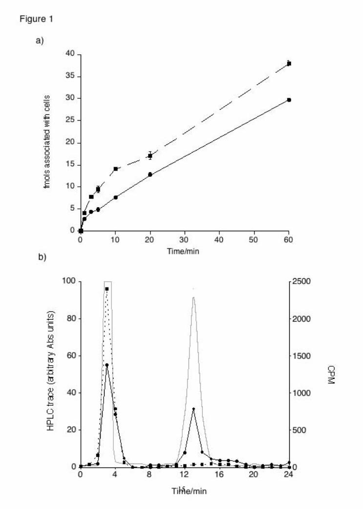
compounds. Cells were fixed in PBS containing 4% paraformaldehyde for 15 min at room temperature, washed with PBS, then permeabilized in PBS containing 0.2% Triton X-100 for 5 min. Cells were washed and then incubated with an anti-cyt-c antibody (1:500; BD, Milano, Italy) or with anti-AIF (1:250; Sigma-Aldrich) antibody in PBS for 1 h at 37°C. After washing, cells were stained with an AlexaFluor488-conjugated donkey anti-mouse-IgG (1:250 ; Molecular Probes, Eugene, OR, USA) in PBS for 1 h at 37°C. Coverslips were inverted and adhered to the glass slides using a mounting solution (DAKO Cytomation, CA, USA). Images from fluorescence patterns of the cells were obtained using a Leika TCS-SP2 microscope. Apoptotic nuclei staining For confocal-microscopy analysis of DNA integrity and AIF nuclear localization counter-staining, neuroblastoma cells were plated on 12 mm glass coverslips and maintained for appropriate length of time with different compounds. DRAQ5 (Biostatus Ltd., Leicestershire, UK) DNA dye was used for nuclear staining of living and apoptotic cells (fragmented nuclei). Briefly, the dye was added to cultured cells at 10 μM for 30 min followed by fixing in a 3.7% PBS/paraformaldehyde for 10 min at room temperature. Cells were washed twice with PBS, stained for AIF protein (as described above) and mounted onto cover slips for visualization with a confocal microscopy (HeNe laser UV 633 nm). Results To test whether NAD is transported into cells, NIH-3T3 cells were incubated for different lengths of time in Locke buffer containing 250 pM [32P]NAD and the reaction was stopped by aspiration of the buffer followed by a wash step with 1 ml Locke buffer. When the amount of [32P]NAD was determined by scintillation counting, a significant amount was recovered from the cellular fraction (Fig. 1A). This amount corresponded to 4.88 ± 0.28 fmols after 5 minutes and 29.81 ± 0.29 fmols (~ 24 % of total [32P]NAD present in the extracellular medium) after one hour. We have previously shown a similar accumulation of NAADP into mast cells (17) and we tested the ability of NIH-3T3 cells to transport [32P]NAADP. When the experiment was repeated with 250 pM [32P]NAADP, 9.44 ± 0.75 fmols of NAADP were recovered after 5 minutes and 38.07 ± 0.57 fmols after one hour.
3
by guest on May 31, 2018
http://ww
w.jbc.org/
Dow
nloaded from

NAD uptake shows at least 2 phases with an initial rapid phase of transport followed by a prolonged phase of steady uptake up to at least an hour. NAD has previously been reported to bind to an extracellular P2Y purinergic receptor (P2Y11; (30)). In order to identify whether a transport mechanism or a receptor mechanism was responsible for the observed NAD accumulation, experiments were performed whereby cells were treated with unlabelled NAD (100 µM) before or after treatment with 250 pM [32P]NAD (Table 1). Cells were washed between treatments. Cells were also treated simultaneously with unlabelled and labelled NAD. The amount of NAD transported was reduced only in the co-treatment experiment indicating direct competition (Table 1). The lack of displacement when excess unlabelled NAD was added after the [32P]NAD indicates that a reversible binding site is not responsible and the lack of inhibition by pre-treatment with unlabelled NAD rules out the presence of an irreversible binding site (Table 1). Furthermore, we can exclude that NAD is metabolised before transport as all of the radioactivity remaining in the extracellular medium co-eluted with authentic NAD when assessed by HPLC (data not shown). We also assessed the fate of the transported [32P]NAD by extracting nucleotides and analysing them by HPLC. We found that 65.4 ± 3.0 % of the transported radioactivity co-eluted with authentic NAD(H) while most of the remaining radioactivity eluted in a single peak co-eluting with NADP (Fig 1B). In order to further investigate transport we constructed a curve using a fixed concentration of labelled NAD (250 pM) and increasing concentration of unlabelled NAD, which allowed us to calculate an IC50 of 66 ± 29 µM (data not shown). Analysis of these data allowed us to construct a Michaelis-Menten plot (Fig. 1C). The apparent Km of NAD transport was ~ 190 µM while the Vmax was 429 pmols/well/10 minutes. In order to determine whether the transport mechanism observed was the same as those previously described for NAADP and cADPR, the pharmacological properties of the mechanism were investigated (Table 2). In particular, NAADP transport has been shown to be Na+- and Ca2+-dependent and sensitive to the nucleoside transport inhibitor dipyridamol (DPR; (17)) while cADPR transport has been shown to be Na+-dependent and via one of three mechanisms: CD38 dimers (31), nucleoside transporters (18) or connexin hemichannels (10).
As NIH-3T3 cells are CD38 negative (32), we concentrated on the other two pathways. When NAD transport was performed in Locke Buffer lacking NaCl or CaCl2 (see materials and methods), uptake was found to be significantly reduced in the absence of extracellular Na+ (NMG Locke) but unaltered in the absence of extracellular Ca2+ (EGTA Locke; Table 2). As NAADP transport has previously been shown to be both Na+- and Ca2+-dependent, we investigated whether the insensitivity of NAD transport to extracellular Ca2+ was a cell specific or nucleotide specific effect (17). When NAADP transport was carried out in the absence of extracellular Na+ or Ca2+, uptake was significantly reduced in both buffers (NMG Locke: 44.8 ± 2.6 % of control NAADP transport; EGTA Locke: 57.2 ± 3.5 % of control NAADP transport). When NAD transport was performed in the presence of 100 µM of the nucleoside transport inhibitors DPR (dipyridamole) and NBMPR (nitrobenzylthioinosine) or with the physiological substrates of these transporters (adenosine or uridine; 100 µM), the only treatment which inhibited transport was DPR (Table 2). NBMPR and uridine were without effect while adenosine significantly increased transport. These results suggest that nucleoside transporters are unlikely to mediate the uptake of NAD and that the effect of DPR is not related to the inhibition of these proteins. In order to evaluate the role of connexins, we used the inhibitors carbenoxelone (100 µM), enoxelone (100 µM) and octanol (1 mM). All three compounds were able to partially block NAD transport (Table 2). Next, we decided to evaluate whether the NAD transport observed could have cellular consequences. To test this, we decided to take advantage of FK866, an inhibitor of the enzyme nicotinamide phosphoribosyltransferase (NMPRTase), which is a key enzyme in the NAD recycling pathway that mediates the production of nicotinamide mononucleotide from dietary nicotinamide (niacin) (6,25,33). Since it induces cell death via the reduction of cellular NAD levels (25), it has now entered clinical trials for cancer treatment (2,34). In our hands, FK866 displayed an IC50 of 4.90 ± 0.55 nM for cell vitality when cells were treated for 72 hours with maximal effect at 100 nM (~ 78% cell death). FK866-induced cell death was time-dependent, with increasing levels of cell death between 24 (no apparent effect; data not shown) and 72 hours (Fig 2A). The IC50 values
4
by guest on May 31, 2018
http://ww
w.jbc.org/
Dow
nloaded from

for cell death at 48 or 72 h were similar (Fig. 2A). Extracellularly applied NAD (100 µM) reverted the effect of FK866 (Fig. 2B). Since the cell vitality assay we employed is based on a respiratory activity which might be biased by changes in NAD levels, we also carried out cell counts to evaluate viability, and all results mirrored those obtained by MTT confirming the validity of the results obtained in the cell vitality assay (Fig. 2B). We then tested whether FK866-induced cell death and rescue by NAD could be explained simply by intracellular NAD levels. To avoid artefacts due to cell death, we incubated cells with 100 nM FK866 for 24 hours (an incubation time that yields no cell death) and evaluated total cellular NAD(P) levels with an NAD(P) cycling assay. Indeed, FK866 dose-dependently reduced cellular NAD(P) levels with an IC50 of 926 ± 130 pM (Fig 3A), while incubation with extracellular NAD replenished these levels in a concentration-dependent manner (Fig 3B). Furthermore, cells that were treated with NAD alone displayed an increase in NAD(P) levels. Alongside NIH-3T3 cells, we investigated whether NAD could be transported also in other cell types. Indeed, in SH-SH5Y (neuroblastoma) cells, NAD transport was present (total transport in 10 minutes - 6.0 ± 0.2 fmols; n = 8) and was sodium dependent (NMG Locke: 28.3 ± 1.1 % of control transport; n = 4) and calcium independent (EGTA Locke: 121 ± 4.5 % of control transport; n = 4). We also found NAD transport in HeLa (epithelial; 8.2 ± 0.3 fmols/10 min), HaCaT (keratinocytes; 2.2 ± 0.2 fmols/10 min), HMEC (endothelial; 2.1 ± 0.1 fmols/10 min) and RAW 264.7 cells (macrophages; 8.5 ± 0.5 fmols/10 min). Transport was significantly reduced in the absence of extracellular Na+ and partially inhibited in the presence of DPR in all cells tested. While in RAW 264.7 cells, transport was insensitive to extracellular Ca2+, in HMEC, HaCaT and HeLa cells, NAD transport was inhibited by ~ 20%, 50% and 80% respectively in EGTA Locke perhaps suggesting diverse mechanisms or diverse regulation (n = 6). NAD transport was not detected in K562 (leukaemia) cells. In order to confirm the data obtained with FK866 in NIH-3T3 cells, we repeated the experiments in SH-SY5Y cells. FK866 displayed a slightly higher IC50 than in NIH-3T3 cells in MTT experiments (0.93 ± 0.06 nM; Fig 4A). This higher potency of FK866 in these cells prompted us to use 10 nM FK866 as this was the lowest concentration which could induce the
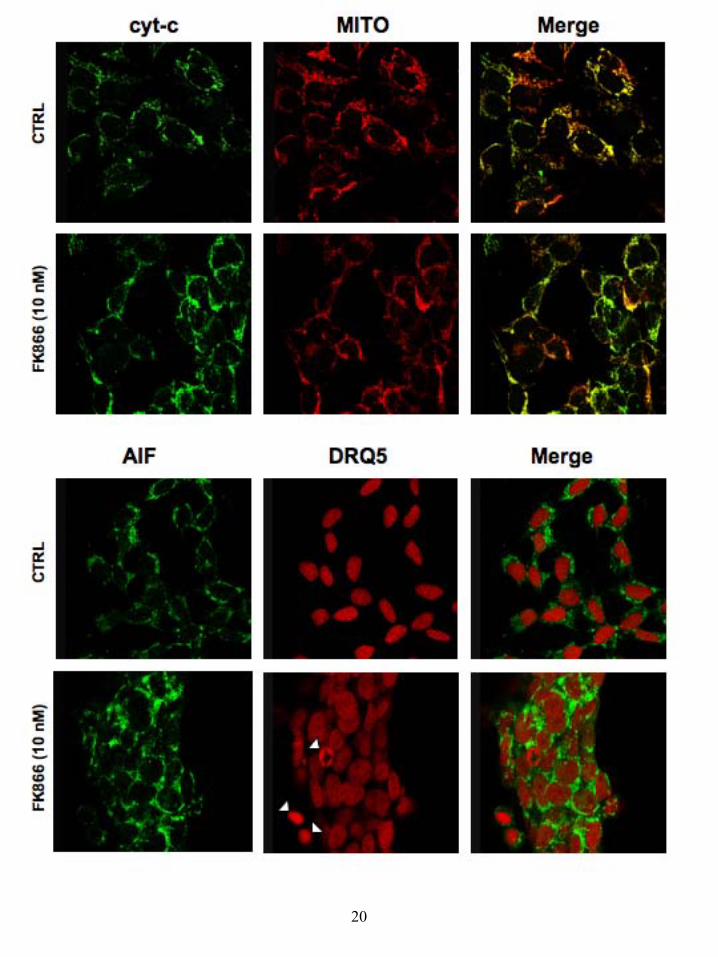
maximal effect (Fig 4). We tested the ability of extracellular NAD to rescue cells from FK866-induced cell death and intracellular NAD depletion, and found near-identical results to those obtained for NIH-3T3 cells (Fig 4B), other than a slightly lower rescue by 100 µM NAD which, along with the higher potency of FK866 perhaps due to a higher NAD turnover in these cells. Similar FK866-induced cell death and rescue by NAD was also observed in HeLa cells (Grolla and Billington, unpublished). In order to investigate whether cell death and subsequent rescue were due to the specific inhibition of NMPRTase and reduction of NAD levels, we performed rescue experiments in NIH-3T3 and SH-SY5Y cells using compounds that enter the recycling pathway either before or after the step catalysed by NMPRTase (Table 3). As might be expected, neither nicotinamide (Nam) or nicotinic acid (NA) were able to rescue cells as they are the substrates for NMPRTase. However, this data is in contrast to the work of Hasmann and Schemainda who reported rescue with both NA and Nam (25). On the contrary, nicotinamide mononucleotide (NMN) and nicotinic acid mononucleotide (NAMN), which feed into the recycling pathway after NMPRTase, were able to rescue cells as efficiently as NAD itself (Table 3) suggesting that the effects of FK866 are specific. As described above, in our cellular models FK866 caused cell death in a peculiar delayed fashion compared to the drop of NAD(P) levels. Consequently, we sought to characterize the type of death induced by FK866 in SH-SY5Y cells by evaluating markers of apoptosis and autophagy. We first assayed the caspase-dependent pathway of apoptosis by evaluating Caspase 3 cleavage. Indeed, this protease is invariably recruited as an effector of the apoptotic caspase machinery, since it is cleaved to enzymatically-active products by the up-stream caspases (Caspase 9, 8 and 10; (35)) in order to dismantle the dying cell. Surprisingly, the Caspase 3 activation fragment (p11) was not detected in FK866-treated cells (10 nM) even after 72 hours of treatment, as assessed by western blot with a p11 specific antibody (data not shown). On the other hand, apoptosis may rely on the activation of mitochondria to release pro-apoptotic mediators such as cytochrome-c (cyt-c) and the apoptosis inducing factor (AIF) (36,37). In order to evaluate this pathway, we studied the cellular distribution of cyt-c and AIF by immuno detection with confocal microscopy. This approach demonstrated that, in the majority of
5
by guest on May 31, 2018
http://ww
w.jbc.org/
Dow
nloaded from

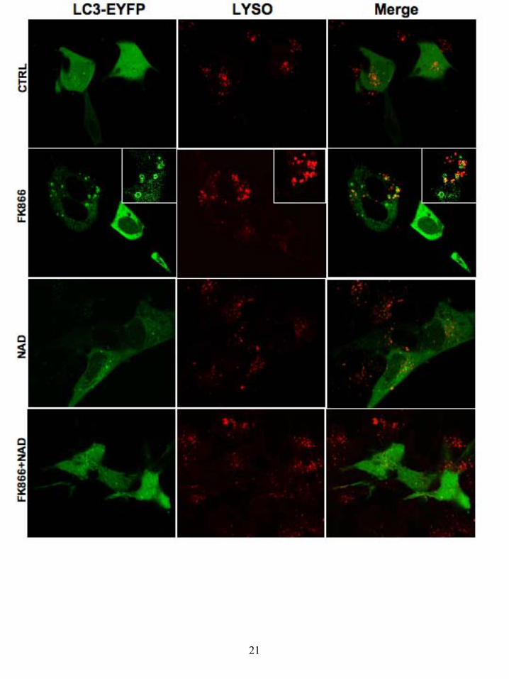
the cells, neither cyt-c or AIF are released from the mitochondria, thereby excluding the engagement of apoptotic death (Fig 5) with the exception of rare populations of cells sporadically detected. Further proof of this came from vital DNA staining with the fluorescent DRQ5 (used as counter-stain for AIF nuclear localization), since typical apoptotic fragmented nuclei were detected in only a very small number of FK866-treated cells (Fig 5). Given the lack of correlation between the intense cytotoxic effect induced by FK866 and the exiguity of the apoptosis detected, we investigated the possibility that FK866 could induce autophagy. Indeed, the length of time required in order to achieve cell-death, abnormally long for a canonical apoptosis, and the energetic relevance of an impaired NAD biosynthesis may be indicative of a metabolic crisis pushing the cell to a compensatory behaviour such as autophagy. To address this hypothesis, we studied, by confocal microscopy, the formation of autophagic vesicles (autophagosomes and autophago-lysosomes), upon FK866-induced NAD depletion. These sub-cellular membranous structures, originating from the coalescence of small vesicles with lysosomes, are characteristically endowed with the Microtubule-associated protein light chain 3 (LC3) specific marker (38). Indeed LC3, which is mainly cytosolic and diffuse in healthy cells, is conjugated to phosphatidylethanolamine upon induction of autophagy and targeted to autophagosomes, thus allowing their detection. Since the commercially available anti-LC3 antibodies are not reliable for immuno-fluorescence studies, we transiently over-expressed EYFP-tagged LC3 in neuroblastoma cells that were treated with FK866 for different times (Fig 6). In such conditions we were able to track the generation of LC3-positive autophagic vacuoles in FK866 treated cells, but not in control cells, already after 24h treatments (Fig 6). Validating the autophagic nature of this LC3 vesicle, the vital staining of lysosomes (LYSOTRACKER) allowed us to detect the presence of these organelles in the LC3-positive structures (Fig 6). In line with data from the cell count and MTT assays, the percentage of cells presenting the LC3-positive vesicles reached 44.3 ± 5.1 % after 36h treatment with 10 nM FK866. More importantly, repletion of the NAD levels with 100 µM extracellular NAD was able to revert the autophagic phenotype almost entirely.
Discussion There has been a recent resurgence in interest in NAD(P) as it is becoming clear that in addition to its role in basic metabolism, these nucleotides play an important role in signal transduction (4). In the present manuscript, we have investigated whether low concentrations of NAD could be transported into the cytoplasm from the extracellular space and could then enter the cellular NAD(H) pool. To accomplish this, we first used radiolabelled NAD to monitor uptake in various cell lines. Six out of seven cell lines tested were able to accumulate different amounts of NAD. The only cell line unable to accumulate NAD were K562 cells which grow in suspension. Interestingly, we have not found NAADP transport in either Jurkat or HL-60 cell lines growing in suspension (RAB and AAG unpublished observation). While these data suggest that NAD transport is not ubiquitous, they may suggest that it is a widespread mechanism. Accumulation was not due to extracellular membrane binding (for example to purinergic receptors) as the radioactivity could not be displaced. The data therefore strongly agree with circumstantial evidence by others suggesting that extracellular NAD can enter cells (10,12-15). NAD uptake has been previously shown at millimolar concentrations, and it has been proposed that connexin hemichannels (e.g. Cx43) might mediate bidirectional NAD transport down a concentration gradient. In our hands, NAD transport occurred at low concentrations of NAD (e.g. 250 pM, far lower than the intracellular concentration), which suggest that NAD is moving up its concentration gradient. NAD was not degraded extracellularly and the majority of the radioactivity transported remained as NAD. A proportion of the transported NAD was found to have been metabolised intracellularly as presumably it had joined the pool of intracellular NAD. A pharmacological profile of transport to elucidate this showed that hemichannel inhibitors were able to partially inhibit NAD transport, while adenosine and uridine, competitors for nucleoside transporters, were devoid of any inhibitory effect. Further evidence against a role for nucleoside transporters was brought by a lack of effect of NBMPR. Yet, dipyridamole, a different inhibitor of nucleoside transporters was partially effective at inhibiting transport. These pharmacological data collectively might suggest that more than one protein family contributes to transport, and that hemichannels play a major
6
by guest on May 31, 2018
http://ww
w.jbc.org/
Dow
nloaded from

role, as proposed by others in different settings (9,12). Yet, in our opinion, a number of issues suggest that canonical hemichannels alone cannot explain the results presented here: (i) The removal of extracellular Ca2+, which should open hemichannels, has no effect on NAD uptake in NIH-3T3, SH-SY5Y or RAW 264.7 cells and in HaCaT, HMEC and HeLa cells, the removal of extracellular Ca2+ reduced transport; (ii) given the large difference between the intracellular and extracellular concentrations of NAD in our experiments, hemichannels would be expected to mediate NAD efflux and not influx; (iii) the removal of extracellular Na+, which almost completely inhibits NAD uptake, has not been shown to regulate the gating of hemichannels. (iv) the time course of NAD uptake would appear to indicate that it is a constitutive mechanism which is incompatible with a canonical channel mechanism. Last, given the high concentrations of pharmacological inhibitors required to obtain effects, we cannot exclude that other related protein families might be affected by these. Transport of the three related nucleotides NAD(H), cADPR and NAADP from the extracellular space has now been demonstrated. Several protein families have been shown to transport these nucleotides including CD38, nucleoside transporters and connexin hemichannels (9,10,12,17,18,31). It is interesting to note that certain similarities exist in the pharmacology of these transport systems such as sodium dependence and inhibition by selected inhibitors of both nucleoside transporters and connexin hemichannels. However, certain differences, such as the sensitivity of NAADP transport to extracellular calcium and discrepancies in the effects of some of the nucleoside transport substrates/inhibitors may point to diverse mechanisms, redundant mechanisms or differential regulation of nucleotide transport (17). Furthermore, it is not clear how or if the extracellular concentration of nucleotides might play a role in the recruitment of pharmacologically distinct uptake mechanisms. Identification of the molecular nature of the transporter(s) will be required in order to resolve these questions and to identify which nucleotide(s)/nucleotide derivative(s) are the physiologically relevant substrates in diverse cell types under diverse physiological or pathological conditions and in an in vivo setting. Independently of the nature of the transporter, the magnitude of transport prompted us to speculate that the transport might be
physiologically relevant. Indeed, there is evidence that NAD is present extracellularly, for example in plasma (23) and our data has confirmed this (concentrations in calf and horse serum were 1.8 ± 0.8 µM and 1.6 ± 0.5 µM respectively; n = 6). It is interesting to note that positive cooperativity of NAD transport was observed in this concentration range (data not shown). To test the physiological relevance, we inhibited the NAD recycling pathway using the NMPRTase inhibitor FK866 (25). Treatment of cells for 24 hours with nM concentrations of this inhibitor resulted in a significant drop in intracellular NAD(P) levels, demonstrating a high turnover of pyridine nucleotides, at least in cultured cells in line with previous reports (25). Extracellular application of NAD was able to counteract this drop and replenish intracellular levels in all three cell types examined (NIH-3T3, SH-SY5Y and HeLa). Interestingly, incubation of cells with NAD alone induced a significant increase in intracellular NAD levels, suggesting that uptake of pyridine nucleotides is constitutive and participates in cellular NAD homeostasis. In parallel, experiments performed on cell vitality showed that NAD uptake could functionally replenish NAD levels as it could save cells from FK866-induced cell death. To rescue cells and replete NAD levels, µM concentrations were required but this is not in contradiction with transport experiments as: (i) intracellular concentrations of NAD are estimated to be in the high micromolar or the millimolar range (25); (ii) abolition of NMPRTase activity does not allow recycling; and (iii) experiments measuring cell vitality or NAD(P) levels after treatment with FK866 suggest a high turnover of NAD(P). Our conclusion that NAD transport is a valid replenishment pathway is supported by previous observation that extracellular NAD can rescue cardiomyocytes (15) and astrocytes (12) from PARP-mediated cell death and can activate sirtuins (15). Along the same lines, it is interesting to note that CD38 knockout mice, which are impaired in their ability to degrade extracellular NAD, display higher endogenous SIRT1 activity (16). Last, we were intrigued by the discrepancy in the latency between FK866-induced NAD depletion and cell death, and decided to investigate the mechanisms leading to cell death. We found that cells did not enter into an apoptotic program, but instead entered into autophagy. Once again, even from a mechanistic viewpoint, extracellular NAD could
7
by guest on May 31, 2018
http://ww
w.jbc.org/
Dow
nloaded from

counteract the effects of intracellular NAD depletion.
8
by guest on May 31, 2018
http://ww
w.jbc.org/
Dow
nloaded from

References 1. Ziegler, M. (2000) Eur J Biochem 267, 1550-1564 2. Khan, J. A., Forouhar, F., Tao, X., and Tong, L. (2007) Expert Opin Ther
Targets 11, 695-705 3. Belenky, P., Bogan, K. L., and Brenner, C. (2007) Trends Biochem Sci 32, 12-
19 4. Berger, F., Ramirez-Hernandez, M. H., and Ziegler, M. (2004) Trends
Biochem Sci 29, 111-118 5. Lee, H. C. (2006) Mol Med 12, 317-323 6. Magni, G., Amici, A., Emanuelli, M., Orsomando, G., Raffaelli, N., and
Ruggieri, S. (2004) Cell Mol Life Sci 61, 19-34 7. Bieganowski, P., and Brenner, C. (2004) Cell 117, 495-502 8. Billington, R. A., Bruzzone, S., De Flora, A., Genazzani, A. A., Koch-Nolte,
F., Ziegler, M., and Zocchi, E. (2006) Mol Med 12, 324-327 9. Bruzzone, S., Guida, L., Zocchi, E., Franco, L., and De Flora, A. (2001) Faseb
J 15, 10-12. 10. Bruzzone, S., Franco, L., Guida, L., Zocchi, E., Contini, P., Bisso, A., Usai,
C., and De Flora, A. (2001) J Biol Chem 276, 48300-48308 11. Smyth, L. M., Bobalova, J., Mendoza, M. G., Lew, C., and Mutafova-
Yambolieva, V. N. (2004) J Biol Chem 279, 48893-48903 12. Ying, W., Garnier, P., and Swanson, R. A. (2003) Biochem Biophys Res
Commun 308, 809-813 13. Zocchi, E., Usai, C., Guida, L., Franco, L., Bruzzone, S., Passalacqua, M., and
De Flora, A. (1999) Faseb J 13, 273-283 14. Zhu, K., Swanson, R. A., and Ying, W. (2005) Neuroreport 16, 1209-1212 15. Pillai, J. B., Isbatan, A., Imai, S., and Gupta, M. P. (2005) J Biol Chem 280,
43121-43130 16. Barbosa, M. T., Soares, S. M., Novak, C. M., Sinclair, D., Levine, J. A.,
Aksoy, P., and Chini, E. N. (2007) Faseb J In Press 17. Billington, R. A., Bellomo, E. A., Floriddia, E. M., Erriquez, J., Distasi, C.,
and Genazzani, A. A. (2006) Faseb J 20, 521-523 18. Guida, L., Bruzzone, S., Sturla, L., Franco, L., Zocchi, E., and De Flora, A.
(2002) J Biol Chem 277, 47097-47105 19. Guida, L., Franco, L., Bruzzone, S., Sturla, L., Zocchi, E., Basile, G., Usai, C.,
and De Flora, A. (2004) J Biol Chem 270, 22066-22075 20. Podesta, M., Benvenuto, F., Pitto, A., Figari, O., Bacigalupo, A., Bruzzone, S.,
Guida, L., Franco, L., Paleari, L., Bodrato, N., Usai, C., De Flora, A., and Zocchi, E. (2004) J Biol Chem 280, 5343-5349
21. Heidemann, A. C., Schipke, C. G., and Kettenmann, H. (2005) J Biol Chem 280, 35630-35640
22. Revollo, J. R., Grimm, A. A., and Imai, S. (2007) Curr Opin Gastroenterol 23, 164-170
23. O'Reilly, T., and Niven, D. F. (2003) Can J Vet Res 67, 229-231 24. Araki, T., Sasaki, Y., and Milbrandt, J. (2004) Science 305, 1010-1013 25. Hasmann, M., and Schemainda, I. (2003) Cancer Res 63, 7436-7442 26. Billington, R. A., and Genazzani, A. A. (2000) Biochem Biophys Res Commun
276, 112-116 27. Morgan, A., Churchill, G., Masgrau, R., Ruas, M., Davis, L., Billington, R.,
Patel, S., Yamasaki, M., Thomas, J., Genazzani, A., and Galione, A. (2005) in Methods in Calcium Signalling (Putney, J. J., ed), 2nd Ed., pp. 265-334, CRC
9
by guest on May 31, 2018
http://ww
w.jbc.org/
Dow
nloaded from
Press, Boca Raton 28. Gasser, A., Bruhn, S., and Guse, A. H. (2006) J Biol Chem 281, 16906 -
16913 29. Biedermann, E., Hasmann, M., Loser, R., Rattel, B., Reiter, F., Schein, B.,
Seibel, K., and Vogt, K. (2002), Klinge Pharma GmbH, USA 30. Moreschi, I., Bruzzone, S., Nicholas, R. A., Fruscione, F., Sturla, L.,
Benvenuto, F., Usai, C., Meis, S., Kassack, M. U., Zocchi, E., and De Flora, A. (2006) J Biol Chem 281, 31419-31429
31. Franco, L., Guida, L., Bruzzone, S., Zocchi, E., Usai, C., and De Flora, A. (1998) Faseb J 12, 1507-1520
32. Zocchi, E., Daga, A., Usai, C., Franco, L., Guida, L., Bruzzone, S., Costa, A., Marchetti, C., and De Flora, A. (1998) J Biol Chem 273, 8017-8024
33. Khan, J. A., Tao, X., and Tong, L. (2006) Nat Struct Mol Biol 13, 582-588 34. Holen, K., Saltz, L. B., Hollywood, E., Burk, K., and Hanauske, A. R. (2007)
Invest New Drugs In Press 35. Cohen, G. M. (1997) Biochem J 326 ( Pt 1), 1-16 36. Cai, J., Yang, J., and Jones, D. P. (1998) Biochim Biophys Acta 1366, 139-149 37. Susin, S. A., Lorenzo, H. K., Zamzami, N., Marzo, I., Snow, B. E., Brothers,
G. M., Mangion, J., Jacotot, E., Costantini, P., Loeffler, M., Larochette, N., Goodlett, D. R., Aebersold, R., Siderovski, D. P., Penninger, J. M., and Kroemer, G. (1999) Nature 397, 441-446
38. Tanida, I., Minematsu-Ikeguchi, N., Ueno, T., and Kominami, E. (2005) Autophagy 1, 84-91
10
by guest on May 31, 2018
http://ww
w.jbc.org/
Dow
nloaded from

Figure legends: Figure 1: NAD transport in NIH-3T3 cells. a) Time-dependent uptake of [32P]NAD (circles, solid line) and [32P]NAADP (squares, dashed line) in NIH-3T3 cells. Data presented as mean ± SEM, n = 8 - 12. b) HPLC analysis of transported nucleotides in NIH-3T3 cells. The grey line represents the HPLC trace for authentic standards (NAD; peak 3 mins and NADP; peak 13 mins). The other lines represent radioactivity remaining extracellularly (dashed line, squares) and recovered intracellularly (solid line, circles) after [32P]NAD incubation. c) Kinetic plot of [32P]NAD uptake in NIH-3T3 cells. Data presented as mean ± SEM, n = 6 - 8. Figure 2: FK866-induced NIH-3T3 cell death and rescue by extracellular NAD. a) Concentration response curve for the effect of FK866 on NIH-3T3 cell vitality after 48 hours (squares, dashed line) or 72 hours (circles, solid line) of treatment; n = 8 - 12. b) Rescue of FK866-induced NIH-3T3 cell death by NAD. Data are presented for both cell vitality experiments (black bars) and cell counts (white bars); n = 4 - 12. Figure 3: FK866-induced reduction in NAD(P) levels in NIH-3T3 cells and rescue by extracellular NAD. a) FK866-induced decrease in intracellular NAD(P) levels in NIH-3T3 cells after 24 hours of treatment. Data presented as mean ± SEM, n = 9. b) Rescue of FK866- induced decrease in intracellular NAD(P) levels in NIH-3T3 cells by extracellularly applied NAD; n = 9. Figure 4: FK866-induced SH-SY5Y cell death and rescue by extracellular NAD. a) Concentration response curve for the effect of FK866 on SH-SY5Y cell vitality after 72 hours of treatment; n = 8 - 16. b) Rescue by extracellular NAD of FK866-induced SH-SY5Y cell death (black bars) and FK866-induced reduction of NAD(P) levels; n = 8 - 16. Nam - nicotinamide. Figure 5: FK866 does not induce apoptosis in SH-SY5Y cells. Cells were co-stained with a cyt-c antibody and Mitotracker or with an AIF antibody and the nuclear marker DRQ5. Cells were treated for 48 hours as indicated. Results are indicative of 4 cultures. Figure 6: FK866-induced autophagy in SH-SY5Y cells. Cells were transiently transfected with LC3-EYFP and stained with lysotracker and treated with FK866 (10 nM) and NAD (100 μM) either alone or together for 36 hours. Results are indicative of 5 cultures.
11
by guest on May 31, 2018
http://ww
w.jbc.org/
Dow
nloaded from
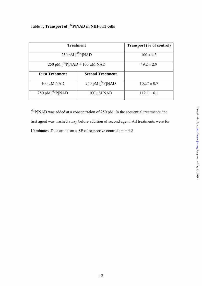
Table 1: Transport of [32P]NAD in NIH-3T3 cells
Treatment Transport (% of control)
250 pM [32P]NAD 100 ± 4.3
250 pM [32P]NAD + 100 µM NAD 49.2 ± 2.9
First Treatment Second Treatment
100 µM NAD 250 pM [32P]NAD 102.7 ± 0.7
250 pM [32P]NAD 100 µM NAD 112.1 ± 6.1
[32P]NAD was added at a concentration of 250 pM. In the sequential treatments, the
first agent was washed away before addition of second agent. All treatments were for
10 minutes. Data are mean ± SE of respective controls; n = 4-8
12
by guest on May 31, 2018
http://ww
w.jbc.org/
Dow
nloaded from

Table 2: Pharmacology of [32P]NAD transport in NIH-3T3 cells.
Treatment Transport (% of control) Control 100 ± 2.4
NMG Locke 21.1 ± 0.8 EGTA Locke 103.7 ± 1.9
NADP 100 µM 87.7 ± 7.0 NAADP 100 µM 78.1 ± 8.9
Dipyridamole 100 µM 53.8 ± 5.4 Nitrobenzylthioinosine 100 µM 169.6 ± 2.1
Adenosine 100 µM 391.1 ± 14.5 Uridine 100 µM 118.9 ± 1.9
Carbenoxelone 100 µM 46.6 ± 0.2 Enoxelone 100 µM 38.5 ± 1.4
Octanol 1 mM 61.1 ± 2.8
Transport experiments were carried out for 10 minutes. Data are presented as the
mean ± SEM, n = 6 - 15.
13
by guest on May 31, 2018
http://ww
w.jbc.org/
Dow
nloaded from

Table 3: Rescue of FK866-induced cell death by intermediates in the NAD recycling
pathway.
Cell Vitality (% of control) Treatment NIH-3T3 SH-SY5Y
Control 100 ± 2.5 100 ± 2.3 FK866 29.8 ± 0.9 25.2 ± 2.4 + NA 100 µM 29.9 ± 0.6 20.3 ± 0.4 + Nam 100 µM 35.2 ± 0.6 23.9 ± 0.6 + NAMN 100 µM 77.2 ± 1.9 69.8 ± 5.0 + NMN 100 µM 77.3 ± 2.7 70.2 ± 4.6 + NAD 100 µM 85.4 ± 2.5 78.6 ± 1.3
Cells were treated for 72 hours with FK866 (100 nM for NIH-3T3 cells and 10 nM
for SH-SY5Y cells) in the presence or absence of various intermediates in the NAD
recycling pathway. Cell vitality was assessed after 72 hours by MTT. Results
expressed as % of control, n = 8 - 16.
14
by guest on May 31, 2018
http://ww
w.jbc.org/
Dow
nloaded from
GenazzaniRoman, Ambra A Grolla, Pier Luigi Canonico, Fabrizio A Condorelli and Armando A
Richard A Billington, Cristina Travelli, Emanuela Ercolano, Ubaldina Galli, Cintia BlasiCharacterization of NAD uptake in mammalian cells
published online January 7, 2008J. Biol. Chem.
10.1074/jbc.M706204200Access the most updated version of this article at doi:
Alerts:
When a correction for this article is posted•
When this article is cited•
to choose from all of JBC's e-mail alertsClick here
by guest on May 31, 2018
http://ww
w.jbc.org/
Dow
nloaded from