MyoD is required for myogenic stem cell in
12
MyoD is required for myogenic stem cell function in adult skeletal muscle Lynn A. ~egeney,' Boris ~ablar,' Kerryn ~ a r r e t t , ~ Judy E. ~nderson,~ and Michael A. ~ u d n i c k i ' . ~ 'Institute for Molecular Biology and Biotechnology, Cancer Research Group, McMaster University, Hamilton, Ontario, Canada; 2Department of Anatomy, University of Manitoba, Winnipeg, Manitoba, Canada To investigate the function of MyoD in adult skeletal muscle, we interbred MyoD mutant mice with mdx mice, a model for Duchenne and Becker muscular dystrophy. Mice lacking both MyoD and dystrophin displayed a marked increase in severity of myopathy leading to premature death, suggesting a role for MyoD in muscle regeneration. Examination of MyoD mutant muscle revealed elevated numbers of myogenic cells; however, myoblasts derived from these cells displayed normal differentiation potential in vitro. Following injury, MyoD mutant muscle was severely deficient in regenerative ability, and we observed a striking reduction in the in vivo proliferation of myogenic cells during regeneration. Therefore, we propose that the failure of MyoD-deficient muscle to regenerate efficiently is not caused by a reduction in numbers of satellite cells, the stem cells of adult skeletal muscle, but results from an increased propensity for stem-cell self-renewal rather than progression through the myogenic program. [Key Words: MyoD; satellite cellj regeneration; Mdx; muscular dystrophy] Received February 27, 1996; revised version accepted April 8, 1996. The myogenic regulatory factors (MRFs] form a group of basic helix-loophelix (bHLHJ transcription factors con- sisting of MyoD, myogenin, Myf-5, and MRF4. The MRFs are expressed exclusively in skeletal muscle, and forcing their expression in a wide range of cultured cells induces the skeletal muscle differentiation program. Therefore, these transcription factors were postulated to have a master regulatory role in the development of the skeletal muscle lineage (for review, see Weintraub et al. 1991). Gene targeting has clearly elucidated the roles of the MRFs during embryogenesis. The introduction of null mutations in Myf-5, MyoD, myogenin, and MRF4 into the germ line of mice has revealed the hierarchical rela- tionships existing among the MRFs and established that functional redundancy is a feature of the MRF regulatory network (for review, see Rudnicki and Jaenisch 1995; Megeney and Rudnicki 1995). The MRF family can be divided into two functional groups: The primary MRFs, MyoD and Myf-5, are required for the determination of skeletal myoblasts; and the secondary MRFs, myogenin and MRF4, act later in the program, likely as differenti- ation factors. Mice lacking a functional MyoD gene were found to have no overt abnormalities in skeletal muscle but ex- pressed about four-fold higher levels of Myf-5 (Rudnicki et al. 1992). Similarly, newborn Myf-5-deficient animals display apparently normal skeletal muscle but die peri- natally because of severe rib abnormalities (Braun et al. 3Corresponding author. 1992, 1994).Newborn mice deficient for both MyoD and Myf-5 are totally devoid of skeletal myoblasts and mus- cle (Rudnicki et al. 1993).Mice lacking myogenin die at birth because of a virtual absence of myofibers; however, normal numbers of MyoD-expressing myoblasts are present (Hasty et al. 1993; Nabeshima et al. 1993; Venuti et al. 1995). In contrast, mice lacking MRF4 are viable with seemingly normal skeletal muscle but display a four-fold increase in myogenin (Patapoutian et al. 1995; Zhang et al. 1995). Vertebrate skeletal muscle is derived from cells in the prechordal and somitic mesoderm that give rise to com- mitted myogenic cells, which become the skeletal mus- cle of the head, trunk, and limbs (for review, see Buck- ingham 1992; Miller 1992; Hauschka 1994).The differ- ent myofiber and myogenic constituents of muscle have been postulated to be formed from different lineages of myogenic cells (Miller 1992; Bischoff 1994). In the mouse, primary myofibers develop first at 8.5 days of gestation, followed by secondary myofibers around day 14. Satellite cells, the stem cells of adult skeletal muscle, arise around day 17 of development as a unique myo- genic cell lineage (Bischoff 1994). Satellite cells lie beneath the basal lamina of adult skeletal muscle closely juxtaposed to the myofiber sar- colemma. Satellite cells make up 2%-7% of the nuclei associated with a particular fiber, and the proportion var- ies with age and muscle group. These adult muscle stem cells are normally mitotically quiescent but are acti- vated (i.e., enter the cell cycle) in response to stress in- duced by weight-bearing or other trauma, such as injury. GENES & DEVELOPMENT 10:1173-1183 0 1996 by Cold Spring Harbor Laboratory Press ISSN 0890-9369196 $5.00 1173 Cold Spring Harbor Laboratory Press on April 3, 2019 - Published by genesdev.cshlp.org Downloaded from
Transcript of MyoD is required for myogenic stem cell in

MyoD is required for myogenic stem cell function in adult skeletal muscle Lynn A. ~egeney , ' Boris ~ablar,' Kerryn ~ a r r e t t , ~ Judy E. ~ n d e r s o n , ~ and Michael A. ~udn ick i ' . ~
'Institute for Molecular Biology and Biotechnology, Cancer Research Group, McMaster University, Hamilton, Ontario, Canada; 2Department of Anatomy, University of Manitoba, Winnipeg, Manitoba, Canada
To investigate the function of MyoD in adult skeletal muscle, we interbred MyoD mutant mice with mdx mice, a model for Duchenne and Becker muscular dystrophy. Mice lacking both MyoD and dystrophin displayed a marked increase in severity of myopathy leading to premature death, suggesting a role for MyoD in muscle regeneration. Examination of MyoD mutant muscle revealed elevated numbers of myogenic cells; however, myoblasts derived from these cells displayed normal differentiation potential in vitro. Following injury, MyoD mutant muscle was severely deficient in regenerative ability, and we observed a striking reduction in the in vivo proliferation of myogenic cells during regeneration. Therefore, we propose that the failure of MyoD-deficient muscle to regenerate efficiently is not caused by a reduction in numbers of satellite cells, the stem cells of adult skeletal muscle, but results from an increased propensity for stem-cell self-renewal rather than progression through the myogenic program.
[Key Words: MyoD; satellite cellj regeneration; Mdx; muscular dystrophy]
Received February 27, 1996; revised version accepted April 8, 1996.
The myogenic regulatory factors (MRFs] form a group of basic helix-loophelix (bHLHJ transcription factors con- sisting of MyoD, myogenin, Myf-5, and MRF4. The MRFs are expressed exclusively in skeletal muscle, and forcing their expression in a wide range of cultured cells induces the skeletal muscle differentiation program. Therefore, these transcription factors were postulated to have a master regulatory role in the development of the skeletal muscle lineage (for review, see Weintraub et al. 1991).
Gene targeting has clearly elucidated the roles of the MRFs during embryogenesis. The introduction of null mutations in Myf-5, MyoD, myogenin, and MRF4 into the germ line of mice has revealed the hierarchical rela- tionships existing among the MRFs and established that functional redundancy is a feature of the MRF regulatory network (for review, see Rudnicki and Jaenisch 1995; Megeney and Rudnicki 1995). The MRF family can be divided into two functional groups: The primary MRFs, MyoD and Myf-5, are required for the determination of skeletal myoblasts; and the secondary MRFs, myogenin and MRF4, act later in the program, likely as differenti- ation factors.
Mice lacking a functional MyoD gene were found to have no overt abnormalities in skeletal muscle but ex- pressed about four-fold higher levels of Myf-5 (Rudnicki et al. 1992). Similarly, newborn Myf-5-deficient animals display apparently normal skeletal muscle but die peri- natally because of severe rib abnormalities (Braun et al.
3Corresponding author.
1992, 1994). Newborn mice deficient for both MyoD and Myf-5 are totally devoid of skeletal myoblasts and mus- cle (Rudnicki et al. 1993). Mice lacking myogenin die a t birth because of a virtual absence of myofibers; however, normal numbers of MyoD-expressing myoblasts are present (Hasty et al. 1993; Nabeshima et al. 1993; Venuti et al. 1995). In contrast, mice lacking MRF4 are viable with seemingly normal skeletal muscle but display a four-fold increase in myogenin (Patapoutian et al. 1995; Zhang et al. 1995).
Vertebrate skeletal muscle is derived from cells in the prechordal and somitic mesoderm that give rise to com- mitted myogenic cells, which become the skeletal mus- cle of the head, trunk, and limbs (for review, see Buck- ingham 1992; Miller 1992; Hauschka 1994). The differ- ent myofiber and myogenic constituents of muscle have been postulated to be formed from different lineages of myogenic cells (Miller 1992; Bischoff 1994). In the mouse, primary myofibers develop first at 8.5 days of gestation, followed by secondary myofibers around day 14. Satellite cells, the stem cells of adult skeletal muscle, arise around day 17 of development as a unique myo- genic cell lineage (Bischoff 1994).
Satellite cells lie beneath the basal lamina of adult skeletal muscle closely juxtaposed to the myofiber sar- colemma. Satellite cells make up 2%-7% of the nuclei associated with a particular fiber, and the proportion var- ies with age and muscle group. These adult muscle stem cells are normally mitotically quiescent but are acti- vated (i.e., enter the cell cycle) in response to stress in- duced by weight-bearing or other trauma, such as injury.
GENES & DEVELOPMENT 10:1173-1183 0 1996 by Cold Spring Harbor Laboratory Press ISSN 0890-9369196 $5.00 1173
Cold Spring Harbor Laboratory Press on April 3, 2019 - Published by genesdev.cshlp.orgDownloaded from

Megeney et al.
The descendants of the activated satellite cells, called myogenic precursor cells (mpc), undergo multiple rounds of division before fusing to existing myofibers or forming new myofibers, leading to repair and/or hypertrophy of the damaged or stressed muscle fibers, respectively. Sat- ellite cells form a stable population, suggesting that self- renewal in the stem-cell compartment maintains the pool of quiescent satellite cells. Moreover, satellite cells and their descendants have a very large potential for pro- liferation, perhaps as much as 80 doublings, as revealed by experiments in which muscle was subjected to repet- itive cycles of injury-induced regeneration (for review, see Grounds and Yablonka-Reuveni 1993; Bischoff 1994).
The expression pattern of MRFs following satellite- cell activation and during mpc proliferation and differ- entiation appears to be distinct from the program mani- fested during the embryonic development of skeletal muscle. Quiescent satellite cells express no detectable MRFs. Upon activation and entrance into the cell cycle, MyoD is rapidly up-regulated concomitant with expres- sion of proliferating cell nuclear antigen (PCNA), a marker for cell proliferation (e.g., Bravo et al. 1987). Myf-5 and MRF4 are expressed at intermediate times when PCNA and MyoD expression are declining. Myo- genin is expressed last during the time associated with fusion and differentiation (Smith et al. 1994; Yablonka- Reuveni and Rivera 1994). This expression of MyoD early after activation is suggestive of an important role for MyoD in satellite cell function.
The viability and apparently normal skeletal muscle of MyoD mutant mice (Rudnicki et al. 1992), and the early expression of MyoD following satellite cell activation (Smith et al. 1994; Yablonka-Reuveni and Rivera 1994), lead us to consider the hypothesis that MyoD may have some role in the postnatal growth and regeneration of skeletal muscle. To investigate the function of MyoD in adult skeletal muscle, we interbred MyoD mutant mice with rndx mice carrying a loss-of-function mutation in the dystrophin gene, and performed injury-induced re- generation experiments on muscle in MyoD-deficient animals. Our data clearly establish a role for MyoD in adult muscle and reveal novel insights into the genetic regulation of muscle stem-cell function.
Results
Mice lacking MyoD and dystrophin exhibit increased myopathy
To determine whether MyoD might have a role in skel- etal muscle regeneration, we interbred MyoD-deficient mice (Rudnicki et al. 1992) with the rndx mouse strain as an in vivo bioassay for skeletal muscle regeneration. The rndx mice carry a loss-of-function point mutation in the X-linked dystrophin gene and are therefore an animal model for human Duchenne and Becker muscular dys- trophy (Sicinski et al. 1989; Ahn and Kunkel 1993). The rndx mice display extensive necrosis of muscle fibers by
2 weeks of age, but unlike humans, maintain skeletal muscle integrity due to a high muscle regenerative ca- pacity. This regeneration results in the formation of ad- ditional muscle fibers, which leads to a significant level of muscle hypertrophy. Hypertrophy in rndx muscle is characterized by a 25% increase in the number of fibers, a four-fold increase in the distribution of the fiber cali- bers, centrally located nuclei within 70%-80% of all fi- bers, and a 1 .-/-fold increase in muscle mass (Anderson et al. 1987; Carnwath and Shotton 1987; Coulton et al. 1988; e.g., see Fig. 2, below; Table 1). If MyoD is required for efficient regeneration of skeletal muscle, mice lack- ing MyoD [designated mdx:MyoD(- / -)] should display more extensive myopathic or dystrophic changes in skel- etal muscle.
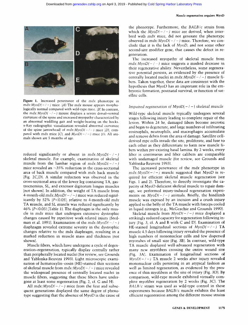
As predicted, mutant mdx:MyoD( - / - ) mice uni- formly displayed several phenotypic traits indicative of a marked increase in the penetrance of the disease (Fig. 1). By 3 to 5 months of age, mdx:MyoD(- / -) mice devel- oped a profound dorsal-ventral curvature of the spine (Fig. 1, cf. C and D), similar to the lordosis and kyphosis of patients with Duchenne muscular dystrophy, and an abnormal waddling gait characterized by weight bearing on the hocks (Fig. 1, compare A and BJ. The animals became progressively less active, with concomitant weight loss, before premature death at -12 months of age. In contrast, rndx mice display virtually normal ex- ternal appearance and normal murine life spans of 1.5-2 years (Coulton et al. 1988).
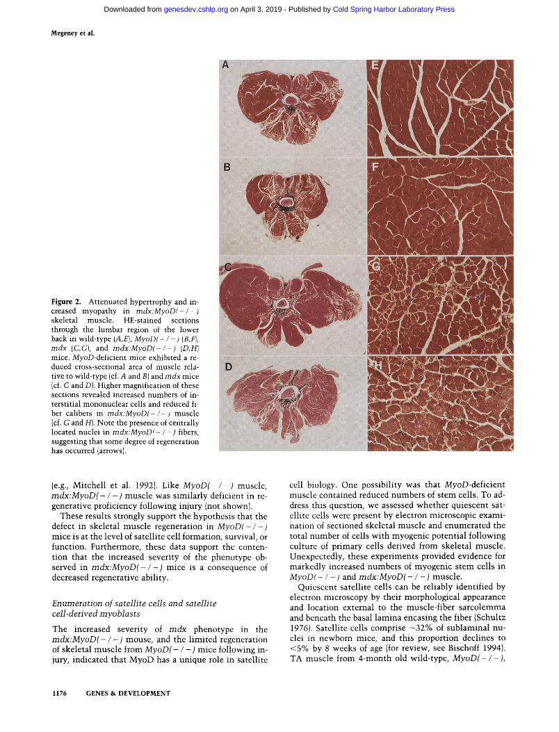
Examination of skeletal muscle from the lumbar re- gion of MyoD( - / -) mice revealed an - 17% reduction in the cross-sectional area of back muscle compared with B A L B I c (hereafter referred to as wild type) back muscle (Fig. 2A,B). Moreover, MyoD( - I - ) tibialis anterior (TA) muscle was significantly reduced in mass relative to wild-type TA by 32% (P<0.05), whereas MyoD(- / -) soleus (SL) muscle was reduced by 8% (Table 1). These data suggest that MyoD has a role in the postnatal growth of skeletal muscle.
The rndx phenotype is characterized by an -1.7-fold increase in the mass of individual skeletal muscles com- pared with wild-type mice (Anderson et al. 1987; Carn- wath and Shotton 1987; Coulton et al. 1988; e.g., see Fig. 2 and Table 1). Importantly, the hypertrophic response is
Table 1. Mean muscle mass from tibialis anterior and soleus hindlimb muscles from 4-month-old animals o f the different genotypes
Mouse strain T A muscle (mg) SL muscle (mg)
Wild type 59.3 -t 1.4 10.1 5 0.5 M y o D ( - I - ) 40.0 + 1.7 9.3 2 0.5 m dx 95.9 + 5.3 17.9 2 1.6 mdx:MyoLl( - / - ) 45.8 + 4.2 9.6 2 0.6
These data reveal the relative hypertrophy in rndx muscle and relative hypotrophy in MyoD( - / - ) and mdx:MyoD( - / - ) muscle. The values above represent the mean and standard error of the mean of muscle weights from five animals ( n = 10).
1174 GENES & DEVELOPMENT
Cold Spring Harbor Laboratory Press on April 3, 2019 - Published by genesdev.cshlp.orgDownloaded from
Muscle regeneration requires MyoD
Figure 1. Increased penetrance of the rndx phenotype in mdx:MyoD(- l - ) mice. (A) The rndx mouse appears morpho- logically normal compared with wild-type mice. IBI In contrast, the mdx:MyoD(- l - ) mouse displays a severe dorsal-ventral curvature of the spine and increased myopathy characterized by an abnormal waddling gait and weight-bearing on the hocks. x-Ray radiographic visualization revealed abnormal curvature of the spine (arrowhead) of mdx:MyoD(-/-) mice [Dl, corn- pared with rndx mice jC), and MyoD(- / -) mice ( E ) . All ani- mals shown are 5 months of age.
reduced significantly or absent in m d x : M y o D ( - / - ) skeletal muscle. For example, examination of skeletal muscle from the lumbar region of m d x : M y o D ( - / -) mice revealed an -35% reduction in the cross-sectional area of back muscle compared with rndx back muscle (Fig. 2C,D). A similar reduction was observed in the cross-sectional area of the lower leg containing TA, gas- trocnemius, SL, and extensor digitorum longus muscles (not shown). In addition, the weight of TA muscle from 4-month-old m d x : M y o D ( - / -) mice was reduced signif- icantly by 52% (P<0.05), relative to 4-month-old rndx TA muscle, and SL muscle was reduced significantly by 46% (P<0.05) (Table 1). The diaphragm is the only mus- cle in rndx mice that undergoes extensive dystrophic changes caused by repetitive work-related injury (Sted- man et al. 1991). Examination of the m d x : M y o D - / - ) diaphragm revealed extreme severity in the dystrophic changes relative to the rndx diaphragm, resulting in a marked reduction in muscle mass and thickness (not shown).
Muscle fibers, which have undergone a cycle of degen- eration-regeneration, typically display centrally rather than peripherally located nuclei (for review, see Grounds and Yablonka-Reuvini 1993). Light microscopic exami- nation of hematoxylin-eosin (HEJ-stained thin sections of skeletal muscle from m d x : M y o D ( - / -) mice revealed the widespread presence of centrally located nuclei in muscle fibers, suggesting that these fibers have under- gone at least some regeneration (Fig. 2, cf. G and H).
All m d x : M y o D ( - / -) mice from the first and subse- quent generations displayed the same degree of pheno- type suggesting that the absence of MyoD is the cause of
the phenotype. Furthermore, the B A L B / c strain from which the M y o D ( - / - 1 mice are derived, when inter- bred with rndx mice, did not generate the phenotype observed in mdx:MyoD( - / - ) mice. Therefore, we con- clude that it is the lack of MyoD, and not some other second-site modifier gene, that causes the defect in re- generatlon.
The increased myopathy of skeletal muscle from m d x : M y o D ( - / - ) mice suggests a marked decrease in thelr regenerative ability. Nevertheless, some regenera- tive potential persists, as evidenced by the presence of centrally located nuclei in m d x : M y o D ( - / -) muscle fi- bers. Taken together, these data are consistent with the hypothesis that MyoD has an important role in the em- bryonic formation, postnatal survival, or function of sat- ellite cells.
Impaired regeneration o f MyoD( - 1 - ) skeletal muscle
Wild-type skeletal muscle typically undergoes several stages following injury leading to complete repair of the lesion. Within 24 hr, damaged fibers become necrotic and begin to degenerate, and large numbers of infiltrating eoslnophils, neutrophils, and macrophages accumulate and remove debris from the area of damage. Satellite cell- derived mpc cells invade the site, proliferate, and fuse to each other as they differentiate to form new muscle fi- bers within pre-existing basal lamina. By 2 weeks, every fiber is continuous and fiber calibers are comparable with undamaged muscle (for review, see Grounds and Yablonka-Reuveni 1993).
The increased penetrance of the rndx phenotype in m d x : M y o D l - / -) muscle suggested that MyoD is re- quired for efficient skeletal muscle regeneration (see Figs. 1 and 2). Therefore, to directly investigate the ca- pacity of MyoD-deficient skeletal muscle to repair dam- age, we performed injury-induced regeneration experi- ments on M y o D ( - / -) animals. Briefly, the hindlimb muscle was exposed by an incision and a crush injury applied to the belly of the TA muscle with forceps cooled in liquid nitrogen (e.g., McGeachie and Grounds 1990).
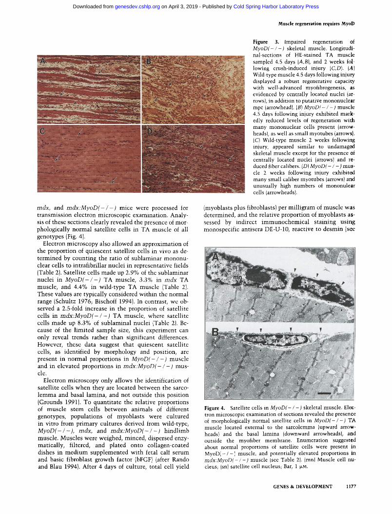
Skeletal muscle from M y o D ( - / -) mice displayed a strikingly reduced capacity for regeneration following in- jury (Fig. 3, cf. A and B with C and D). Examination of HE-stained longitudinal sections of M y o D ( - / -) T A muscle 4.5 days following injury revealed the presence of high numbers of mononuclear cells and few dispersed myotubes of small size (Fig. 3B). In contrast, wild-type TA muscle displayed well-advanced regeneration with many new myofibers crossing the entire wound site (Fig. 3A). Examination of longitudinal sections of M y o D ( - / -) TA muscle 2 weeks after injury revealed mononuclear cells persisting in an atypical fashion as well as limited regeneration, as evidenced by the pres- ence of thin myofibers at the site of injury (Fig. 3D). By comparison, wild-type muscle exhibited virtually com- plete myofiber regeneration by 2 weeks (Fig. 3C). The B A L B / c strain was used as wild-type control in these experiments because BALBIc muscle exhibits the least efficient regeneration among the different mouse strains
GENES & DEVELOPMENT 1175
Cold Spring Harbor Laboratory Press on April 3, 2019 - Published by genesdev.cshlp.orgDownloaded from
Megeney et al.
Figure 2. Attenuated hypertrophy and in- creased myopathy in mdx:MyoD(- / -) skeletal muscle. HE-stained sections through the lumbar region of the lower back in wild-type (A,E], MyoD(- / ) (B,F), mdx (C,G], and mdx:MyoD(- l - ) (D,H) mice. MyoD-deficient mice exhibited a re- duced cross-sectional area of muscle rela- tive to wild-type (cf. A and B ] and mdx mice jcf. C and Dl. Higher magnification of these sections revealed increased numbers of in- terstitial mononuclear cells and reduced fi- ber calibers in mdx:MyoD(- l - ) muscle jcf. G and HI. Note the presence of centrally located nuclei in mdx:MyoD(- / -) fibers, suggesting that some degree of regeneration has occurred (arrows).
(e.g., Mitchell et al. 1992). Like MyoD(- I - ) muscle, mdx:MyoD(- / -) muscle was similarly deficient in re- generative proficiency following injury (not shown).
These results strongly support the hypothesis that the defect in skeletal muscle regeneration in MyoD(- / -) mice is at the level of satellite cell formation, survival, or function. Furthermore, these data support the conten- tion that the increased severity of the phenotype ob- served in mdx:MyoD(- / -) mice is a consequence of decreased regenerative ability.
Enumeration of satellite cells and satellite cell-derived myoblasts
The increased severity of mdx phenotype in the mdx:MyoD(- / -) mouse, and the limited regeneration of skeletal muscle from MyoD(- / -) mice following in- jury, indicated that MyoD has a unique role in satellite
GENES & DEVELOPMENT
cell biology. One possibility was that MyoD-deficient muscle contained reduced numbers of stem cells. To ad- dress this question, we assessed whether quiescent sat- ellite cells were present by electron microscopic exami- nation of sectioned skeletal muscle and enumerated the total number of cells with myogenic potential following culture of primary cells derived from skeletal muscle. Unexpectedly, these experiments provided evidence for markedly increased numbers of myogenic stem cells in MyoDl- / -) and mdx:MyoD(- / -) muscle.
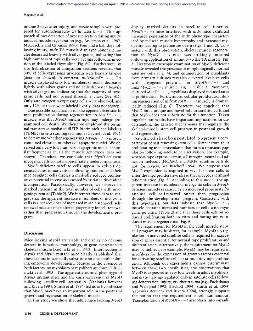
Quiescent satellite cells can be reliably identified by electron microscopy by their morphological appearance and location external to the muscle-fiber sarcolemma and beneath the basal lamina encasing the fiber (Schultz 1976). Satellite cells comprise -32% of sublaminal nu- clei in newborn mice, and this proportion declines to <5% by 8 weeks of age (for review, see Bischoff 1994). TA muscle from 4-month old wild-type, MyoD(- / -),
Cold Spring Harbor Laboratory Press on April 3, 2019 - Published by genesdev.cshlp.orgDownloaded from
Muscle regeneration requires MyoD
Figure 3. Impaired regeneration of MyoD(- / - ) skeletal muscle. Longitudi-
mdx, and mdx:MyoD(- / -) mice were processed for transmission electron microscopic examination. Analy- sis of these sections clearly revealed the presence of mor- phologically normal satellite cells in TA muscle of all genotypes (Fig. 4). - .. . -
Electron microscopy also allowed an approximation of the proportion of quiescent satellite cells in vivo as de- termined by counting the ratio of sublaminar mononu- clear cells t o intrafibrillar nuclei in representative fields (Table 2). Satellite cells made up 2.9% of the sublaminar nuclei i n MyoD(-/-) TA muscle, 3.3% in mdx TA muscle, and 4.4% in wild-type TA muscle (Table 2). These values are typically considered within the normal range (Schultz 1976; Bischoff 1994). In contrast, we ob- served a 2.5-fold increase in the proportion of satellite cells i n mdx:MyoD(- / -) TA muscle, where satellite cells made up 8.3% of sublaminal nuclei /Table 21. Be- cause of the-limited sample size, this experiment can only reveal trends rather than significant differences. However, these data suggest that quiescent satellite cells, as identified by morphology and position, are present in normal proportions in MyoD(- / -) muscle and in elevated proportions in mdx:MyoD(- / -) mus- cle.
Electron microscopy only allows the identification of satellite cells when they are located between the sarco- lemma and basal lamina, and not outside this position (Grounds 1991). T o quantitate the relative proportions of muscle stem cells between animals of different genotypes, populations of myoblasts were cultured in vitro from primary cultures derived from wild-type, MyoD(- / -), mdx, and mdx:MyoD(- / -) hindlimb muscle. Muscles were weighed, minced, dispersed enzy- matically, filtered, and plated onto collagen-coated dishes in medium supplemented with fetal calf serum and basic fibroblast growth factor (bFGF) (after Rando and Blau 1994). After 4 days of culture, total cell yield
nil-sections of HE-stained TA muscle sampled 4.5 days [A,B], and 2 weeks fol- lowing crush-induced injury (C,D). ( A ) Wild-type muscle 4.5 days following injury displayed a robust regenerative capacity with well-advanced myofibrogenesis, as evidenced by centrally located nuclei [ar- rows], in addition to putative mononuclear mpc (arrowhead). (B) MyoD(- / - ) muscle 4.5 days following injury exhibited mark- edly reduced levels of regeneration with many mononuclear cells present (arrow- heads), as well as small myotubes (arrows). (C) Wild-type muscle 2 weeks following injury, appeared similar to undamaged skeletal muscle except for the presence of centrally located nuclei (arrows) and re- duced fiber calibers. ID) MyoD(- / -) mus- cle 2 weeks following injury exhibited many small caliber myotubes (arrows) and unusually high numbers of mononulear cells (arrowheads).
(myoblasts plus fibroblasts) per milligram of muscle was determined, and the relative proportion of myoblasts as- sessed by indirect immunochemical staining using monospecific antisera DE-U-10, reactive to desmin (see
Figure 4. Satellite cells in MyoD(- / -) skeletal muscle. Elec- tron microscopic examination of sections revealed the presence of morphologically normal satellite cells in MyoD(- / -) TA muscl; located external to the sarcolemma (upward arrow- heads) and the basal lamina (downward arrowheads), and outside the myofiber membrane. Enumeration suggested about normal proportions of satellite cells were present in MyoD( - 1 - ) muscle, and potentially elevated proportions in mdx:MyoD(- / - ) muscle (see Table 2). (mn) Muscle cell nu- cleus; (sn) satellite cell nucleus; Bar, 1 p ~ .
GENES & DEVELOPMENT 1177
Cold Spring Harbor Laboratory Press on April 3, 2019 - Published by genesdev.cshlp.orgDownloaded from

Megeney et al.
Table 2. MyoD(-/-) muscle contains elevated numbers of myogenjc cells
Genotype Percent satellite
cells in vivod
Total number cellslmg
wet weighth Percent cells desmin +veC
Total number myoblastslmg wet weightd
Percent nuclei MHC + vee
Wild type 4.4 1531 2 174 22.4 2 1.8 343 6.5 2 0.8 MyoD( - I - ) 2.9 3356 2 599 18.5 2 1.3 62 1 11.0 2 1.8 mdx 3.3 1506 2 174 4.8 2 0.7 72 N.D. mdx:MyoD( -1 - ) 8.3 8314 2 1509 11.7 2 1.4 972 N.D.
The values represent the mean and standard error of the mean for five independent experiments (n > 2000). (N.D.] Not determined. "The proportion of satellite cells in TA muscle as determined by electron microscopy (n > 200). b ~ f t e r 4 days in culture, the total number of cells recovered per mg of muscle was determined by monodispersing the cultures and counting cells in a hemocvtometer. "
"The proportion of myoblasts was determined by immunodetection with anti-desmin antibody DE-U-10. d ~ h e relative number of cells with myogenic potential per mg wet weight was determined by multiplying the percent ( "1 by the total number of cells recovered I h ) . 'The proportion of differentiated myocytes was determined by immunodetection with anti-myosin heavy chain antibody MF20.
Fig. 5). Desmin is a n intermediate filament protein ex- pressed i n m p c i n vivo, and myoblasts i n vitro, but not satellite cells (George-Weinstein e t al. 1993; Bischoff 1994).
Immunochemical analysis revealed that t h e Dercent- age of myoblasts observed in 4-day cultures derived from wild-type skeletal muscle was 22.4%) whereas the per- centage of myoblasts i n cultures from M y o D l - / - ) mus- cle was 18.5%. However. because the total vield of cells per milligram was increased 2.2-fold i n the MyoDl- / - 1 muscle relative t o wild-type muscle (P<0.05), the nurn- ber of myogenic cells recovered per milligram of muscle was therefore increased 1.8-fold i n M y o D ( / ) muscle relative t o wild-type muscle (Table 2) . T h e pcrcentagc of myoblasts observed i n cultures from m d x muscle was 4.8'70, whereas t h e percentage of myoblasts i n cultures from m d x : M y o D ( - / -) muscle was 11.7%. Similarly, because t h e total number of cells per muscle weight was
Figure 5. Cultured myoblasts from pri- maw cultures of satellite cells. Immuno- chemical detection of desmin-expressing myoblasts from wild-type (A) and MyoD- deficient (B] muscle indicated that myo- genic cel!s (arrows) were present in ele- vated proportions in M y o D ( / -) skeletal muscle (see Table 2). Differentiated myo- cytes expressing myosin heavy chain in- duced from myoblasts derived from wild- type (C) and MyoD-deficient (Dl muscle displayed similar morphology and were found at similar frequencies (see Table 2). Myoblasts were detected with anti-desmin antibody DE-U-10, and myocytes with anti-myosin heavy chain antibody MF20. Magnification in A and B is twice that in C and D.
GENES & DEVELOPMENT
increased 5.5-fold i n t h e m d x : M y o D ( - / -) muscle rela- tive to m d x muscle (P<0.05), t h e number of myogenic cells recovered per milligram was therefore increased 13- fold in m d x : M y o D ( - / -) muscle relative t o m d x muscle (Tablc 2 ) .
Interestingly, wild-type and M y o D m u t a n t myoblasts differcd both i n morphological appearance and intensi ty of anti-desmin antibody labeling. A s described elsewhere [e.g., Ontel l e t al. 1992), >90% of the desmin-labeled myoblasts from wild-type muscle were typically spheri- cal, phase dark, and appeared t o express high levels of desmin (Fig. 5AJ. In contrast, t h e majority of myoblasts f rom M y o D ( - / - ) muscle displayed a flat stellate mor- phology and expressed lower levels of desmin (Fig. 5B).
T h e differentiation potential of myoblasts derived from primary cultures from wild-type and M y o D ( - I - ) muscle was also investigated. Cultured myoblasts were expanded under growth conditions for 1 week and trans-
Cold Spring Harbor Laboratory Press on April 3, 2019 - Published by genesdev.cshlp.orgDownloaded from

Muscle regeneration requires MyoD
ferred to differentiation medium for a second week. Dif- ferentiated myocytes were detected using indirect im- munochemistry with mouse monoclonal antibody MF20 reactive with striated muscle myosin heavy chain (Bader et al. 1982). M y o D mutant myocytes were morpho- logcally indistinguishable from wild-type myocytes (Fig. 5, cf. C and D). The proportion of nuclei within immunoreactive myocytes was found to be 11.0% in M y o D ( - / -) cultures and 6.5% in wild-type cultures (Table 2). The increased proportions of differentiated MyoD-deficient myocytes observed in vitro (P<0.05) suggests that desmin may be expressed below the limit of detection in some portion of M y o D mutant myo- blasts; hence, the numbers of myogenic cells per milli- gram in M y o D ( - / -) muscle may represent an underes- timate (Table 2).
The increased numbers of myoblasts recovered from M y o D ( - / - ) muscle raised the possibility that myo- blasts lacking M y o D may display increased rates of pro- liferation. To determine the doubling time of myoblasts in vitro, we measured the increase in numbers of desmin-staining cells over a period of 8 days. Impor- tantly, we observed the in vitro doubling time of myo- blasts derived from M y o D ( - / - ) muscle to be about three-fold longer than wild-type myoblasts. Further- more, purified populations of M y o D ( - / -) myoblasts in- corporated three-fold less tritiated thymidine. These ob- servations support the notion that MyoD has an impor- tant role in positively inducing myoblast proliferation. Furthermore, these data suggest that we have underesti- mated the numbers of myoblasts isolated from M y o D ( - / -) muscle by at least two-fold.
In v ivo proliferation of MyoDi - / - ) myogenic cells
Skeletal muscle from m d x : M y o D ( - / - ) mice relative to m d x mice contained a 2.5-fold increase in numbers of
satellite cells by morphology and position (Fig. 4, Table 21, and at least a 13-fold increase in numbers of myogenic cells per milligram of muscle (Fig. 5; Table 2). If MyoDi - / - ) mpc proliferate normally in vivo, we should observe a 13-fold increase in the proportion of cells undergoing cell division in m d x : M y o D ( - / -) mus- cle relative to m d x muscle. Therefore, we examined cel- lular proliferation in vivo in m d x : M y o D ( - l -) muscle containing elevated numbers of myogenic cells under conditions that normally lead to high-level satellite cell activation, namely by performing injury-induced regen- eration as described above. We examined the in vivo pro- liferative potential of myogenic cells in m d x vs. m d x : M y o D ( - / -) skeletal muscle using two indepen- dent approaches.
Replicating cells express PCNA at high levels through- out S phase (e.g., Bravo et al. 1987). Consequently, im- munostaining of PCNA in sections of skeletal muscle with monospecific antibodies allows detection of myo- genic cell proliferation (Johnson and Allen 1993). The TA muscle in 4-month-old m d x , and m d x : M y o D ( - / -) mice was subjected to crush injury, and 24 hr after in- jury, samplcs were prepared for immunohistochemical staining with antibody reactive to PCNA (n =3]. Exam- ination of PCNA expression 24 hr after injury should allow detection of muscle stem-cell activation. Enumer- ation of peripherally located nuclei expressing PCNA in surviving areas immediately adjacent the site of injury revealed similar proportions of PCNA-expressing nuclei in m d x and m d x : M y o D ( - / -) muscle (Fig. 6A,B). Be- cause m d x : M y o D ( - / -) muscle contains 13-fold higher numbers of myogenic cells relative to m d x muscle (Ta- ble 2), these data therefore suggest that MyoD-deficient satellite cells exhibit reduced rates of activation, or entry into the cell cycle.
In a second approach, mice were injected intraperito- nealy with 2.0 ~Ci lg r am of body weight of tritiated thy-
GENES & DEVELOPMENT 1179
Cold Spring Harbor Laboratory Press on April 3, 2019 - Published by genesdev.cshlp.orgDownloaded from
Megeney et al.
midine 3 days after injury, and tissue samples were pre- pared for autoradiography 24 hr later (n =5) . This ap- proach allows detection of rnpc replication during injury- induced muscle regeneration (e.g., Anderson et al. 198 7; McGeachie and Grounds 1990). Four and a half days fol- lowing injury, mdx TA muscle displayed abundant nu- clei decorated heavily with silver grains, indicating that high numbers of rnpc cells were cycling following injec- tion of the labeled thymidine (Fig. 6C). Furthermore, in situ hybridization of companion sections revealed that 30% of cells expressing myogenin were heavily labeled (data not shown). In contrast, mdx:MyoD( - / - ) TA muscle displayed only low numbers of nuclei decorated lightly with silver grains and no cells decorated heavily with silver grains, indicating that the majority of myo- genic cells had not passed through S phase (Fig. 6D). Only rare myogenin-expressing cells were observed, and only 12% of these were labeled lightly (data not shown).
One possible explanation for the apparent decrease in rnpc proliferation during regeneration in MyoD(- / -) muscle, was that MyoD mutant rnpc may undergo pro- grammed cell death. We therefore employed the termi- nal transferase-mediated dUTP-biotin nick end labeling (TUNEL) in situ staining technique (Gavrieli et al. 1992) to determine whether regenerating MyoD(- / -) muscle contained elevated numbers of apoptotic nuclei. We ob- served only very low numbers of apoptotic nuclei at sim- ilar frequencies in all the muscle genotypes (data not shown). Therefore, we conclude that MyoD-deficient myogenic cells do not inappropriately undergo apoptosis.
MyoD-deficient satellite cells appear to exhibit de- creased rates of activation following trauma, and their rnpc daughter cells display a markedly reduced prolifer- ative potential in vivo as assessed by tritiated thymidine incorporation. Paradoxically, however, we observed a marked increase in the total number of cells with myo- genic potential (Table 2). We interpret these data to sug- gest that the apparent increase in numbers of myogenic cells is a consequence of increased muscle stem-cell self- renewal because of an altered propensity for self-renewal rather than progression through the developmental pro- gram.
Discussion
Mice lacking MyoD are viable and display no obvious defects in function, morphology, or gene expression in skeletal muscle (Rudnicki et al. 1992). Interbreeding of MyoD and Myf-5 mutant mice clearly established that these factors functionally substitute for one another dur- ing embryonic development, because in the absence of both factors, no myoblasts or myofibers are formed (Rud- nicki et al. 1993). The apparently normal phenotype of MyoD mutant mice and the early expression of MyoD following satellite-cell activation (Yablonka-Reuveni and Rivera 1994; Smith et al. 1994) led us to hypothesize that MyoD may have an important role in the postnatal growth and regeneration of skeletal muscle.
In this study we show that adult mice lacking MyoD
display marked deficits in satellite cell function. MyoDl- , -) mice interbred with mdx mice exhibited increased penetrance of the mdx phenotype character- ized by reduced muscle hypertrophy and increased my- opathy leading to premature death (Figs. 1 and 2). Con- sistent with this observation, skeletal muscle regenera- tion in iVyoD(- / -) mice was strikingly impaired following application of an injury to the TA muscle (Fig. 3). Electron microscopic examination of MyoD-deficient muscle revealed the presence of morphologically normal satellite cells (Fig. 4), and enumeration of myoblasts from primary cultures revealed elevated levels of cells with myogenic potential in MyoD(-/-) and mdx:MyoD(- / -) muscle (Fig. 5, Table 2). Moreover, cultured MyoD(- / -) myoblasts displayed reduced rates of proliferation. Furthermore, cellular proliferation dur- ing regeneration of mdx:MyoD(- / -) muscle is dramat- ically reduced (Fig. 6). Therefore, we conclude that MyoD has a unique and novel role in satellite cells and that Myf-5 does not substitute for this function. Taken together, our results have important implications for un- derstanding the genetic mechanisms that regulate the skeletal-muscle stem-cell program in postnatal growth and regeneration.
Satellite cells have been postulated to represent a com- partment of self-renewing stem cells distinct from their proliferating rnpc descendants that form a transient pop- ulation following satellite cell activation. For example, whereas rnpc express desmin, a7-integrin, neural cell ad- hesion molecule (NCAM), and MRFs, satellite cells do not (for review, see Bischoff 1994). We propose that MyoD expression is required in vivo for stem cells to enter the rnpc proliferative phase that precedes terminal differentiation (Fig. 7). According to this model, the ap- parent increase in numbers of myogenic cells in MyoD- deficient muscle is caused by an increased propensity for satellite cell self-renewal rather than progression through the developmental program. Consistent with this hypothesis, our data indicate that MyoD(- / -) muscle contains increased numbers of cells with myo- genic potential (Table 2) and that these cells exhibit re- duced proliferation both in vitro and during injury-in- duced muscle regeneration (Fig. 6).
The requirement for MyoD in the adult muscle stem- cell program may be direct, for example, MyoD up-reg- ulation in activated satellite cells is required for expres- sion of genes essential for normal rnpc proliferation and differentiation. Alternatively, the requirement for MyoD may be indirect, for example, MyoD may be required in myofibers for the expression of growth factors essential for activating satellite cells or stimulating rnpc prolifer- ation. Although our experiments cannot discriminate between these two possibilities, the observations that MyoD is expressed at very low levels in adult myofibers, and is strongly up-regulated only in satellite cells follow- ing denervation, injury, or other trauma (e.g., Fuchtbauer and Westphal 1992; Bischoff 1994; Smith et al. 1994; Yablonka-Reuveni and Rivera 1994), strongly supports the notion that the requirement is cell autonomous. Transplantation of MyoD(- / -) myoblasts into yirradi-
1180 GENES & DEVELOPMENT
Cold Spring Harbor Laboratory Press on April 3, 2019 - Published by genesdev.cshlp.orgDownloaded from

Muscle regeneration requires MyoD
Figure 7. A unique role for MyoD in sat- ellite-cell function. Our data suggest a model in which up-regulation of MyoD is required for satellite cells to enter the rnpc proliferative phase that precedes terminal
Satellite Cell M P ~ Differentiated differentiation. Normally, MyoD is up-reg- Activation Prol~feration Myofiber ulated with entry into the cell cycle, Myf-5
cs. Q and MRF4 are expressed at intermediate times, and myogenin is expressed last dur- ing the time associated with differentia-
Wildtype -@+@*) tion (Smith et al. 1994; Yablonka-Reuveni MyoD !iE Myogenln and Rivera 1994). In this model, the ab-
sence of MyoD leads to an apparent in- crease In numbers of myogenlc cells as a
-\>\%-f=-,
> > -- consequence of increased muscle stem-cell MyoD(-I-) self-renewal because of an altered propen-
? Myogenin slty for self-renewal rather than progres- EZZl slon through the developmental program.
ated wild-type muscle should clarify whether the re- quirement for MyoD is cell autonomous.
MyoD is expressed early during the program as satel- lite cells enter the cell cycle and become mpc, Myf-5 and MRF4 are expressed at intermediate times, and myoge- nin is expressed on terminal differentiation (Smith et al. 1994; Yablonka-Reuveni and Rivera 1994). The coex- pression of MyoD with PCNA suggests that MyoD has an early role in the satellite cell developmental program. Such a role appears inconsistent with the well-estab- lished capacity of MyoD to arrest the proliferation of cultured cells (e.g., see Olson 1992) but is consistent with the expression of Myf-5 and MyoD in proliferating rnpc during embryogenesis and in proliferating myoblast cell lines (for review, see Rudnicki and Iaenisch 1995). Several possible roles can be hypothesized. (1) The in- duction of MyoD is required for the specification of rnpc from satellite cells; (2) expression of MyoD positively regulates rnpc proliferation; and (3) MyoD expression acts to counter the proliferation induced by activation and leads to differentiation. Clearly, identification of the genes that are regulated by MyoD in this context should elucidate the function of MyoD in satellite cells.
Satellite cells are postulated to arise from a unique cell population that forms late in development and is sepa- rate from the lineages believed to give rise to primary and secondary myofibers (Cossu and Molinaro 1987; Stockdale 1992). Our results indicate that MyoD mutant muscle contains morphologically normal satellite cells in elevated numbers as evidenced by their recovery in cell culture (Fig. 4; Table 2). Therefore, these data sug- gest that MyoD is not required for the determination of the satellite-cell lineage during embryonic development. Potentially, other MRFs, for example, Myf-5, could be considered candidates for such a role, but at this time the genetic mechanisms that determine the embryonic ori- gin of satellite cells remain unknown.
Relatively little is known of the pathways regulating the quiescence, activation, and proliferation of satellite cells. Clearly, our data indicate that induction of MyoD expression is required for appropriate satellite cell func-
tion. Transgenic and transfection analysis of MyoD reg- ulatory sequences has indicated that at least two enhanc- ers are involved in regulating MyoD transcription. Be- tween - 18 and - 22 kb upstream from the transcription start site there is a strong enhancer that is capable of directing appropriate muscle-specific expression (Gold- hamer et al. 1992) and has a conserved core sequence of 258 bp that is functional even after mutation of E-boxes (Goldhamer et al. 1995). A second, less robust MyoD enhancer between - 4.73 and - 5.39 kb upstream from the transcription start site (Tapscott et al. 1992) can also direct appropriate myotomal-specific expression (Asakura et al. 1995). Whether either of the two MyoD enhancers is required for appropriate induction of MyoD following satellite cell activation remains to be deter- mined. However, such an analysis should elucidate the regulatory pathways involved in satellite cell activation.
The interbreeding of mdx and MyoD mutant mice has revealed a role for MyoD in the satellite cell program and has provided an experimental animal model that more closely resembles Duchenne and Becker muscular dys- trophy. Except in the diaphragm, mdx mice do not dis- play the muscle fiber loss and extensive interstitial fi- brosis observed in humans (Stedman et al. 1991). More- over, unlike mdx muscle, human dystrophin deficiency leads to severe reduction in satellite cell populations and rnpc proliferative potential (Webster and Blau 1990). Therefore, the reduced proliferative potential of myo- genic cells and the increased myopathic phenotype sug- gests that the mdx:MyoD(-/ -) mice will be a useful system for a wide variety of studies relevant to the un- derstanding of different aspects of muscular dystrophy. For example, physiological studies of mdx:MyoD(- / -) skeletal muscle, determination of isometric twitch ki- netics, metabolic perturbations, and so forth, will allow insight into the progressive degenerative consequences produced as a result of impaired satellite cell function. Importantly, the mdx:MyoD(- / -) mice should be a useful model for testing the impact of different viral or DNA gene transfer, myoblast transfer, or pharmacologic treatment modalities. Clearly, the more severe pheno-
GENES 8 DEVELOPMENT 1181
Cold Spring Harbor Laboratory Press on April 3, 2019 - Published by genesdev.cshlp.orgDownloaded from
Megeney et al.
type evident in mdx:MyoD(- l - ) mice will allow a more relevant testing of the biological efficacy of these different approaches.
Materials and methods
Induced regeneration of skeletal muscle
To induce regeneration of skeletal muscle, the TA muscle was subjected to a single freeze-crush injury as described by McGeachie and Grounds (1990). Briefly, mice were anesthetized through an intraperitoneal injection of Avertin (0.016 ml of 2.5% avertinlgram body weight]. An incision was made (frontal portion of lower hindlimb, knee joint to ankle joint) exposing the TA muscle and a pair of serrated forceps (jaw width 2.5 mm), cooled in liquid nitrogen, were used to apply a crush injury to the TA. The contralateral limb was used as an undamaged con- trol.
Cell culture and immunodetection of desmin and myosin
Satellite cell-derived myoblasts were isolated from the skeletal muscle of the lower hindlimb of adult mice as described previ- ously (Rando and Blau 1994). Growth medium consisted of Ham's F-10 (GIBCO BRL) supplemented with 20% fetal calf serum (GIBCO BRL], 2.5 nglml of bFGF (Boehringer Mann- heim], 200 Unitslml of penicillin (GIBCO BRL), 200 pglml of streptomycin (GIBCO BRL], and 0.002% Fungizone (GIBCO BRL]. Differentiation medium consisted of Dulbecco's modified Eagle medium (DMEM) (GIBCO BRL) supplemented with 2% horse serum (GIBCO BRL) and antibiotics listed above. Growth medium was changed twice every day, and differentiation me- dium every second day.
Cultured myoblasts were detected with mouse anti-desmin antibody DE-U-10 (Sigma, St. Louis, MO] after 4 days of culture. Differentiated myocytes were detected with mouse anti-myosin heavy chain antibody MF20 (Developmental Studies Hybrid- oma Bank, Iowa City, IA) in primary cells cultured for 7 days in growth medium and then switched to differentiation medium for 7 days. Briefly, immunochemistry was performed as follows. Culture dishes rinsed in PBS were fixed in 90% methanol for 5 min at - 2WC, rinsed three times in PBS containing 5% skim milk powder, incubated with primary antibody (1: 10 dilution) for 1 hr, and rinsed three times with PBS. Culture dishes were then incubated with secondary antibody [goat-anti-mouse IgG HRP conjugate (Bio-Rad) diluted 1: 10001 for 1 hr, rinsed three times in PBS, and developed with 0.6 mglml of diaminobenzi- dine (Sigma) for 20 min. Nuclei were counterstained with Gill's hematoxylin.
Histological analysis
TA muscle was prepared for electron microscopy by fixation in 2% glutaraldehydel0.1 M cacodylate (pH 7.41, for 2 hr at 4°C and processed using standard procedures as descibed previously (Ka- blar 1995). Randomly chosen fields for each section were viewed and assessed with a Jeol 1200 EX Biosystem transmission elec- tron microscope. TA muscle was prepared for immunohisto- chemistry by overnight fixation in 4% paraformaldehyde at 4°C. Immunohistochemistry, as described previously (Rudnicki et al. 1992) was performed on paraffin-embedded TA sections with mouse monoclonal PC10 antibody reactive with PCNA (Dako- patts]. For HE staining, muscles were fixed for 1 week in 10% formalin and prepared as described previously (Bancroft and Stevens 1990).
Detection of cell proliferation by tritiated thymidine incor-
poration in vivo was performed as described previously (Ander- son et al. 1987). Mice were intraperitonealy injected 3.5 days following injury with tritiated thymidine (Amersham) at a dose of 2.0 pCi/gram body weight. Frozen sections were prepared 4.5 days following injury for autoradiography. Sections were fixed in 10% formalin for 10 min, rinsed, dipped in K5 emulsion (Ilford), and exposed for 6 weeks at 4°C. Slides were developed in D-19 (Kodak, Rochester, NY), fixed, and counterstained in Go- mori's trichrome stain.
Acknowledgments
We thank lohn Hassell, William Muller, Luc Sabourin, and Jim Smiley for critical comments on the manuscript. This work was supported by grants to M.A.R. from the Medical Research Coun- cil of Canada, and the Muscular Dystrophy Association, and to 1.E.A. from the P.H.T. Thorlakson Foundation and the Manitoba Health Research Council. L.A.M. held a Postdoctoral Fellow- ship from the Natural Science and Engineering Research Coun- cil of Canada and is currently a Postdoctoral Fellow of the Med- ical Research Council of Canada. K.L.G. held a combined Post- doctoral Fellowship from the Muscular Dystrophy Association of Canada and Medical Research Council of Canada. M.A.R. is a Research Scientist of the National Cancer Institute of Canada, and is a member of the Canadian Genetic Disease Network of Excellence.
The publication costs of this article were defrayed in part by payment of page charges. This article must therefore be hereby marked "advertisement" in accordance with 18 USC section 1734 solely to indicate this fact.
References
Ahn, A.H. and L.M. Kunkel. 1993. The structural and functional diversity of dystrophin. Nature Genet. 3: 283-291.
Anderson, J.E., W.K. Ovalle, and B.H. Bressler. 1987. Electron microscopic and autoradiographic characterization of hind- limb muscle regeneration in the mdx mouse. Anat. Rec. 219: 243-257.
Asakura, A., G.E. Lyons, and S.J. Tapscott. 1995. The regulation of MyoD gene expression: Conserved elements mediate ex- pression in embryonic axial muscle. Dev. Biol. 171: 386- 398.
Bader, D., T. Masaki, and D.A. Fischmann. 1982. Immuno- chemical analysis of myosin heavy chain during avian myo- genesis in vivo and in vitro. J. Cell Biol. 95: 763-770.
Bancroft, 1.D. and A. Stevens. 1990. Theory and practice of his- tological techniques. Churchill Livingston, Edinburgh, UK.
Bischoff, R. 1994. The satellite cell and muscle regeneration, In Mj7010gy 2nd ed. (ed. A.G. Engel and C. Franzi~ti-Armstrong), pp. 97-133. McGraw-Hill, New York, NY.
Braun, T., M.A. Rudnicki, H.H. Arnold, and R. Jaenisch. 1992. Targeted inactivation of the mouse regulatory gene Myf5 results in abnormal distal rib development and early postna- tal death in homozygous mouse mutants. Cell 71: 369-382.
Braun, T., E. Bober, M.A. Rudnicki, R. Jaenisch, and H.H. Ar- nold. 1994. MyoD expression marks the onset of skeletal myogenesis in homozygous Myf-5 mutant mice. Develop- ment 120: 3083-3092.
Bravo, R., R. Frank, P.A. Blundell, and H. MacDonald-Bravo. 1987. CyclinIPCNA is the auxiliary protein of DNA poly- merase-8. Nature 326: 5 15-5 17.
Buckingham, M. 1992. Making muscle in mammals. Trends Genet. 8: 144-148.
Carnwath, J.W. and D.M. Shotton. 1987. Muscular dystrophy in
1182 GENES & DEVELOPMENT
Cold Spring Harbor Laboratory Press on April 3, 2019 - Published by genesdev.cshlp.orgDownloaded from
Muscle regeneration requires MyoD
the mdx mouse: Histopathology of the soleus and extensor digitorum longus muscles. Neurol. Sci. 80: 39-54.
Cossu, G. and M. Molinaro. 1987. Cell heterogeneity in the myogenic lineage. Curr. Top. Dev. Biol. 23: 185-208.
Coulton, G.R., J.E. Morgan, T.A. Partridge, and 1.C. Sloper. 1988. The mdx mouse skeletal muscle myopathy: I. A his- tochemical, morphometric, and biochemical investigation. Neuropathol. Applic. Neurobiol. 14: 53-70.
Fuchtbauer, E.-M. and H. Westphal. 1992. MyoD and myogenin are coexpressed in regenerating skeletal muscle of the mouse. Dev. Dynam. 193: 34-39.
Gavrieli, Y., Y. Sherman, and S.A. Ben-Sasson. 1992. Identifica- tion of programmed cell death in situ via specific labeling of nuclear DNA fragmentation. 1. Cell Biol. 119: 493-501.
George-Weinstein, M., R.F. Foster, J.V. Gerhart, and S.J. Kaufman. 1993. In vitro and in vivo expression of a 7 integrin and desmin define primary and secondary myogenic lin- eages. Dev. Biol. 156: 209-229.
Goldhamer, D.J., A. Faerman, M. Shani, and C.P. Emerson. 1992. Regulatory elements that control the lineage specific expression of MyoD. Science 256: 538-541.
Goldhamer, D.J., B.P. Brunk, A. Faerman, A. King, M. Shani, and C.P. Emerson. 1995. Embryonic activation of the myoD gene is regulated by a highly conserved distal control element. Development 121: 637-649.
Grounds, M.D. 1991. Towards understanding skeletal muscle regeneration. Pathol. Res. Pract. 118: 1-22.
Grounds, M.D. and Z. Yablonka-Reuvini. 1993. Molecular and cellular biology of muscle regeneration. In Molecular and cellular biology o f muscular dystrophy (ed. T. Partridge), pp. 210-256. Chapman and Hall, London, UK.
Hasty, P., A. Bradley, J.H. Morris, D.G. Edmondson, 1. Venuti, E.N. Olson, and W.H. Klein. 1993. Muscle deficiency and neonatal death in mice with targeted mutation in the myo- genin gene. Nature 364: 501-506.
Hauschka, S.D. 1994. The embryonic origin of muscle. In M y - ology 2nd ed. (ed. A.G. Engel and C. Franzini-Armstrong), pp. 3-73. McGraw-Hill, New York, NY.
Johnson, S.E. and R.E. Allen. 1993. Proliferating cell nuclear antigen (PCNA) is expressed in activated skeletal muscle satellite cells. I. Cell. Physiol. 154: 39-43.
Kablar, B. 1995. Structural study on the appearance of innerva- tion in the stomach of mouse and rat embryos. Tiss. Cell 27: 309-315.
McGeachie, J.K. and M.D. Grounds. 1990. Applications of an autoradiographic model of skeletal muscle myogenesis in vivo. In Pathogenesis and therapy of Duchenne and Becker muscular dystrophy (ed. B.A. Kakukas and F.L. Mastaglial, pp. 15 1-1 70. Raven Press, NY.
Megeney, L.A. and M.A. Rudnicki. 1995. Determination versus differentiation and the MyoD family of transcription factors. Biochem. Cell. Biol. 73: 723-732.
Miller, J.B. 1992. Myoblast diversity in skeletal myogenesis: How much and to what end? Cell 69: 1-3.
Mitchell, C.A., J.K. McGeachie, and M.D. Grounds. 1992. Cel- lular differences in the regeneration of murine skeletal mus- cle: A quantitative histological study in SJLIJ and BALBIC mice. Cell Tissue Res. 269: 159-166.
Nabeshima, Y., K. Hanaoka, M. Hayasaka, E. Esumi, S. Li, I. Nonaka, and Y. Nabeshima. 1993. Myogenin gene disrup- tion results in perinatal lethality because of severe muscle defect. Nature 364: 532-535.
Olson, E.N. 1992. Interplay between proliferation and differen- tiation within the myogenic lineage. Dev. Biol. 154: 261- 272.
Ontell, M.P., D. Hughes, S.D. Hauschka, and M. Ontell. 1992.
Transient neonatal denervation alters the proliferative ca- pacity of myosatellite cells in dystrophic ( 1 2 9 ~ e ~ ~ ~ ' ~ ~ ) mus- cle. 1. Neurobiol. 23: 407-419.
Patapoutian, A., J.K. Yoon, J.H. Miner, S. Wang, K. Stark, and B. Wold. 1995. Disruption of the mouse MRF4 gene identifies multiple waves of myogenesis in the myotome. Develop- ment 121: 3347-3358.
Rando, T.A. and H.M. Blau. 1994. Primary mouse myoblast pu- rification, characterization, and transplantation for cell-me- diated gene therapy. I. Cell Biol. 125: 1275-1287.
Rudnicki, M.A. and R. Jaenisch. 1995. The MyoD family of transcriptior, factors and skeletal myogenesis. BioEssays 17: 203-209.
Rudnicki, M.A., T. Braun, S. Hinuma, and R. Jaenisch. 1992. Inactivation of MyoD in mice leads to upregulation of the myogenic HLH gene Myf5 and results in apparently normal muscle development. Cell 71: 383-390.
Rudnicki, M.A., P.N.J. Schnegelsberg, R.H. Stead, T. Braun, H.H. Arnold, and R. Iaenisch. 1993. MyoD or Myf-5 is re- quired for the formation of skeletal muscle. Cell 75: 1351- 1359.
Schultz, E. 1976. Fine structure of satellite cells in growing skeletal muscle. A m . I. Anat. 147: 49-60.
Sicinski, P., Y. Geng, A.S. Ryder-Cook, E.A. Barnard, M.G. Dar- lison, and P.J. Barnard. 1989. The molecular basis of muscu- lar dystrophy in the mdx mouse: A point mutation. Science 244: 1578-1580.
Smith, C.K., M.J. Janney, and R.E. Allen. 1994. Temporal ex- pression of myogenic regulatory genes during activation, proliferation, and differentiation of rat skeletal muscle sat- ellite cells. /. Cell. Physiol. 159: 379-385.
Stedman, H.H., H.L. Sweeney, J.B. Shrager, H.C. Maguire, R.A. Panettieri, B. Petrof, M. Narusawa, J.M. Leferovitch, J.T. Sladky, and A.M. Kelly. 1991. The mdx mouse diaphragm reproduces the degenerative changes of Duchenne muscular dystrophy. Nature 352: 536-539.
Stockdale, F.E. 1992. myogenic cell lineages. Dev. Biol. 154: 284-298.
Tapscott, S.J., A.B. Lassar, and H. Weintraub. 1992. A novel myoblast enhancer element mediates MyoD transcription. Mol. Cell. Biol. 12: 4994-5003.
Venuti, J.M., J. Hsi-Morris, J.L. Vivian, E.N. Olson, and W.H. Klein. 1995. Myogenin is required for late but not early as- pects of myogenesis during mouse development. \. Cell Biol. 128: 563-576.
Webster, C. and H.M. Blau. 1990. Accelerated age-related de- cline in replicative life-span of Duchenne muscular dystro- phy myoblasts: Implications for cell and gene therapy. So- matic. Cell Mol. Genet. 16: 557-565.
Weintraub, H., R. Davis, S. Tapscott, M. Thayer, M. Krause, B. Benezra, T.K. Blackwell, D. Turner, R. Rupp, S. Hollenberg, Y. Zhuang, and A.B. Lasser. 1991. The myoD gene family: Nodal point during specification of the muscle cell lineage. Science 251: 761-766.
Yablonka-Reuveni, Z. and A.J. Rivera. 1994. Temporal expres- sion of regulatory and structural muscle proteins during myogenesis of satellite cells on isolated adult rat fibers. Dev. Biol. 164: 588-603.
Zhang, W., R.R. Behringer, and E.N. Olson. 1995. Inactivation of the myogenic bHLH gene MRF4 results in upregulation of myogenin and rib anomalies. Genes & Dev. 9: 1388-1399.
GENES & DEVELOPMENT 1183
Cold Spring Harbor Laboratory Press on April 3, 2019 - Published by genesdev.cshlp.orgDownloaded from
10.1101/gad.10.10.1173Access the most recent version at doi: 10:1996, Genes Dev.
L A Megeney, B Kablar, K Garrett, et al. muscle.MyoD is required for myogenic stem cell function in adult skeletal
References
http://genesdev.cshlp.org/content/10/10/1173.full.html#ref-list-1
This article cites 42 articles, 12 of which can be accessed free at:
License
ServiceEmail Alerting
click here.right corner of the article or
Receive free email alerts when new articles cite this article - sign up in the box at the top
Copyright © Cold Spring Harbor Laboratory Press
Cold Spring Harbor Laboratory Press on April 3, 2019 - Published by genesdev.cshlp.orgDownloaded from

![Skeletal Muscle-derived Hematopoietic Stem Cells: Muscular ... · injury or exercise, they initiate proliferation and give rise to daughter myogenic precursor cells [1-3]. After multiple](https://static.fdocuments.in/doc/165x107/5e77ef32e6c8d136ca3ba872/skeletal-muscle-derived-hematopoietic-stem-cells-muscular-injury-or-exercise.jpg)