Mycotic Aneurysm in Angiitis Associated with Herpes Zoster ... - AJNR · herpes zoster ophthalmicus...
5
James M. O'Donohue' Dieter R. Enzmann 2 Received June 23 , 1986; accepted after revision February 24 , 1987. 'Department of Radiology, Good Samaritan Hospital, 2425 Samaritan Drive, San Jose, CA 95124 . Address reprint requests to J. O'Donohue. 2 Department of Diagnostic Radiology and Nu· clear Medicine, Stanford University School of Med- iCine, Stanford, CA 94305. AJNR 8:615-619, July/August 1987 0195-6108/87/0804-0615 © American Society of Neuroradiology 615 Mycotic Aneurysm in Angiitis Associated with Herpes Zoster Ophthalmicus Varicella zoster (VZ) is an unusual cause of CNS angiitis, usually occurring in older patients and immunocompromised hosts, The infection most commonly presents as herpes zoster ophthalmicus with contralateral hemiplegia, Mycotic aneurysm formation associated with VZ angiitis is rare. We report two cases of VZ angiitis with mycotic aneurysm formation (both aneurysms eventually ruptured) and one case of probable VZ angiitis with distal carotid occlusion and cerebral infarction. The CT and angiographic appearances, clinical course, and histopathology are presented. Cerebral angiitis is an unusual complication of herpes zoster ophthalmicus (HZO) and is most often referred to as the syndrome of HZO with contralateral hemiplegia, or HZO-associated cerebral angiitis [1]. Hemiplegia results from infarction caused by cerebral angiitis ipsilateral to the herpes zoster infection. Only one case of mycotic aneurysm formation has been reported in HZO-associated cerebral angiitis [2]. Over the past year we studied three patients with the syndrome of HZO- associated cerebral angiitis, two of whom had an ipsilateral aneurysm. Case Reports Case 1 A 67-year-old white man with a 5-year history of chronic renal failure was admitted with sudden onset of left hemiparesis. Three weeks before admission HZO involving the right eye and ophthalmic division of the fifth cranial nerve was noted. There was no history of systemic corticosteroid treatment. The patient was mildly hypertensive and presented with lethargy and a dense left hemiparesis. CSF analysis showed a protein of 108 mg/dl and glucose of 75 mg/dl; cell count showed 21 RBCs and 25 WBCs with 82% lymphocytes, 12% monocytes, and 6% granulocytes. A clinical diagnosis of varicella zoster cerebral angiitis associated with HZO was made. An initial CT scan obtained a few hours after admission (Fig. 1 A) showed asymmetrical ventricular size but no evidence of infarction or hemorrhage. Cerebral arteriog- raphy 6 hr after admission showed a 3-mm aneurysm of the right anterior choroidal artery (Fig . 1 B). There was no other angiographic evidence of cerebral angiitis, and there was no evidence of carotid ulceration, carotid stenosis, or intracranial emboli. The patient's lethargy and hemiparesis were unchanged for 2 weeks when a marked deterioration in mental status occurred. A repeat CT scan (Fig. 1 C) showed a new area of subarachnoid hemorrhage and surrounding contrast enhancement in the area of the right anterior choroidal artery (Fig. 10). Repeat CSF analysis showed 18,000 RBCs, 33 WBCs, protein of 122 mg/dl, and glucose of 95 mg/dl. The patient's neurologic condition deteriorated and he died 1 week later. No autopsy was performed . Case 2 A 33-year-old black woman with end-stage renal disease secondary to systemiC lupus erythematosus nephritis was admitted with a 12-hr history of progressive obtundation following the sudden onset of severe headache. No hemiparesis or other focal neurologic
Transcript of Mycotic Aneurysm in Angiitis Associated with Herpes Zoster ... - AJNR · herpes zoster ophthalmicus...
James M. O'Donohue' Dieter R. Enzmann2
Received June 23 , 1986; accepted after revision February 24 , 1987.
'Department of Radiology, Good Samaritan Hospital, 2425 Samaritan Drive, San Jose, CA 95124 . Address reprint requests to J. O'Donohue.
2 Department of Diagnostic Radiology and Nu· clear Medicine, Stanford University School of MediCine, Stanford, CA 94305 .
AJNR 8:615-619, July/August 1987 0195-6108/87/0804-0615 © American Society of Neuroradiology
615
Mycotic Aneurysm in Angiitis Associated with Herpes Zoster Ophthalmicus
Varicella zoster (VZ) is an unusual cause of CNS angiitis, usually occurring in older patients and immunocompromised hosts, The infection most commonly presents as herpes zoster ophthalmicus with contralateral hemiplegia, Mycotic aneurysm formation associated with VZ angiitis is rare. We report two cases of VZ angiitis with mycotic aneurysm formation (both aneurysms eventually ruptured) and one case of probable VZ angiitis with distal carotid occlusion and cerebral infarction. The CT and angiographic appearances, clinical course, and histopathology are presented.
Cerebral angiitis is an unusual complication of herpes zoster ophthalmicus (HZO) and is most often referred to as the syndrome of HZO with contralateral hemiplegia, or HZO-associated cerebral angiitis [1]. Hemiplegia results from infarction caused by cerebral angiitis ipsilateral to the herpes zoster infection. Only one case of mycotic aneurysm formation has been reported in HZO-associated cerebral angiitis [2]. Over the past year we studied three patients with the syndrome of HZOassociated cerebral angiitis, two of whom had an ipsilateral aneurysm.
Case Reports
Case 1
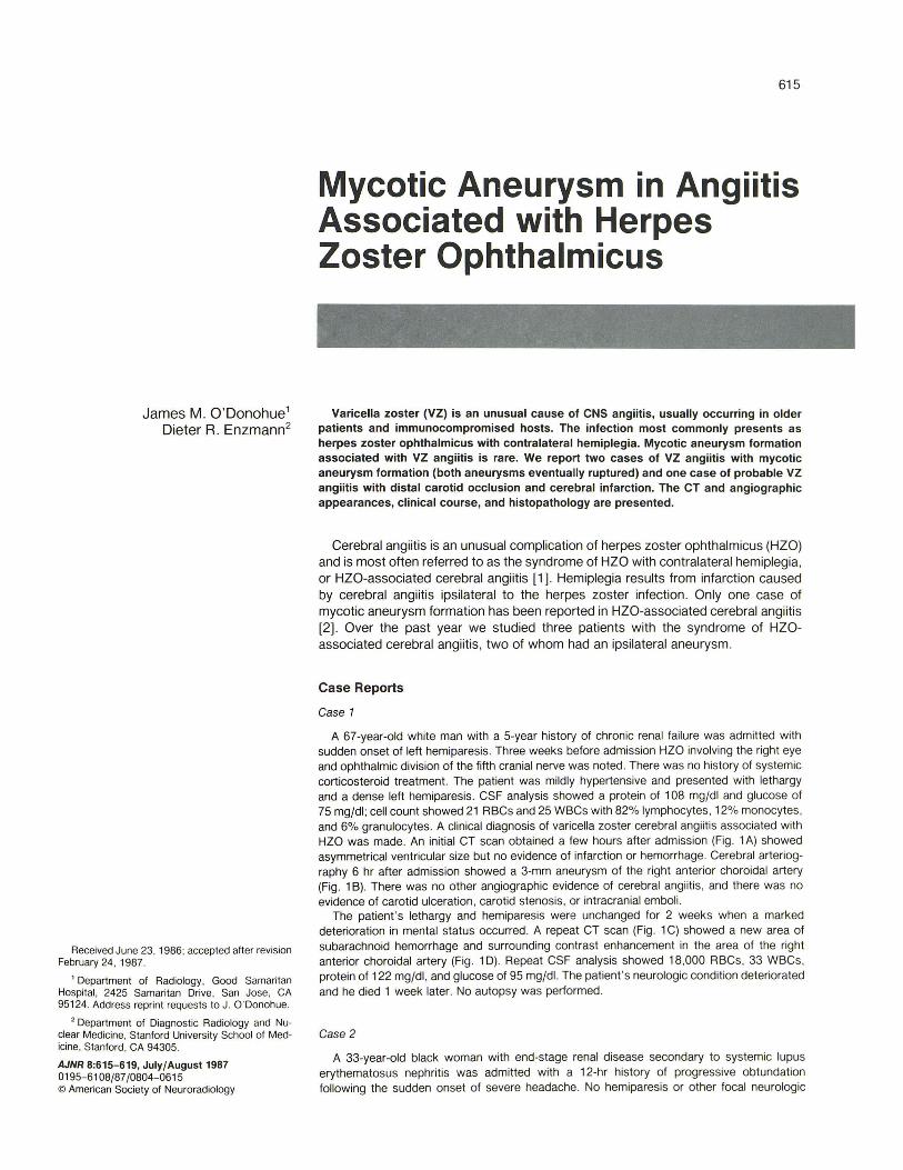
A 67-year-old white man with a 5-year history of chronic renal failure was admitted with sudden onset of left hemiparesis. Three weeks before admission HZO involving the right eye and ophthalmic division of the fifth cranial nerve was noted. There was no history of systemic corticosteroid treatment. The patient was mildly hypertensive and presented with lethargy and a dense left hemiparesis. CSF analysis showed a protein of 108 mg/dl and glucose of 75 mg/dl; cell count showed 21 RBCs and 25 WBCs with 82% lymphocytes, 12% monocytes, and 6% granulocytes. A clinical diagnosis of varicella zoster cerebral angiitis associated with HZO was made. An initial CT scan obtained a few hours after admission (Fig. 1 A) showed asymmetrical ventricular size but no evidence of infarction or hemorrhage. Cerebral arteriography 6 hr after admission showed a 3-mm aneurysm of the right anterior choroidal artery (Fig . 1 B). There was no other angiographic evidence of cerebral angiitis , and there was no evidence of carotid ulceration, carotid stenosis , or intracranial emboli.
The patient 's lethargy and hemiparesis were unchanged for 2 weeks when a marked deterioration in mental status occurred. A repeat CT scan (Fig. 1 C) showed a new area of subarachnoid hemorrhage and surrounding contrast enhancement in the area of the right anterior choroidal artery (Fig. 10). Repeat CSF analysis showed 18,000 RBCs, 33 WBCs, protein of 122 mg/dl , and glucose of 95 mg/dl. The patient 's neurologic condition deteriorated and he died 1 week later. No autopsy was performed .
Case 2
A 33-year-old black woman with end-stage renal disease secondary to systemiC lupus erythematosus nephritis was admitted with a 12-hr history of progressive obtundation following the sudden onset of severe headache. No hemiparesis or other focal neurologic

616 O'DONOHUE AND ENZMANN AJNR :8, July/August 1987
A B
c o
deficits were present. HZO distributed along the left ophthalmic nerve had been diagnosed 4 months earlier. An initial CT scan on admission (Fig. 2A) revealed subarachnoid hemorrhage and a focal hematoma in the anterior interhemispheric fissure and a smaller hematoma just above the internal carotid artery bifurcation on the left. Cerebral angiography revealed a 5-mm aneurysm arising from the proximal mid A 1 segment of the left anterior cerebral artery (Fig . 2B). There was no other angiographic evidence of cerebral angiitis. At surgery 2 weeks later, the aneurysm was described as having a thin , friable, necrotic, and "mycotic looking appearance," with similar changes affecting the adjacent segments of the anterior cerebral artery . Histopathologic examination of the aneurysm wall showed inflammatory cells and granulation tissue. Bacierial and fungal cultures and stains were negative. Viral cultures and electron microscopy were not performed. The patient recovered and was discharged 10 days after surgery and has continued to do well.
Case 3
Fig. 1.-Patient 1. A, Initial CT scan shows no evidence of infarct
or hemorrhage. 8, Right carotid arteriogram. 3·mm aneurysm
of cisternal segment of right anterior choroidal artery.
C, Repeat CT scan performed after sudden deterioration shows subarachnoid hemorrhage in right suprasellar cistern.
D, Contrast enhancement is seen surrounding region of right anterior choroidal artery aneurysm.
A 22-year-old Oriental man was admitted with a right hemiparesis that had evolved in a stuttering fashion over the preceding 2 weeks . Five months before admission he had experienced HZO on the left side. An initial CT scan at another hospital reportedly showed effacement of sulci of the left hemisphere. Angiography revealed complete occlusion of the left internal carotid artery just distal to the ophthalmic artery origin (Fig . 3A). The patient was transferred to our hospital for evaluation 3 weeks after the onset of symptoms. On physical examination there was moderate weakness of the right upper extremity, mild right central facial palsy, and a slightly spastic gait on the right. Rheumatoid factor and FANA were both negative. Lumbar puncture showed an opening pressure of 140 mm H20 , CSF glucose of 61 mg/dl , protein of 36 mg/dl , 70 RBC per JlI , and 2 WBC per JlI (82% lymphocytes, 18% monocytes). CSF, bacterial and fungal stains and

AJNR :8. July/August 1987 MYCOTIC ANEURYSM 617
Fig. 2.-Patient 2. A, Initial unenhanced CT scan shows sub
arachnoid hemorrhage with focal collections in anterior interhemispheric fissure, left sylvian fissure, and near left internal carotid artery bifurcation.
B, Left carotid arteriogram shows 5-mm aneurysm superimposed on proximal A 1 segment of left anterior cerebral artery.
cultures, and CSF VDRL were all negative. Repeat CT scans (Figs. 3C and 3D) showed new areas of low density in the left internal capsule, posterior temporal lobe, and centrum semiovale consistent with evolving infarctions. A diagnosis of HZO-associated cerebral angiitis with cerebral infarction was made. The patient improved and was discharged for rehabilitation .
Discussion
The varicella zoster (VZ) virus, a member of the human herpes virus family , causes a wide range of infections in man, the most common being chicken pox. After an episode of chicken pox , the VZ virus may remain dormant in spinal or cranial nerve ganglia. Herpes zoster results from reactivation of the dormant VZ virus and is characterized by a painful rash in the radicular distribution of the involved nerve. Herpes zoster ophthalmicus (HZO) results from involvement of the ophthalmic division of the fifth cranial nerve, which is involved 20 times more commonly than either the second or third divisions. The frontal branch of the ophthalmic nerve is affected more frequently than either the lacrimal or nasociliary branches [3, 4] .
An unusual complication of HZO, with only 54 cases reported in the world 's literature, is the syndrome of HZO with contralateral hemiplegia [1 , 5, 6]. The hemiplegia results from infarction of the cerebral hemisphere ipsilateral to the side of the HZO infection, due to a cerebral angiitis associated with the VZ infection. Because the cerebral angiitis can cause a wide range of neurologic manifestations that do not always include hemiplegia, a more comprehensive designation of HZO-associated cerebral angiitis (HZO-ACA) may be more appropriate [7].
The syndrome of HZO-ACA usually occurs in older patients (mean age, 58 years) , but the age range is wide (7-96 years) [8]. Relative impairment of cell-mediated immunity with advancing age may account for the increased prevalence of
herpes zoster and the associated cerebral angiitis in the older population [9]. Immunocompromised hosts-especially those with impaired cell-mediated immunity such as occurs with systemic lupus erythematosus, lymphoma, and chronic renal failure-are at higher risk. Onset of the hemiplegia or other neurologic symptoms may be from 1-42 weeks after the onset of HZO, with an average delay of 7 weeks [8]. Alteration of mental status, lethargy, or an acute confusional state may precede the focal deficits.
Angiography was performed in 22 of the 54 cases reported in the literature, and was abnormal in 21 [2 , 8, 10-16] . Changes of cerebral vasculitis , such as segmental constrictions and occlusions, are typically unilateral and limited to the proximal segments of the ipsilateral middle cerebral artery and anterior cerebral artery or the carotid siphon . More widespread and even bilateral vasculitic changes have been described [1 , 13]. One case of basilar artery involvement has also been described [17].
The exact pathogenesis of the angiitis remains controversial, but local spread of the virus from the gasserian ganglionitis to the adjacent carotid artery, middle cerebral artery, and anterior cerebral artery is generally accepted [15] . There is both anatomic and clinical evidence supporting this mechanism. In studies with cats, intracranial branches of the ophthalmic division of the trigeminal nerve have been shown to supply sensory branches to the perivascular plexus of the proximal middle cerebral artery and anterior cerebral artery as well as the supraclinoid internal carotid artery [18] . In humans, stretching or chemical stimulation of the circle of Willis and proximal segments of the middle cerebral artery and anterior cerebral artery causes pain mediated by the ophthalmic division of the trigeminal nerve [19]. Neural spread of the viral infection along these intracranial branches of the ophthalmic nerve to this perivascular plexus is currently the most favored mechanism explaining the adjacent cerebral angiitis [15]. The distribution of sensory branches of the

618 O'DONOHUE AND ENZMANN AJNR:8, July/August 1987
A 8
c D
ophthalmic nerve closely matches the distribution of the lesions of the vasculitis . Bilateral and more widespread angiitis suggests that direct spread via CSF pathways or a hematogenous route may also occur [17 , 20).
Evidence of direct viral invasion of blood vessel walls has come from histologic studies showing viral particles, giant cells , and lymphocytic and monocytic cellular infiltrates involving the media and adventitia of the involved vessels [8 , 17, 21). VZ virus antigens have been found in the media of vessels involved by the angiitis following HZO, but only on the side ipsilateral to the involved trigeminal nerve (22). Granulomatous angiitis of the CNS following HZO has also been described [22-24).
There has only been one previous report of mycotic aneurysm format ion in herpes zoster-associated cerebral angiitis. In 1980, Gursoy et al. (2) reported the case of 24-year-old woman who presented with right-sided hemiplegia after an episode of HZO involving the left eye 1 week earlier. The initial cerebral arteriogram was normal. Five weeks later the patient had recurrent symptoms, and a repeat arteriogram revealed a large aneurysm of the intrapetrous portion of the left internal carotid artery . No histopathologic examination of the aneurysm wall was performed .
In our two patients with ruptured aneurysms, one had
Fig. 3.-Patient 3. A, Left carotid arteriogram shows complete
occlusion of left internal carotid artery just distal to origin of ophthalmic artery.
B, Right carotid arteriogram shows retrograde filling of left middle cerebral artery branches via pial collaterals.
C and D, Follow-up unenhanced CT scans show focal low-density regions in left internal capsule, posterior temporal lobe, and centrum semiovale consistent with infarction.
histopathologic evidence for a "mycotic" inflammatory etiology. In the other patient, the evidence is circumstantial. Several features of the aneurysm were unusual for a berry aneurysm: the location of the aneurysm in the cisternal segment of the choroidal artery, the rupture of the aneurysm temporally associated with the HZO infection, contrast enhancement around the ruptured aneurysm, and the proximity to the infected trigeminal ganglion all strongly suggest a mycotic etiology.
In conclusion, it seems most likely that both these aneurysms represent "mycotic" aneurysms secondary to inflammatory necrosis of the media of arteries involved by VZ virus angiitis. Mycotic aneurysm formation may complicate the syndrome of HZO-associated cerebral angiitis more commonly than previously reported . The long delay that often occurs between the episode of HZO and the cerebral infarction , hemorrhage, or other neurologic event, may mask the association of the acute clinical syndrome, or the presence of an aneurysm, with the previous HZO infection. In older patients and immunocompromised hosts, unilateral cerebral angiitis or aneurysms in atypical locations should raise the possibility of an association with a prior episode of herpes zoster ophthalmicus. With greater use of high-resolution magnification angiography, we may see an increase in the detec-

AJNR :8, July/August 1987 MYCOTIC ANEURYSM 619
tion of small aneurysms. Treatment may be more urgent since no antibiotic therapy is available.
REFERENCES
1. Bourdette ON. Rosenberg NL, Yatsu FM. Herpes zoster ophthalmicus and delayed ipsilateral cerebral infarction. Neurology 1983;33: 1428-1432
2. Gursoy G, Aktin E, Bahar S, Tolun R, Ozden B. Post-herpetic aneurysm in the intrapetrosal portion of the internal carotid artery. Neuroradiology 1980;19 : 279-282
3. Raber I, Laibson P. Herpes zoster ophthalmicus. In Leibowitz H, ed . Corneal disorders: clinical diagnosis & management. Philadelphia: Saunders, 1984: 404-419
4. Grayson M. Diseases of the cornea . St. Louis: Mosby, 1979:148-151 5. Reshef E, Greenberg SB, Jankovic J. Herpes zoster ophthalmicus followed
by contralateral hemiparesis: report of two cases and review of literature. J Neurol Neurosurg Psychiatry 1985;48: 122- 127
6. Gasperetti C, Song SK. Contralateral hemiparesis following herpes zoster ophthalmicus. J Neurol Neurosurg Psychiatry 1985 ;48:338-341
7. Bourdette ON , Yatsu FM, Rosenberg NL. Reply . Neurology 1980;35 :443 8. Hilt DC , Buchholz 0 , Krumholz A, Weiss H, Wolinsky JS. Herpes zoster
ophthalmicus and delayed contralateral hemiparesis caused by cerebral angiitis: diagnosis and management approaches. Ann Neurol 1983; 14 :543-553
9. Miller AE. Selective decline in cellular immune response to varicella-zoster in the elderly. Neurology 1980;30 :582-587
10. Tabbaa M, Selhorst JB. Angiography in herpes zoster ophthalmicus and delayed contralateral hemiparesis. Neurology 1985;35:442- 443
11 . Kuroiwa Y, Furukawa T. Hemispheric infarction after herpes zoster ophthal-
micus: computed tomography and angiography. Neurology 1981 ;31: 1030- 1032
12. Womack LW, Liesegang T J. Complications of herpes zoster ophthalmicus. Arch Ophthalmo/1983 ;101 :42-45
13. Pratesi R, Freemon FR , Lowry JL. Herpes zoster ophthalmicus with contralateral hemiplegia. Arch Neural 1977;34 :640-641
14. Walker RJ III , EI Gammal T, Allen MB Jr. Cranial arteritis associated with herpes zoster. Radiology 1973;107: 109-110
15. MacKenzie RA, Forbes GS, Karnes WE. Angiographic findings in herpes zoster arteritis. Ann Neural 1981 ;10:458-464
16. Gilbert GJ . Herpes zoster ophthalmicus & delayed contralateral hemiparesis . JAMA 1974;229 :302- 304
17. Linnemann CC Jr, Alvira MM. Pathogenesis of varicella-zoster angiitis in the CNS. Arch Neuro/1980;37 :239-240
18. Mayberg M, Langer RS, Zervas NT, Moskowitz MA. Perivascular meningeal projections from cat trigeminal ganglia: possible pathway for vascular headaches in man . Science 1981;213:228-230
19. Moskowitz MA. The neurobiology of vascular head pain. Ann Neural 1984;16:157-168
20. Blue MG, Rosenblum WI. Granulomatous angiitis of the brain with herpes zoster & varicella encephalitis . Arch Pathol Lab Med 1983;107: 126-128
21 . Doyle PW. Gibson G, Dolman CL. Herpes zoster ophthalmicus with contralateral hemiplegia: identification of cause. Ann Neurol 1983; 14 : 84-85
22. Eidelberg 0 , Sotrel A. Horoupian OS, Neumann PE, Pumarola-Sune T, Price RW. Thrombotic cerebral vasculopathy associated with herpes zoster. Ann Neural 1986;19:7-14
23 . Kolodny EH, Rebeiz JJ , Caviness VS, Richardson EP Jr. Granulomatous angiitis of the central nervous system. Arch Neuro/1 968 ;19 :510- 524
24 . Rosenblum WI, Hadfield MG. Granulomatous angiitis of the nervous system in cases of herpes zoster and lymphosarcoma. Neurology 1972; 22 :348-354