
Protein Mutational Analysis Using Statistical Geometry Methods

PRECLINICAL STUDY
Mutational studies on single circulating tumor cells isolatedfrom the blood of inflammatory breast cancer patients
Catherine Bingham1• Sandra V. Fernandez1,3 • Patricia Fittipaldi1 •
Paul W. Dempsey2 • Karen J. Ruth1 • Massimo Cristofanilli1,4 • R. Katherine Alpaugh1,3
Received: 1 December 2016 / Accepted: 25 February 2017
� The Author(s) 2017. This article is published with open access at Springerlink.com
Abstract
Purpose The molecular characterization of circulating
tumor cells (CTCs) is critical to identify the key drivers of
cancer metastasis and devising therapeutic approaches,
particularly for inflammatory breast cancer (IBC) which is
usually diagnosed at advance stages and progresses rapidly.
Methods Genomic alterations in tumor tissue samples were
studied using Foundation OneTM. Single CTCs were iso-
lated using CellSearch followed by single-cell isolation by
DEPArrayTM. Samples with 20 or more CTCs were chosen
to isolate single CTCs using the DEPArrayTM.
Results Genomic alterations were studied in primary
tumor or metastatic sites from 32 IBC patients. Genes with
high-frequency mutations were as follows: TP53 (69%),
RB1 (16%), PIK3CA (13%), and also ErbB2 (3%). At least
once during treatment, CTCs were detected in 26 patients
with metastatic IBC, in two patients with locally advanced
IBC, and four patients had no detectable CTCs. Per 7.5 mL
of blood, fifteen patients (47%) had C20 CTCs and six of
them were chosen at random to isolate single CTCs. These
cells were tested for the presence of TP53, RB1, PIK3CA,
and/or ErbB2 mutations previously found in matching tis-
sue biopsies. The isolated CTCs showed the same muta-
tions as primary or metastatic tumor samples. Intra-patient
CTC heterogeneity was found by the presence of different
CTC subclones, with some CTCs harboring different
combinations of mutated and wild-type genes.
Conclusions Our results indicate that CTCs could repre-
sent a non-invasive source of cancer cells from which to
determine genetic markers as the disease progresses and
identify potential therapeutic targets in IBC patients.
Keywords CTC � Single-cell analysis � Tumor
heterogeneity � IBC
Abbreviations
CK Cytokeratin
CTC Circulating tumor cell
EpCAM Epithelial cell adhesion molecule
ErbB2 or
Her2
v-erb-b2 avian erythroblastic leukemia viral
oncogene homolog 2
FFPE Formalin- fixed paraffin embedded
Fs Frameshift
IBC Inflammatory breast cancer
NGS Next-generation sequencing
OS Overall survival
PE Phycoerythrin
PgR Progesterone receptor
RB1 Retinoblastoma tumor suppressor
TN Triple-negative
TNBC Triple-negative breast cancer
WBC White blood cells
WGA Whole genome amplification
Electronic supplementary material The online version of thisarticle (doi:10.1007/s10549-017-4176-x) contains supplementarymaterial, which is available to authorized users.
& Sandra V. Fernandez
& R. Katherine Alpaugh
1 Fox Chase Cancer Center, Philadelphia, PA 19111, USA
2 Cynvenio Biosystems, Westlake Village, CA, USA
3 Protocol Support Laboratory, Fox Chase Cancer Center,
333 Cottman Ave., Philadelphia, PA 19111, USA
4 Present Address: Robert H Lurie Comprehensive Cancer
Center of Northwestern University, Chicago, IL, USA
123
Breast Cancer Res Treat
DOI 10.1007/s10549-017-4176-x

Introduction
Inflammatory breast cancer (IBC) is a very aggressive type
of advanced breast cancer with a poor prognosis. The
clinical symptoms of IBC involve the rapid onset of
changes in the skin overlaying the breast, including edema,
redness, and swelling, exhibiting a wrinkled, orange peel-
like appearance of the skin known as peau d’orange [1].
This peculiar presentation is associated with the invasion of
aggregates of tumor cells, defined as tumor emboli, into the
dermal lymphatics, where they obstruct the lymph channels
[2, 3]. IBC currently accounts for only 2–6% of all breast
cancer cases in the United States and up to 20% of all
breast cancer cases globally [4–7]. Due to its propensity to
rapidly metastasize, it is responsible for a disproportionate
number (15%) of breast cancer-related deaths [7–9]. IBC is
either stage III or IV; at the time of diagnosis, virtually all
patients have lymph node metastases and one third of the
patients have metastases in distant organs such as the brain,
the bones, and/or the visceral organs [6].
Metastatic disease is the most common cause of cancer-
related death in patients with solid tumors and it is often
associated with the presence of circulating tumor cells
(CTCs) in the peripheral blood of cancer patients [10].
CTCs have been detected in a majority of epithelial can-
cers, including prostate [11], colorectal [12], and breast
cancers [13]. CTCs are tumor cells shed from either the
primary tumor or its metastases and can thus be regarded as
‘‘liquid biopsies’’ of metastasizing cells. Although their
exact composition is unknown, a fraction of these cells is
thought to be viable metastatic precursors capable of ini-
tiating a clonal metastatic lesion [14, 15]. Little is known
about the timing of CTC release from primary tumors, their
functional properties, or their heterogeneity. Intra-tumor
heterogeneity denotes the coexistence of subpopulations of
cancer cells that differ in their genetic, phenotypic, or
behavioral characteristics within a given primary tumor
and between a given primary tumor and its metastasis.
Thus, intra-tumor heterogeneity poses a tremendous chal-
lenge for the characterization of biomarkers and treatments
selection. In this work, we isolated single CTCs from the
blood of IBC patients in order to analyze the presence of
different mutations found in the primary tumor or meta-
static sites and determine the heterogeneity of these cells.
Materials and methods
Patients
Thirty-two patients with inflammatory breast cancer (IBC)
undergoing systemic treatment for their disease were
included in this study. At the time of the first CTC enu-
meration, 29 patients had metastatic IBC (Stage IV) and
three patients had locally advance IBC (Stage III). Clinical
details and treatment timelines for the 32 patients are given
in Supplementary Information. Targeted treatment out-
comes have also been reported elsewhere on patients
D84455 [16] and I77438 [17].
Genomic studies in tumor samples
Formalin-fixed paraffin embedded (FFPE) tumor tissues
(breast, chest wall, lymph node, bone marrow, liver biopsy,
abdominal skin punch, brain biopsy, and/or pleural fluids)
were used for genomic studies. Ten unstained sections
were cut (5–10 lm) and placed on charged slides and
submitted to Foundation Medicine (Cambridge, MA) for
genetic analysis. Briefly, DNA was isolated from the fixed
tumor cells and genomic analysis was performed using
next-generation sequencing (NGS) (Foundation OneTM).
CTCs enumeration from the blood using CellSearch
One tube of 7.5 mL blood from the IBC patients was drawn
and the CellSearchTM System was used for CTC enrich-
ment and enumeration. After running the blood in the
CellSearchTM system for CTC enumeration, the cells were
recovered from the cassettes in order to be used for single
CTC selection using the DEPArrayTM System (Silicon
Biosystems, San Diego, CA). The standard protocol used
for CTC enrichment is described in Supplemental Materi-
als and Methods. Samples containing a minimum of 20
CTCs were selected and prepared for single-cell selection
using the DEPArrayTM.
Isolation of single CTCs using the DEPArrayTM
system
After the CellSearch enrichment, single CTCs were
selected and isolated using the DEPArrayTM (Silicon
Biosystems) as described in Supplemental Materials and
Methods. Individual CTCs or clustered cells that were a-cytokeratin (PE)-positive, CD45 (APC)-negative, and
DAPI-positive were recovered in several tubes for genomic
analysis. In addition, individual white blood cells (WBC)
classified as CD45 (APC)-positive, CK (PE)-negative, and
DAPI-positive were selected and recovered as single cells
to use as controls in the genomic studies. Selected cells
were stored at -80 �C for genomic analyses.
TP53, ErbB2, PIK3CA, and RB1 mutations in CTCs
Whole genome amplification (WGA) was performed using
the Ampli1TM WGA Kit (Silicon Biosystems). The
Breast Cancer Res Treat
123

Table 1 Mutations in tumor samples and number of CTCs detected during disease progression in triple-negative IBC patients
Patient ID Age at diagnosis
Tissue Number of CTCs ( in 7.5 ml blood) andIBC stage at that point
Survivaltime
Tissue source Mutation Amplification Baseline Month1-10
Month11-20
Month21-30
Month31-40
Month41-50
J73299 34 Liver (month 13)
TP53 P190_H193 >*EBRCA1 E23 fs*17
AKT1, RPTORMCL1,MYC
NDA(III)
ND(III)
†
101(IV)
†
201(IV)
†
21 months
T77549 47 Chest wall (month 22)
RB1 splice 607+1 G>C NDA(III)
NDA 60(IV)
†
2,502(IV)
†
27 months
D84455 57 Chest wall (month 21)
TP53 C229 fs*10ERBB2 V777LERBB2 S310F PIK3CA K111E
NDA(IV)
NDA 25+
(IV)†
46+
(IV)†
19+
(IV)†
31 months
R85453 45 Breast (month 8)
TP53 R110 fs*13 MCL1, MYC,JUN
NDA(III)
33(IV)
†
222(IV)
†
15 months
L67504 43 Chest wall(month 44)
TP53 R110 fs*13BRAC2 A1326 fs*4RB1 K720*
CCNE1,MYC
NDA(III)
NDA NDA 20(IV)
†
12+
(IV)†
90+
(IV)†
51 months
T81354 45 Lymph node(month 19)
TP53 splice site 782+1C>TRB1 P777 fs*33
NDA(IV)
9(IV)
†
4(IV)
†
NDA NDA NDA 45 months
S80274 61 Chest wall(month 20)
TP53 G245S, G245D NDA(III)
NDA 3(IV)
†
9(IV)
†
ND
†
31 months
J64403 52 Skin central upper abdomen
(month 43)
BRAC2 L1768 fs*5 NDA(III)
NDA NDA 1(IV)
†
0(IV)
†
9(IV)
†
48 months
D89802 44 Chest wall(month 1)
TP53 R248QMEN1 E496*
CCND1 NDA(IV)
2(IV)
†
ND
†
19 months
C65525 55 Chest wall (month 30)
TP53 S241 fs*23 AKT2 NDA(III)
NDA 0(IV)
†
12(IV)
†
4(IV)
†
38 months
B87480(male)
65 Chest wall (month 13)
Kras G12DNOTCH1 E424K
NDA(IV)
NDA 0(IV)
†
NDA NDA 36 months
R67904 72 Lymph node(month 1)
TP53 W146*,ATM E672 fs*31
CCND1, MCL1MYC,
NKX2-1
6(IV)
†
0(IV)
†
34(IV)
†
ND(IV)
†
22 months
M76085 51 Chest wall(month 27)
NOTCH1 loss NDA(IV)
NDA NDA 3(IV)
†
ND
†
32 months
M67752 61 Skin of right upper back medial
(month 27)
TP53 R342* FGFR1 NDA(IV)
0(IV)
†
0(IV)
†
28(IV)
†
ND
†
32 months
E91111 40 Bone marrow(month 34)
TP53 G245C, BRCA1 R1583 fs*39, EPHA3 E237K
AKT3, JAK2
NDA(III)
NDA NDA 0(IV)
†
8(IV)
†
39 months
L95781 44 Chest wall(month 8)
TP53 L257fs*8, splice site 920-2 A>GNOTCH1 H2428 fs*6
MCL1 NDA(IV)
117(IV)
†
9 months
L92225 53 Pleural fluid (month 14)and
Abdominal skin punch(month 16)
AKT1 E17KTP53 D259YRB1 E693*CDH1 S337_L343 del
NDA(IV)
NDA 178(IV)
†
20 months
T89857 42 Right breast (month 7)
TP53 R156fs*14PIK3CA E8_L15>G
MCL1MYC
NDA(III)
3(III)
†
0(IV)
†
19 months
Breast Cancer Res Treat
123

Ampli1TM WGA kit uses a polymerase with proofreading
activity with a lower error rate (4.8 9 10-6) than standard
Taq DNA polymerases. Global amplification consisting of
DNA isolation, restriction digestion, adaptor ligation, and
PCR amplification was performed as described in Supple-
mental Materials and methods [18]. To study TP53, ErbB2,
and PIK3CA mutations in CTCs, reverse and forward
primers were used (Supplementary Table 1). The PCR
products were cleaned using the QIAquick PCR purifica-
tion kit and sequenced using the ABI 3130XL capillary
genetic analyzer. As we were unsuccessful in studying RB1
K720* mutation using specific primers as described before
for other genes, this mutation was studied using next-
generation sequencing (NGS) as described in Supplemental
Materials and Methods.
Results
Mutation analysis of tissue samples from metastatic
IBC patients
A total of 32 patients with IBC were included in the study
(Tables 1 and 2). All the patients were females save one
male (B87480, Table 1), and all of them were at an
advanced clinical stage at time of diagnosis (stage III or
IV). Their median age at diagnosis was 48 years with a
range of 32–72 years old. From the 32 patients, 20 patients
(62.5%) had triple-negative (ER-negative, PgR-negative,
and Her2-negative) IBC (Table 1); five patients (15.6%)
had ER-positive Her2-negative IBC; three patients (9.4%)
had ER-negative Her2-positive IBC; and four patients
(12.5%) had ER-positive Her2-positive IBC at time of
diagnosis (Table 2).
The genomic alterations in the primary tumor or meta-
static sites were determined using the NGS-based cancer
gene test, Foundation OneTM (Tables 1 and 2). IBC
patients showed mutations in the following: TP53 (22/32;
69%), RB1 (5/32; 16%), PIK3CA (4/32; 13%), BRCA1 (3/
32; 9%), BRCA2 (3/32; 9%), and Notch1 (3/32; 9%). Other
mutated genes were ErbB2 (or Her2; 3%), ATM, Kras,
ESR1, EGFR, and PAX5. Also IBC tumor samples showed
amplifications in the following: MYC (8/32; 25%),
CCND1 (7/32; 22%), ErbB2 (5/32; 16%), MCL1 (6/32;
19%), and FGFR1 (3/32; 9%). Interestingly, patient
D66122, who was found to have a triple-negative disease
by the analysis of her first biopsy, a second chest wall
biopsy in month 26 showed amplification of the ErbB2
gene. Based on these results, this patient was subsequently
treated with Herceptin (Table 1 and Supplementary
Information).
Circulating tumor cells (CTCs) enumeration
The number of CTCs present in 7.5 mL of blood was
determined at different points during the patients’ treat-
ments using the CellSearchTM system. From the 32 IBC
patients, CTCs were detected in 28 patients (C5 CTCs/
7.5 mL blood in 24 patients;\5 CTCs/7.5 mL blood in
four patients) at least once during treatment, while four
patients had no detectable CTCs at any point during
treatment (Tables 1 and 2). CTCs were detected in 26
patients with metastatic IBC and two patients with locally
advance IBC. From 20 patients with triple-negative IBC,
Table 1 continued
D66122 44 Breast (month 8)
TP53 P98 fs*18SOX10 A361V
RAF1 NDA(III)
ND
†
21+
(IV)†
140+
(IV)†
39+
(IV)†
34 months
Skin chest wall(month 26)
TP53 Q104 fs*19 ERBB2
K93878 42 Left breast(month 43)
TP53 S99 fs*44RB1 L779*
MYC, MDM4
NDA(III)
NDA NDA NDA NDA 127(IV)
†
44 months
Patient ID Age at diagnosis
Tissue Number of CTCs ( in 7.5 ml blood) andIBC stage at that point
Survivaltime
Mutations and gene amplifications in tumor samples are shown. Time of tissue collection (t = 0; time of diagnosis) is indicated in each case. The
survival time since IBC diagnostic is indicated. Several blood samples were run in order to determine the number of CTCs along the patient’s
treatment; in the table, only the highest numbers of CTCs during each 10 month period are indicated (in Supplementary Information, all the
points are shown). Also, the disease stage at the time of CTC enumeration is indicated
In some patients, CTCs were present as clusters; presence of CTCs clusters are indicated as (?). Baseline: CTCs number before neoadjuvant
treatment; NDA no data available because the patient was treated at another institution; ND not-done
The time when patients came to Fox Chase Cancer Center for treatment recommendations and/or blood samples draw for CTC enumeration are
indicated as (�)
Breast Cancer Res Treat
123

16 patients had C5 CTCs/7.5 mL blood, and four patients
had 0–4 CTCs/7.5 mL blood. Patients with triple-negative
IBC had significantly worse overall survival compared to
those with ER-positive Her2-positive IBC (p = 0.011),
ER-positive Her2-negative IBC (p = 0.001), and ER-neg-
ative Her2-positive IBC (p = 0.027) (Fig. 1). Patients in
the ER-positive Her2-negative group had the longest
median survival (ER-positive Her2-negative = 76 months
(95% CI 39–101), Triple-Negative = 31.5 months (95%
CI 20–38), ER-negative Her2-positive = 39 months (95%
CI 23–59.), ER-positive Her2-positive = 49.5 months
(95% CI 21-undet.) (Figure 1). In the ER-positive Her2-
positive group at the time of diagnosis (Table 2), patient
K76386 developed a ER-positive (weak) Her2-negative
component in month 9; and a tumor biopsy performed from
patient B62630 in month 49 showed that the tumor was
ER-positive Her2-negative (Supplementary Information).
CTCs were detected in 26 patients with metastatic IBC
(Stage IV) and in two patients with locally advance IBC
(Stage III) at least once during treatments (Tables 1 and 2).
CTCs were not detected in patient B87480 with triple-
negative IBC (Table 1) nor in patients S71769, I77438, and
J70105 with Her2-positive IBC (Table 2).
In order to successfully isolate single CTC using the
DEPArray, samples from the CellSearch that contain at
least 20 CTCs were used. Per 7.5 mL of blood from the 32
Table 2 Mutations in tumor samples and number of CTCs detected during disease progression in ER-positive Her2-negative, ER-negative Her2-
positive, and ER-positive Her2-positive IBC patients
ER-positive Her-2 negative
Patient ID
Age ER PgR Her2 *
Tissue Number of CTCs / 7.5 ml blood(IBC stage at that point)
Survivaltime
Tissue source Mutation Amplific.Baseline
Month 1-10
Month 11-20
Month 21-30
Month 31-40
Month 41-50
Month51-60
Month 61-70
Month71-80
M71182 34 pos pos neg Chest wall(month 69)
PIK3CA H1047RESR1 D538G
CCND1 NDA(III)
NDA NDA NDA NDA NDA 3(IV)
†
11(IV)
†
48(IV)
†
76 months
D84055 58 pos pos neg Left breast (month 1)
CCND1,CDK4, MDM2
NDA(IV)
NDA NDA NDA NDA NDA 2(IV)
†
0(IV)
†
NDA 82 months
M85099 44 pos pos neg Pleural fluid(month 29)
TP53 R273H, R181C FGFR2, IKBKE, CCND1, MCL1
NDA(III)
NDA 1(IV)
†
6(IV)
†
11(IV)
†
ND
†
50 months
A89555 44 pos neg neg Right breast(month 2)
TP53 splice site 993+1 C>T CCND1, MYC
NDA(III)
NDA NDA NDA ND 1(IV)
†
0(IV)
†
NDA NDA 101 months
M66830 45 pos neg neg Left breast(month 6)
FGFR1 345(III)
†
ND
†
4(IV)
†
189(IV)
†
NDA 39 months
ER-negative Her-2 positiveN88166 60 neg neg pos Chest wall
(month 19)TP53 H179R,PIK3R1 441N_452Ydel
ERBB2 NDA(III)
NDA 1(IV)
†
7(IV)
†
23 months
S71769 37 neg neg pos Brain (month 18)
ERBB2 NDA(III)
ND
†
0(IV)
†
ND
†
NDA NDA NDA 59 months
I77438 51 neg pos pos Chest wall (month 22)
TP53 K132NPIK3CA H1047REGFR L858R
ERBB2 NDA(IV)
NDA 0(IV)
†
ND
†
NDA 39 months
ER-positive Her2-positive
L88046 32 pos neg pos Breast (month 13)
TP53 A159VPAX5 I301T
ERBB2 NDA(IV)
NDA NDA NDA 5(IV)
†
ND 43 months
J70105 50 pos neg pos Chest wall (month 28)
AKT1, AURKA, FGFR1,
MYC
ND(III)
NDA NDA 0(III)
†
ND(III)
†
NDA NDA NDA NDA More than 80 months-Alive-
K76386 56 pos neg pos Chest wall (month 15)
PTEN D107YBRAC1 truncationBRAC2 truncation
NDA(III)
111(IV)
†
32(IV)
†
ND
†
21 months
B62630 37 pos pos pos Chest wall (month 49)
TP53 S215G CCNE1 ND(III)
NDA NDA NDA NDA 29+
(IV)†
43+
(IV)†
56 months
Pleural fluid(month 50)
CCND1, IGF1R, MDM2,
AURKA,SRC
Mutations and amplifications in tumor samples are shown. The age of the patients, the ER/PgR/Her2 status at diagnosis, and survival time since
IBC diagnosis are indicated
The number of CTCs present in 7.5 mL of blood was determined using the CellSearch system
Blood samples were run in order to determine the number of CTCs during the disease progression; only the highest numbers of CTCs, every
10-month period since time of start of treatment, are showed
NDA no data available because the patient was treated in another institution, ND non-done, ? presence of clusters
Baseline number of CTCs before treatment
Breast Cancer Res Treat
123

IBC patients, fifteen (47%) patients had C20 CTCs, two
(6%) patients had 10–20 CTCs, five patients (16%) had
between 5 and 10 CTC, and six patients (19%) had 1–5
CTCs. Of the 20 patients with triple-negative IBC, CTCs
were detected in 19 of them at some point during the course
of their disease, and 11 of these patients (55%) had more
than 20 CTCs per 7.5 mL of blood (Table 1). CTCs were
detected in all five patients with ER-positive Her2-negative
IBC at least once during their treatment and two of these
patients had more than 20 CTCs per 7.5 mL of blood
(Table 2). Of the seven patients with Her2- positive IBC,
three patients did not show any CTCs (although CTCs were
enumerated only once in them), and two patients had more
than 20 CTCs (Table 2). The two ER-positive Her2-posi-
tive IBC patients with a high number of CTCs were ini-
tially responsive to Herceptin therapy but during disease
progression they failed to respond to the treatment
(K76386 and B62630; Table 2).
Mutations in single circulating tumor cell (CTC)
From the fifteen patients that had C20 CTCs, six were
chosen at random in order to isolate single CTCs using the
DEPArrayTM (Table 3). Among these patients, all but
patient B62630 had triple-negative IBC at diagnosis but,
developed a Her2-negative component during disease
progression as it was previously mentioned (Supplemen-
tary Information). For molecular characterization of single
CTCs, cells were recovered from the CellSearch cassettes
after enumeration as it was described in Supplementary
Materials and Methods, washed and loaded in the
DEPArray cassettes for single-cell isolation. The DEPAr-
rayTM system (Silicon Biosystems, San Diego, CA) is an
automated platform that uses dielectrophoresis and a high-
quality image-based cell selection system that allows for
the identification and recovery of individual cells from
heterogeneous samples [18]. CTCs that were discrete sin-
gle cells were seen in the blood of three patients (J73299,
R85453 and T77549), whereas the remaining three patients
(B62630, L67504, and D84455) had both individual single
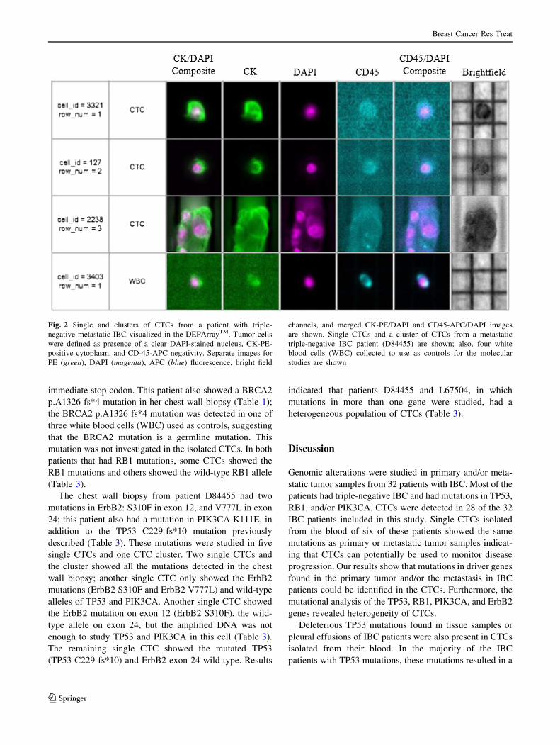
CTCs and clusters of associated CTCs (Table 3). Usually,
CTCs clusters were composed by five to 14 associated cells
(Fig. 2). Samples containing single CTCs, pooled single
CTCs, and/or CTCs clusters were selected and recovered
using DEPArrayTM, and the samples were tested for the
presence of mutations previously revealed in tumor sam-
ples from the same patient. WGA was performed on the
isolated CTCs and regions of TP53, ErbB2, and PIK3CA
shown to be mutated in matching tumor tissue were
amplified and sequenced using Sanger’s method. In five of
the six IBC patients where TP53 was mutated in the tumor
tissues, the isolated CTCs showed the same TP53 muta-
tions (Table 3). However, a TP53 S215G mutation was
present in a chest wall biopsy in patient B62630, which was
not observed in either the isolated CTCs or in tumor cells
collected from pleural effusion (Table 2, pleural effusion
Fig. 1 Survival curves
according to ER (estrogen
receptor) and Her2 (ErbB2)
status. Overall survival in the
triple-negative group was lower
than the other groups (TN vs
ER- Her2?, p = 0.011; TN vs
ER? Her2?, p = 0.001; TN vs
ER? Her2-, p = 0.027).
Patients from the ER? Her2-
group had the longest median
survival (76 months, 95% CI
39–101). In the
ER? Her2? group, one patient
was still alive and shown as
censored (?) at the end of the
curve
Breast Cancer Res Treat
123

from month 50). The TP53 S215G mutation is a missense
mutation in exon 6 (Table 3). The other patients, J73299,
R85453, L67504, and D84455, had TP53 mutations that
produced a premature stop codon in the protein (Table 3).
The liver biopsy of patient J73299 showed a mutation in
TP53 exon 6, TP53 P190_H193[ *E; this mutation was
also found in CTCs isolated from the patient’s blood
(Table 3). Two patients (R85453 and L67504) had a TP53
mutation in exon 4, TP53 R110 fs*13 (Table 3). In patient
L67504, a C deletion (nucleotide 328, delC) was detected
in the chest wall biopsy and the same mutation was found
in the CTCs (Table 3). Patient R85453 showed a G dele-
tion (nucleotide 329, delG) in p53 exon 4 in a breast biopsy
sample (Table 1); while this mutation was also found in
one CTC isolated from the patient, a second CTC revealed
a C deletion (nucleotide 328, delC) that was not previously
detected in the tissue biopsy (Table 3). The chest wall
biopsy of patient D84455 showed a TP53 C229 fs*10
mutation that was also found in three of five single CTCs
analyzed and in one CTC cluster isolated from the patient’s
blood; one single CTC showed the TP53 wild-type allele
and one CTC provided no data as we were unable to suc-
cessfully amplify the region of interest (Table 3). In
Table 4, the deleterious mutations in TP53 are shown.
The RB1 gene was mutated at high frequency in IBC
patients; CTCs isolated from patients T77549 and L67504
were chosen to further study RB1 mutations in single CTCs
(Table 3). RB1 splice mutation 607 ? 1 G[C was
detected in the chest wall biopsy from T77549 and this
mutation was detected in one single CTC and in one pool
of 6 CTCs isolated from the blood of that patient (Table 3).
A second pooled cell sample containing 4 CTCs harbored
RB1 wild type. The RB1 607 ? 1 G[C denotes the G to
C substitution at nucleotide ?1 of the intron (between
exons 6 and 7) in the coding DNA positioned between
nucleotide 607 and 608, resulting in low expression or
partial inactivation of the RB1 protein. Patient L67504
showed a RB1 K720* mutation in her chest wall (Table 1);
the RB1 K720* mutation was also found in two of five
single CTCs and a cluster containing three CTCs that were
isolated from the blood (Table 3). The RB1 K720* muta-
tion is a nonsense variant, a substitution that produces an
Table 3 Mutation analyses in single, pooled, and/or CTCs clusters
Pa�entID #
Muta�ons in �ssue samples
CTCs (single, pools or clusters) WBC control
B62630 TP53 S215G(chest wall)
5 single CTCs: • 5 CTCs: TP53 w. t.
1 cluster of CTCs: TP53 w. t.
5 single WBCs: • 5 WBCs: TP53 w. t.
J73299 TP53 P190_H193>*E(liver)
2 single CTCs: • 2 CTCs: TP53 P190_H193>*E
4 single WBCs: • 4 WBCs: TP53 w. t.
R85453 TP53 R110 delG fs*13(breast biopsy)
6 single CTCs: • 1 CTCs: TP53 R110 delG fs*13• 1 CTC: TP53 R110 delC fs*13• 4 CTCs: TP53 w. t.
1 pool of 14 CTCs: TP53 R110 delC fs*13
4 single WBCs: • 4 WBCs: TP53 w. t.
T77549 RB1 607+1 G>C(chest wall)
1 single CTC:• RB1 607+1 G>C
2 pools of CTCs:• 1 pool of 6 CTCs: RB1 607+1 G>C • 1 pool of 4 CTCs: RB1 w.t.
1 single WBC: • RB1 w. t.
1 pool of 5 WBCs:• RB1 w. t.
L67504TP53 R110 delC fs*13RB1 K720*BRCA2 pA1326 fs*4
(chest wall)
5 single CTCs: • 2 CTCs: TP53 R110 delC fs*13; RB1 K720* • 2 CTCs: TP53 w. t.; RB1 w.t.• 1 CTC: TP53 R110 delC fs*13; RB1 w.t.
1 cluster of 3 CTCs:• TP53 R110 delC fs*13; RB1 K720*
3 single WBCs:•2 WBCs: TP53 w. t.; RB1 w.t.;
BRCA2 w.t.• 1 WBC: TP53 w.t.; RB1 w.t.;
BRCA2 pA1326 fs*4
D84455TP53 C229 fs*10ERBB2 S310FERBB2 V777LPIK3CA K111E
(chest wall)
5 single CTCs: • 2 CTCs: TP53 C229 fs*10; ERBB2 exon 12 S310F; ERBB2 exon 24 V777L; PIK3CA K111E • 1 CTC: TP53 w.t.; ERBB2 S310F; ERBB2 V777L; PIK3CA w.t.• 1 CTC: : TP53 C229 fs*10; ERBB2 exon 12 n.d.; ERBB2 exon 24 w.t.; PIK3CA n.d.• 1 CTC: : TP53 n.d.; ERBB2 exon 12 S310F; ERBB2 exon 24 w.t.; PIK3CA n.d.
1 cluster of CTCs: TP53 C229 fs*10; ERBB2 S310F; ERBB2 V777L; PIK3CA K111E
4 single WBC:• 4 WBCs: TP53 w.t.; ERBB2 w.t.;
PIK3CA w.t.
Single CTCs, pooled single CTCs, and/or CTCs clusters were recovered using DEPArray. Mutations in TP53, ErbB2, PIK3CA, and RB1 found
in tumor tissue samples were detected in the CTCs
Intra-patient heterogeneous CTCs populations were found; w.t. wild type; n.d. non-done (because not enough amplified DNA)
Breast Cancer Res Treat
123
immediate stop codon. This patient also showed a BRCA2
p.A1326 fs*4 mutation in her chest wall biopsy (Table 1);
the BRCA2 p.A1326 fs*4 mutation was detected in one of
three white blood cells (WBC) used as controls, suggesting
that the BRCA2 mutation is a germline mutation. This
mutation was not investigated in the isolated CTCs. In both
patients that had RB1 mutations, some CTCs showed the
RB1 mutations and others showed the wild-type RB1 allele
(Table 3).
The chest wall biopsy from patient D84455 had two
mutations in ErbB2: S310F in exon 12, and V777L in exon
24; this patient also had a mutation in PIK3CA K111E, in
addition to the TP53 C229 fs*10 mutation previously
described (Table 3). These mutations were studied in five
single CTCs and one CTC cluster. Two single CTCs and
the cluster showed all the mutations detected in the chest
wall biopsy; another single CTC only showed the ErbB2
mutations (ErbB2 S310F and ErbB2 V777L) and wild-type
alleles of TP53 and PIK3CA. Another single CTC showed
the ErbB2 mutation on exon 12 (ErbB2 S310F), the wild-
type allele on exon 24, but the amplified DNA was not
enough to study TP53 and PIK3CA in this cell (Table 3).
The remaining single CTC showed the mutated TP53
(TP53 C229 fs*10) and ErbB2 exon 24 wild type. Results
indicated that patients D84455 and L67504, in which
mutations in more than one gene were studied, had a
heterogeneous population of CTCs (Table 3).
Discussion
Genomic alterations were studied in primary and/or meta-
static tumor samples from 32 patients with IBC. Most of the
patients had triple-negative IBC and had mutations in TP53,
RB1, and/or PIK3CA. CTCs were detected in 28 of the 32
IBC patients included in this study. Single CTCs isolated
from the blood of six of these patients showed the same
mutations as primary or metastatic tumor samples indicat-
ing that CTCs can potentially be used to monitor disease
progression. Our results show that mutations in driver genes
found in the primary tumor and/or the metastasis in IBC
patients could be identified in the CTCs. Furthermore, the
mutational analysis of the TP53, RB1, PIK3CA, and ErbB2
genes revealed heterogeneity of CTCs.
Deleterious TP53 mutations found in tissue samples or
pleural effusions of IBC patients were also present in CTCs
isolated from their blood. In the majority of the IBC
patients with TP53 mutations, these mutations resulted in a
Fig. 2 Single and clusters of CTCs from a patient with triple-
negative metastatic IBC visualized in the DEPArrayTM. Tumor cells
were defined as presence of a clear DAPI-stained nucleus, CK-PE-
positive cytoplasm, and CD-45-APC negativity. Separate images for
PE (green), DAPI (magenta), APC (blue) fluorescence, bright field
channels, and merged CK-PE/DAPI and CD45-APC/DAPI images
are shown. Single CTCs and a cluster of CTCs from a metastatic
triple-negative IBC patient (D84455) are shown; also, four white
blood cells (WBC) collected to use as controls for the molecular
studies are shown
Breast Cancer Res Treat
123
non-functional protein. A non-functional TP53 has been
shown to offer survival advantages to the cancer cells by
facilitating growth, anoikis resistance, and the emergence
of a potentially more aggressive malignancy [19]. TP53
mutations are exceptionally frequent in cancer and are
among the key driving factors in triple-negative breast
cancer (TNBC) [20]. Furthermore, TP53 mutations are
more frequent in inflammatory breast cancer (50%) than in
non-inflammatory breast cancer (20–30%) [21, 22]. TP53
mutations have been shown to predict a poor response to
anthracycline-based neoadjuvant chemotherapy [23–25];
others suggested that TP53 mutations confer sensitivity to
taxane [26, 27]. A recent study suggested that patients with
TP53 mutations are more likely to respond to anthracy-
cline/cyclophosphamide-based neoadjuvant chemotherapy
[28]. Several clinical trials are ongoing to study TP53-
mutated breast cancer sensitivity to different chemothera-
peutic agents. Other clinical trials are targeted towards
either expression of the wild-type TP53, suppressing
expression of mutated TP53, or strategies that involve
targeting of the cell cycle regulator Wee-1 tyrosine kinase
inhibitors (clinicaltrials.gov).
RB1 mutations were found in only the triple-negative
IBC patients from this study, and all of these mutations
render a premature stop codon and a non-functional RB1
protein. RB1 mutations were also detected in the CTCs.
RB1 mediates cell cycle control and is frequently inacti-
vated in human TNBC [29–31]. There are targeted inhi-
bitors that are currently in advanced clinical testing for
tumors harboring RB1 and PIK3CA mutations [32, 33].
One triple-negative IBC patient (D84455) with a dele-
terious TP53 mutation also showed two ErbB2 mutations
(V777L and S310F). Both mutations activate ErbB2 by
either affecting its auto-phosphorylation or phosphoryla-
tion of downstream substrates in breast cancer cells
[34–36]. Initially, this patient had ER? Her2? invasive
ductal carcinoma (IDC) in the left breast that was treated
with standard local therapies over 2 years and this patient
subsequently developed triple-negative IBC in the same
breast [16] (Supplementary Information). This suggests
that the ErbB2 pathway could be the driver of this patient’s
disease even in the absence of ErbB2 amplification. Fur-
thermore, this patient had a PIK3CA K111E mutation
which is also an activating mutation [37, 38]. All the
mutations detected in the chest wall biopsy of patient
D84455 were detected in the CTCs isolated from their
blood. A heterogeneous population of CTCs were found in
this patient with some CTCs showing the mutated genes
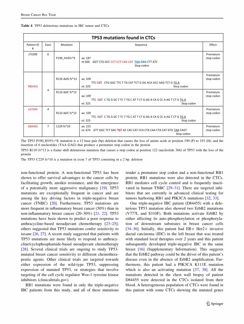
Table 4 TP53 deleterious mutations in IBC tumor and CTCs
TP53 mutations found in CTCsPatient ID
#Exon Mutation Sequence Effect
J73299 6P190_H193>*E aa: 187
nt:566 GGT CTG GCC CCT CCT CAG CAT TAA GAG CTT ATCStop codon
Premature stop codon
R854534
R110 delG fs*13 aa: 109 TTC CGT CTG GGC TTC T TG CAT TCT G GG ACA GCC AAG TCT G TG A
nt: 325 Stop codon
Premature stop codon
R110 delC fs*13aa: 109
TTC CGT C TG G GC T TC T TG C AT T CT G GG A CA G CC A AG T CT G TG Ant: 325 Stop codon
Premature stop codon
L67504 4R110 delC fs*13 aa: 109
TTC CGT C TG G GC T TC T TG C AT T CT G GG A CA G CC A AG T CT G TG Ant: 325 Stop codon
Premature stop codon
D84455 7 C229 fs*10 aa: 225 nt: 674 GTT GGC TCT GAC TGT AC CAC CAT CCA CTA CAA CTA CAT GTG TAA CAGT
Stop codon
Premature stop codon
The TP53 P190_H193[*E mutation is a 12 base pair (bp) deletion that causes the loss of amino acids at position 190 (P) to 193 (H), and the
insertion of 6 nucleotides (TAA GAG) that produce a premature stop codon in the protein
TP53 R110 fs*13 is a frame shift deleterious mutation that causes a stop codon at position 122 (nucleotide 366) of TP53 with the loss of the
protein
The TP53 C229 fs*10 is a mutation in exon 7 of TP53 consisting in a 2 bp. deletion
Breast Cancer Res Treat
123

and others showing different combinations of the mutated
and wild-type genes.
It has been successfully demonstrated by single-cell
sequencing that many breast cancers are composed of
multiple distinct subclones [39]. Intra-patient cellular
heterogeneity is widely reported in epithelial malignancies
and it is expected that CTCs will also be heterogeneous
[40–42]. Our results are consistent with previous findings
which showed a heterogeneous pattern of genomic muta-
tions on single CTCs obtained from breast, esophageal, and
colorectal cancer patients [42–44].
Our results demonstrate that we were able to select
uncontaminated CTCs by combining the CellSearch and
DEPArrayTM systems. However, one disadvantage of the
DEPArrayTM is that there is approximately 40% cell-loss,
although the DEPArray cartridge is manually loaded with
14 lL of sample, only 9.26 lL of sample is injected into
the micro-chamber of the cartridge. We found that in order
to successfully isolate single CTC, samples with 20 CTCs
or more should be used to load in the DEPArray cassette. In
order to perform molecular analysis of CTCs in samples
with less than 20 CTCs per 7.5 mL blood, multiple samples
from the same patient could potentially be combined after
the CellSearch and loaded in the DEPArray cassette.
However, despite the promise of CTCs as multifunctional
biomarkers, there are still numerous challenges that hinder
their incorporation into standard clinical practice. Some
patients showed little to no CTCs even though their disease
was progressing. This is the case for most of the patients with
Her2-positive IBC, with the exception of patients K76386
and B62630 who developed a Her2-negative component
during their disease progression; both had high number of
CTCs. Patients with Her2-positive IBC were treated with
Herceptin for a long period of time, so this could explain why
CTCs were not present in their blood due to the high speci-
ficity of these antibodies. In addition, the CellSearchTM,
relies on the detection of the surface epithelial cell adhesion
molecule EpCAM, and the existence of an EpCAM-negative
subpopulation of CTCs had been described in patients with
Her2-positive metastatic breast cancer [45]; therefore, it will
be interesting to combine different pre-enrichment strategies
with the DEPArray in order to study these cells, especially in
Her2-positive IBC.
It has been shown that elevated CTC at baseline or at
any time through the course of metastatic breast cancer is
associated with worse prognosis; patients with C5 CTCs/
7.5 mL blood had a shorter overall survival compared with
the patients with\5CTCs/7.5 mL blood, and elevated
CTCs while on treatment ultimately are predictive of an
ineffective therapy [13, 46]. Our data showed that the
distribution of CTCs in the patients with serial blood draws
during the treatments varied; many patients were initially
CTCs negative but converted to positive and vice versa.
Based on our results, single time-point measurements of
CTCs seem to be inadequate, and could result in incorrect
microscopic disease staging. Collecting sequential blood
samples for real-time monitoring of the efficacy of sys-
temic therapies would offer new possibilities in evaluating
targeted therapies based on genomic profiling of CTCs and
improving the clinical management of patients with IBC.
In order to implement these studies in future clinical
practice, we are developing protocols in order to study
mutations in single, pools, and clusters of CTCs using NGS
and a panel of 15 genes frequently mutated in IBC.
This work demonstrates that the isolation and pooling of
CTCs from IBC patients can be used for genomic analysis,
both to initially identify targetable mutations where solid
tumor samples are unavailable and to be used as a bio-
marker to reveal which cell populations are affected by the
current or previous therapy. Our results suggest that CTCs
represent the entire spectrum of the primary tumor and
distal metastases for patients with IBC. Furthermore, our
studies showed the presence of different CTCs subclones in
the peripheral blood of IBC patients.
Acknowledgements We thank Andreas Papoutsis and Erich Klem
from Cynvenio Biosystems for the RB1 K720* mutation analysis by
NGS, and Francesca Fontana from Menarini Silicon Biosystems for
her help with DEPArray protocols. We thank Dr. Jennifer Winn for
the revision of patient clinical histories in Supplementary Informa-
tion. We thank the Fox Chase Cancer Center Sequencing Facility.
Funding This work was performed under the grant NIH RO1 CA
138239-02 (PI: Cristofanilli).
Compliance with ethical standards
Conflict of interest Paul W. Dempsey is an employee of Cynvenio
Biosystems. All remaining authors have declared no competing
interests.
Research involving human participants Patients signed an
informed consent and HIPAA certification from the Human Subject
Protection Committee prior to sample collection. This study was
approved by both the research review committee (RRC) and institu-
tional research board (IRB) at Fox Chase Cancer Center.
Open Access This article is distributed under the terms of the
Creative Commons Attribution 4.0 International License (http://crea
tivecommons.org/licenses/by/4.0/), which permits unrestricted use,
distribution, and reproduction in any medium, provided you give
appropriate credit to the original author(s) and the source, provide a
link to the Creative Commons license, and indicate if changes were
made.
References
1. Cristofanilli M, Valero V, Buzdar AU, Kau SW, Broglio KR,
Gonzalez-Angulo AM, Sneige N, Islam R, Ueno NT, Buchholz
TA, Singletary SE, Hortobagyi GN (2007) Inflammatory breast
Breast Cancer Res Treat
123

cancer (IBC) and patterns of recurrence: understanding the
biology of a unique disease. Cancer 110(7):1436–1444
2. Alpaugh ML, Tomlinson JS, Kasraeian S, Barsky SH (2002)
Cooperative role of E-cadherin and sialyl-Lewis X/A-deficient
MUC1 in the passive dissemination of tumor emboli in inflam-
matory breast carcinoma. Oncogene 21(22):3631–3643
3. Molckovsky A, Fitzgerald B, Freedman O, Heisey R, Clemons M
(2009) Approach to inflammatory breast cancer. Can Fam
Physician 55(1):25–31
4. Cristofanilli M, Buzdar AU, Hortobagyi GN (2003) Update on
the management of inflammatory breast cancer. Oncologist
8(2):141–148
5. Dawood S, Cristofanilli M (2011) Inflammatory breast cancer:
what progress have we made? Oncology (Williston Park)
25(3):264–273
6. Robertson FM, Bondy M, Yang W, Yamauchi H, Wiggins S,
Kamrudin S, Krishnamurthy S, Le-Petross H, Bidaut L, Player
AN, Barsky SH, Woodward WA, Buchholz T, Lucci A, Ueno
NT, Cristofanilli M (2010) Inflammatory breast cancer: the dis-
ease, the biology, the treatment. CA Cancer J Clin 60(6):351–375
7. Hance KW, Anderson WF, Devesa SS, Young HA, Levine PH
(2005) Trends in inflammatory breast carcinoma incidence and
survival: the surveillance, epidemiology, and end results program
at the National Cancer Institute. J Natl Cancer Inst 97(13):
966–975
8. Anderson WF, Schairer C, Chen BE, Hance KW, Levine PH
(2005) Epidemiology of inflammatory breast cancer (IBC).
Breast Dis 22:9–23
9. Levine PH, Veneroso C (2008) The epidemiology of inflamma-
tory breast cancer. Semin Oncol 35(1):11–16
10. Alunni-Fabbroni M, Sandri MT (2010) Circulating tumour cells
in clinical practice: methods of detection and possible charac-
terization. Methods 50(4):289–297
11. Danila DC, Heller G, Gignac GA, Gonzalez-Espinoza R, Anand
A, Tanaka E, Lilja H, Schwartz L, Larson S, Fleisher M, Scher HI
(2007) Circulating tumor cell number and prognosis in progres-
sive castration-resistant prostate cancer. Clin Cancer Res
13(23):7053–7058
12. Cohen SJ, Alpaugh RK, Gross S, O’Hara SM, Smirnov DA,
Terstappen LW, Allard WJ, Bilbee M, Cheng JD, Hoffman JP,
Lewis NL, Pellegrino A, Rogatko A, Sigurdson E, Wang H,
Watson JC, Weiner LM, Meropol NJ (2006) Isolation and char-
acterization of circulating tumor cells in patients with metastatic
colorectal cancer. Clin Colorectal Cancer 6(2):125–132
13. Cristofanilli M, Budd GT, Ellis MJ, Stopeck A, Matera J, Miller
MC, Reuben JM, Doyle GV, Allard WJ, Terstappen LW, Hayes
DF (2004) Circulating tumor cells, disease progression, and
survival in metastatic breast cancer. N Engl J Med 351(8):
781–791
14. Yu M, Ting DT, Stott SL, Wittner BS, Ozsolak F, Paul S,
Ciciliano JC, Smas ME, Winokur D, Gilman AJ, Ulman MJ,
Xega K, Contino G, Alagesan B, Brannigan BW, Milos PM,
Ryan DP, Sequist LV, Bardeesy N, Ramaswamy S, Toner M,
Maheswaran S, Haber DA (2012) RNA sequencing of pancreatic
circulating tumour cells implicates WNT signalling in metastasis.
Nature 487(7408):510–513
15. Lee JS, Magbanua MJ, Park JW (2016) Circulating tumor cells in
breast cancer: applications in personalized medicine. Breast
Cancer Res Treat 160(3):411–424
16. Ali SM, Alpaugh RK, Downing SR, Stephens PJ, Yu JQ, Wu H,
Buell JK, Miller VA, Lipson D, Palmer GA, Ross JS, Cristofanilli
M (2014) Response of an ERBB2-mutated inflammatory breast
carcinoma to human epidermal growth factor receptor 2-targeted
therapy. J Clin Oncol 32(25):e88–91
17. Ali SM, Alpaugh RK, Buell JK, Stephens PJ, Yu JQ, Wu H,
Hiemstra CN, Miller VA, Lipson D, Palmer GA, Ross JS,
Cristofanilli M (2014) Antitumor response of an ERBB2 ampli-
fied inflammatory breast carcinoma with EGFR mutation to the
EGFR-TKI erlotinib. Clin Breast Cancer 14(1):e14–16
18. Fernandez SV, Bingham C, Fittipaldi P, Austin L, Palazzo J,
Palmer G, Alpaugh K, Cristofanilli M (2014) TP53 mutations
detected in circulating tumor cells present in the blood of meta-
static triple negative breast cancer patients. Breast Cancer Res
16(5):445
19. Zhang Y, Lu H, Dazin P, Kapila Y (2004) Squamous cell car-
cinoma cell aggregates escape suspension-induced, p53-mediated
anoikis: fibronectin and integrin alphav mediate survival signals
through focal adhesion kinase. J Biol Chem 279(46):
48342–48349
20. Walerych D, Napoli M, Collavin L, Del Sal G (2012) The rebel
angel: mutant p53 as the driving oncogene in breast cancer.
Carcinogenesis 33(11):2007–2017
21. Sawaki M, Ito Y, Akiyama F, Tokudome N, Horii R, Mizunuma
N, Takahashi S, Horikoshi N, Imai T, Nakao A, Kasumi F,
Sakamoto G, Hatake K (2006) High prevalence of HER-2/neu
and p53 overexpression in inflammatory breast cancer. Breast
Cancer 13(2):172–178
22. Turpin E, Bieche I, Bertheau P, Plassa LF, Lerebours F, de
Roquancourt A, Olivi M, Espie M, Marty M, Lidereau R, Vidaud
M, de The H (2002) Increased incidence of ERBB2 overex-
pression and TP53 mutation in inflammatory breast cancer.
Oncogene 21(49):7593–7597
23. Aas T, Borresen AL, Geisler S, Smith-Sorensen B, Johnsen H,
Varhaug JE, Akslen LA, Lonning PE (1996) Specific P53
mutations are associated with de novo resistance to doxorubicin
in breast cancer patients. Nat Med 2(7):811–814
24. Berns EM, Foekens JA, Vossen R, Look MP, Devilee P, Henzen-
Logmans SC, van Staveren IL, van Putten WL, Inganas M,
Meijer-van Gelder ME, Cornelisse C, Claassen CJ, Portengen H,
Bakker B, Klijn JG (2000) Complete sequencing of TP53 predicts
poor response to systemic therapy of advanced breast cancer.
Cancer Res 60(8):2155–2162
25. Kandioler-Eckersberger D, Ludwig C, Rudas M, Kappel S, Jan-
schek E, Wenzel C, Schlagbauer-Wadl H, Mittlbock M, Gnant M,
Steger G, Jakesz R (2000) TP53 mutation and p53 overexpression
for prediction of response to neoadjuvant treatment in breast
cancer patients. Clin Cancer Res 6(1):50–56
26. Gluck S, Ross JS, Royce M, McKenna EF Jr, Perou CM, Avisar
E, Wu L (2012) TP53 genomics predict higher clinical and
pathologic tumor response in operable early-stage breast cancer
treated with docetaxel-capecitabine ± trastuzumab. Breast Can-
cer Res Treat 132(3):781–791
27. Wahl AF, Donaldson KL, Fairchild C, Lee FY, Foster SA,
Demers GW, Galloway DA (1996) Loss of normal p53 function
confers sensitization to Taxol by increasing G2/M arrest and
apoptosis. Nat Med 2(1):72–79
28. Wang Y, Xu Y, Chen J, Ouyang T, Li J, Wang T, Fan Z, Fan T,
Lin B, Xie Y (2016) TP53 mutations are associated with higher
rates of pathologic complete response to anthracycline/cy-
clophosphamide-based neoadjuvant chemotherapy in operable
primary breast cancer. Int J Cancer 138(2):489–496
29. Sellers WR, Novitch BG, Miyake S, Heith A, Otterson GA, Kaye
FJ, Lassar AB, Kaelin WG Jr (1998) Stable binding to E2F is not
required for the retinoblastoma protein to activate transcription,
promote differentiation, and suppress tumor cell growth. Genes
Dev 12(1):95–106
30. Whitaker LL, Su H, Baskaran R, Knudsen ES, Wang JY (1998)
Growth suppression by an E2F-binding-defective retinoblastoma
protein (RB): contribution from the RB C pocket. Mol Cell Biol
18(7):4032–4042
31. Jiang Z, Jones R, Liu JC, Deng T, Robinson T, Chung PE, Wang
S, Herschkowitz JI, Egan SE, Perou CM, Zacksenhaus E (2011)
Breast Cancer Res Treat
123

RB1 and p53 at the crossroad of EMT and triple-negative breast
cancer. Cell Cycle 10(10):1563–1570
32. Cancer Genome Atlas N (2012) Comprehensive molecular por-
traits of human breast tumours. Nature 490(7418):61–70
33. Carey L, Winer E, Viale G, Cameron D, Gianni L (2010) Triple-
negative breast cancer: disease entity or title of convenience? Nat
Rev Clin Oncol 7(12):683–692
34. Bose R, Kavuri SM, Searleman AC, Shen W, Shen D, Koboldt
DC, Monsey J, Goel N, Aronson AB, Li S, Ma CX, Ding L,
Mardis ER, Ellis MJ (2013) Activating HER2 mutations in HER2
gene amplification negative breast cancer. Cancer Discov
3(2):224–237
35. Greulich H, Kaplan B, Mertins P, Chen TH, Tanaka KE, Yun CH,
Zhang X, Lee SH, Cho J, Ambrogio L, Liao R, Imielinski M,
Banerji S, Berger AH, Lawrence MS, Zhang J, Pho NH, Walker
SR, Winckler W, Getz G, Frank D, Hahn WC, Eck MJ, Mani DR,
Jaffe JD, Carr SA, Wong KK, Meyerson M (2012) Functional
analysis of receptor tyrosine kinase mutations in lung cancer
identifies oncogenic extracellular domain mutations of ERBB2.
Proc Natl Acad Sci U S A 109(36):14476–14481
36. Herter-Sprie GS, Greulich H, Wong KK (2013) Activating
mutations in ERBB2 and their impact on diagnostics and treat-
ment. Front Oncol 3:86
37. Rudd ML, Price JC, Fogoros S, Godwin AK, Sgroi DC, Merino
MJ, Bell DW (2011) A unique spectrum of somatic PIK3CA
(p110alpha) mutations within primary endometrial carcinomas.
Clin Cancer Res 17(6):1331–1340
38. Zheng G, Tseng LH, Chen G, Haley L, Illei P, Gocke CD,
Eshleman JR, Lin MT (2015) Clinical detection and categoriza-
tion of uncommon and concomitant mutations involving BRAF.
BMC Cancer 15:779
39. Robinson DR, Wu YM, Vats P, Su F, Lonigro RJ, Cao X,
Kalyana-Sundaram S, Wang R, Ning Y, Hodges L, Gursky A,
Siddiqui J, Tomlins SA, Roychowdhury S, Pienta KJ, Kim SY,
Roberts JS, Rae JM, Van Poznak CH, Hayes DF, Chugh R, Kunju
LP, Talpaz M, Schott AF, Chinnaiyan AM (2013) Activating
ESR1 mutations in hormone-resistant metastatic breast cancer.
Nat Genet 45(12):1446–1451
40. Klein CA (2009) Parallel progression of primary tumours and
metastases. Nat Rev Cancer 9(4):302–312
41. Marusyk A, Almendro V, Polyak K (2012) Intra-tumour hetero-
geneity: a looking glass for cancer? Nat Rev Cancer
12(5):323–334
42. Stoecklein NH, Klein CA (2010) Genetic disparity between pri-
mary tumours, disseminated tumour cells, and manifest metas-
tasis. Int J Cancer 126(3):589–598
43. Ignatiadis M, Rothe F, Chaboteaux C, Durbecq V, Rouas G,
Criscitiello C, Metallo J, Kheddoumi N, Singhal SK, Michiels S,
Veys I, Rossari J, Larsimont D, Carly B, Pestrin M, Bessi S,
Buxant F, Liebens F, Piccart M, Sotiriou C (2011) HER2-positive
circulating tumor cells in breast cancer. PLoS ONE 6(1):e15624
44. Riethdorf S, Muller V, Zhang L, Rau T, Loibl S, Komor M,
Roller M, Huober J, Fehm T, Schrader I, Hilfrich J, Holms F,
Tesch H, Eidtmann H, Untch M, von Minckwitz G, Pantel K
(2010) Detection and HER2 expression of circulating tumor cells:
prospective monitoring in breast cancer patients treated in the
neoadjuvant GeparQuattro trial. Clin Cancer Res 16(9):
2634–2645
45. Giordano A, Gao H, Anfossi S, Cohen E, Mego M, Lee BN, Tin
S, De Laurentiis M, Parker CA, Alvarez RH, Valero V, Ueno NT,
De Placido S, Mani SA, Esteva FJ, Cristofanilli M, Reuben JM
(2012) Epithelial-mesenchymal transition and stem cell markers
in patients with HER2-positive metastatic breast cancer. Mol
Cancer Ther 11(11):2526–2534
46. Swaby RF, Cristofanilli M (2011) Circulating tumor cells in
breast cancer: a tool whose time has come of age. BMC Med 9:43
Breast Cancer Res Treat
123


![Inflammatory, and Metastatic Breast Cancer NIH Public ... Biol Blood... · Patients with inflammatory breast cancer (IBC) have the worst prognosis, with a 5-year OS of 40% [4]. Once](https://static.fdocuments.in/doc/165x107/5f024e187e708231d4039ba2/inflammatory-and-metastatic-breast-cancer-nih-public-biol-blood-patients.jpg)