Mutational dynamics of murine angiogenin duplicates
14
RESEARCH ARTICLE Open Access Mutational dynamics of murine angiogenin duplicates Francisco M Codoñer 2 , Silvia Alfonso-Loeches 3 , Mario A Fares 1,4* Abstract Background: Angiogenin (Ang) is a protein involved in angiogenesis by inducing the formation of blood vessels. The biomedical importance of this protein has come from findings linking mutations in Ang to cancer progression and neurodegenerative diseases. These findings highlight the evolutionary constrain on Ang amino acid sequence. However, previous studies comparing human Angiogenin with homologs from other phylogenetically related organisms have led to the conclusion that Ang presents a striking variability. Whether this variability has an adaptive value per se remains elusive. Understanding why many functional Ang paralogs have been preserved in mouse and rat and identifying functional divergence mutations at these copies may explain the relationship between mutations and function. In spite of the importance of testing this hypothesis from the evolutionarily and biomedical perspectives, this remains yet unaccomplished. Here we test the main mutational dynamics driving the evolution and function of Ang paralogs in mammals. Results: We analysed the phylogenetic asymmetries between the different Ang gene copies in mouse and rat in the context of vertebrate Ang phylogeny. This analysis shows strong evidence in support of accelerated evolution in some Ang murine copies (mAng). This acceleration is not due to non-functionalisation because constraints on amino acid replacements remain strong. We identify many of the amino acid sites involved in signal localization and nucleotide binding by Ang to have evolved under diversifying selection. Compensatory effects of many of the mutations at these paralogs and their key structural location in or nearby important functional regions support a possible functional shift (functional divergence) in many Ang copies. Similarities between 3D-structural models for mAng copies suggest that their divergence is mainly functional. Conclusions: We identify the main evolutionary dynamics shaping the variability of Angiogenin in vertebrates and highlight the plasticity of this protein after gene duplication. Our results suggest functional divergence among mAng paralogs. This puts forward mAng as a good system candidate for testing functional plasticity of such an important protein while stresses caution when using mouse as a model to infer the consequences of mutations in the single Ang copy of humans. Background Angiogenin (Ang) is a 14 kDa protein that belongs to the pancreatic ribonuclease A (RNase) superfamily [1-3], and is involved in angiogenesis by inducing the formation of blood vessels [3,4]. Ang is over-expressed in tumoral cancer cells [5] and inhibition of Ang function through protein-protein interactions blocks the establishment, progression and metastasis in mice [6-11]. Ang may function as a tRNA-specific ribonuclease that binds to actin on the surface of endothelial cells; once bound, angiogenin is translocated to the nucleus, promoting the endothelial invasiveness necessary for blood vessel forma- tion. The biomedical importance of this protein has been recently pinpointed by studies that have associated point mutations in Ang to neuro-degenerative disease as in the case of amyotrophic lateral sclerosis [12-14]. Human Ang (hAng) has been widely studied and has been the first to be isolated from human colon adeno- carciroma cells [15]. Crystallization of the hAng protein in 1994 [16] has been instrumental for many molecular and biomedical studies, however little insight has been * Correspondence: [email protected] 1 Evolutionary Genetics and Bioinformatics Laboratory, Department of Genetics, Smurfit Institute of Genetics, University of Dublin, Trinity College, Dublin, Ireland Full list of author information is available at the end of the article Codoñer et al. BMC Evolutionary Biology 2010, 10:310 http://www.biomedcentral.com/1471-2148/10/310 © 2010 Codoñer et al; licensee BioMed Central Ltd. This is an Open Access article distributed under the terms of the Creative Commons Attribution License (http://creativecommons.org/licenses/by/2.0), which permits unrestricted use, distribution, and reproduction in any medium, provided the original work is properly cited.
Transcript of Mutational dynamics of murine angiogenin duplicates
RESEARCH ARTICLE Open Access
Mutational dynamics of murineangiogenin duplicatesFrancisco M Codoñer2, Silvia Alfonso-Loeches3, Mario A Fares1,4*
Abstract
Background: Angiogenin (Ang) is a protein involved in angiogenesis by inducing the formation of blood vessels.The biomedical importance of this protein has come from findings linking mutations in Ang to cancer progressionand neurodegenerative diseases. These findings highlight the evolutionary constrain on Ang amino acid sequence.However, previous studies comparing human Angiogenin with homologs from other phylogenetically relatedorganisms have led to the conclusion that Ang presents a striking variability. Whether this variability has anadaptive value per se remains elusive. Understanding why many functional Ang paralogs have been preserved inmouse and rat and identifying functional divergence mutations at these copies may explain the relationshipbetween mutations and function. In spite of the importance of testing this hypothesis from the evolutionarily andbiomedical perspectives, this remains yet unaccomplished. Here we test the main mutational dynamics driving theevolution and function of Ang paralogs in mammals.
Results: We analysed the phylogenetic asymmetries between the different Ang gene copies in mouse and rat inthe context of vertebrate Ang phylogeny. This analysis shows strong evidence in support of accelerated evolutionin some Ang murine copies (mAng). This acceleration is not due to non-functionalisation because constraints onamino acid replacements remain strong. We identify many of the amino acid sites involved in signal localizationand nucleotide binding by Ang to have evolved under diversifying selection. Compensatory effects of many of themutations at these paralogs and their key structural location in or nearby important functional regions support apossible functional shift (functional divergence) in many Ang copies. Similarities between 3D-structural models formAng copies suggest that their divergence is mainly functional.
Conclusions: We identify the main evolutionary dynamics shaping the variability of Angiogenin in vertebrates andhighlight the plasticity of this protein after gene duplication. Our results suggest functional divergence amongmAng paralogs. This puts forward mAng as a good system candidate for testing functional plasticity of such animportant protein while stresses caution when using mouse as a model to infer the consequences of mutations inthe single Ang copy of humans.
BackgroundAngiogenin (Ang) is a 14 kDa protein that belongs to thepancreatic ribonuclease A (RNase) superfamily [1-3], andis involved in angiogenesis by inducing the formation ofblood vessels [3,4]. Ang is over-expressed in tumoralcancer cells [5] and inhibition of Ang function throughprotein-protein interactions blocks the establishment,progression and metastasis in mice [6-11]. Ang may
function as a tRNA-specific ribonuclease that binds toactin on the surface of endothelial cells; once bound,angiogenin is translocated to the nucleus, promoting theendothelial invasiveness necessary for blood vessel forma-tion. The biomedical importance of this protein has beenrecently pinpointed by studies that have associated pointmutations in Ang to neuro-degenerative disease as in thecase of amyotrophic lateral sclerosis [12-14].Human Ang (hAng) has been widely studied and has
been the first to be isolated from human colon adeno-carciroma cells [15]. Crystallization of the hAng proteinin 1994 [16] has been instrumental for many molecularand biomedical studies, however little insight has been
* Correspondence: [email protected] Genetics and Bioinformatics Laboratory, Department ofGenetics, Smurfit Institute of Genetics, University of Dublin, Trinity College,Dublin, IrelandFull list of author information is available at the end of the article
Codoñer et al. BMC Evolutionary Biology 2010, 10:310http://www.biomedcentral.com/1471-2148/10/310
© 2010 Codoñer et al; licensee BioMed Central Ltd. This is an Open Access article distributed under the terms of the CreativeCommons Attribution License (http://creativecommons.org/licenses/by/2.0), which permits unrestricted use, distribution, andreproduction in any medium, provided the original work is properly cited.
achieved regarding the structural and functional con-straints on Ang mutational dynamics. Despite theimportant function of Ang, and therefore its expectedevolutionary conservation, many research groups foundthis protein to be evolutionarily variable, probably linkedto the divergent function between hAng and angiogeninfrom other organisms. For example, hAng exhibits aribonucleolytic activity that is weaker than bovine pan-creatic RNase A, around 105 to 106 times less efficient[17-19], probably due to a single amino acid substitutionat position 117 of the protein [20]. It cleaves preferen-tially on the 3’ side of pyrimidines and follows a trans-phosphorylation/hydrolysis mechanism when inducingangiogenesis, differing not only in magnitude but also inthe specificity for the bovine pancreatic RNaseA.Whether the angiogenin functional plasticity is corre-lated with an evolutionary plasticity remains to betested. To conduct this test it is important to define theset of functional domains and amino acid sites that pro-vide Ang its function and to identify the evolutionary/functional potential of this protein–which refers to thepotential of this protein to evolve towards novel func-tions. Functional and comparative structural analyseshave been paramount to unravel key sites for Ang func-tion (See for example, [16,21-23]). Many of these studieshave specifically assigned functions to particular aminoacid sites within the Ang protein. For example, His13,Lys40 and His114 have been shown to be essential inthe catalytic activity of Ang [24-26].Mouse is the model used to study the implications of
mutations at Angiogenin in some human illnesses andstudies of murine Angiogenin (mAng) have highlighteda burst of other amino acid sites essential for its activityincluding: i) the B1 binding site comprising Thr44 andSer118 [16]; ii) the poorly conserved B2 binding site,that binds a purine ring on the opposite side of the scis-sile bond, with Glu108 being key at this functionaldomain [16,26]; iii) the P2 site that facilitates, in con-junction with B2 binding site, the binding of Ang to thenucleus of the cell owing this activity to the amino acidsArg5 and His8 [27]; and iv) another putative bindingsite that has been described to be required for Angactivity, covering the range of residues Asn59 to Asn68,residues Ala108 to Phe110 and residue Asn119. Previousstudies pointed to the possible implication of some ofthese residues (for example residues Glu58 to Lys70) inthe binding of Ang to the cell and in causing aggrega-tion rather than purine binding as in the case of RNa-seA [28,29]. In addition to these regions, there is anuclear localization signal that spans amino acidsArg31-Leu35 of mAng [30].Due to the variable copy numbers for mAng generated
by gene duplication, mAng become a questionablemodel to infer the effect of mutations in hAng, because
the functional constraints on amino acid sites in Angmay have changed after gene duplication. This problemis magnified in mice for which six different Ang para-logs genes have been so far described (mAng1 tomAng6), all resulting from tandem duplications of thehAng ortholog (mAng1). Only four out of the six copies(mAng1 to mAng4) have been tested for activity.Among these, mAng1, mAng3 and mAng4 present aribonuclolytic and angiogenic activity (Nobile et al.,1996; Fu et al., 1999; Crabtree et al., 2007a).Sites involved in Ang activity have been identified
through comparative structural analyses and functionaldata [31-33]. Moreover, mAng2 has been reported to lackangiogenic activity and has been considered to be apseudo-gene [31]. To date, no function has been reportedfor either mAng5 or mAng6. Recent studies have providedevidence for the action of diversifying selection post-datingthe duplication events that gave rise to five of the mAng1paralogs [34-37]. Adaptive evolution has also been foundin the duplicated gene of Ang from rats (rAng) [36] andprimates [38-40]. Aside from these studies, exhaustive ana-lyses of evolutionary dynamics and structural constraintsat this gene in mice remain unperformed.Here we present an evolutionary study of the duplicated
Ang genes in mice to identify amino acid regions that mayhave played key roles in its functional diversification. Wetest for the fixation of adaptive amino acid replacementsafter mAng duplication events to identify shifts on thefunctional constraints of amino acids after gene duplica-tion and we explore the structural consequences of suchshifts. We finally discuss on the putative functional rolesof the different Ang proteins in mouse based on our evo-lutionary analyses.
Results and DiscussionEvolutionary history of AngThe aim of this study was to understand the evolutionarydynamics post-dating the different duplication events inmAng. The phylogenetic position of mAng was para-mount to infer accurately the evolutionary processes cor-responding to each of the multiple duplication events.Maximum-likelihood approach identified JTT with a het-erogeneous distribution of substitution rates among sites(gamma: Γ, with a shape parameter a = 0.939) as the bestevolutionary model to use in Ang alignments. We testedthe support of the phylogenetic position of mAng in twoways. In the first method we inferred the bootstrap sup-port values for each of the node using 1000 alignmentpseudo-replicates and inferring the support for each ofthe nodes. The resulting phylogenetic tree (Figure 1) sup-ported five mouse specific repeated duplication events,while a single duplication could be reliably assigned tothe rat lineage (Figure 1). The second approach involvedthe comparison of the likelihoods for the four alternative
Codoñer et al. BMC Evolutionary Biology 2010, 10:310http://www.biomedcentral.com/1471-2148/10/310
Page 2 of 14
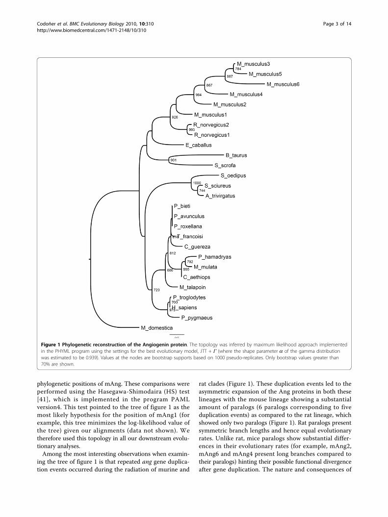
phylogenetic positions of mAng. These comparisons wereperformed using the Hasegawa-Shimodaira (HS) test[41], which is implemented in the program PAMLversion4. This test pointed to the tree of figure 1 as themost likely hypothesis for the position of mAng1 (forexample, this tree minimizes the log-likelihood value ofthe tree) given our alignments (data not shown). Wetherefore used this topology in all our downstream evolu-tionary analyses.Among the most interesting observations when examin-
ing the tree of figure 1 is that repeated ang gene duplica-tion events occurred during the radiation of murine and
rat clades (Figure 1). These duplication events led to theasymmetric expansion of the Ang proteins in both theselineages with the mouse lineage showing a substantialamount of paralogs (6 paralogs corresponding to fiveduplication events) as compared to the rat lineage, whichshowed only two paralogs (Figure 1). Rat paralogs presentsymmetric branch lengths and hence equal evolutionaryrates. Unlike rat, mice paralogs show substantial differ-ences in their evolutionary rates (for example, mAng2,mAng6 and mAng4 present long branches compared totheir paralogs) hinting their possible functional divergenceafter gene duplication. The nature and consequences of
Figure 1 Phylogenetic reconstruction of the Angiogenin protein. The topology was inferred by maximum likelihood approach implementedin the PHYML program using the settings for the best evolutionary model, JTT + Γ (where the shape parameter a of the gamma distributionwas estimated to be 0.939). Values at the nodes are bootstrap supports based on 1000 pseudo-replicates. Only bootstrap values greater than70% are shown.
Codoñer et al. BMC Evolutionary Biology 2010, 10:310http://www.biomedcentral.com/1471-2148/10/310
Page 3 of 14
this functional divergence are elusive and more analysesare needed to determine whether such divergence led toneo-functionalisation or sub-functinalisation of the para-log copies. Mice present large effective population sizes incomparison to human and hence the probability for neo-functionalisation in mice is greater than in humans. Theo-retical and population genetics data predict that in largepopulations strong constraints act against slightly deleter-ious mutations, hindering the subsequent fixation of com-pensatory mutations, and consequently the probability ofsub-functionalisation is lower than that of neo-functionali-sation [42]. Regardless the final outcome, asymmetrybetween mice paralogs point to the fixation of burst ofmutations by adaptive evolution, which may have drivenmAng copies to angiogenin functional diversification. Theasymmetry in mice angiogenin paralogs is substantial,but what is the selective value of this asymmetry? andWhat changes have been essential for mAng functionaldiversification?
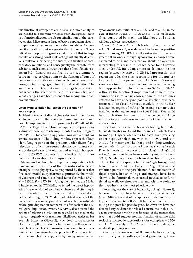
Diversifying selection has driven the evolution ofmAng copiesTo identify events of diversifying selection in the murineangiogenin, we applied the maximum likelihood basedmodels implemented in the program CODEML of thePAML package. In addition, we applied the parsimonysliding window approach implemented in the programSWAPSC. This second approach was convenient forseveral reasons: i) The sliding window approach allowsidentifying regions of the proteins under diversifyingselection, or other non-neutral selective constraints suchas accelerated rates of evolution and mutation hotspots;and ii) SWAPSC accounts for nucleotide bias and fornon-neutral evolution of synonymous sites.Maximum likelihood based approach supported a het-
erogeneous distribution of the intensities of selectionthroughout the phylogeny, as pinpointed by the fact thatfree-ratio model outperformed significantly the modelof Goldman and Yang (Likelihood Ratio Test value LRT =c2 = 115.12, P = 4.77×10⁻⁷). Using the intermediate ModelB implemented in CODEML, we tested the direct hypoth-esis of the evolution of each branch before and after dupli-cation events in mice (branches that were tested areindicated in Figure 2). Model B identified several of thebranches to have undergone different selection constraintsbefore gene duplication compared to after each of the sev-eral gene duplication events. SWAPSC also detected theaction of adaptive evolution in specific branches of thetree convergently with maximum likelihood analyses. Forexample, Branch A (Figure 2), which leads to the ancestorof all the duplication events in the murine lineage, andBranch G, which leads to mAng6, were found to be underpositive selection using both approaches. Positive selectionat these branches was supported by non-synonymous-to-
synonymous rates ratio of ω = 2.3058 and ω = 5.65 in thecase of Branch A and ω = 1.731 and ω = 1.16 for BranchG, as computed by maximum likelihood and slidingwindow analyses, respectively.Branch F (Figure 2), which leads to the ancestor of
mAng3 and mAng5, was detected to be under positiveselection using CODEML as the estimated ω = ∞ wasgreater than one, although synonymous changes wereestimated to be 0 and therefore we should be careful ininterpreting this result. In Branch A we found severalsites under PS, including amino acids spanning theregion between Met30 and Gly34. Importantly, thisregion includes the sites responsible for the nuclearlocalization of the protein [43]. At Branch G severalsites were found to be under positive selection usingboth approaches, including residues Ser52 to Gly62.Although the functional importance of some of theseamino acids is as yet unknown, some of the residuesdetected to have undergone positive selection have beenreported to be close or directly involved in the nuclearlocalization region of mAng (for example amino acidsincluded in the region Arg31 to Leu35) [43]. This couldbe an indication that functional divergence of mAng6was due to positively selected amino acid replacementsat these sites.Inspecting the rest of the branches leading to the dif-
ferent duplicates we found that branch H, which leadsto mAng3 (Figure 2), seems to have been evolvingunder negative selection, with ω = 0.1912 and ω =0.1329 for maximum likelihood and sliding window,respectively. In contrast some branches such as branchD, which leads to the ancestor of mAng3, mAng5 andmAng6, seems to have been evolving neutrally (ω =0.951). Similar results were obtained for branch E (ω =1.015), that corresponds to the mAng4 lineage andbranch I (ω = 0.964), that leads to mAng5. This neutralevolution points to the possible non-functionalization ofthese copies, but as mAng4 and mAng3 have beenshown to be functional, we expected mAng5 to be func-tional as well; we show further analysis that point tothis hypothesis as the most plausible one.Interesting was the case of branch C, mAng2 (Figure 2),
because it seems to have been evolving at the same rate(ω = 0.618) as the rest of the species included in the phy-logenetic analysis (ω = 0.556). It has been described thatmAng2 is a possible pseudo-gene, however we have notdetected any evidence for relaxed constraints in this line-age in comparison with other lineages of the mammaliantree that could suggest neutral fixation of amino acidreplacing nucleotide substitutions (for example ω = 0.556< < 1). Conversely, mAng2 seem to have undergonemoderate purifying selection.Gene’s expression is one of the main factors affecting
evolutionary rates, with expressed genes being those
Codoñer et al. BMC Evolutionary Biology 2010, 10:310http://www.biomedcentral.com/1471-2148/10/310
Page 4 of 14

highly conserved. To account for this when comparingnon-synonymous-to-synonymous rates ratios amongmAng gene copies we investigated the expression ofeach of the copies using codon adaptation index (CAI)as a proxy to gene expression. CAI was calculated usingthe webpage http://www.cbib.u-bordeaux2.fr/pise/cai.html. The values of non-synonymous-to-synonymousnucleotide substitutions are not due to different expres-sion levels of the gene copies because, on average, thedifferent mAng copies presented similar expressionlevels (CAI was estimated to be 0.245, 0.235, 0.232,
0.247, 0.243 and 0.237 for mAng copies 1 to 6, respec-tively). These gene copies also presented similar expres-sion levels to that of hAng (CAI = 0.262). Difference inevolutionary rates therefore was not due to differencesin expression levels among duplicates.The fact that these copies remain in the proteome of
mouse argues against previous studies suggesting non-functionalisation [31]. In addition, all post-duplicationlineages presented similar intensities of selection exceptthe pairs of post-duplication lineages F-G (leading tomAng6 and ancestor of mAng5-mAng3 respectively)
Figure 2 Selective constraints analysis of pre- and post-duplication lineages. We used the branch-site model implemented in the programCODEML from the PAML package version 4.0. Branches labeled are those tested in a search for evidence of constraints different from those ofthe background constraint.
Codoñer et al. BMC Evolutionary Biology 2010, 10:310http://www.biomedcentral.com/1471-2148/10/310
Page 5 of 14
and H-I that lead to mAng5 and mAng3, respectively(Figure 2). The elevated ω values are more consistentwith shifts in the evolutionary rates after gene duplica-tion and with the possible functional divergence of theresulting paralogous copies. In the first pair (F-Glineages), both post-duplication lineages underwentadaptive evolution (for example ω > 1) indicating thepossible functional divergence type II (as defined in[44]). Functional divergence type II involves a change ofthe ancestral amino acid at a particular amino acid siteof the protein after gene duplication. This replacementinvolves the fixation of two different residues in thepost-duplication lineages and their high conservationafter the speciation of each of the copies due to theirdifferent but equally important functional role in eachof the paralogs. Conversely, both post-duplicationlineages in the second pair (H-I) evolved under purifyingselection, although mAng5 presented significantly accel-erated rates of evolution compared to mAng3, indicatingpossible functional divergence type I (as defined by[45]). Unlike functional divergence type II, type Iinvolves the fixation of a function conferring residuemutation in one of the paralogs where it becomes highlyconstrained, while this amino acid sites evolves neutrallyin the other where amino acid replacements occur withno functional consequences.
Co-evolution between residues proximal to functionalregions in AngRelaxed selection is a common phenomenon after geneduplication and it can take place in one or both copiesof the gene because of gene redundancy [46,47]. One ofthe gene copies therefore may accumulate deleteriousmutations while the other copy can remain under strongpurifying selection to preserve the ancestral function.The most expected fate for one of the gene copies isnon-functionalization followed by its disintegrationwithin few million years of evolution depending on theeffective population sizes of the organism [48]. The twocopies of a gene can persist in the genome either if thecombined function of both paralogs performs the ances-tral function (sub-functionalization) or if one copyreproduces the ancestral function while the otherdiverges towards other functions (neo-functionalisation).Survival of a pseudo-gene in the genome for long evolu-tionary periods is very unlikely, and therefore copiesthat remain are likely to be functional. However, evolu-tion of gene copies after duplication can be very com-plex and up to twelve models have been recentlyproposed to account for all possible evolutionary scenar-ios [49]. Based on this assumption, we examinedwhether the mAng gene copies that were kept in thegenome were followed by functional divergence afterduplication. Functional divergence is likely to happen in
two ways: i) classic functional divergence involves theaccumulation of functionally innovative advantageousmutations in one of the gene copies [44,45]; or alterna-tively ii) after gene duplication functionally innovativebut structurally destabilizing mutations may havebecome fixed once they have been compensated for byother mutations (compensatory co-evolution): in a nor-mal physiological background the effect of both twomutations is neutral but the phenotypic advantage ofthe destabilizing mutation may be expressed undernovel environmental conditions.Applying the method of Gu [45] we could not identify
classical functional divergence in any of the consideredclusters. To identify the second type of functionallydivergent mutations we first performed analysis of co-evolution (see Material and methods for details). Theco-evolution method identified several pairs of aminoacids showing correlated changes. Groups of coevolution–with each group including only amino acids that pre-sent correlated evolution with each and all the membersof that group (Table 1)– highlighted several amino acidsites to be correlated in their evolutionary patterns(Table 1). Most of the sites are close (for examplewithin 4Å) to essential amino acids of the active site(His13, Lys40 and His114), or to the binding sites, or tothe domain responsible for the nuclear translocation(Arg31-Leu35). These proximities support the possiblecompensatory relationship between such amino acidsites because their proximity to important functionalregions makes it likely that mutations at these sites canhave deleterious effects. The next question we askedwas whether these constraints have undergone substan-tial changes after gene duplication. To answer this ques-tion and the hypothesis of compensatory effects weanalysed the distribution of co-evolving pairs of aminoacid sites in the protein structure and tested theirproximities.
Detection of compensatory mutationsIn order to understand the relationships between co-evolving amino acids in Ang, we plotted these in thecrystal structure of hAng and asked whether pairs of co-evolving residues presented evidence of interaction withone another. In spite of the fact that mAng1 andmAng4 have been isolated and crystallized, we usedhAng as a reference structure due to the medical rele-vance of this protein for humans and because it wasidentical to mAng. All other mouse Ang structurecopies have been synthetically modified from themAng1 in previous studies. The procedure utilized toanswer this question consisted in determining whetherthe pair of co-evolving residues was located within 4Åfrom each other, being indicative of their possible func-tional or structural interactions. Alternatively, for those
Codoñer et al. BMC Evolutionary Biology 2010, 10:310http://www.biomedcentral.com/1471-2148/10/310
Page 6 of 14
distantly located amino acids (presenting a distancefrom one another greater than 4Å), we asked whetherthey were contacting common amino acids that showedhighly conserved evolutionary pattern (see material andmethods for details). Many of the co-evolving aminoacids presented distances greater than 4Å (Figure 3).Importantly, most of these amino acids were proximalto residues that showed a significantly conserved evolu-tionary pattern compared to the rest of the alignment(pairwise Poisson distances in the lower 99% tail of thedistances distribution, see methods for details). Thismethod was used previously with significant success toidentify compensatory relationships between mutations[50]. The highly conserved sites identified nearby co-evolving residues are close to the sites responsible forthe ribonuclease activity and to those involved in thetranslocation of the protein to the nucleus.After examining the different co-evolution groups we
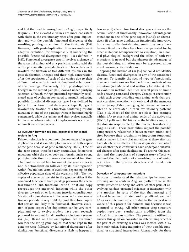
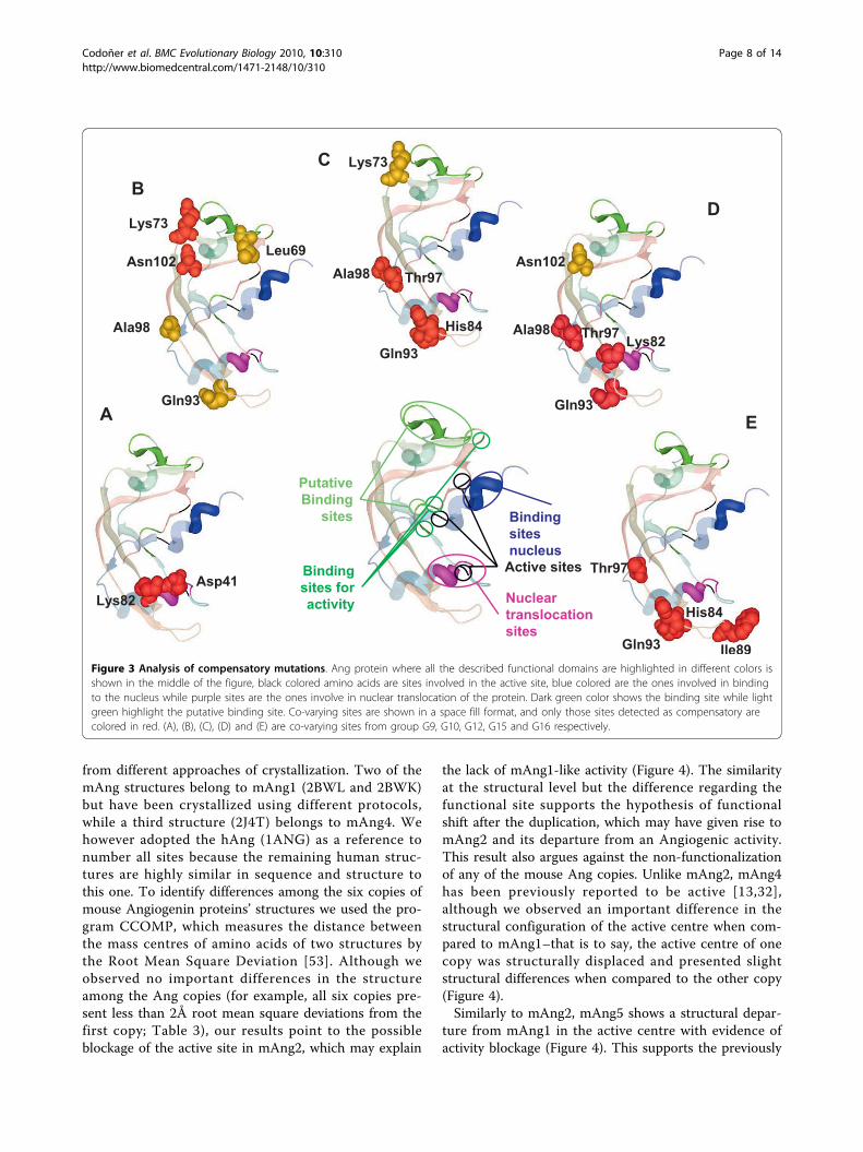
found that groups G9, G10, G12, G15 and G16 (Table 1)presented pairs of amino acids with strong evidence fortheir compensatory effects–that is to say they fall within4 Å of each other in the protein structure and are there-fore likely to present interacting effects. The pair ofamino acid sites Asp41-Lys82 (Figure 3A), classifiedwithin co-evolution group G9 (Table 2), presented evi-dence of compensatory effects. Asp41 and Lys82 areinvolved in dimerization of angiogenine and are closelylocated to amino acid regions that interact with inhibitors
and the catalytic centre (Table 2). Another example ofpossible compensatory interaction is that presented bythe pair of amino acid sites Lys73-Asn102 (Figure 3B)that are classified within group G10 (Table 2). Impor-tantly, this pair of amino acids is located structurallyclose to the putative binding site (Figure 3). His84-Gln93and His84-Thr97 where detected as compensatory andthey are in two co-evolutionary groups, G12 (Figure 3Cand Table 2) and G16 (Figure 3E and Table 2). Thesesites are close to catalytic sites and are both involved inbinding the inhibitor of angiogenin. The pair His84-Ile89was also found as a coadaptation pair in G16 (Figure 3Eand Table 2). Another pair Thr97-Ala98, which has beenconsistently detected to co-evolve in many of the groups,also presented evidence for a compensatory relationshipin G12 (Figure 3C and Table 2) and G15 (Figure 3D andTable 2). Thr97 and Ala98 are involved in angiogenindimerization (Table 2). In G15 (Figure 3D and Table 2)we also found other compensatory mutations Lys82-Gln93 and Lys82-Thr97. All compensatory mutationshave been highlighted in red in Figure 3. It is interestingto notice that, although many of the sites detected to beunder adaptive evolution fall within the same domains ofthose co-evolving (for example, sites 30 to 35 which arewithin the nuclear localization signal peptide), there wasno match between these two sets of sites. One possiblereason may be purely methodological because both selec-tion and functional divergence analyses have been per-formed in a qualitatively different manner. In selectionanalyses we focused the detection of adaptation on parti-cular lineages of the tree. Conversely, in co-evolution theentire tree was used which makes it more difficult toidentify selection at co-evolving amino acid sites: pairs ofamino acids that changed in a correlated way in fewlineages may have undergone strong purifying selectionin most of the remaining lineages of the tree. This wouldimply that on average these sites would be under strongnegative selection most of the time alternating withpunctual episodic adaptive evolution, which would beunlikely to be detected by actual selection methods. Weconsider therefore both the selection methods and co-evolutionary analyses to be complementary approachesto identify adaptive evolutionary events.
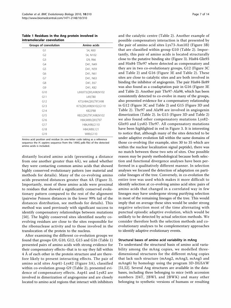
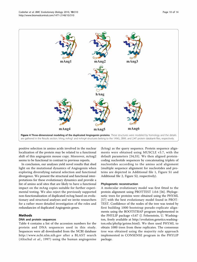
Structural bases of amino acid variability in mAngTo understand the structural basis of amino acid varia-bility among the mAng copies, we modelled three-dimensional structures for the different mAng copiesthat lack such structure (mAng2, mAng3, mAng5 andmAng6) by homology using the program 3D-JIGSAW[51,52]. Several Ang structures are available in the data-bases, including three belonging to mice (with accessionnumbers 2J4T, 2BWL and 2BWK) and many otherbelonging to synthetic versions of humans or resulting
Table 1 Residues in the Ang protein involved inintramolecular coevolution
Groups of coevolution Amino acids
G1 S4, K60
G2 S4, N102
G3 I29, R66
G4 D41, N49
G5 D41, N59
G6 D41, N61
G7 D41, N63
G8 D41, E67
G9 D41, K82
G10 L69,K73,Q93,A98,N102
G11 L69,T80
G12 K73,H84,Q93,T97,A98
G13 K73,Q93,A98,N102,G110
G14 K82,P88
G15 K82,Q93,T97,A98,N102
G16 H84,W89,Q93,T97
G17 H84,A98,G110
G18 H84,W89,121
G19 W89,G110
Amino acid position and residue (in one-letter code taking as a referencesequence the H. sapiens sequence from the 1ANG pdb file) of the detectedamino acids is included.
Codoñer et al. BMC Evolutionary Biology 2010, 10:310http://www.biomedcentral.com/1471-2148/10/310
Page 7 of 14
from different approaches of crystallization. Two of themAng structures belong to mAng1 (2BWL and 2BWK)but have been crystallized using different protocols,while a third structure (2J4T) belongs to mAng4. Wehowever adopted the hAng (1ANG) as a reference tonumber all sites because the remaining human struc-tures are highly similar in sequence and structure tothis one. To identify differences among the six copies ofmouse Angiogenin proteins’ structures we used the pro-gram CCOMP, which measures the distance betweenthe mass centres of amino acids of two structures bythe Root Mean Square Deviation [53]. Although weobserved no important differences in the structureamong the Ang copies (for example, all six copies pre-sent less than 2Å root mean square deviations from thefirst copy; Table 3), our results point to the possibleblockage of the active site in mAng2, which may explain
the lack of mAng1-like activity (Figure 4). The similarityat the structural level but the difference regarding thefunctional site supports the hypothesis of functionalshift after the duplication, which may have given rise tomAng2 and its departure from an Angiogenic activity.This result also argues against the non-functionalizationof any of the mouse Ang copies. Unlike mAng2, mAng4has been previously reported to be active [13,32],although we observed an important difference in thestructural configuration of the active centre when com-pared to mAng1–that is to say, the active centre of onecopy was structurally displaced and presented slightstructural differences when compared to the other copy(Figure 4).Similarly to mAng2, mAng5 shows a structural depar-
ture from mAng1 in the active centre with evidence ofactivity blockage (Figure 4). This supports the previously
Figure 3 Analysis of compensatory mutations. Ang protein where all the described functional domains are highlighted in different colors isshown in the middle of the figure, black colored amino acids are sites involved in the active site, blue colored are the ones involved in bindingto the nucleus while purple sites are the ones involve in nuclear translocation of the protein. Dark green color shows the binding site while lightgreen highlight the putative binding site. Co-varying sites are shown in a space fill format, and only those sites detected as compensatory arecolored in red. (A), (B), (C), (D) and (E) are co-varying sites from group G9, G10, G12, G15 and G16 respectively.
Codoñer et al. BMC Evolutionary Biology 2010, 10:310http://www.biomedcentral.com/1471-2148/10/310
Page 8 of 14
observed poor angiogenic activity and the suggestedinvolvement of mAng in other processes [31].Finally, our selective constraints analyses show evidence
of adaptive evolution in mAng6. The fact that our struc-tural modelling detects a structurally different active cen-tre in this protein compared to other protein copies wherewe detect adaptive evolution suggests functional diver-gence after the split between mAng6 and the remainingAng copies. Furthermore, intra-molecular co-evolutionaryanalyses show evidence of compensatory mutations eventslocated nearby important functional regions of the Angprotein. For example, Pro18 and Thr36, frequently identi-fied as coevolving with many other residues in the
structure, are probably responsible of the operability of theactive site as its location suggests its role in maintainingthe proper orientation of sites His13 and Thr44 thatbelong to the active site. The rest of the pairs of coevolu-tion are mostly surrounding the active site–which com-prises amino acids His13, Thr44 and His114. Others areeither included within or surrounding the nuclear peptidesignal (Arg31-Leu35). These results indicate thereforethat, in general, amino acid sites close or includedin important protein domains may have coevolved tomaintain the structural features necessary for the properfunctional activity of Angiogenin. We have also detectedtwo groups of compensatory mutations in mAng afterduplication. The importance of these sites is furtherenhanced by their location in or nearby amino acidsArg31 to Leu35 that have been described to be involved inthe nuclear localization of the protein in mouse [30].
ConclusionsEven though all the duplicates in mouse are different frommAng1, and that nothing has been described for mAng5and mAng6, there is no reason to think that these copiesare non-functional. Remarkably it has been reported thenon-nuclear localization of mAng6. The identification of
Table 2 Identification of functionally important residues using co-evolutionary analyses
Pairs CoevolutionGroup
Conserved Sites (4Å close) Active Site Binding Site PutativeBindingSite
NuclearImplications
D41-K82 G9 H84, Q93, R95 K40, D41, I42, C92 T80, C81, K82, R121 - -
K73-N102 G10, G13 S74, S75, N102 - R101 I56, N63,R70, I71,S72, K73,R101
K82-Q93 G15 D23, H84, Q93, R95, A96 K40, D41, C92 C81, K82 - C26, E27
K82-T97 G15 D23, H84, R95, A96, T97, R122 - T44, T79, C81, K82, F120 - C26
H84-Q93 G12, G16 D23, H84, G86, P91, Q93, R95, A96 C39, K40, D41, S87, C92 C81, K82 - E27, C39
H84-T97 G12, G16 D23, H84, R95, A96, T97 - C81, K82 - -
W89-Q93 G16 T36, G86, W89, P90, P91, Q93 C39, K40, S87, C92 - - -
T97-A98 G12, G15 R21, D22, H47, Q77, R95, A96, T97,A98, G99, F100
- V78, T79, T80, C81 V78 C26
We identify highly conserved amino acid sites (labeled in one-letter code and using as reference the H. Sapiens sequence) that are structurally close to or withinfunctionally important regions of Ang protein and that are close (within 4Å) to coevolving pairs with potential compensatory relationships. In bold we highlightthose co-evolving amino acid positions reported to be involved in dimmer formation of hAng, in Italic we remark positions implicated in the interaction withangiogenin-inhibitor in Human. We finally underscore those positions identified to be important as catalytic sites in hAng, as described in NCBI-IBIS database [78].
Table 3 Root Mean Square Deviation (RMSD) betweenthe modeled structures for murine ANG protein paralogs
Comparison RMSD
mAng1 vs mAng2 1.078
mAng1 vs mAng3 0.772
mAng1 vs mAng4 0.814
mAng1 vs mAng5 0.744
mAng1 vs mAng6 1.038
hAng vs mAng1 1.306
Codoñer et al. BMC Evolutionary Biology 2010, 10:310http://www.biomedcentral.com/1471-2148/10/310
Page 9 of 14
positive selection in amino acids involved in the nuclearlocalization of the protein may be related to a functionalshift of this angiogenin mouse copy. Moreover, mAng2seems to be functional in contrast to previous reports.In conclusion, our analyses yield novel results that shed
light on the mutational dynamics of Angiogenin whenexploring diversifying natural selection and functionaldivergence. We present the structural and functional inter-pretations for these evolutionary dynamics and provide alist of amino acid sites that are likely to have a functionalimpact on the mAng copies suitable for further experi-mental testing. We also reject the previously supportednon-functionalization of duplicated mAng based on evolu-tionary and structural analyses and we invite researchersfor a rather more detailed investigation of the roles andredundancies of duplicated Angiogenin genes.
MethodsDNA and protein sequencesTable 4 contains a list of the accession numbers for theprotein and DNA sequences used in this study.Sequences were all downloaded from the NCBI databasehttp://www.ncbi.nlm.nih.gov after a BLAST search(Altschul et al., 1997) using the human angiogenine
(hAng) as the query sequence. Protein sequence align-ments were obtained using MUSCLE v3.7, with thedefault parameters [54,55]. We then aligned protein-coding nucleotide sequences by concatenating triplets ofnucleotides according to the amino acid alignment(multiple sequence alignment for nucleotides and pro-teins are depicted in Additional file 1, Figure S1 andAdditional file 2, Figure S2, respectively).
Phylogenetic reconstructionA molecular evolutionary model was first fitted to theprotein alignment using PROTTEST 1.0.6 [56]. Phyloge-netic trees for proteins were obtained using the PHYML[57] with the best evolutionary model found in PROT-TEST. Confidence of the nodes of the tree was tested byfirst building 1000 bootstrap pseudo-replicate align-ments using the BOOTSTRAP program implemented inthe PHYLIP package v3.67 (J. Felsenstein, U. Washing-ton, freely available at http://evolution.genetics.washing-ton.edu/phylip/getme.html). We then used PHYML toobtain 1000 trees from these replicates. The consensustree was obtained using the majority rule approachimplemented in CONSENSE program in the PHYLIPpackage.
Figure 4 Three-dimensional modeling of the duplicated Angiogenin proteins. These structures were modeled by homology and the detailsare gathered in the Results section. hAng, mAng1 and mAng4 structures belong to the 1ANG, 2BWL and 2J4T protein databank files, respectively.
Codoñer et al. BMC Evolutionary Biology 2010, 10:310http://www.biomedcentral.com/1471-2148/10/310
Page 10 of 14
Identification of selective constraintsTo identify the main functional diversifying events inAng during the evolutionary radiation of mammals weanalyzed the change in the dynamics of synonymous(dS) and of non-synonymous (dN) nucleotide replace-ments. In our study we assumed that dS accumulatesneutrally on average since they produce no amino acidreplacements and are therefore not seen by selection.Taking into account this assumption, we estimated theintensity of selection by obtaining the ratio between dNand dS (ω = dN/dS). This ratio has been regarded as themost stringent way to identify selection, with ω = 1, ω< 1 and ω > 1, indicating neutral evolution, purifyingselection and diversifying selection, respectively [58-60].However, caution is required when measuring selectionusing this approach because the stability of RNA mole-cule secondary structure as well as translational selec-tion may impose constraints on synonymous sitesleading to lower dS values and consequently to inflatedω estimates [61-64].To ameliorate the effects of these limitations, we tested
for the presence of diversifying selection following two
main ways. First we used maximum-likelihood models toidentify selective constraints as implemented in the pro-gram CODEML of the PAML package v4.0 [65]. Usingthis approach, we compared a model assuming homoge-nous distribution of selective constraints along the pro-tein and the phylogeny (model M0: one ω value for theentire tree and alignment) to a model assuming an inde-pendent ω for each lineage of the tree [66]. These twonested models (the more complex model includes para-meters of the simple model) were compared by the likeli-hood ratio test (LRT) [67], with twice the differencebetween the log-likelihood values of the two modelsbeing compared to a c2 distribution with as many degreesof freedom as number of branches in the tree -1. Secondwe used a parsimony-based approach robust to devia-tions from the assumption of neutrality of synonymoussubstitutions. This parsimony approach was based on thesliding window procedure previously published [68] andis implemented in the program SWAPSC version 1.0[69]. This program uses a statistically optimized windowsize to detect selective constraints in specific codonregions of the given alignment at a particular branch ofthe phylogenetic tree that show the evolutionary historyof the sequences under study [68].Briefly, SWAPSC estimates the expected distribution
of dS and dN by Li’s method [70] from simulated align-ments and assuming a Poisson distribution of substitu-tions. A statistically optimum windows size is thenestimated that makes the detection of adaptive evolutionindependent of the windows size. The empirical valuesof dS and dN obtained by using the optimal window sizeare contrasted with the expected distributions, and sev-eral hypotheses regarding the selective constraints actingon codon regions are tested. We obtained the simulatedalignments needed for the analysis with the EVOLVERprogram implemented in the PAML package version4.0, with the parameters estimated from the truesequence alignment after running the most appropriatedcodon based model in PAML. Finally, we consideredonly regions and branches detected under adaptive evo-lution by those approaches as the true positive results.
Detection of intra-molecular co-evolutionTo test for intra-molecular coevolution, we used arecently developed parametric model [71] implementedin the program CAPS v1 [72]. The sensitivity of CAPSto identify coevolution between pairs of amino acidsites that are functionally linked has been shown tooutperform other methods based on mutual informa-tion content or on other models of coevolution [71].We considered therefore the method to be appropriatefor an accurate detection of co-evolution. This methodhas been applied in numerous case studies similar tothe one here conducted [50,71,73,74].
Table 4 Accession numbers for the DNA and proteinsequences of the Angiogenin protein used in the analysis
Species Protein DNA
Mus musculus1 NP_031473 NM_007447.2
Mus musculus2 NP_031475 NM_007449.2
Mus musculus3 AAC05794 U72672
Mus musculus4 NP_808212 NM_177544
Mus musculus5 AAV87188 AY665820
Mus musculus6 AAV87189 AY665821
Rattus norvegicus1 NP_001006993 NM_001006992.1
Rattus norvegicus2 NP_001012359 NM_001012359.1
Homo sapiens NP_001136 NM_001145.2
Trachypithecus francoisi AAO41336 AY221129
Pygathrix avunculus AAO41339 AY221132
Pygathrix bieti AAO41338 AY221131
Pygathrix roxellana AAO41337 AY221130
Pongo pygmaeus AAL61645 AF441663.1
Chlorocebus aethiops AAL61646 AF441664
Sus scrofa NP_001038038 NM_001044573
Miopithecus talapoin AAL61647 AF441665
Pan troglodytes NP_001009159 NM_001009159
Macaca mulatta AAL61649 AF441667
Equs caballus NP_001075368 NM_001081899
Saguinus oedipus AAL61650 AF441668
Bos Taurus NP_001071612 NM_001078144
Saimiri sciureus AAL61652 AF441670
Aotus trivirgatus AAL61651 AF441669
Papio hamadryas AAL61648 AF441666
Colobus guereza AAO41335 AY221128
Monodelphis domestica XP_001379328 XM_001379291
Codoñer et al. BMC Evolutionary Biology 2010, 10:310http://www.biomedcentral.com/1471-2148/10/310
Page 11 of 14
Briefly, CAPS compares the correlated variance of theevolutionary rates at 2 amino acid sites in a proteinalignment, corrected by the time since the divergence ofthe 2 sequences they belong to. The algorithm estimatesthe synonymous nucleotide pairwise sequence diver-gence as a proxy for their divergence time. This methodcompares the amino acid transition probability scoresbetween 2 sequences at 2 particular sites, using theblocks substitution matrix [75]. The significance of theCAPS correlation values was assessed by randomly pair-ing sites of the alignment and building a distribution ofcorrelation coefficients for 1,000,000 randomly pairedsites against which we compared real correlation values.To correct for multiple tests and data non-independenceCAPS performs a step-down permutation procedure[76] and corrects the probabilities for the correlationcoefficients of co-evolving pairs of sites accordingly [72].For co-evolution analyses we used the protein-coding
sequence of Ang and minimized type I error using aconfidence value of 0.01. The structural PDB file forhANG (1ang, [16]) was used to identify the co-evolvingamino acid positions in the structure (for example, allthe amino acid positions in this study refer to their loca-tion in the hAng three-dimensional structure).Molecular co-evolution between amino acids can be
the result of their structural, functional, interaction, phy-logenetic, or stochastic link [77]. Disentangling the dif-ferent types of coevolution is a difficult task, although aphylogenetic approach has been suggested as a feasibleway to remove amino acids covariation due to stochasticnoise [71]. Distinguishing between structural, functional,and interaction co-evolution requires biological informa-tion in addition to the mathematical adjustments madeby the method. Accordingly, we used correlated varia-tion in the physico-chemical properties of the aminoacids as a further filter to our co-evolutionary analyses.
Identifying Compensatory Mutational Dynamics inAngiogeninEach one of the amino acid sites identified as coevolvingwas plotted in the crystal structure of the protein andthe Euclidean distance between them was calculated.We calculated this distance as the average distancebetween the atoms of the amino acid sites as follows:
dNK
X X Y Y Z Zi j i j i j
j
K
i
N
= − + − + −==
∑∑1 2 2 2
11
( ) ( ) ( )
Here N is the number of atoms in amino acid i while Kis that number in amino acid j. X, Y and Z represent thethree-dimensional coordinates of the atoms correspondingto each of the amino acids. We considered two aminoacids to contact each other when the distance betweentheir closest atoms was equal or less than 4Å.
Two mutations were considered to have compensatedeach other if, in addition to presenting the same phylo-genetic pattern (be coevolving), they were located within4Å from each other in the protein crystal structure.Also, two amino acid sites can compensate each otherindirectly. For example, if site “A” and site “B” are atmore than 8Å distance but are surrounding (within 4Å)an important functional site “C”, then changes at site“A” may affect site “C” which has to be compensated bychanges at site “B”. We also considered these cases tobe in support of a compensatory relationship betweensites “A” and “B”. However, caution must be taken inmaking such assumptions because close amino acids,even though are likely to influence one another, maynot have a compensatory relationship. Conversely,amino acid sites distantly located in the protein struc-ture may have indirect compensatory effects upon oneanother. The other limitation of this approach is thatproteins can undergo dramatic conformational changesduring their interactions with other proteins, which isnot reflected in the crystal (static) structure of proteins.Under these circumstances, amino acids that are dis-tantly located may interact and hence influence oneanother. Nonetheless, we adopted the very conservativeview that amino acids interacting in our crystal struc-tures are true interactors at the particular conditionsunder which the protein was crystallized. To identifycompensatory relationships under our assumptionsbetween amino acids at distances greater than 4Å, wesearched for sites contacting both covarying amino acidsites in the structure showing very low divergence levelsin comparison with the rest of the molecule. We mea-sured divergence levels per site by estimating the Pois-son amino acid distances for each amino acid site in themultiple sequence alignments. The level of divergencewas compared to the distribution of divergence levelsbuilt using a pseudo-random sample of 1,000,000 aminoacid site columns sampled with replacement from thealignment (one site could be sampled more than once).
Three-dimensional analysis in Angiogenin3D-JIGSAW program [51,52] was used to model the 3Dstructure of the different duplicates. To identify differ-ences among the six copies of mouse Angiogenin pro-teins’ structures we used the program CCOMP [53],that measures the Mean Root Square Deviation betweenthe different structures.
Additional material
Additional file 1: Figure S1. Multiple sequence alignment fornucleotides and proteins
Additional file 2: Figure S2. Multiple sequence alignment fornucleotides and proteins
Codoñer et al. BMC Evolutionary Biology 2010, 10:310http://www.biomedcentral.com/1471-2148/10/310
Page 12 of 14
AcknowledgementsThis work was supported by Science Foundation Ireland to M.A.F (04/YI1/M518). F.M.C. was supported by Marie Curie European Reintegration GrantFP7 actions (238885). S.A.L. is supported by a FPI PhD grant from theSpanish Ministerio de Ciencia y Tecnologia.
Author details1Evolutionary Genetics and Bioinformatics Laboratory, Department ofGenetics, Smurfit Institute of Genetics, University of Dublin, Trinity College,Dublin, Ireland. 2IrsiCaixa, Laboratori de Retrovirologia, Hospital UniversitariGermans Trias i Pujol, Ctra. Canyet s/n 08916 Badalona Spain. 3Departmentof Cellular Pathology, Centro de Investigaciones Principe Felipe, Avda.Autopista del Saler, 16-3 (junto Oceanográfico), 46012 Valencia Spain.4Laboratory of Integrative and Systems Biology, Instituto de BiologiaMolecular y Celular de Plantas (CSIC- Universidad Politécnica de Valencia(UPV)), Valencia, Spain.
Authors’ contributionsMAF and FMC conceived the idea in collaboration with SAL. FMC and MAFdid the analyses. FMC drafted the manuscript and MAF wrote the finalversion of the manuscript. All authors read and approved the final version ofthe manuscript.
Received: 28 May 2010 Accepted: 15 October 2010Published: 15 October 2010
References1. Strydom DJ, Fett JW, Lobb RR, Alderman EM, Bethune JL, Riordan JF,
Vallee BL: Amino acid sequence of human tumor derived angiogenin.Biochemistry 1985, 24(20):5486-5494.
2. Beintema JJ, Breukelman HJ, Carsana A, Furia A: Evolution of vertebrates.Ribonucleases: Structure and Function New York: Academic Press New York1997.
3. Adams SA, Subramanian V: The angiogenins: an emerging family ofribonuclease related proteins with diverse cellular functions. Angiogenesis1999, 3(3):189-199.
4. Riordan JF: Structure and function of angiogenin. Ribonucleases: Structureand Function New York: Academic Press New York 1997.
5. Tello-Montoliu A, Patel JV, Lip GY: Angiogenin: a review of thepathophysiology and potential clinical applications. J Thromb Haemost2006, 4(9):1864-1874.
6. Fett JW, Olson KA, Rybak SM: A monoclonal antibody to humanangiogenin. Inhibition of ribonucleolytic and angiogenic activities andlocalization of the antigenic epitope. Biochemistry 1994, 33(18):5421-5427.
7. Olson KA, French TC, Vallee BL, Fett JW: A monoclonal antibody to humanangiogenin suppresses tumor growth in athymic mice. Cancer Res 1994,54(17):4576-4579.
8. Olson KA, Fett JW, French TC, Key ME, Vallee BL: Angiogenin antagonistsprevent tumor growth in vivo. Proc Natl Acad Sci USA 1995, 92(2):442-446.
9. Olson KA, Byers HR, Key ME, Fett JW: Prevention of human prostate tumormetastasis in athymic mice by antisense targeting of humanangiogenin. Clin Cancer Res 2001, 7(11):3598-3605.
10. Piccoli R, Olson KA, Vallee BL, Fett JW: Chimeric anti-angiogenin antibodycAb 26-2F inhibits the formation of human breast cancer xenografts inathymic mice. Proc Natl Acad Sci USA 1998, 95(8):4579-4583.
11. Kao RY, Jenkins JL, Olson KA, Key ME, Fett JW, Shapiro R: A small-moleculeinhibitor of the ribonucleolytic activity of human angiogenin thatpossesses antitumor activity. Proc Natl Acad Sci USA 2002,99(15):10066-10071.
12. Greenway MJ, Andersen PM, Russ C, Ennis S, Cashman S, Donaghy C,Patterson V, Swingler R, Kieran D, Prehn J, et al: ANG mutations segregatewith familial and ‘sporadic’ amyotrophic lateral sclerosis. Nat Genet 2006,38(4):411-413.
13. Crabtree B, Thiyagarajan N, Prior SH, Wilson P, Iyer S, Ferns T, Shapiro R,Brew K, Subramanian V, Acharya KR: Characterization of humanangiogenin variants implicated in amyotrophic lateral sclerosis.Biochemistry 2007, 46(42):11810-11818.
14. Wu D, Yu W, Kishikawa H, Folkerth RD, Iafrate AJ, Shen Y, Xin W, Sims K,Hu GF: Angiogenin loss-of-function mutations in amyotrophic lateralsclerosis. Ann Neurol 2007, 62(6):609-617.
15. Fett JW, Strydom DJ, Lobb RR, Alderman EM, Bethune JL, Riordan JF,Vallee BL: Isolation and characterization of angiogenin, an angiogenicprotein from human carcinoma cells. Biochemistry 1985, 24(20):5480-5486.
16. Acharya KR, Shapiro R, Allen SC, Riordan JF, Vallee BL: Crystal structure ofhuman angiogenin reveals the structural basis for its functionaldivergence from ribonuclease. Proc Natl Acad Sci USA 1994,91(8):2915-2919.
17. Shapiro R, Riordan JF, Vallee BL: Characteristic ribonucleolytic activity ofhuman angiogenin. Biochemistry 1986, 25(12):3527-3532.
18. Shapiro R, Strydom DJ, Weremowicz S, Vallee BL: Sites of modification ofhuman angiogenin by bromoacetate at pH 5.5. Biochem Biophys ResCommun 1988, 156(1):530-536.
19. Harper JW, Vallee BL: A covalent angiogenin/ribonuclease hybrid with afourth disulfide bond generated by regional mutagenesis. Biochemistry1989, 28(4):1875-1884.
20. Russo N, Shapiro R, Acharya KR, Riordan JF, Vallee BL: Role of glutamine-117 in the ribonucleolytic activity of human angiogenin. Proc Natl AcadSci USA 1994, 91(8):2920-2924.
21. Shapiro R: Structural features that determine the enzymatic potency andspecificity of human angiogenin: threonine-80 and residues 58-70 and116-123. Biochemistry 1998, 37(19):6847-6856.
22. Holloway DE, Chavali GB, Hares MC, Baker MD, Subbarao GV, Shapiro R,Acharya KR: Crystallographic studies on structural features thatdetermine the enzymatic specificity and potency of human angiogenin:Thr44, Thr80, and residues 38-41. Biochemistry 2004, 43(5):1230-1241.
23. Leonidas DD, Shapiro R, Subbarao GV, Russo A, Acharya KR:Crystallographic studies on the role of the C-terminal segment ofhuman angiogenin in defining enzymatic potency. Biochemistry 2002,41(8):2552-2562.
24. Shapiro R, Fox EA, Riordan JF: Role of lysines in human angiogenin:chemical modification and site-directed mutagenesis. Biochemistry 1989,28(4):1726-1732.
25. Shapiro R, Vallee BL: Site-directed mutagenesis of histidine-13 andhistidine-114 of human angiogenin. Alanine derivatives inhibitangiogenin-induced angiogenesis. Biochemistry 1989, 28(18):7401-7408.
26. Curran TP, Shapiro R, Riordan JF: Alteration of the enzymatic specificity ofhuman angiogenin by site-directed mutagenesis. Biochemistry 1993,32(9):2307-2313.
27. Russo N, Acharya KR, Vallee BL, Shapiro R: A combined kinetic andmodeling study of the catalytic center subsites of human angiogenin.Proc Natl Acad Sci USA 1996, 93(2):804-808.
28. Hallahan TW, Shapiro R, Vallee BL: Dual site model for the organogenicactivity of angiogenin. Proc Natl Acad Sci USA 1991, 88(6):2222-2226.
29. Hallahan TW, Shapiro R, Strydom DJ, Vallee BL: Importance of asparagine-61 and asparagine-109 to the angiogenic activity of human angiogenin.Biochemistry 1992, 31(34):8022-8029.
30. Moroianu J, Riordan JF: Identification of the nucleolar targeting signal ofhuman angiogenin. Biochem Biophys Res Commun 1994, 203(3):1765-1772.
31. Nobile V, Vallee BL, Shapiro R: Characterization of mouse angiogenin-related protein: implications for functional studies on angiogenin. ProcNatl Acad Sci USA 1996, 93(9):4331-4335.
32. Fu X, Roberts WG, Nobile V, Shapiro R, Kamps MP: mAngiogenin-3, atarget gene of oncoprotein E2a-Pbx1, encodes a new angiogenicmember of the angiogenin family. Growth Factors 1999, 17(2):125-137.
33. Crabtree B, Holloway DE, Baker MD, Acharya KR, Subramanian V: Biologicaland structural features of murine angiogenin-4, an angiogenic protein.Biochemistry 2007, 46(9):2431-2443.
34. Brown WE, Nobile V, Subramanian V, Shapiro R: The mouse angiogeningene family: structures of an angiogenin-related protein gene and twopseudogenes. Genomics 1995, 29(1):200-206.
35. Strydom DJ: The angiogenins. Cell Mol Life Sci 1998, 54(8):811-824.36. Singhania NA, Dyer KD, Zhang J, Deming MS, Bonville CA,
Domachowske JB, Rosenberg HF: Rapid evolution of the ribonuclease Asuperfamily: adaptive expansion of independent gene clusters in ratsand mice. J Mol Evol 1999, 49(6):721-728.
37. Cho S, Beintema JJ, Zhang J: The ribonuclease A superfamily of mammalsand birds: identifying new members and tracing evolutionary histories.Genomics 2005, 85(2):208-220.
38. Osorio DS, Antunes A, Ramos MJ: Structural and functional implications ofpositive selection at the primate angiogenin gene. BMC Evol Biol 2007, 7:167.
Codoñer et al. BMC Evolutionary Biology 2010, 10:310http://www.biomedcentral.com/1471-2148/10/310
Page 13 of 14
39. Zhang J, Rosenberg HF: Diversifying selection of the tumor-growthpromoter angiogenin in primate evolution. Mol Biol Evol 2002,19(4):438-445.
40. Wildman DE, Uddin M, Liu G, Grossman LI, Goodman M: Implications ofnatural selection in shaping 99.4% nonsynonymous DNA identitybetween humans and chimpanzees: enlarging genus Homo. Proc NatlAcad Sci USA 2003, 100(12):7181-7188.
41. Shimodaira H, Hasegawa M: Multiple comparisons of log-likelihoods withapplications to phylogenetic inference. Mol Biol Evol 1999, 16:1114-1116.
42. Lynch M, O’Hely M, Walsh B, Force A: The probability of preservation of anewly arisen gene duplicate. Genetics 2001, 159(4):1789-1804.
43. Moroianu J, Riordan JF: Nuclear translocation of angiogenin inproliferating endothelial cells is essential to its angiogenic activity. ProcNatl Acad Sci USA 1994, 91(5):1677-1681.
44. Gu X: Maximum-likelihood approach for gene family evolution underfunctional divergence. Mol Biol Evol 2001, 18(4):453-464.
45. Gu X: Statistical methods for testing functional divergence after geneduplication. Mol Biol Evol 1999, 16(12):1664-1674.
46. Ohno S: Evolution by Gene duplication. Berlin: Springer-Verlag 1970.47. Nowak MA, Boerlijst MC, Cooke J, Smith JM: Evolution of genetic
redundancy. Nature 1997, 388(6638):167-171.48. Lynch M, Conery JS: The evolutionary fate and consequences of
duplicate genes. Science 2000, 290(5494):1151-1155.49. Innan H, Kondrashov F: The evolution of gene duplication: classifying and
distinguishing between models. Nat Rev Genet 2010, 11:97-108.50. Tully DC, Fares MA: Shifts in the selection-drift balance drive the
evolution and epidemiology of foot-and-mouth disease virus. J Virol2009, 83(2):781-790.
51. Bates PA, Sternberg MJ: Model building by comparison at CASP3: usingexpert knowledge and computer automation. Proteins 1999, 3:47-54.
52. Contreras-Moreira B, Bates PA: Domain fishing: a first step in proteincomparative modelling. Bioinformatics 2002, 18(8):1141-1142.
53. Sicinska W, Rotkiewicz P: Computational analysis of the active sites inbinary and ternary complexes of the vitamin D receptor. J SteroidBiochem Mol Biol 2007, 103(3-5):305-309.
54. Edgar RC: MUSCLE: a multiple sequence alignment method with reducedtime and space complexity. BMC Bioinformatics 2004, 5:113.
55. Edgar RC: MUSCLE: multiple sequence alignment with high accuracy andhigh throughput. Nucleic Acids Res 2004, 32(5):1792-1797.
56. Abascal F, Zardoya R, Posada D: ProtTest: selection of best-fit models ofprotein evolution. Bioinformatics 2005, 21(9):2104-2105.
57. Guindon S, Gascuel O: A simple, fast, and accurate algorithm to estimatelarge phylogenies by maximum likelihood. Syst Biol 2003, 52(5):696-704.
58. Akashi H: Inferring the fitness effects of DNA mutations frompolymorphism and divergence data: statistical power to detectdirectional selection under stationarity and free recombination. Genetics1999, 151(1):221-238.
59. Crandall KA, Kelsey CR, Imamichi H, Lane HC, Salzman NP: Parallelevolution of drug resistance in HIV: failure of nonsynonymous/synonymous substitution rate ratio to detect selection. Mol Biol Evol1999, 16(3):372-382.
60. Sharp PM: In search of molecular darwinism. Nature 1997,385(6612):111-112.
61. Chamary JV, Parmley JL, Hurst LD: Hearing silence: non-neutral evolutionat synonymous sites in mammals. Nat Rev Genet 2006, 7(2):98-108.
62. Mayrose I, Doron-Faigenboim A, Bacharach E, Pupko T: Towards realisticcodon models: among site variability and dependency of synonymousand non-synonymous rates. Bioinformatics 2007, 23(13):i319-327.
63. Parmley JL, Chamary JV, Hurst LD: Evidence for purifying selection againstsynonymous mutations in mammalian exonic splicing enhancers. MolBiol Evol 2006, 23(2):301-309.
64. Resch AM, Carmel L, Marino-Ramirez L, Ogurtsov AY, Shabalina SA,Rogozin IB, Koonin EV: Widespread positive selection in synonymous sitesof mammalian genes. Mol Biol Evol 2007, 24(8):1821-1831.
65. Yang Z: PAML 4: phylogenetic analysis by maximum likelihood. Mol BiolEvol 2007, 24(8):1586-1591.
66. Goldman N: Variance to mean ratio, R(t), for poisson processes onphylogenetic trees. Mol Phylogenet Evol 1994, 3(3):230-239.
67. Yang Z: Statistical properties of a DNA sample under the finite-sitesmodel. Genetics 1996, 144(4):1941-1950.
68. Fares MA, Elena SF, Ortiz J, Moya A, Barrio E: A sliding window-basedmethod to detect selective constraints in protein-coding genes and itsapplication to RNA viruses. J Mol Evol 2002, 55(5):509-521.
69. Fares MA: SWAPSC: sliding window analysis procedure to detectselective constraints. Bioinformatics 2004, 20(16):2867-2868.
70. Li WH: Unbiased estimation of the rates of synonymous andnonsynonymous substitution. J Mol Evol 1993, 36(1):96-99.
71. Fares MA, Travers SA: A novel method for detecting intramolecularcoevolution: adding a further dimension to selective constraintsanalyses. Genetics 2006, 173(1):9-23.
72. Fares MA, McNally D: CAPS: coevolution analysis using proteinsequences. Bioinformatics 2006, 22(22):2821-2822.
73. Travers SA, Fares MA: Functional coevolutionary networks of the Hsp70-Hop-Hsp90 system revealed through computational analyses. Mol BiolEvol 2007, 24(4):1032-1044.
74. Travers SA, Tully DC, McCormack GP, Fares MA: A study of thecoevolutionary patterns operating within the env gene of the HIV-1group M subtypes. Mol Biol Evol 2007, 24(12):2787-2801.
75. Henikoff S, Henikoff JG: Amino acid substitution matrices from proteinblocks. Proc Natl Acad Sci USA 1992, 89(22):10915-10919.
76. Westfall P, Young S: Resampling-based multiple testing. New York: JohnWiley & Sons 1993.
77. Atchley WR, Wollenberg KR, Fitch WM, Terhalle W, Dress AW: Correlationsamong amino acid sites in bHLH protein domains: an informationtheoretic analysis. Mol Biol Evol 2000, 17(1):164-178.
78. Shoemaker BA, Zhang D, Thangudu RR, Tyagi M, Fong JH, Marchler-Bauer A, Bryant SH, Madej T, Panchenko AR: Inferred BiomolecularInteraction Server–a web server to analyze and predict proteininteracting partners and binding sites. Nucleic Acids Res 2010,38(D):518-24.
doi:10.1186/1471-2148-10-310Cite this article as: Codoñer et al.: Mutational dynamics of murineangiogenin duplicates. BMC Evolutionary Biology 2010 10:310.
Submit your next manuscript to BioMed Centraland take full advantage of:
• Convenient online submission
• Thorough peer review
• No space constraints or color figure charges
• Immediate publication on acceptance
• Inclusion in PubMed, CAS, Scopus and Google Scholar
• Research which is freely available for redistribution
Submit your manuscript at www.biomedcentral.com/submit
Codoñer et al. BMC Evolutionary Biology 2010, 10:310http://www.biomedcentral.com/1471-2148/10/310
Page 14 of 14