Multi-User TAMU Facilities: · Web viewOur laboratory develops multi-scale, multi-modal,...
37
QUICK LINKS TO NAVIGATE DEPARTMENT FACILITIES MULTI-USER FACILITIES OTHER FACILITIES BIOMEDICAL ENGINEERING DEPARTMENT FACILITIES BIOMEDICAL ENGINEERING INDIVIDUAL FACULTY RESEARCH LABORATORIES (https://engineering.tamu.edu/biomedical/research/labs) Advanced Spectroscopy Laboratory Contact: Vladislav Yakovlev Dr. Vladislav V. Yakovlev is professor in the Department of Biomedical Engineering at Texas A&M University and Fellow of the Optical Society of America, the American Institute of Medical and Biological Engineering and the International Society for Optics and Photonics. His research focuses on the development of new instrumentation for biomedical diagnostics and imaging. Dr. Yakovlev’s primary research interests include biomechanics on a microscale level; nanoscopic optical imaging of molecular and cellular structures; protein spectroscopy and structural dynamics; bioanalytical applications of optical technology and spectroscopy; and deep-tissue imaging and sensing. Bioinspired Translational Microsystems Contact: Abhishek Jain Development of patient-specific and physiologically-relevant diagnostics and therapeutics has been a long-standing clinically unmet need. Our lab aspires to develop smart analytical medical devices for making long-lasting impact in improving patient care and reducing healthcare costs. To achieve these goals, we are developing several student-driven projects that leverage the organs-on-a-chip technology. We are uniquely more focused on building physiologically-relevant in vitro models to solve the most demanding problems that arise in human blood vessels and their surrounding environment (extracellular matrix). By applying the biophysics and transport phenomena of whole blood within organs-on-chip, we aim to reproduce pathophysiological
Transcript of Multi-User TAMU Facilities: · Web viewOur laboratory develops multi-scale, multi-modal,...

QUICK LINKS TO NAVIGATEDEPARTMENT FACILITIESMULTI-USER FACILITIESOTHER FACILITIES
BIOMEDICAL ENGINEERING DEPARTMENT FACILITIES
BIOMEDICAL ENGINEERING INDIVIDUAL FACULTY RESEARCH LABORATORIES (https://engineering.tamu.edu/biomedical/research/labs)
Advanced Spectroscopy LaboratoryContact: Vladislav YakovlevDr. Vladislav V. Yakovlev is professor in the Department of Biomedical Engineering at Texas A&M University and Fellow of the Optical Society of America, the American Institute of Medical and Biological Engineering and the International Society for Optics and Photonics. His research focuses on the development of new instrumentation for biomedical diagnostics and imaging. Dr. Yakovlev’s primary research interests include biomechanics on a microscale level; nanoscopic optical imaging of molecular and cellular structures; protein spectroscopy and structural dynamics; bioanalytical applications of optical technology and spectroscopy; and deep-tissue imaging and sensing.
Bioinspired Translational MicrosystemsContact: Abhishek JainDevelopment of patient-specific and physiologically-relevant diagnostics and therapeutics has been a long-standing clinically unmet need. Our lab aspires to develop smart analytical medical devices for making long-lasting impact in improving patient care and reducing healthcare costs. To achieve these goals, we are developing several student-driven projects that leverage the organs-on-a-chip technology. We are uniquely more focused on building physiologically-relevant in vitro models to solve the most demanding problems that arise in human blood vessels and their surrounding environment (extracellular matrix). By applying the biophysics and transport phenomena of whole blood within organs-on-chip, we aim to reproduce pathophysiological interactions between the blood cells and the vessel wall (endothelium) to draw a systems perspective of the vascular and immune function, and pathology of hemostasis and thrombosis.
Bio-Instructive Materials LabContact: Daniel AlgeThe goals of Alge’s research group are to understand how biomaterial properties influence biological processes and to simply engineer better biomaterials. The lab specializes in hydrogels due to their diverse biomedical applications. Current projects are focused on the chemistries used to fabricate and functionalize hydrogels and the incorporation of stimulus-responsive chemical functionalities to enable tunable, dynamic changes to the material properties. These materials are being used to engineer cellular microenvironments for musculoskeletal tissue engineering, to understand the role of microenvironmental factors in infectious disease, for drug delivery and to develop implantable biosensors.
Biomaterials – Grunlan Research Group Contact: Melissa GrunlanOur lab is experienced in a variety of polymer characterization techniques. We have the equipment and resources to assist external users in testing at the rates listed below. Additional expenses for consumables (e.g. solvents, gases, liquid nitrogen) may apply.
We design new polymeric biomaterials to improve the performance of medical devices as well as regenerative therapies. A distinction of our lab is inorganic-organic hybrid materials prepared by combining inorganic silicon-containing polymers with organic polymers.
Polymer Characterization Techniques Used Spectroscopy (e.g. NMR, FTIR) Chromatography (e.g. GPC) Microscopy (e.g. SEM) Thermal analysis (e.g. TGA, DSC, DMTA) Surface analysis (e.g. contact angle analysis, ellipsometry, XPS, QCM) Mechanical analysis (e.g. tensile and compression tests, DMA)
In-lab Equipment Dynamic Mechanical (Thermal Analyzer (DMA) TA Instruments Q800 Thermal Gravimetric Analyzer (TGA) TA Instruments Q50 Fourier Transform-InfraRed Spectrometer (FTIR) Tensor 27 FTIR Polymerization Reactor Differential Scanning Calorimeter (DSC) TA Instruments Q100 Gel Permeation Chromatography (GPC) Viscotek 270 Triple Detector System Polymer Synthesis Equipment Rotary Evaporator
Biomedical DeviceContact: Duncan MaitlandThe Biomedical Device Laboratory (BDL) resides within the Department of Biomedical Engineering at Texas A&M University, a higher education research institution that is committed to solving health problems through exploration of new ideas, integrated research and innovation. The BDL develops novel technologies to improve health care through translation of research and proof-of-concept devices to clinical trials. A current focus of the BDL is shape memory polymer materials and devices. This includes material synthesis and characterization in addition to design, fabrication, and verification of innovative implantable medical devices. Through collaborations with industrial, clinical, and academic partners, the BDL enables commercialization of its materials and device.
Research Equipment: Anprolene Gas - Sterilizer Ethylene Oxide Gas, Sterilization Apparatus Heated Molding Press - Heated Compression Plate Molding Press FlackTek Speed Mixer - Centrifugal Rapid Mixer

Fortus 360mc 3D Printer - Extrudable Filament Printer for 3D Molds and Support Material
Q800 Dynamic Mechanical Analysis (DMA) - Materials Tensile and Compression Analysis
Q200 Differential Scanning Calorimetry (DSC) - Heat Flow Measurements for Transition Regions of Materials
SC250 Stent Crimper - Concentric Compression Device LaVision Particle Image Velocimetry (PIV) System - Fluid Dynamic Microparticle
Analysis Aurora Plasma System Ultraviolet Crosslinker - Supplies UV Exposure to Materials Labconco Precise Glovebox Instron Model 5966 Dual Column Test System J-Crimp Radial Compressor Symphony Gravity Oven Inframetrics PM250 High Resolution (12.8 MP) - Thermal Camera FreeZone Freeze Tray Dryer iWeld 300 Laser Welder Symphony Vacuum and Convection Oven Labconco Centrivap, Cold Trap, and Vacuum Concentrator MTS Synergie 400 Tensile Test Machine Inframetrics PM250 Software FreeZone Freeze Dryer Babylock Esante Sewing Machine - Graft Development Cubify Cube 3D Printer - Rapid Prototyping MTS Insight Robo3D R1 3D Printer - Rapid Prototyping Ultrasonic Atomizer Benchtop Injection Molder
Biomedical OpticsContact: Dr. Kristen MaitlandOur laboratory develops multi-scale, multi-modal, multi-dimensional, and high-speed optical imaging systems for basic science, preclinical, and clinical research. Specific projects include FLIM and reflectance confocal microscopy for early cancer detection, large-area confocal imaging to investigate inflammation and cancer pathogenesis, fiber-based fluorescence imaging to study bacterial pathogenesis, and head-mounted fluorescence imaging to investigate neural coding in songbirds.
Biosensing Systems and Materials LabContact: Dr. Mike McShaneOur research mission is to develop miniature analytical tools (e.g. biosensors) and associated implementation protocols for medical research and clinical application. While aiming to produce
useful and commercially valuable research findings, we value discipline, integrity, responsibility, and teamwork.
Our training mission is to prepare members to be contributors and leaders in an interdisciplinary research and development environment. This is accomplished by a combination of relevant and challenging biomedical engineering projects, specific professional development activities, and an atmosphere that encourages individual creativity as well as collaborative work.
We apply the following fundamental and advanced techniques to design, fabricate, characterize, and refine "smart materials" as biosensors for metabolic monitoring, studying neurochemical dynamics, and investigating physiological transport processes: The use of nanotechnology allows us to assemble, at the nanoscale, materials that have
unique and specific physical, chemical, optical, and biological functionality. The use of fluorescence spectroscopy and microscopy allows us to noninvasively monitor the
environment within micro/nanostructures, particularly those designed to be miniature biochemical probes.
The use of mathematical modeling methods allows us to appropriately design micro/nanoscale systems and interfaces as well as predict chemical and optical behavior under different conditions.
The use of microtechnology allows us to precisely define chemical microenvironments for characterizing spatial and temporal responses of probes.
Lab Equipment: Inverted Phase/Fluorescence Scope: Make: Nikon Model: TS2000 Accessories: CCD
Camera CoolPix Color Camera Dual-View Beamsplitter Biochemistry Analyzer: Make: Yellow Springs Instruments Model: 2700 Select Particle Counter/Sizer: Make: Micromeritics Model: ElZone 2 5390 Fluorescence spectrometer: Make: ISS Model: PC1 Accessories: Fiber bundle and
connecting interface chamber UV-Vis spectrophotometer: Make: Cary/Agilent Model: Accessories: Thermostatted 6-
Position Cuvette Carousel Oxygen Microsensor: Make: Unisense Model: OX-500 Electrode and PA-2000
picoammeter
BMEN Prototyping LaboratoryDr. Balakrishna Haridas
Cardiac MechanicsDr. John Criscione
Cellular Biomechanics LabContact: Dr. Roland KaunasThis laboratory is focused on determining the stresses and strains generated in vascular cells in response to hemodynamic stimuli such as tensile stretch and fluid shear stress, and how these

mechanical signals are transduced into intracellular signals leading to changes in cell behavior. Past and current projects include studies on the role of cytoskeletal organization on mechanotransduction, estimation of the mechanical properties of endothelial and smooth muscle cells, and the effects of flow on angiogenesis.
Center for Bioelectronics, Biosensors and BiochipsContact: Dr. Guiseppi-ElieAn integrative research and education enterprise that is oriented toward service, is actively managed and directed by the constituency it serves, and is focused on providing leadership and excellence to the scientific and technological area of bioelectronics, biosensors and biochips.
Embedded Signal Processing Laboratory (http://jafari.tamu.edu) Contact: Dr. Roozbeh JafariThe Embedded Signal Processing lab formed to investigate how embedded processing and sensing systems employing advanced signal processing techniques can improve medical care and enhance lives. Injuries, wounds, diseases, and learning disabilities deny people the freedom and opportunities they crave. By partnering with world class medical research teams, we have access to data and expertise which allows us to help return some of these freedoms. We are developing platforms to monitor the progression of disease, provide feedback to aid in rehabilitation, and even identify actions and postures which can lead to injury.
We explore theoretical properties of our problems and platforms. These problems include power optimization techniques, developing compact models to represent our problems, new techniques for classification in resource constrained environments, and signal processing methodologies for reducing data and identifying key signals. Our ultimate objective is to bridge the gap between theory and implementation.
This research requires an inherently multi-disciplinary approach, exploiting ideas from fields as diverse as pattern recognition, signal processing, and embedded system design. In most cases, we build our systems from scratch which involves hardware and software design. We use the systems to collect data. The design techniques mostly are derived from case study on data, and by exploiting specific properties of the signal processing.
We are involved in cutting-edge research focusing primarily on design and development of wearable computers. Our team collaborates with health-science researchers as well as many other top research programs in the US.
Musculoskeletal Tissue EngineeringCosgriff-Hernandez LaboratoryContact: Dr. Cosgriff-HernándezThe primary research focus of our lab is the development of biomaterial scaffolds for tissue engineering applications.

Our laboratory specializes in the development of hybrid material systems that combine the advantages of synthetic and natural polymers (e.g., collagen) to advance tissue engineering design. Biomaterial synthesis is complemented by the development of new fabrication strategies that improve our ability to manipulate 3D scaffold architecture. In addition to providing improved scaffolds for tissue repair, these innovative biomaterials and fabrication strategies provide new tools to probe the complex process of tissue remodeling in order to enhance the rational design of biomaterial scaffolds and guide tissue regeneration strategies.
Molecular Biomechanics LaboratoryContact: Dr. Wonmuk HwangAn essential aspect of the cellular machinery is mechanics – motor proteins convert chemical energy to generate mechanical work, and cells also bear, exert, and transmit forces by dynamical assembly and remodeling of the cytoskeleton and the extracellular matrix (ECM). Mechanical force is thus a key mediator of many biological processes. Our research interests are in understanding the properties of the mechanical elements of the cell and macromolecular assemblies, where the current focus is on motor proteins and biofilaments. They are different from macroscopic mechanical bodies thus our research also leads to the discovery of novel biomechanical and biophysical phenomena. We mainly use computer simulations that range from atomistic molecular dynamics to multiscale modeling via coarse-graining. Corresponding experiments are carried out either in our lab and/or through collaborations with other labs. The outcome of this work hence ranges from the role of individual amino acids for the dynamics of individual proteins, up to the continuum-level description of their assemblies. Our broader goal is to understand how the cellular ‘hardware’ works over multiple length and time scales. Fundamental knowledge gained from our research will add a new mechanical dimension to the more widely known chemical universe of biomolecules and cells. Elucidating the relatively under-explored aspect of the cell will also be important for developing novel therapeutic strategies, biomaterial, and bio-nanotechnology applications.
Nanomaterials, Stem Cells and Tissue EngineeringContact: Dr. Akhilesh K. Gaharwar Our research spans diverse fields, including materials science, chemistry, stem cells biology and microfabrication of polymeric biomaterials and nanocomposites. Specifically, the lab is developing biomimetic nanomaterials with native interface tissue-like gradient in physical and chemical properties, integrating advanced micro- and nano- fabrication technologies to mimic native interface tissue architecture and directing stem cell behavior to obtain regionalized tissue constructs in vitro and in vivo.
This integrated approach brings together a range of seemingly disparate disciplines that will address some of the complexity associated with engineering functional tissue interfaces in a manner that is otherwise not possible. Our research approach is focused on innovation and translational research in Biomedical Engineering, Materials Science and Stem Cells.
The mission of the iNanoTE is to create a research environment that stimulates and fosters a broad range of interdisciplinary approaches to understand nanomaterials-stem cell interactions
and bridge the gap between scientific discovery and clinical translation. In particular, the lab will:
(a) Develop and support a highly collaborative research environment in the area of materials science, stem cell biology, and engineering;(b) Focus on both fundamental and translational research in regenetative medicine; and(c) Encourage rapid translation of discoveries into innovative and practical clinical applications.
The overarching goal of our lab is to engineer functional human tissue that will drive the engine of biomedical innovations for the next century enabling better understanding of diseases, drug discovery and therapeutics. Our research interests are nanocomposite hydrogels, stem cell engineering, stimuli-responsive biomaterials and 3D Bioprinting. To address the grand engineering challenge, our research focuses on:
(a) developing biomimetic nanomaterials with native interface tissue-like gradient in physical and chemical properties, (b) integrating advanced micro- and nano- fabrication technologies to mimic native interface tissue architecture and(c) in vitro and in vivo evaluation of engineered constructs.
The objective of the lab is to generate a cohesive approach for directing stem cell differentiation and fabricating functional artificial tissue interfaces. We hypothesize that the proposed integrated approach will bring together a range of seemingly disparate disciplines that will enable us to address the complexity associated with engineering functional tissue interfaces in a manner that is otherwise not possible.
Nuclear Magnetic Resonance Radiofrequency LabContact: Dr. Mary McDougall and Dr. Steven WrightThe NMR RF Lab functions as part of the Magnetic Resonance Systems Lab at Texas A&M University. Our research focuses on the development of hardware and methodologies to address challenges in the field of magnetic resonance imaging and spectroscopy.
Novel RF coil design for high field imaging and spectroscopy Transmit and receive parallel imaging coils and methodology
o Small animal imagingo Non-periodic and one-time eventso High field human applications
Full wave modeling and simulations
The NMR RF lab primarily operates in a 500 sq ft lab space in the Emerging Technologies Building on the main campus of Texas A&M University. The lab houses
state-of-the art Agilent network analyzers reflow soldering system printed circuit board prototyping capabilities small instrumentation for RF coil fabrication

3D printing capabilities computing support for Remcom XFdtd®
Scanner access is located off campus in the MRSL in 9000 sq ft of research space in the University Services Building.The lab houses
4.7T/33cm MRI scanner, supported by Varian Unity/Inova spectrometer 4.7T/40cm scanner with RF screen room, supported by Varian Unity/Inova spectrometer 1T/20cm Samsung extremity scanner light machine shop small animal handling area, anesthesia, gating, and monitoring equipment
High field imaging is performed off-site in collaboration with the Advanced Imaging Research Center at UT Southwestern Medical Center in Dallas, which houses a Philips 7.0T Achieva System.
Laboratory for Optical and Molecular ImagingContact: Dr. Brian Applegate"Developing advanced technologies for disease diagnosis, monitoring, and research.”Areas of Research
Inner Ear Structure and Function: Understanding cochlear pathophysiology and function using picometer sensitive, spatially resolved vibrometry in the ear
Coronary Artery Disease: Multimodal intravascular imaging for diagnosis and monitoring of atherosclerosis
Molecular Imaging: New technologies for ultra-high resolution Photoacoustic Microscopy and Molecular Contrast Optical Coherence Tomography
Optical Bio-Sensing LaboratoryContact: Dr. Gerard L. CotéThe Optical Bio-Sensing Laboratory (OBSL) focuses on the development of biological sensors and diagnostic systems using optical techniques such as Raman and fluorescence spectroscopy, optical polarimetry, total internal reflectance, and particle image/tracking velocimetry.
Our five main projects include: Glucose Sensing Using Polarimetry Fluorescent Glucose Implantable Biosensor Diagnostic Sensor using Surface Enhanced Raman Spectroscopy Velocity and Shear Measurements in the Lymphatic System Implantable Perfusion and Oxygenation Optical Sensor
Laboratory for Optical Diagnosis and ImagingContact: Dr. Javier A. JoThe Laboratory for Optical Diagnosis and Imaging was established within the Department of Biomedical Engineering at Texas A&M University in 2007. The overall mission of our laboratory is to develop technologies that could aid to the understanding of the underlying

physiology and biology in various pathological conditions, and to translate such technologies into the clinical arena.
The focus of the laboratory is to develop optical spectroscopy and imaging technologies and related signal and image processing tools for quantifying nondestructively the structure, molecular composition, and physiological state of biological tissues with both macroscopic and microscopic resolutions. Early tissue pathological transformation are accompanied by subtle changes in tissue microstructure, biochemical composition and physiological regulations. Thus, our hope is that some of our developing technologies will help to clinically detect diseases during their early stages as well as to guide, monitor and personalize clinical interventions.
Our interdisciplinary research involves the coordination and integration of numerous disciplines, including electrical and computer engineering, physics and biochemistry, and physiology and medicine. Most of our research projects have three main components: a) Instrumentation Development, b) Signal and Image Processing, and c) Clinical Validation Studies. Our ultimate goal is to develop clinical tools that would aid in the understanding and the early diagnosis of various diseases, including cancer and cardiovascular conditions.
Pharmacoengineering LaboratoryContact: Dr. Corey BishopThe Pharmacoengineering Laboratory objectives are to: engineer drug delivery platforms for personalized and targeted medicine, such as cancer and infectious disease vaccines; augment, restore, or inhibit cellular functions of interest via gene modulation; as well as elucidate structure-function relationships to enable the design of superior drug carriers.
Therapeutic Drug Development amd High-Resolution ImagingWard Ober LabContact: Dr. Raimund OberOur research is directed towards taking an interdisciplinary approach to generate effective therapeutics for autoimmunity and cancer. This involves a combination of antibody/protein engineering, fluorescence microscopy and in vivo studies in mice. We have very active multi- and interdisciplinary research projects in:
Antibody engineering Development of therapeutics for cancer and autoimmunity Study of novel therapeutics on the cellular level and in-vivo Molecular basis for pharmacokinetics and FcRn Single molecule microscopy, development of novel methodologies Multifocal plane microscopy Signal and image processing with biomedical applications Surface plasmon resonance for biomolecular interactions Systems biology
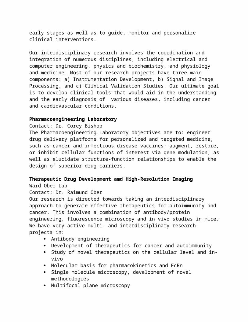
We have developed an imaging modality, multifocal plane microscopy (MUM), to allow for 3D sub-cellular trafficking studies within a live cell environment. In MUM, the sample is simultaneously imaged at distinct focal planes (Fig. 1), which enables the visualization of intracellular events that occur at different focal planes.

Figure 2. The photograph shows an implementation of a MUM setup on a Zeiss Axiovert 200 microscope. The setup is configured to simultaneously image four distinct planes within the specimen. VA1-VA3 - Zeiss video adaptors, CAM1-CAM4: CCD cameras, CC - C-mount/spacer and custom camera coupler assembly.
Tissue Microscopy LaboratoryContact: Dr. Alvin T. YehThe Tissue Microscopy Laboratory is a multidisciplinary research group at Texas A&M University in the Department of Biomedical Engineering. Our research group specializes in developing laser scanning microscopy based on sub-10-femtosecond pulses, particularly Nonlinear Optical Microscopy (NLOM, that is, two-photon and multiphoton microscopy), for the microscopic characterization of living tissues. Our work integrates enabling tools and reagents to develop novel insights into fundamental biological processes. Our recent research efforts have concentrated in tissue engineering and regenerative medicine and developmental biology.
BME Shared Facilities:
List of shared equipment available: (https://engineering.tamu.edu/media/2795925/shared-equipment-updated-list.pdf on https://engineering.tamu.edu/biomedical/safety)
Interdisciplinary Manufacturing Facility (IMF)Established through TAMU research development funds, this shared facility represents TAMU Engineering, Architecture, Veterinary Medicine, Science, and the Health Science Center. Housed in a 5000 sq. ft. space in the Emerging Technologies Building, this facility is equipped
with a hybrid manufacturing setup 3D printer and a 3D-bioplotter. The Hybrid Manufacturing printer is perhaps the only platform that allows “sculpting” objects by a concurrent combination of material deposition (through laser sintering), removal (machining) and shaping (forming). It is capable of working with multiple metals simultaneously, being able to mix and match four simultaneous metallic powders to create complex shapes and combinations of materials that can be precisely tailored locally to create next-generation artifacts. One of the most important and promising recent advancements at the intersection of medicine and engineering is the ability to “print” tissues and organs. The 3D-Bioplotter® can fabricate scaffolds using a wide range of materials including soft hydrogels over polymer melts, and hard ceramics. The system is designed to support the development of tissues and devices vital to the success of regenerative therapies, controlled drug release, and patient-specific implants.
Biomedical Engineering Faculty Research Laboratories (https://engineering.tamu.edu/biomedical/research/labs)
Gel Doc Ez Imager Automated gel imaging instrument for DNA gels and fluorsence imaging on a couple of different filter options.
UPLC
Used in biomolecule analysis with a flow-through needle injector and quaternary solvent delivery system. This machine is optimized for the analysis of protein, peptides, nucleic acids, and glycans using ion exchange, size exclusion, hydrophilic interaction, or reversed phase.
UltracentrifugeThis centrifuge delivers fast, efficient separations from samples as small as 175 [u]l up to 32.4 mL at speeds up to 150,000 RPM and more than 1,000,000 x g.
Slide Scanner Digitalizes slide images to scan, view, and share on a cloud server
Plate ReaderUsed for common detection of absorbance, fluorescence intensity, luminescence, time-resolved fluorescence, and fluorescence polarization of samples in well plates.
UV Transilluminators Provide brilliant back illumination of transparent materials placed on filter area designed to illuminate and increase fluorescence
NI Elvis II+ BoardsHands-on design and prototyping platform with an oscilloscope, digital multimeter, function generator, variable power supply, and Bode analyzer.
Gravograph CO2 Laser
This laser chamber system is used for basic cutting/etching of samples.
Excimer Laser A form of ultraviolet laser of the noble gas halide type used for micromachining.
Lyophilizers
Benchtop freeze dryer used to freeze the material and reduce the surrounding pressure so that the frozen water will sublimate from solid phase to gas phase. This is most often used in samples that will be imaged under SEM and need to preserve their structure
RheometerUsed to find stress, strain, modulus, or viscosity data using a cone and plate with set parameters such as shear, temperature, oscillation, etc.
Goniometer Contact angle measurements in order to know surface properties of materials when interacting with water.
Sputter Coater Used to coat non-conducting samples with a conductive gold layer
for standard SEM imagingUV-Vis Miniature fiber optic spectrometer preconfigured for 200-850 nm.
Spin Coater Used to deposit uniform thin films to flat substrates by way of spinning
Ethylene Oxide Sterilizer
Used for sterilization of medical devices including plastic and heat sensitive materials by exposing ethylene oxide gas.
Autoclaves Pressure chamber used to sterilize equipment and supplies by subjecting to high pressure saturated steam.
CO2 Incubators Chamber used to grow and maintain cell cultures at a certain temperature and carbon dioxide level.
Scanning Electron Microscope
The surface of a specimen is scanned by a beam of electrons that are reflected by a secondary or backscattered detector to form an image.
Atomic Force Microscope
Used for mapping atomic-scale topography of a surface by means of a repulsive electronic force between the surface and tip of a probe moving over a surface.
Confocal MicroscopeEnables reconstruction of three-dimensional structures from obtained images by use of laser scanning (also contains brightfield and fluorescent options).
Fluorescent Microscope
Uses fluorescence and phosphorescence of a substance instead of reflection and absorption.
Instron Mechanical testing of samples using 1000N load cell to test compression and tension
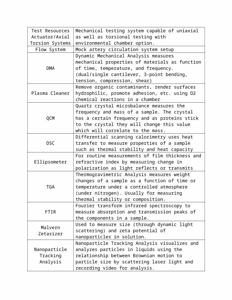
Test Resources Actuator/Axial
Torsion Systems
Mechanical testing system capable of uniaxial as well as torsional testing with environmental chamber option.
Flow System Mock artery circulation system setup
DMADynamic Mechanical Analysis measures mechanical properties of materials as function of time, temperature, and frequency. (dual/single cantilever, 3-point bending, tension, compression, shear)
Plasma Cleaner Remove organic contaminants, render surfaces hydrophilic, promote adhesion, etc. using O2 chemical reactions in a chamber
QCM
Quartz crystal microbalance measures the frequency and mass of a sample. The crystal has a certain frequency and as proteins stick to the crystal they will change this value which will correlate to the mass.
DSC Differential scanning calorimetry uses heat transfer to measure properties of a sample such as thermal stability and heat capacity
Ellipsometer For routine measurements of film thickness and refractive index by measuring change in polarization as light reflects or transmits
TGA
Thermogravimetric Analysis measures weight changes of a sample as a function of time or temperature under a controlled atmosphere (under nitrogen). Usually for measuring thermal stability or composition.
FTIR Fourier transform infrared spectroscopy to measure absorption and transmission peaks of the components in a sample.
Malvern Zetasizer Used to measure size (through dynamic light scattering) and zeta
potential of nanoparticles in solution.
Nanoparticle Tracking Analysis
Nanoparticle Tracking Analysis visualizes and analyzes particles in liquids using the relationship between Brownian motion to particle size by scattering laser light and recording video for analysis.
Hotplate/StirrersVortex MixersVacuum Ovens
CentrifugesAnalytical BalanceHot Water Baths
UV Cure BoxDI Water
Dishwasher
Multi-User TAMU Facilities:
Supercomputing FacilitiesTAMU HIGH PERFORMANCE RESEARCH COMPUTINGThis resource for research and discovery has four available clusters for faculty research:
(1) Ada is a 874-node hybrid cluster from IBM/Lenovo with Intel Ivy Bridge processors and a Mellanox FDR-10 Infiniband interconnect. Ada includes 68 NVIDIA K20 GPUs supporting applications already ported to GPUs, and 24 Intel Xeon Phi 5110P co-processors supporting applications benefiting from Knights Corner Phi cards.
(2) Terra is a 320-node heterogeneous Intel cluster from Lenovo with an Omni-Path Architecture (OPA) interconnect and 48 NVIDIA K80 dual-GPU accelerators. There are 304 nodes based on the Intel Broadwell processor and 16 nodes based on the Intel Knights Landing processor.
(3) Curie is a 75-node IBM Power7+ cluster with a 10Gb Ethernet interconnect. Each node has two IBM 64-bit 8-core POWER7+ processors and 256 GB of memory. Curie's file systems and batch scheduler are shared with the Ada cluster.
(4) LoneStar5 is the latest cluster hosted by the Texas Advanced computing Center. Jointly funded by the University of Texas System, Texas A&M University and Texas Tech University, it provides additional resources to TAMU researchers. LoneStar5 has: 1252 Cray XC40 compute nodes, each with two 12-core Intel® Xeon® processing cores for a total of 30,048 compute cores; 2 large memory compute nodes, each with 1TB memory; 8 large memory compute nodes, each with 512GB memory; 16 Nodes with NVIDIA K-40 GPUs; 5 Petabyte DataDirect Networks storage system; and Cray-developed Aries interconnect.
The HPRC group provides its users with access to several specially configured "HPRC Lab" Linux workstations at two separate locations on the TAMU campus, and can assist with: debugging, code optimization and parallelization, batch processing, and collaborative advanced program support.
MICROSCOPY & IMAGING CENTER (MIC)The mission of the Microscopy & Imaging Center (MIC) is to provide current and emerging technologies for teaching and research involving microscopy and imaging in Life and Physical Sciences on the Texas A&M campus and beyond, training and support services for microscopy, sample preparation, in situ elemental/molecular analyses, as well as digital image analysis and processing. This facility promotes cutting edge research in basic and applied sciences through research and development activities, as well as quality training and education through individual training, short courses and formal courses that can be taken for credit.Instruments available at the MIC include:
Light Microscopyo Zeiss Axiophoto Olympus FV1000 confocal microscopeo Multiphoton non-linear optical microscopeo Deconvolutiono Nikon Stereo Photo Microscope
Scanning Electron Microscopyo FEI Quanta 600 FE-SEMo Tescan Vega3 SEMo Zyvex S100 Nanomanipulator
Transmission Electron Microscopyo FEI Tecnai G2 F20 FE Cryo-TEMo FEI Tecnai G2 F20 ST FE-TEM - Materialso JEOL 1200 EX TEMo JEOL JEM-2010 TEMo Analog & Digital Image Analysiso Ancillary Equipment
Correlative Light and Electron Cryo-Microscopyo FEI cryo-fluorescence stage on the Olympus microscope
MATERIALS CHARACTERIZATION FACILITYThe Materials Characterization Facility (MCF) at Texas A&M University is a multi-user facility located in the Frederick E. Giesecke Engineering Research Building (GERB) housing the fabrication and characterization instrumentation essential for the development, understanding, and study of new materials and devices. Specific instrumentation available include:Electron Microscopy:
Field Emission-Scanning Electron Microscope (FE-SEM)(JEOL JSM-7500F), Lyra Focused Ion Beam-Scanning Electron Microscope (FIB-SEM) with an EDS
Microanalysis System, Fera Focused Ion Beam-Scanning Electron Microscope (FIB-SEM) with EBSD and
Integrated Time-of-Flight Mass Spectrometer (ToF-SIMS), and Electron microprobe with Wavelength Dispersive Spectroscopy (WDS)
Electron Microscopy Field Emission Scanning Electron Microscope (FE-SEM with EDS)
Lyra Focused Ion Beam Microscope (FIB-SEM with EDS ) Fera FIB-SEM with EBSD and Time of Flight Mass Spectrometer (TOF-SIMS ) Electron Microprobe with EDS and WDS
Thermal and Electrical Analysis Thermal mechanical analysis (TMA) Dynamic mechanical analysis (DMA) Differential scanning calorimetry (DSC) Dielectric spectroscopy Hot Disc thermal conductivity analysis Dielectric Spectrometer
Surface Analysis X-ray Photoelectron Spectroscopy (XPS)/Ultraviolet Photoelectron Spectroscopy (UPS) Nanoindenter Imaging ellipsometer Cameca ion microprobe Icon Atomic Force Microscope (AFM ) Atomic Force Microscopy – Infrared Spectroscopy (AFM-IR)
In-Situ Mechanical Testing PI 95 PicoIndenter for TEM PI 85 PicoIndenter for SEM Tensile Stage 500 N (in situ/ex situ) Tensile Stage 10 kN (in situ/ex situ)
Spectroscopy & Microscopy Spectrofluorometer UV-Vis-NIR spectrophotometer Raman confocal microscope FTIR spectrometer Fluorescent confocal microscope Optical Microscope
Sample Preparation Tools LADD carbon evaporator Struers LaboPol-5 polishing table Diamond band saw Powder press Buehler hot mounting press Nikon SMZ800N stereomicroscope Nikon LV100 petrographic microscope Epoxy disk preparation Oven
ENGINEERING DESIGN CENTER (EIC) (https://engineering.tamu.edu/academics/eic/index.html) The SuSu and Mark A. Fischer ’72 Engineering Design Center is an open space where engineering students have access to state-of-the-art prototyping tools, equipment, material and support staff. Through partnerships with industry and non-profit sponsors, the center is an environment where concepts become solutions to real-world problems and student teams come together to build new prototypes, acquire new
skills and develop new relationships. It is supported with differential tuition funds and it is open to engineering undergraduate students.
TAMU CENTER FOR CHEMICAL CHARACTERIZATION AND ANALYSIS (CCCA), Department of Chemistry
Nuclear Magnetic Resonance (NMR) Facility - The NMR Facility includes 10 superconducting spectrometer magnet systems, 4 LINUX workstations dedicated to data processing, and 3 full time staff to support them with maintenance, user training, and spectroscopic service. Although this facility is physically housed within the Chemistry Department, it provides services to the entire campus community.
X-Ray Diffraction Laboratory - The lab is a full service X-ray Diffraction laboratory offering state of the art instrumentation for the analysis of solid materials. Our services include single-crystal and powder diffraction for Chemistry, Material Sciences and Pharmaceuticals. We are staffed by trained Ph.D. scientists who employ the most up to date diffraction and X-ray techniques. Please feel free to contact us about your diffraction needs.
Chemistry Mass Spectrometry Facility - The services available include analyses of compounds ranging from small organic molecules to macromolecules including proteins, oligonucleotides, polymers and dendrimers. Instruments available include: Applied Biosystems PE SCIEX QSTAR; Thermo Scientific DSQ II GCMS; and Thermo Scientific LCQ-DECA
Center for Mass Spectrometry - is dedicated to providing cutting-edge technology and expertise for the characterization of molecules to fulfill the needs of researchers at TAMU. Mass spectrometry (MS) plays an increasingly important role in molecular level research, and it is central to ‘omics’ research, i.e., petroleomics, proteomics, metabolomics, lipidomics, glycomics, etc and the CMS provides expert staff with modern instrumentation to complete these tasks . Instruments available include: Thermo Scientific Fusion; Bruker 9.4T FT-ICR MS; MDS-Sciex 4000 Qtrap; and Applied Biosystems 4800+ MALDI TOF/TOF MS.
Elemental Analysis - The laboratory provides research support in the area of elemental and trace analysis as well as service analyses to TAMU users, other university and government agencies and private industry. It is unique in that it features fast neutron activation analysis (FNAA) capabilities in addition to thermal instrumental neutron activation (INAA) using the University's Nuclear Science Center 1 MW TRIGA research reactor. In addition, the laboratory has recently added inductively-coupled plasma - mass spectrometry to its stable of facilities. The ICP-MS has been fitted with both conventional sample introduction hardware for solution work as well as a 213 nm laser ablation system for studying solids and surfaces.
AGGIEFABThe AggieFab Nanofabrication Facility (AggieFab) is a shared nano/microfabrication facility that recently moved to the 1st floor of the Frederick E. Giesecke Engineering Research Building at Texas A&M University. The facility has over 6,500 sq. ft. of class 100/1000 cleanroom space with raised access floor and vertical laminar flow, and an additional 4,500 sq. ft. of support space, totaling 11,000 sq. ft. The facility and equipment have been built up over the past 20 years by many faculties in the Department of Electrical and Computer Engineering, with support from

the department and the College of Engineering. The recent move to the newly built GERB was enabled by a $12M investment from the Texas A&M Engineering Experiment Station (TEES), as well as additional support by the Department of Electrical and Computer Engineering. The facility has state-of-the-art equipment for full ranges of micro and nano-scale fabrication on diverse materials. In addition, the facility recently (Fall 2017) acquired $5M worth of new nanopatterning and deposition instruments through a donation from the Hewlett Packard Enterprise (HPE). These include the Zeiss Orion HeliumIon Microscope/Nanofab, FEI Helios DualBeam Focused Ion Beam (FIB), ASM Atomic Layer Deposition (ALD) tool, and the Clustex Multi-target deposition tool. The facility also has recently received $1.5M from the Texas A&M University Research Development Fund to purchase several new tools to improve micropatterning and advanced substrate development capabilities.
MICRO/NANO MANUFACTURING LABORATORY (http://mnmlab.tamu.edu) The MNM lab is directed by Dr. Wayne Nguyen P. Hung. The 1556 ft2 Micro/nano Laboratory (μnM Lab) is part of the Department of Engineering Technology and Industrial Distribution at TAMU. Its vision is to (i) serve as the key center for micro/nano manufacturing at TAMU, and (ii) to integrate TAMU with national micro/ nano manufacturing network. The missions of μnM lab are to provide expertise and initiate synergistic collaboration between various departments at TAMU, industry, and international institutions and also to inspire and prepare our students for further study in nanotechnology as well as to expose local high school teachers with the state-of-the-art micro/nano manufacturing technology. μnM Lab focuses to developing new technologies to fabricate 3D micro/nano components using novel materials such as super alloys, engineering polymers, electroactive polymers, and nanocomposites for engineering applications. A dedicated team of undergraduate and graduate students and faculty are working together in this group to develop innovative technologies using micro-machining, micro electrical discharged machining, laser micro machining/welding, micro-molding, investment micro-casting, micro-extrusion, microassembly, and focused ion sputtering. The group has extensive research collaborations with industries and faculties across the university as well as with renowned research institutions in Mexico and Singapore.
Laboratory Animal Resources and Research (LARR) – College of Veterinary Medicine, Comparative Medicine Program (http://vpr.tamu.edu/resources/research-infrastructure-support/cmp/comparative-medicine-program). The Comparative Medicine Program (CMP) is the centrally administered support service for animal research and teaching programs at Texas A&M University, College Station. The program's facilities and services are available for all Texas A&M campus affiliated faculty, staff, and students who have been approved to conduct animal research by the Institutional Animal Care and Use Committee (IACUC). CMP is accredited by the Association for the Assessment and Accreditation of Laboratory Animal Care (AAALAC) through its affiliation with other AAALAC-accredited Texas A&M programs.
CMP facilities offer housing and care for most standard laboratory animals. Specialized housing can be provided for biohazard projects and hazardous chemical projects. Varying degrees of animal isolation are available. Housing for large animal species is limited; however, various other campus animal care facilities can provide housing for large animals. CMP also offers a variety of services to institutional personnel.
Texas A&M Institute for Preclinical Studies (TIPS) (http://tips.tamu.edu/) The Texas A&M Institute for Preclinical Studies (TIPS) provides large animal Good Laboratory Practices (GLP) and translational research studies with unique access to expertise in all major medical and scientific disciplines including surgery, biomedical engineering, advanced imaging, pathology, radiography, interventional cardiology, neurology, animal behavior, chemistry, and engineering.
With unique resources and collaboration with the Texas A&M College of Veterinary Medicine and Biomedical Sciences, TIPS facilitates the inclusion of animals with naturally occurring disease as viable research models. TIPS provides unique opportunities to develop new therapeutics in the fields of oncology, cardiology, immunology, endocrinology, and an array of genetic disorders common to humans and companion animals.
The TIPS state-of-the-art 112,000 square-foot facility is an exceptional setting for GLP or non-GLP studies and physcian training utilizing large animals and includes:
Animal Housing - 240 individual large animal housing, plus additional long-term housing with barn and pasture support
GLP compliant clinical pathology laboratory Three 600 square-foot surgical suites, plus a hybrid imaging suite Conference rooms Sponsor work rooms Large 150 person auditorium for meetings/training Advanced imaging facilities including an MRI/Interventional suite equipped with a 3T
MRI and fluoroscopy, 128 slice PET/CT
Sponsor workrooms are designed for privacy and equipped with full length polyvision glass, wifi, large split screen flat panel monitors for viewing from the surgical suite's headcam, light cam, room cam, and digital monitoring equipment.
Services: Advanced Diagnostic Imaging Naturally Occurring Disease Models Cardiovascular Research Cardiovascular Pathology Clinical Pathology Cardiac Catheterization Surgical Training Facilities Large Animal Models
TAMHSC Institute for Regenerative Medicine (IRM) (https://medicine.tamhsc.edu/irm/) DNA Core instruments
o Laser capture microdissection (LCM)o Microarrays

o PCR – 2 systems available Flow Cytometry Core
o a two laser (20mW Blue/488nm & 25mW Red/635nm), seven parameter (5-color) Beckman-Coulter FC500 bench top analyzer with a dedicated operator or with training may be used individually.
o three laser (200mW Blue/488nm, 100mW UV/351nm & 25mW Red/633nm) eleven parameter (9-color) Beckman-Coulter MoFlo XDP fluorescence activated cell sorter. This instrument is capable of sterilely separating and depositing up to four purified subsets of cells into tubes based on any of the eleven parameters at a rate of about 25,000 cells/second.
Microscopy Coreo Nikon xenon-based fluorescence microscope coupled with an environmental
chambero Olympus spinning disk confocal microscope with Slidebook, Neuroleucida and
Stereo Investigator software
Facilities from other TAMU Departments that are used by BMEN Faculty:
GENOMICS AND BIOINFORMATICS FACILITY (www.txgen.tamu.edu/), Texas A&M AgriLife Research. This facility is housed approximately 5 miles south of the Texas A&M University campus. The facility is a subsidiary of the TAMU Institute of Plant Genomics and Biotechnology and is headed by Dr. Charles Johnson. The facility recently added an Illumina HiSeq 2000 and a new computer system and is upgrading to the HiSeq 2500 platform prior to the proposed funding period. These systems supplant their previous Illumina GAIIx systems and complements their Roche 454 sequencing system. The AgriLife Genomics and Bioinformatics Service Facility brings together a team of genomic, bioinformatic, molecular and computational scientists to address the needs of the Texas A&M Univerisity system. The Facility has several computer clusters and access to the Texas A&M Supercomputer as needed. The Facility is experienced at preparing sequencing libraries for Next Gen Sequencing from RNA or DNA samples, and provides this service in addition to the sequencing at a competitive cost. They also have expert bioinformaticians that can provide bioinformatic analysis of sequence datasets at a reasonable hourly fee.
Housed in the Genomics and Bioinformatics Facility is an Illumina HiSeq 4000, PacBio Sequal and MiSeq systems. This facility is a short driving distance south of the main Texas A&M campus.
CENTER FOR STRUCTURAL BIOLOGY (CSB) (http://www.sacclab.com/) High Throughput Screening Lab The High Throughput Screening (HTS) laboratory provides for automated liquid-handling robots and instruments for conducting biochemical, and phenotypic whole cell assays. These assays include bacteria, parasites, fungi, eukaryotic cells and viruses. There are two CyBio Vario systems which handle all high throughput plate assays, and for many cell-based assays
processing exceeds 20,000 samples per day. The systems are housed in biosafety cabinets to maintain sterility, and to protect staff members from potential pathogens. Other equipment includes an integrated robotic system provides automated liquid handling with hit picking, serial dilution, plate sealing, plate incubation, plate washing, and plate reading capabilities. A GE Healthcare INCell Analyzer 2000 allows us to incorporate high-throughput, high-content imaging into assays for phenotyping at the single cell and even subcellular level. Coupled with the image analysis software, it is possible to perform assays involving cell or organelle morphology, subcellular localization and/or heterogeneous cell populations. The laboratory is also equipped with BMG Labtech Polarstar Omega plate readers that provide detection capabilities in absorbance, fluorescence, and fluorescence polarization.
Our facility currently includes 2 CyBio Well Vario Systems- housed in Biosafety cabinets
Cybio 8plus1 integrated robotic system epMotion 5075 liquid handler GE Healthcare INCell 2000 Thermo Multiskan 2 BMG Labtech Polarstar Omega plate reader
Our custom in-house diversity library is designed to offer compounds with drug-like properties but low redundancy for exploring a wide range of chemical space to identify inhibitors with novel structures. This library was constructed such that each compound has a non-redundant chemical structure, minimizing the bias of many existing libraries which often contain many variations of common scaffolds. Thus, we can achieve similar breadth of screening with minimal time and resources compared to a very large ultra-high-throughput compound collection. This diversity library was designed by combining structures from over 3 million compounds, and selecting a diverse and representative subset using a clustering algorithm, which guarantees that no pair of compounds have a Tentimoto score greater than 0.7.
Libraries Include 1,000 compound fragment library
~104,000 compound diversity library ~160,000 sample natural products library
X-Ray Crystallography Lab X-ray crystallography is a method of seeing the structure of molecules that can’t be seen with a microscope. To do this, first the molecule of interest, such as a protein, must be arranged in crystal form. This is accomplished by putting the protein into a solution that encourages the molecules to align themselves in an ordered fashion. This solution usually has a high salt or polymer concentration and often is pH regulated to keep the protein folded in a physiologically relevant way. Care must be taken not to over concentrate this solution or the protein will precipitate out of solution in a disordered form which can not be analyzed. The crystallization step is often the most time consuming and some proteins are not amenable to crystallization, making this the rate limiting step in crystallography.

Center for Structural Biology Chemists have made great strides in understanding how molecular structure influences the reactivity of molecular systems, and biolchemists have exploited chemical processes such as site-directed mutagenesis and polymerase chain reaction to probe biological systems. The aim of structural biology is to develop a molecular level understanding of these structure-function relationships. One of the aims of structural biology is to understand the structural changes introduced by chemical modifications as well as to determine how such changes alter the biology, thereby developing a more complete understanding of structure-function relationships. Another important application is in the practical areas of rational drug discovery Impact of ILSB Structural Biology Program TAMU has strong structural biology research programs that independently operate as single-investigator programs, and unquestionably several of these programs are highly successful as judged by publications, funding, and national-international visibility in the various research communities.
Other FacilitiesStructure Based Drug Design (SBDD) is a process of developing drugs against a target based on the structure of the target. Traditional high through-put methods of drug development are based on luck - screening a library of random compounds in the hopes of finding a compound that acts against the target. This method is expensive and has a very low success rate. However, it is possible to tailor a potential drug to a target by using the wealth of structural information currently available, this is the basis for SBDD.
PROTEIN CHEMISTRY LAB (PCL) (Director, Larry Dangott) (https://pcl.tamu.edu/) The Protein Chemistry Laboratory is a core resource facility created and partially funded under the auspices of the Office of the Vice President for Research of Texas A&M University.
Automated Edman Protein Sequence AnalysisAutomated Edman chemistry is performed on an Applied Biosystems 492 Procise Protein Sequencer. The Procise is a complete, automated chemistry and analytical system for amino acid sequence analysis of protein and peptide samples in picomole to nanomole amounts.
The Procise 492 Protein Sequencer performs automated Edman sequencing chemistry on a Biobrene-coated Glass Fiber Filter (for liquid samples) or on proteins blotted to PVDF after PAGE separation. Sensitivity is in the low pmol range.
Amino Acid AnalysisAmino acid analysis is performed using the old Hewlett Packard AminoQuant method adapted to a newer system. The system consists of a Agilent 1260 liquid chromatograph that is equipped with a programmable autosampler for automated sample handling, derivatization and injection, a variable wavelength UV detector for low-moderate sensitivity (Agilent G1365D) analyses and an in-line Agilent G1321B fluorescence detector for high-sensitivity analyses (fluorescent detection). All system control and data analysis is performed by Agilent Chemstation software. Liquid and vapor phase hydrolysis protocols have been developed in order to service a wide range of sample types (liquid, dried and electroblotted proteins on membranes). We offer routine validated (qualitative and quantitative) amino acid compositions of purified proteins and synthetic peptides as well as (quantitative ) free-amino acid (or total amino acid) compositions of
feeds, flours and physiological fluids. Hydrolyzed proteins samples and free amino acids are derivatized pre-column with o-phthalaldehyde (OPA) and 9-fluoromethyl-chloroformate (FMOC) prior to separation and quantitation by reverse phase HPLC. Sensitivity is in the picomole to nanomole range depending upon sample matrix contributions.
Protein Gel Electrophoresis and ElectroblottingElectrophoresis has been a valuable method of protein analysis for decades. In recent years, the increased sensitivity of automated sequencers and analyzers has enabled the use of electrophoretic separation techniques to be used as preparative methods for subsequent stages of high-sensitivity protein analysis. The Laboratory is fully equipped to perform a variety of routine electrophoretic separations of proteins in polyacrylamide or agarose matrices. Sodium dodecyl sulfate polyacrylamide (SDS) and non-denaturing gels are available in standard and mini-gel format (linear and gradient format) as are 2-dimensional electrophoretic separations. Analytical isoelectric focusing (IEF) is performed on an Amersham Pharmacia Biotec IPGPhor electrophoresis module (first dimension) using immobilized pH gradients in polyacrylamide strips (DryStrips). The second dimension of resolution is obtained on a vertical slab gel electrophoresis unit equipped with an external re-circulating cooling device.Electroblotting of proteins to solid membrane supports has been used for many years in protocols used for immunodetection of antigens. Recent developments in membrane support technology have led to the production of high-capacity, chemically inert matrices that are resistant to N-terminal sequencing chemistries. Proteins blotted to these matrices can be excised from a mixture of proteins and subjected directly to sequence analysis or preliminary manipulations such as direct In situ chemical or proteolytic cleavage with subsequent peptide extraction and purification. The Laboratory is equipped to perform electroblotting procedures as well as In situ or "in gel" digestion procedures as required to obtain sequence information and protein identity.
DALTSixThe DALTSix is a modular electrophoresis system for large format 2D gel electrophoresis. It can run up to 6 lab-cast or pre-cast gels (26 x 20 cm) simultaneously. It is used with 18 and 24 cm IPG DryStrips.
Typhoon Trio Fluorescent ImagerThe Typhoon Trio (GE Healthcare) is a high performance gel and blot imager. The imager has powerful laser excitation sources and high-quality confocal optics that allow for the sensitive detection of low-abundance targets. Red, Green and Blue excitation wavelengths (488 nm, 532 nm, 633 nm) and a wide choice of emission filters enable imaging of an extensive variety of fluorophores. The Trio is also useful for Storage Phosphor Autoradiography, Chemiluminescence and Chemifluorescence detection. (Client must have their own Phosphor screens. The PCL does not own any.) Please call ahead with specific questions. The instrument is used on a first-come, first-served basis, but reservations are recommended for analyses that will require extensive periods of time.
Ettan Robotic SystemsThe PCL houses three robotic systems for High-throughput Proteomics analysis; Ettan Spot Picker, Ettan Digester and the Ettan (MALDI plate) Spotter (GE Healthcare).

Picker automatically picks selected protein spots from stained and unstained gels. It is designed for backed as well as unbacked gels. This instrument is seamless with our DeCyder 2-D Differential Analysis software.The Picker accurately picks protein spots from 2D gels and transfers them into 96 well microplates for further treatment.The Digester is a highly versatile instrument designed to perform in-gel digestion of proteins captured in 2D gel electrophoresis spots. Default digestion protocols can be customized. The instrument performs automatic tracking of samples.The Ettan Spotter is designed to automatically mix and spot digested proteins and matrix onto any MALDI sample plate.
High Pressure Liquid Chromatography Liquid chromatography is used extensively to purify proteins and peptides from mixtures of natural materials. The PCL has a stand-alone Hewlett Packard 1100 HPLC system is equipped with an autosampler and a diode array detector (DAD) for spectral analysis and is used for narrowbore, microbore and capillary LC peptide mapping and purification using reverse phase chromatographic techniques. This instrument is used in conjunction with the MALDI-TOF and Hewlett Packard G1005A Protein Sequencer to obtain internal protein sequence information from protein digests that can allow investigators to identify their proteins of interest and aid in the design of the appropriate probes for their isolation and, possible, cloning. An LC Packings Probot is attached to the HP1100 HPLC to aid in automated spotting of peptides to a sample plate for MALDI-TOF mass spectrometry. In addition, the facility has a Pharmacia Acta Explorer 10 Protein Purification and a GE Healthcare AKTA Purifier system that are capable of running aqueous and organic mobile phases for native protein purification as well as peptide isolation. These instruments are being used to develop pilot protein purification protocols using a library of columns that includes hydrophobic interaction, ion exchange, metal chelation and affinity matrices. In addition, the systems are used for Immuno-Depletion of contaminating proteins from physiological samples prior to Proteomic analysis.
MALDI-TOF Mass SpectrometerThe PCL has a Shimadzu/Kratos MALDI-TOF mass spectrometer equipped with delayed extraction and reflectron capabilities which is used for routine high mass accuracy analysis of peptides (<3 kDa). The instrument can also be operated in linear mode for protein analysis (>3 kDa).
Electrospray Mass SpectrometerWe are equipped with a ThermoFisher DecaXP ion trap mass spectrometer that is used for peptide mass measurements and fragmentation (de novo sequencing). Samples can be applied by direct infusion or after in-line reversed phase chromatographic separation. Automated search algorithms (Sequest and MASCOT) are available to perform database searches to aid in protein identification. This instrument is for peptide/protein analyses only.
BIOMOLECULAR NMR Facility (http://biobionmr.tamu.edu/)The Biomolecular NMR facility of the Department of Biochemistry and Biophysics is hosted in newly built NMR wing of the BioBio building located on the West Campus of Texas A&M University. The facility consists of four state of the art NMR spectrometers including a Bruker AVANCE III 800 MHz. At 18.8 Tesla, this modern spectrometer represent one of the very few ultra-high field instruments currently operational in southern United States and Texas. It is
equipped with 5mm TCI cryoprobe, which has exceptional proton detection sensitivity of 10,677:1.Instrumentation:Bruker AVANCE III 800Bruker AVANCE III HD 600Bruker AVANCE III HD 500
LABORATORY FOR MOLECULAR SIMULATION (http://lms.chem.tamu.edu/) The Laboratory for Molecular Simulation (LMS) brings molecular modeling and computational chemistry closer to the experimental scientist by offering training to both new and advanced users. Advanced modeling software is available for researchers at Texas A&M University to perform quantum calculations on small molecular or solid systems and molecular mechanics/dynamics modeling for large systems such as proteins, DNA, nanomolecules, polymers, solids, and liquids. The LMS also provides support for faculty that wish to incorporate molecular modeling in their course material.
Publications and/or presentations that result from the use of the software and/or hardware provided by the LMS needs to include the following statement in the acknowledgments: We would like to thank the Laboratory for Molecular Simulation at Texas A&M University for providing software(, support, and computer time).
WORKSHOPS - The LMS offers, free of charge, three types of workshops throughout the year: 1) Linux, 2) Molecular Modelling, 3) Quantum Mechanics Short Course, and 4) CHEM 658 Introduction to Molecular Modeling. For more information please see the workshops link to the left. Most of the programs available through the LMS are only available on Linux based machines, therefore, the Linux workshop is a pre-requisite for all other workshops.
HARDWARE – The LMS has hardware solutions to fit your computation research needs. In addition, LMS hardware can host ITAR and EAR software.
INSTITUTE FOR PLANT GENOMICS AND BIOTECHNOLOGYThe Institute for Plant Genomics & Biotechnology is a multi-unit, multi-disciplinary organization with participating faculty, students, and scientists from fourteen units affiliated with Texas A&M University, Texas A&M AgriLife Research, and the USDA-ARS. The Borlaug Center provides the Institute for Plant Genomics and Biotechnology with a base of operations including specialized research laboratories, infrastructure, and equipment for the plant science community at Texas A&M University.
Labs: Bioenergy, Systems Biology and Bioinformatics Bioseparations Laboratory Functional Genomics of Plants Laboratory Laboratory for Crop Transformation
Laboratory for Genome Technology MIPI LAB || (Molecular Insect Plant Interact) Plant Microbe Interact Lab Plant-Fungal Interaction Laboratory PMG Lab || (Plant Molecular Genetics) Sorghum Genomics Lab USDA Laboratory||Genetics/Genomics Borlaug Plant Growth Chamber Facility
LABORATORY FOR GENOME BIOTECHNOLOGY (http://lpgb.tamu.edu). ABI 3130xl Genetic Analyzer
A capillary sequencer for service sequencing and DNA marker analysis $2.50/sample Please see the DNA Sequecing Information
Real-Time PCR Systems CFX 384 and ABI 7900HT SDS Real-Time PCR machine for allelic discrimination, quantification and dissociation curve
analysisComputing system
DNA Sequencing Analysis and Sequencer software for sequence analysis Primer Express software to design TaqMan primers and probes Genescan, Genotyper and GeneMaper
Additional equipment Genogrinder Photo-documentation System: ChemiDoc XRS+, Gel Doc XR+ Beckman DU-640 spectrophotometer, NanoDrop ND 1000 Spectrophotometer Victor
Plate Reader and Alpha Imager Mini