MOVEMENT ANALYSIS OF THE UPPER LIMB IN CHILDREN WITH UNILATERAL CEREBRAL PALSY ... ·...
66
1 MOVEMENT ANALYSIS OF THE UPPER LIMB IN CHILDREN WITH UNILATERAL CEREBRAL PALSY: FOREARM PRO- AND SUPINATION. Zoë CASIER Student number: 01208014 Annelore DEFRANCQ Student number: 01203208 Promotor: Prof. Dr. Frank Plasschaert Co-promotor : Dr. Sophie Lauwagie Supervisor: Prof. Dr. Ir. Malcolm Forward A dissertation submitted to Ghent University in partial fulfilment of the requirements for the degree of Master of Medicine in Medicine. Academic year: 2016 – 2018
Transcript of MOVEMENT ANALYSIS OF THE UPPER LIMB IN CHILDREN WITH UNILATERAL CEREBRAL PALSY ... ·...

1
MOVEMENT ANALYSIS OF THE UPPER
LIMB IN CHILDREN WITH UNILATERAL
CEREBRAL PALSY: FOREARM PRO-
AND SUPINATION.
Zoë CASIER Student number: 01208014
Annelore DEFRANCQ Student number: 01203208
Promotor: Prof. Dr. Frank Plasschaert
Co-promotor : Dr. Sophie Lauwagie
Supervisor: Prof. Dr. Ir. Malcolm Forward
A dissertation submitted to Ghent University in partial fulfilment of the requirements for the degree of
Master of Medicine in Medicine.
Academic year: 2016 – 2018

2
Permission to use

3
Preface
We enjoyed working in the paediatric orthopaedics department of the University Hospital of
Ghent, where we have spent numerous hours figuring out marker placements, protocol
exercises, parameters and so on. We could not have done this without the help of several
important people, who we would like to thank in this part of our scription.
When we faced problems of any nature, we could always count on Ellen Dedobbelaere. She
was always there to answer all our questions and to provide us with tips and tricks on how to
work our way in and around the lab.
It wouldn’t have been possible to write this thesis without the help of Prof. Dr. Ir. Forward. His
knowledge in kinematics and expertise in the lab and in engineering was of great importance.
He helped us to understand every step of the process of developing a kinematic protocol while
giving us the space to come up with our own ideas. We really appreciate all the hard work and
time he has put into this protocol.
We also would like to thank our co-promotor dr. Lauwagie, on who we could count for details
on the clinical background of cerebral palsy. When we encountered practical problems, dr.
Lauwagie was always prepared to meet up and support our process.
We would like to thank prof. dr. Plasschaert, the promotor of this thesis, for giving us the
opportunity to work on this project. We have learned a lot by being able to develop the protocol
by ourselves and having the opportunity and the freedom to think of our own exercises. We
were allowed to use the resources of the Cerebral Palsy Reference Centre and the gait
analysis lab.
We are very thankful for the subjects who participated in this study and their families, for taking
the time to come to the gait analysis lab. We really enjoyed working with all these children.
They gave us an extra reason to be motivated and enthusiast to go to the lab for assessing.
At last a thank you to our friends and family for understanding the amount of time and work
that slipped into this scription, their support during these two years was not to be
underestimated. Also Marie Depoortere and Maxine Winter for making the numerous hours we
have spent together in the lab even more fun. Off course we would like to thank each other as
well for the excellent atmosphere of cooperation. Being able to do this work together was one
of the best parts of this thesis as we both know we could not have done without each other’s
support.

4
Table of content
Abstract .................................................................................................................................................. 1
Nederlandstalige samenvatting .......................................................................................................... 2
Introduction ............................................................................................................................................ 4
What is Cerebral Palsy? .............................................................................................................. 4
Neuroplasticity ............................................................................................................................... 4
Cerebral palsy subtypes .............................................................................................................. 6
Upper limb involvement in cerebral palsy ................................................................................. 7
Interventions .................................................................................................................................. 8
Classification of the functional ability of the upper extremity ............................................... 10
Kinematics in the evaluation of the upper limb ...................................................................... 12
The role of EMG .......................................................................................................................... 13
Conclusion ................................................................................................................................... 14
Materials and Methods ....................................................................................................................... 15
The lab and materials..................................................................................................................... 15
Subjects ............................................................................................................................................ 16
Retroreflective marker locations ................................................................................................... 17
EMG sensors ................................................................................................................................... 18
The protocol ..................................................................................................................................... 19
General ......................................................................................................................................... 19
Static trial ..................................................................................................................................... 19
Analytical trial .............................................................................................................................. 20
Functional trial ............................................................................................................................. 22
Maximal voluntary contractions (MVC) and EMG .................................................................. 27
Processing the data ........................................................................................................................ 27
Results .................................................................................................................................................. 28
Clinical assessment of patient ...................................................................................................... 28
Results of the analytical trials ....................................................................................................... 29
Results of the functional trials ....................................................................................................... 29
Comparing functional AROM to analytical AROM ..................................................................... 29
Discussion ............................................................................................................................................ 31
The lab and materials..................................................................................................................... 31
Subjects ............................................................................................................................................ 32
Retroreflective marker locations ................................................................................................... 32
EMG sensors ................................................................................................................................... 33
The protocol ..................................................................................................................................... 34

5
General ......................................................................................................................................... 34
Analytical trial .............................................................................................................................. 36
Calibration .................................................................................................................................... 37
Functional trials ........................................................................................................................... 38
Processing ....................................................................................................................................... 42
Future ............................................................................................................................................... 43
Conclusion ........................................................................................................................................... 44
References .......................................................................................................................................... 45
Attachments ........................................................................................................................................... 1
Attachment 1: Alphabetical list of abbreviations .......................................................................... 1
Attachment 2: House classification and MRC-scale ................................................................... 2
Attachment 3: Informed consent for a child above the age of 12 .............................................. 3
Attachment 4: Informed consent for a child younger than 12 years old ................................... 5
Attachment 5: Approval of the Ethics Committee ........................................................................ 7
Attachment 6: Tables of the parameters and the results ............................................................ 9

1
Abstract
INTRODUCTION Cerebral Palsy (CP) is a non-progressive neurological disorder often leading
to motor impairments. It is caused by damage to the brain in the early stages of its
development, this is before, during or after birth. The upper limb is involved in 60% of the
children with CP, leading to difficulties in handling objects and independence in daily life
activities. There are numerous classification systems for scoring the upper limb function.
However none of these are objective. Therefore this study focusses on developing an objective
movement analysis with the aid of kinematic and EMG registrations. Because the upper limb
is very complex regarding its degrees of freedom in which movement is possible, it is chosen
to only focus on the pro- and supination.
METHOD This study is designed to assess the difficulties encountered during pro- and
supination objectively incorporating kinematics and EMG data. 12 Patients and 12 age- and
gender matched controls are evaluated in the gait analysis lab of the University Hospital of
Ghent. To create the kinematic axissystems and graphs, retroreflective markers are placed on
the subject's arms. EMG electrodes are placed on the Pronator Teres muscle and the Biceps
Brachii, to retrieve information about muscle activity. After this the subjects perform analytical
as well as functional tasks as instructed. With the help of parameters selected for each
exercise, data on the pro- and supination performance is gathered.
RESULTS Due to the limited timeframe the data of only one patient with a rather severe form
of CP and its control are compared. The clinical assessment of the patient describes a nearly
impossible active supination. These findings are confirmed by the results of the protocol. It is
clear that the patient's affected arm had an inferior performance for almost all parameters
compared to its control and to his own dominant hand.
DISCUSSION There are a few points of discussion concerning the protocol, its execution and
the results. However, these points are not insurmountable and can be easily adjusted in the
future. Key is that the kinematic model is capable of assessing the pro- and supination rotation
in an objective and quantifiable way.
CONCLUSION By creating an objective assessment of the upper limb and describing its pro-
and supination function on a rather small scale, it is hoped that clinical and therapeutic decision
making will be possible using this protocol. Future studies should concentrate on proving the
protocol’s value in the everyday practice.

2
Nederlandstalige samenvatting
Deze thesis handelt over onderzoek naar de bovenste lidmaat functie van kinderen met een
cerebraal parese, meer bepaald naar hun pro- en supinatie capaciteiten. Cerebraal parese is
een niet-progressieve neurologische aandoening die veroorzaakt wordt door hersenschade
die opgelopen werd tijdens de ontwikkeling van de hersenen. Dit kan voor, tijdens of na de
geboorte zijn. Deze schade heeft belangrijke implicaties op het functioneren van de patiënt,
voornamelijk door motorische moeilijkheden. De prevalentie bedraagt 1-2 op duizend
levendgeborenen. Het bovenste lidmaat is bij 60% van de kinderen betrokken, wat belangrijke
beperkingen in het uitvoeren van activiteiten in het dagelijks leven met zich meebrengt.
Er zijn 4 verschillende CP subtypes: spastisch, dyskinetisch, ataxisch en gemengd. De
subtypes worden bepaald op basis van de klinische neurologische presentatie, die afhankelijk
is van de plaats en het tijdstip van het hersenletsel. Het spastische subtype is het meest
voorkomende (80%). Door de grote variabiliteit in het tijdstip van het optreden van het letsel
en in klinische presentaties ervan, is steeds een persoonlijke behandeling nodig voor elke
patiënt. De aanpak is bovendien multifunctioneel en langdurig, waarbij voornamelijk nieuwe
complicaties worden voorkomen en de beperkingen op het functioneren aangepakt worden.
Er bestaan verschillende classificatiesystemen om de bovenste lidmaatfunctie van patiënten
te scoren, al deze systemen zijn echter subjectief en berusten louter op de beoordeling van de
ouders of een therapeut. Verschillende onderzoeken kaarten aan dat het nodig is om een
objectieve test te ontwerpen die de therapiekeuze kan begeleiden en duidelijk verschil kan
aantonen voor en na therapie. Het implementeren van een kinematische analyse en de
registratie van spieractivatie met behulp van elektromyografie (EMG) zou zo’n analyse
objectiever kunnen maken.
In deze studie werd een protocol ontworpen die aan deze objectieve eisen zou kunnen
voldoen. Zo werd de pro- en supinatie vergeleken tussen 12 normaal ontwikkelende kinderen
en 12 kinderen met unilaterale cerebraal parese. Alle kinderen waren tussen 6 en 14j jaar en
werden aan elkaar gekoppeld op basis van leeftijd en geslacht. De studie vond plaats in het
ganglabo van het UZ Gent. Dit labo beschikt over 24 3D camera’s en 4 videocamera’s die aan
de hand van retroreflectieve markers de beweging van de testpersoon kunnen volgen. De
plaatsing van deze markers werd nauwkeurig uitgewerkt zodat optimale tracking en resultaten
konden bereikt worden. De EMG elektrodes werden geplaatst op de musculus Pronator Teres
en musculus Biceps Brachii. Dit alles op beide armen van de testpersonen.
Het protocol dat werd ontworpen bestond uit twee delen. Een eerste deel bestond uit een set
van analytische opnames. Hierbij werd de testpersoon, zittend en met de ellebogen
ondersteund, gevraagd om pro- en supinatie cycli uit te voeren. Dit werd zowel bimanueel als

3
voor elke arm apart gedaan. Het tweede deel bestond uit 3 functionele oefeningen. Elk van
deze 3 oefeningen werd met een apart doel ontwikkeld. Zo werd een oefening met een
pingpong racket ontworpen die de focus legde op snelheid en het vasthouden van een object.
Een volgende oefening waarbij de testpersoon gevraagd werd een marker op het hoofd te
bedekken, liet toe om snel een inschatting van de ernst van de beperking op het dagelijks
functioneren te maken. Tot slot een bimanuele oefening waarbij de persoon gevraagd werd te
klappen op de middellijn die kon aantonen dat de dominante hand de mindere prestatie van
de andere hand kan compenseren.
Voor deze oefeningen werden verschillende toepasbare parameters gekozen. Zo werd steeds
de gemiddelde bewegingsboog bepaald alsook de maximale pro-en supinatie graden. Verder
werd er naar de snelheid, versnelling en vlotheid van beweging gekeken voor de analytische
oefeningen. Bij de pingpong oefening werden alle parameters overgenomen behalve degene
voor vlotheid. Voor de oefening waarbij het hoofd wordt aangeraakt werd naast de
bewegingsboog ook naar het succesvol uitvoeren van de oefening gekeken. De klapoefening
werd besproken aan de hand van de afstand tot de middellijn van beide handen op het moment
van de klap. Voor al deze parameters werden de resultaten vergeleken tussen de patiënt en
zijn controle voor dominante handen en niet-dominante handen. Ook binnen de proefpersonen
zelf, werd links met rechts vergeleken.
Al deze zaken werden toegepast op één patiënt met een ernstige vorm van CP en zijn controle.
Hier werden volgende zaken opgemerkt. De bewegingsboog was duidelijk veel beperkter aan
de aangetaste zijde van de patiënt in vergelijking met zijn andere zijde en met de niet-
dominante zijde van zijn controle. Deze boog werd voornamelijk beperkt door het onvermogen
een supinatie uit te voeren. Ook de snelheid en acceleratie gaven een sterke beperking weer.
De vlotheid was duidelijk beperkter voor de aangetaste zijde. Wanneer de unimanuele
oefening werd vergeleken met de bimanuele viel het op dat de dominante zijde van de patiënt
over het algemeen beter scoorde tijdens de unimanuele oefeningen. In de klapoefening werd
duidelijk aangetoond dat de beperkte rotatie van de aangetaste kant werd gecompenseerd
door een grotere supinatieboog aan de dominante zijde.
Het ontworpen protocol en zijn bijhorend kinematisch model hebben aangetoond in staat te
zijn op een objectieve en kwantitatieve manier de beperkingen van patiënten met CP vast te
stellen. In de toekomst is het belangrijk te bewijzen dat dit protocol ook klinische relevant is.
Therapeutische beslissingen moeten kunnen gemaakt worden op basis van informatie
verzameld uit dit onderzoek.

4
Introduction
What is Cerebral Palsy?
Cerebral palsy (CP) is caused by damage to the developing brain during fetal development or
during the first years of life (1–11). The result of this damage is a non-progressive neurological
disorder with motor impairments, called cerebral palsy (1–5,8–11). The damage can be due to
maternal infections like rubella, chorioamnionitis and cytomegalovirus or pre-eclampsia
(5,6,10). Other risk factors for the acquisition of brain damage are low weight or hypoxic events
at birth, multiple pregnancy, premature birth, neonatal seizures, infection, restricted
intrauterine growth, vascular incidents and placental abnormalities (4–6,10). In 10% of the
cerebral palsy patients, damage to the developing brain occurred in the postneonatal period,
with infections of the central nervous system, seizures and (non-)accidental head trauma as
potential causes (6). However this subgroup is often excluded from trials investigating risk
factors during pregnancy and birth (4).
As a consequence of the brain lesion, movement and posture of these children can be
disturbed, caused by one or several of the following symptoms: spasticity, loss of selective
motor control, co-contractions, muscle weakness, hypertonia and hyperreflexia, mirror
movements and decreased velocity of movements (2,8,11–13). In addition they often display
sensory deficits, cognitive impairments and problems with communicative and social skills,
together with secondary musculoskeletal problems and epilepsy (2,5,7,12). The secondary
musculoskeletal problems are caused by hypertonia and weakness of the involved muscles,
leading to muscle contractures. The muscle imbalance in combination with skeletal growth
causes bony deformities (2,8).
CP is a descriptive term that covers the largest group of movement disorders in children
(2,4,5,7,10,14). It is not a diagnosis, as the term covers nothing about the location, type or
timing of the injury (4). Out of 1000 live births, 1 to 2 children are born with CP (2,5,6,10,14,15).
In children born before 28 weeks gestational age the prevalence of CP even increases to 40-
100 per 1000 (2,14). These premature babies have a higher risk of intraparenchymal or
intraventricular bleeding and damage to the periventricular white matter, which could harm the
developing brain (5).
Neuroplasticity
The developing brain is most vulnerable in the most rapid phase of its growth, which is from in
utero until approximately 5 months after birth (2). In this stage, infection, malformation or
disruption of the blood supply can damage the brain (2). In the first 3 months of development,
the blood supply originates from the front and the back of the brain and stretches out to the
more central and deeper structures. These structures in the periventricular zone of the motor

5
cortex are far away from the developing blood vessels and thus more susceptible to be
damaged by disruption of the developing blood supply (2). However, at full term, the most
vulnerable cells in the brain are the basal ganglia. These are the cells with the highest
metabolic needs and are thus compromised in a hypoxic situation in the perinatal stage (2).
The various possible stages of development of the central nervous system at the time of the
injury and different possible locations of the brain lesion lead to different clinical presentations
and have a harmful effect on the functional capacities of the child (2,6,12,16,17). The ability of
the brain to compensate for this injury, depends on its developmental stage at the time of the
insult (14,16). The developing brain has a greater reorganizing capacity than the adult brain
because it can react by taking over the affected brain functions in different, atypical locations
(16,18).
Approximately 20 weeks post conception, the corticospinal axons of the motor cortex of both
hemispheres have reached the cervical spinal cord, where they connect mainly with alfa-
motoneurons (16). Because of the bilateral projections of both hemispheres at this stage of the
development, competition between the ipsilateral and contralateral projections to the
extremities occurs (16). This means that the extremities receive motor signals from both the
ipsilateral and contralateral hemisphere (16,18). Neuronal activity during the third trimester and
the first years of life in normal brain development then leads to the disintegration of the
ipsilateral projections and the consolidation of the contralateral projections to the extremities
(16). The ipsilateral innervation of the extremities is thus transient (14,16). A lesion that
occurred during the early developmental stages of the brain, resulting in less neuronal activity
coming from one hemisphere, leads to the persistence of the ipsilateral projections from the
contralesional hemisphere to the extremities because of its dominant neuronal activity during
the competition (16). This process is called neuroplasticity or reorganization and is responsible
for the innervation of the paretic extremities (14,16). However, normal function of the paretic
limb has not yet been reported with only ipsilateral projections (16).
Because the contralesional hemisphere now innervates both the ipsilateral and contralateral
extremities, mirror movements can persist. Mirror movements are symmetrical movements of
the contralateral limb and are a normal phenomenon in typically developing children until 8
years old. It starts to disappear by the age of 9 and should be gone at 12. By this age, all
ipsilateral projections should have disintegrated as a result of the dominant neuronal activity
coming from the contralateral hemisphere (19). Due to damage to the developing brain, both
the ipsilateral and contralateral projections of the contralesional hemisphere persist in children
with unilateral CP, leading to the occurrence of mirror movements (19).

6
Cerebral palsy subtypes
There are four subtypes of CP according to the Surveillance of Cerebral Palsy in Europe
(SCPE): spastic CP, dyskinetic CP, ataxic CP and mixed or unclassified CP (9,10,15,17).
These subtypes are based on the clinical features and clear neurological signs (9). Only when
the patient reaches the minimum age of 4 or 5, the diagnosis of CP can be confirmed as
progressive neurological diseases or other causes of motor impairment will have become
apparent by then (4,9,14,17).
Spastic CP is by far the most common subtype, diagnosed in more than 80% of the CP patients
(7,14,20). Damage to the motor cortex of the brain affects the corticospinal or pyramidal
system, which is responsible for organising goal oriented, complicated movements. An injury
to this system can induce spasticity, hypertonia, abnormal patterns of movement and/or
posture as well as hyperreflexia in the contralesional side of the body (5,9,10,21). Damage to
the cortex can also lead to focal epilepsy (9,21).
The increased muscle tone is explained by the inhibition of the release of -aminobutyric acid
(GABA). The release of this inhibiting neurotransmitter is normally stimulated by the
descending corticospinal tracts and leads to the interruption of reflex arcs (2). A lesion of the
corticospinal tracts, as occurs in spastic CP, prevents the release of GABA and thus prevents
the interruption of these reflex arcs (2). The muscles become overactivated and show an
increase in tonic stretch reflexes, called spasticity, which are velocity dependent (2,7,9). Young
children often display spasticity, which is dynamic and mostly interferes with function. Non-
surgical interventions can be used to reduce spasticity while older children might have
developed more fixed contractions, which require surgery (7).
The prevalence of dyskinetic CP is approximately 6.5%. The symptoms of this CP subtype, as
defined by SCPE, are loss of coordination, disturbed posture and abnormal movement
patterns. Movements are often uncontrolled and involuntary and they can be provoked by a
sensory overload such as anxiety or noise (2,5,9,10,21). This CP subtype can be subdivided
into dystonic and choreo-athetotic CP, in which the latter consists of hyperkinesia with
hypotonia and the first is typed by hypokinesia and hypertonia (9,10). These symptoms can be
explained by a lesion to the extrapyramidal system, consisting of the basal ganglia (2,21).
The basal ganglia are responsible for the coordination of reflex patterns. This leads to fast and
smooth movements, which are adjusted by visual and proprioceptive input. Damaging this
complex interaction of motor control can affect the fluidity of movement and lead to this subtype
of CP (2).
Patients who are categorized in the ataxia group have most difficulties coordinating
movements, displaying an abnormal rhythm, force and accuracy (2,9,10). These symptoms

7
can be explained by lesions in the cerebellum, which role is to coordinate movements that are
initiated by the corticospinal system (2,21). Hypotonia and intention tremor can also be
observed in this subtype (9). The prevalence is approximately 4% (20).
When the characteristics of more than one subtype are seen, classifications are made based
on the most dominant movement pattern or clinical feature (9). Classifications can also be
made into bilateral or unilateral CP, based on the distribution of the limb involvement. Bilateral
CP indicates involvement of the limbs of both sides of the body, while only one side of the body
is affected in unilateral CP. In unilateral CP, the involvement of the upper limb is often more
severe than the involvement of the lower limb. On the contrary in bilateral CP, the involvement
of the lower limbs is most severe (9,17). Bilateral spastic CP is the most common CP subtype
(10,20). Other possible classifications are based on the timing (prenatal, during birth or
postnatal) or the location (pyramidal, extrapyramidal, cerebellum, cerebral cortex) of the injury
(5,6).
Upper limb involvement in cerebral palsy
In 60% of CP patients, the upper limb is involved resulting in a compromised bimanual function
as demonstrated by the MACS (see infra) (1,3,11,17,22,23). The combination of an internal
rotation contracture of the shoulder, a flexion contracture of the elbow and flexion-pronation
contracture at the wrist with ulnar deviation is frequently seen in the upper limb (1,2,7,15,24).
Next to these, a thumb in palm deformity, in which the thumb abduction is insufficient, and a
swan neck deformity, with hyperextension of the proximal interphalangeal joints and flexion of
the distal interphalangeal joints, can be seen (7,15). In addition, studies comparing unilateral
CP patients to typically developing children show longer movement durations, reduced speed
and smoothness of movements in combination with increased trunk anteflexion (24).
These contractures and aberrant movement patterns have great impact on the patients’
functional abilities and their independence in daily life activities, as they interfere with accurate
positioning of the hand, grip, reaching, release and bimanual coordination (1–
3,7,11,13,15,22,24,25). Adequate function of the upper extremity is also important for
communication, hygiene and social contact (15,17). In addition, especially older patients, might
worry about the appearance of the hand (7,15).
The effect of both muscle weakness and spasticity, leading to an imbalance between agonist
and antagonist muscles is for example seen in the forearm where the Biceps Brachii muscle
is a supination agonist and the Pronator Teres muscle is a pronation agonist. In the forearm,
the Supinator muscle and the Biceps Brachii (BB) muscle are often weak, while the Pronator
Teres (PT) and Quadratus (PQ) muscles are often spastic, leading to a pronation contracture
in the forearm (1,7). Because of this, the BB becomes overactivated in functional tasks, when

8
both supination of the forearm and extension of the elbow are required, to conquer the
spasticity of the PT and PQ. This overactivation will subsequently lead to a flexion contracture
and less supination in the elbow (1). The pronator spasticity, the difficult active supination and
the possible secondary flexion contracture obviously have a great impact on the children’s
functional abilities and performance in daily life activities and should therefore be prevented,
detected and addressed by regular follow-up and treatment when necessary. The focus of this
study is on objectively describing pro- and supination in children with cerebral palsy, in order
to focus therapy, improve function in daily life activities and integration into society.
Interventions
The follow-up and treatment of children with CP requires a multidisciplinary approach and
involves paediatricians, physiotherapists, social workers, orthopaedic surgeons, neurologists,
teachers and so on (2,5–7). They work together to limit the impact of the brain lesion, the
physical impairment and secondary deformities on the daily life activities and well-being of the
child (2,8). The goal of the multidisciplinary approach is to improve participation and function
of the child by strengthening weakened antagonist muscles, reducing spasticity of the agonist
muscles and by prevention and correction of secondary deformities (2,6–8). Systematic and
regular re-assessment is important, to evaluate the impact of interventions and for the early
detection of secondary deformities (2).
The physiotherapist and the occupational therapist work together to improve the patient’s
functional motor abilities and play a central role in the treatment of the CP patient (7,8). The
type of intervention used and the activities that are trained differ per child, taking into account
their age, cognitive abilities, treatment goals and the type and severity of the motor impairment.
To maximise treatment effects, the techniques and strategies that are learned in therapy must
be applied at home and in school as well (7). The physiotherapist mostly works on gross motor
skills and aims to improve mobility of children with cerebral palsy, while the focus of the
occupational therapist is more on fine motor skills with a task specific approach. Children in
physiotherapy practice sitting, standing, walking and displacements with the help of a
wheelchair, orthoses or other tools (8).
One of the approaches applied for the treatment of learned non-use is constraint induced
movement therapy (CIMT) (7,8). The phenomenon of learned non-use is frequently seen in
unilateral CP, when the child has learned to complete bimanual tasks mainly using the
unaffected arm and hand. This non-use also aggravates the muscle weakness in the affected
arm (7). In CIMT, the unaffected or least affected arm is restricted during several periods per
day so children are obligated to use their affected arm to complete tasks (7,8). Furthermore
9
the weakened muscles are trained during progressive resisted exercises, spastic muscles are
stretched and the joints are passively mobilised (7)
Other approaches in physio- and occupational therapy are the cognitive approach, in which
the several movement components that are part of difficult functional tasks are practised, in
order to become skilled in these tasks with a focus on optimal functionality instead of good
quality (7). In neurodevelopmental therapy the desired movements are guided by the
occupational therapist. By stretching and traction, pathological movement patterns are
inhibited while mature and more functional movement patterns are promoted (7,8). This
approach is based on the process of neuroplasticity (8). Physio- and occupational therapy are
an essential part of the management of the CP patient (7).
In addition to physical and occupational therapy, splints and orthoses are used to reduce and
prevent muscle contracture caused by spasticity and to improve function (7,8,23). By their
external application, they support the weakened musculoskeletal system of the patient, not
only of the upper limb. Some splints are mostly worn overnight, because they interfere with
upper limb and hand function. Their main goals are to prevent and reduce muscle contractures,
to inhibit high muscle tone or to stretch and lengthen specific muscles but attention must be
paid to the effect of immobilization on muscle atrophy. Other splints can be worn during specific
daily life tasks, as they provide an optimal positioning of the hand, the thumb and the upper
limb (7,8,23). This use of splints is part of the activity and participation-centred approach of CP
and enhance the patient’s independence (23).
Next to this, the neurotoxin botulinum toxin A can be used to reduce local spasticity when no
fixed deformities are present (2,7). The goal of reducing spasticity is to reduce the risk of
developing muscle contractures and secondary bony deformities (7,8). Guided by
electromyography (EMG) or ultrasound, the intramuscular injection of this neurotoxin induces
an irreversible block of the release of acetylcholine at the neuromuscular junction and thus
leads to temporary muscle relaxation (2,6–8). Because of the subsequent synaptic regression,
and in combination with splints and intensive physical therapy, this relaxation lasts
approximately 3 to 5 months. In this period, an improvement of function, range of motion and
a reduction of spasticity is seen (2,7,8). This strategy to attack spasticity is frequently used in
the PT, the Flexor Carpi Radialis and Ulnaris muscles, the BB and the Brachialis muscle.
Adverse effects following the injection can be pain and bruising at the site of injection and
possible grip weakness which resolves within 3 months (7).
The aim of surgery in the upper limb is to improve range of motion, function and appearance
and to release contractures of spastic muscles, but it can never induce normal upper limb
function. One of the strategies used is tendon transfer, where for example the Flexor Carpi

10
Ulnaris is transferred to the Extensor Carpi Radialis Brevis to improve wrist function while it
also slightly improves forearm pronation. Rotational deficits of the forearm or the upper arm
can be corrected by an osteotomy, but this not a frequent intervention. Other possible
procedures are the releases or the lengthening of tight structures such as the BB, the bicipital
aponeurosis, the PT or the Brachioradialis muscle. When possible, surgical interventions are
postponed until the patient reaches skeletal maturity as growth could induce the recurrence of
the corrected deformity. Pending surgical treatment, conservative measures such as splinting
can be used. Careful selection of patients who could benefit from these interventions is
important, as the upper limb function of many CP patients will not improve by surgical
interventions. Of all subtypes, the best surgical results are obtained in spastic CP (7).
Classification of the functional ability of the upper extremity
Because of the disturbed movement patterns, CP can affect functional tasks and integration
into society (1–3,5,6,11,15). To describe the functional ability of the hand, which can change
over time, different classification systems such as the Manual Ability Classification System, the
Gross Motor Functional Classification system, the House Scale, the Jebsen-Taylor Hand
Function Test, the Melbourne Assessment of unilateral Upper Limb Function and the Assisting
Hand Assessment are often used.
The Manual Ability Classification System (MACS) categorizes the ability of the arm and hand
to handle objects in daily activities into five levels, based on the child’s overall daily
performance (2,13,15,17,19,26). Level 1 is the highest score and means the child is completely
independent in handling objects manually. When the child is fully dependent in handling
objects, hand function is at level 5. It is scored by an assessor while observing the child or by
someone who knows the child well (2,13,15,17,26). The MACS focusses on bimanual
capacities, with no specific focus on one hand. Many unilateral CP patients have learnt to
compensate for the lack of gross and fine motor skills in the impaired upper limb and are able
to perform daily life activities without using both hands, resulting in a MACS score of 1 or 2
(2,13,15,17). The Gross Motor Functional Classification System or GMFCS is the equivalent
of the MACS and scores the mobility levels of the patient. It divides patients into 5 groups
based on their level of mobility. Level 5 are children who are completely dependent for
mobilization and level 1 children who are completely independent for mobilization (2,15,19).
For the performance of the affected hand in bimanual tasks, the Assisting Hand Assessment
(AHA) is developed for children between 18 months and 12 years of age with congenital
unilateral impairment of the upper limb (13,19,27). It involves a video recorded session in which
the children participate in a play session or a board game, according to their age. It is guided
by a therapist and the session is scored afterwards using the video recording. The performance

11
of the assisting hand receives a score between 1 and 4 on 22 components of hand use such
as pace of movement, general use, coordination and fine motor skills. The maximum score of
88 points means the impaired upper limb is used as a typically developed non-dominant hand
while the minimum score of 22 points means the assisting hand is not used at all (13,26,27).
The spontaneous use of the affected upper extremity is scored between 0 and 8 by the House
Scale (7,26). It is designed for children between 2 and 20 years old and relies on observation
of the patient. There are no specific tasks to be executed and no time requirements. The
spontaneous use is scored as absent (grade 0), passive or active (grade 8). The Modified
House Classification (MHC) describes 32 additional points (26).
Starting from the age of 5, patients can be scored on the Jebsen-Taylor Hand Function Test.
This test measures the effective use of the hand in daily life activities. Both the dominant and
non-dominant hand have to complete 7 timed subtests. (Sub)total scores can be compared to
data retrieved from a reference population with the same age and gender or to the subject’s
previous performances, to measure change in effective use. Completion of the Jebsen-Taylor
Hand Function test takes 15 to 30 minutes. However, its reliability and validity to detect change
is controversial (26).
To describe unilateral limb movements in CP patients or children with neurological impairment,
the Melbourne Assessment of unilateral Upper Limb Function (MUUL) is used. For children
between 5 to 15 years old, this test can describe unilateral upper limb movements and its
characteristics like active range of motion, fluency and accuracy on a 122 point-scale (7,19,26).
It scores 16 items based on the video evaluation of several performed activities. Both the
implementation and the scoring afterwards take 30 minutes. The obtained score is then
converted into a percentage. Greater quality of the upper limb movement gives a higher
percentage score (19,26).
The existing classification systems to score the upper limb function in children with CP are
numerous. Although these scales are already defined, validated and standardized, they have
a highly subjective component and do not incorporate EMG or kinematic data. They rely on
the observer who visually scores the test or on the patient and the parents who fulfill the
questionnaires (11,28). In these tools, quality of movement is often scored with ordinal ratings
(25). Another point of discussion is their sensitivity and ability to detect meaningful
improvement of function after intervention (11,25). Furthermore these tools tend to score the
patient’s capacities in a test situation, while therapeutic interventions focus on what the child
really does in order to improve their level of participation (3,19,27). Therefore the use of these
classification systems in combination with other more objective measures such as 3D
movement analysis and EMG is necessary (25,28). This combination could tell more about
12
pathological upper limb movement strategies, which makes it possible to direct the treatment
to the patient’s functional needs (1,25).
Kinematics in the evaluation of the upper limb
To make upper limb movement analysis repeatable and comparable to one another, a standard
set of guidelines must be defined. These would enable the generation of a normative data set
which can then be used as a reference (29,30). The guidelines must incorporate EMG data
and 3D analysis of movement, consisting of a selection of relevant tasks and an accurate
biomechanical model (25,31). By objectively quantifying the patient’s motor performances and
establishing a functional pathological profile of the patient, kinematic evaluation could support
the decision making process in treatment of CP patients (24,31). Early and adequate treatment
planning is essential to improve the arm and hand function of patients and thus their
independence in life. The kinematic study, including both spatiotemporal and kinematic
parameters, in combination with EMG registration potentially offers quantitative objective
information about (pathological) motor strategies associated with specific tasks (24,28).
In the lower limb, gait analysis is used to support the clinician in treatment planning and to
monitor intervention outcomes (25,29,30). Normal gait is a well described, cyclic and
repeatable motion with little interindividual differences which makes it possible to define a
normal, reference gait pattern (29,30). Parameters extracted from the gait analysis are, among
other things, the number of steps per unit of time, the distance covered per unit of time, step
length and step width (30). Joint angles can be calculated from the retroreflective markers
applied to the lower limb and combining this information with the ground reaction forces
measured by forceplates, makes the description of kinetic parameters in the lower limb
possible. All of these parameters enable the clinician to detect and describe an abnormal gait
pattern, so therapy can be adjusted to the patient’s needs (30).
The various possible movements in the upper limb, their complexity and the greater range of
motion make the interpretation of its kinematic evaluation more challenging than it is in the
lower limb (29,30). The cyclic, repetitive and reproducible movement patterns seen in gait, are
absent in the upper limb (29,30). The greater range of motion of the upper limb introduces a
bigger problem of skin and soft tissue movement and the multiple degrees of freedom lead to
different possible ways to perform a certain movement (29,30). In contrast to the lower limb,
wide rotations around longitudinal segmental axes are seen in the upper limb, which is an
additional obstacle in the interpretation of its three dimensional movement analysis (30). Most
of the range of motion of the lower limb movements occurs in the sagittal plane which allows
a good approximation with a two dimensional approach. This is not the case for the upper limb,
where the movements cannot be described in 2D (30). The force plates used in gait analysis

13
to calculate joint moments and joint forces, cannot be used in the evaluation of the upper limb,
where external forces are frequently absent and gravitational and inertial forces can only be
estimated to describe the joint forces and joint moments. This leads to a less accurate
description of kinetics in the upper limb (30). Only a few palpable anatomical reference points
can be used when defining the ulna and radius, so other bony landmarks are needed (32). The
applied markers on the skin can’t distinguish between the radius and the ulna which decreases
the accuracy of the calculations as well (30).
That’s why up to now, no consensus is reached on which biomechanical model should be used
for the kinematic evaluation of the upper limb (29–31,33). This shows the high need for a
standardized procedure in the quantitative assessment of upper limb movements to monitor
the outcomes of interventions in the upper limb, evaluate treatment and monitor follow up
(29,30,34). Several options have been proposed but so far, none of them have been
implemented in routine clinical assessments. These proposed models differ in the number of
segments used, in a single or multijoint approach, in marker placements, in the selected set of
tasks, in a segmental or functional approach,… which makes it hard to compare these models
and their results. The complexity of the arm and its movements make it difficult to define a
standard, easily applicable biomechanical model and a set of functional and clinically relevant
tasks (29–31). These tasks can be extracted from daily life activities or they can be composed
for the assessment and can consist of one specific movement or several cyclic repeats of a
movement. Recommendations on, among other things, the joint coordinate systems have
already been published by The International Society of Biomechanics (ISB), but these
guidelines are often not applied in upper limb kinematic studies (31).
The role of EMG
Involving EMG electrodes in the 3D motion analysis could provide objective information on
muscle activation and coordination (24,30,35). This way, more insight on the timing of and the
interaction between agonist and antagonist muscles can be obtained, which is clinically
relevant information for the treatment and follow-up of CP patients. Also, the occurrence of co-
contractions during movement and the ability of selective activation of muscles can be
observed.
The surface EMG electrodes, chosen because of the superficial position of the muscles and
the non-invasive intentions of this study, are attached to the skin, on top of the muscle of
interest. It registers the summation of action potentials fired by a contracting muscle as
electrical activity (30,36). Attention must be paid to the possibility of cross talk. The surface
electrode registers the action potentials within its field of interest. However, these action
potentials could come from muscle fibers that are located next to the muscle of interest (30,36).

14
In this protocol the muscles of interest are the PT and the BB, because of their role in pro- and
supination. Only these two muscles are provided with EMG sensors. Co-contractions of
surrounding muscles like the Brachialis muscle for the BB and the Flexor Carpi Radialis, the
Palmaris Longus and the Brachialis muscles for the PT could thus lead to the presence of
crosstalk at the BB and PT electrode sites. Applying electrodes to these surrounding muscles
as well could provide more information on co-contractions and (pathological) movement
strategies. The activity of the Supinator and the PQ muscles was not explored, since these are
deep muscles and are not accessible with surface EMG. Besides, even in typically developed
subjects, the BB is a stronger supinator than the supinator muscle (1). Because the muscle of
interest was only verified by palpation during a resisted movement, placement of the EMG
electrode could be inaccurate.
Surface EMG electrodes are only semi-quantitative. The amplitude of the registration depends
on the thickness of the intermediate layers separating the electrodes from the muscle as well
as on the impedance of the skin, interelectrode space and their position relative to the
underlying muscle (30).
It is clear that adding EMG registration to the quantitative assessment of the upper limb has
important consequences for treatment planning too, as it could serve as a guidance for training
weakened muscles or for botulinum toxin A injections into an overactive or spastic muscle. It
offers a better perspective in understanding the impact of altered muscle function on the
movement deficits and eventually will add to the clinical decision-making process (24,30).
Conclusion
This study aims to establish a protocol to evaluate pro-and supination in an analytical and
functional context in children with CP. Differences in approaches between published studies
make it difficult to compare results as different kinematic models and different tasks are used.
At this point, there is no reliable and objective outcome measurement, comparable to gait
analysis in the lower extremity, available for the upper extremity (3,12). No consistent
guidelines and routines are provided in the clinical literature about how upper extremity
movements should be investigated. This suggests the need for standardized protocols and a
common approach to evaluate the upper extremity function, using kinematics and EMG data
to support clinical and therapeutic decisions to improve forearm rotation and consequently
upper limb function in children with unilateral cerebral palsy (3,13,22).
By breaking down the complex movement patterns of the upper limb and focusing only on pro-
and supination, this study hopes to obtain a clearer view on rotation deficits of the forearm and
their consequences on functional limitations in the patient’s daily life. The goal is for the
methodology to be used to offer quantitative objective information to the task of examining and

15
scoring patients’ upper limb performance and ability. This necessitates comparison of several
well defined parameters between typically developing children and CP patients during
analytical pro- and supination cycles and during functional tasks that require forearm rotation.
The goal is to obtain a better understanding on how the pro- and supination ability varies
between patients, between the dominant and the non-dominant hand and between patients
and typically developing children. This in order to focus therapy, improve function in daily life
activities and integration into society.
Materials and Methods
The lab and materials
For the analysis of the movement of the upper limb a 24 camera 3D movement analysis system
(VICON Motion Systems, Oxford, UK) was used. These 3D system cameras are hanging 2.2m
above the ground in a 2.5m diameter circular composition. The cameras have a resolution of
16 megapixel and data was sampled at 100 frames per second. The camera incorporate LEDs
which send out pulsed infrared light which reflects off the 3D retroreflective markers positioned
on the patient’s skin. In addition to the 3D cameras, 4 video cameras are used to capture video
footage of the actual movement synchronously with the 3D and EMG data. Three of these
cameras are used to record the view of the subject in the anatomical planes. The fourth camera
films in an approximate 45° angle to the subject’s frontal and sagittal plane in order to record
the subject’s ventral right side. This camera is placed approximately 1m above the ground. A
clear view of the details of both analytical and functional trials is thus assured. To record
muscle activity 4 bipolar surface EMG sensors (Trigno® sensors, Delsys™ Inc, Massachusetts,
USA) are used, 2 on each arm (see infra) in conjunction with re-usable miniature bipolar
surface electrodes (Gerionics™ Inc, USA).
For the functional trial a ping-pong racket is used. The racket has a diameter of 10cm and a
full length of 16cm. The length of the grip, which diameter is 1.5cm, is 5cm. The racket has two
different colours, with 3 retroreflective markers placed on the border, 2 opposite to each other
and one on top, to track the rotation of the racket with the 3D system (Figure 1).
Figure 1: Positioning of the ping-pong markers

16
Both a Tripp Trapp (Stokke, Ålesund, Norway) and a height-adjustable stool (Swippo,
Hamburg, Germany) are used (Figure 2) as appropriate for the patient’s pathology. This in
order to optimally control the patient’s posture as they both can be easily adjusted in height
and provide appropriate pelvic or trunk support. The Tripp Trapp chair can also support the
feet. When seated on the stool, the feet are supported by the ground. The armrests consist of
a proximal and distal arm support (manufactured in house, see Figure 2). They are made in
such a way that the height and width of both supports can be adjusted to fit the subject. The
maximal height of the armrests is 83cm and the minimal height is 51cm. The minimal distance
between the two supports is 10cm and the maximal distance is 38cm.
A triangular wooden wedge shaped box (manufactured in house, Figure 3 ) is used to calibrate
the pro- and supination angles provided by the kinematic model. The slope angles of the
triangle are 60°, 30° and 90°.
Subjects
Participants were 12 patients (6 boys and 6 girls) with unilateral CP, between 6 and 14 years
old. They were recruited in the Cerebral Palsy Reference Centre in Ghent, Belgium. Inclusion
criteria were the diagnosis of unilateral CP with a minimal ‘House’ classification of 4 on the
affected side. Details of the House classification can be found Table 1 in Attachment 2. Patients
were excluded if they had a botulinum toxin A injection or surgery of the upper limb in the last
6 months. There were no specific exclusion criteria for patients with contractures in the upper
limb, however none of the patients had contractures. The control group consisted of twelve
age- and gender matched typically developing children, who had no history of musculoskeletal
or neurological disorders. All subjects had to be able to comprehend the protocol instructions
and have no impaired hearing or vision (Table 2). The informed consent form was signed by
the subjects (older than 12 years old, Attachment 3) or their parents/guardian (when younger
than 12 years old, Attachment 4) before starting the assessment. Ethical approval was granted
by the local Ethics Committee of the UZ Gent (Attachment 5). Clinical information of the CP
Figure 3: Triangular calibration device
Figure 2: Height-adjustable stool and armrests

17
patients is provided by Dr. Lauwagie. This information is necessary to be linked to the results
of this protocol.
Table 2: Inclusion criteria of CP patients
✓ Cognitive capacity to understand the given tasks
✓ No significant visual restrictions
✓ No significant auditory restrictions
✓ Capacity to perform tasks (Minimum House 4 on affected
side)
✓ 6-14 years old
✓ Informed consent
✓ Unilateral cerebral palsy
Retroreflective marker locations
To define and reconstruct the upper limb in the 3D movement analysis program (Nexus® 2.6.1,
VICON™ Motion Systems, Oxford, UK), 10 retroreflective markers are applied to each arm.
The styloid process of the radius (RST) and the ulna (UST) are chosen as anatomical
landmarks and are defined on the left side by markers LRST and LUST and by RRST and
RUST on the right side. On the dorsal side of the forearm (FA), at the midline between the
styloid processes and approximately 3cm more proximal, marker [R\L]FA is applied. The last
forearm marker [R\L]FA2 is placed on the forearm slightly more lateral than [R\L]FA and more
proximal than the line between the styloid process in comparison to [R\L]FA, as seen in Figure
4.
Three markers ([R\L]HUM1, [R\L]HUM2 & [R\L]HUM3) are placed on the upper arm of both
sides to form a triangular cluster from which to define a technical segment for the upper arm.
HUM1 and HUM2 are placed towards the posterior and anterior part of the upper arm while
the arms are relaxed in a normal standing posture. HUM3 is placed on the lateral aspect of the
upper arm, below HUM1 and HUM2, as seen in Figure 5.
On the medial (MEP) and lateral (LEP) epicondyles of the humerus and on the tip of the
acromion, markers [R\L]MEP, [R\L]LEP and [R\L]SHL are applied. The acromion marker is
used to estimate the location of the glenohumeral joint centre in the horizontal plane. The
position of these 3 markers in the reference frame of the technical upper arm segment is
recorded in a static standing trial. ‘Virtual’ versions of [R\L]MEP, [R\L]LEP and [R\L]SHL are
then created in the dynamic trials. 14 mm markers are used on the epicondyles, acromion and

18
upper arm whilst on the styloid process and forearm, the smallest retroreflective markers are
used (3 mm diameter) so as not to impede functional tasks.
EMG sensors
Next to the 10 retroreflective markers, the arm is provided with 2 Trigno® wireless EMG sensor
units (Trigno® sensors, Delsys™ Inc, Massachusetts, USA). The EMG registration of BB and
PT muscle activity is recorded because of their role in supination and pronation (see supra).
The sensors have a 10 mm inter-electrode spacing and are positioned according to the
placement protocol defined by Basmajian and Blumenstein in 1980 and modified by Blanc,
presented at the SIAMOC meeting in 2013. The EMG sensor that registers BB activity is
centred on the belly of this target at the greatest bulge (see A in Figure 5). The electrodes on
the PT are placed a short distance (typically 5 cm in an adult) along a line which starts in the
medial epicondyle of the humerus and which is 45° to a line drawn through both epicondyles
(see B in Figure 5). When placing the electrodes, it is important to palpate the muscle and to
provoke a resisted movement, to make sure to obtain a correct placement of the electrodes as
there always is the possibility of an anatomical variation of muscle location between subjects.
The attachment sites are marked and shaved to reduce the electrical impedance at the skin–
electrode junction. Positions of the retroreflective markers and EMG sensors are illustrated in
Figure 4 and Figure 5.
The wireless pre-amplifier/signal conditioner units (2 Delsys Trigno™ Mini and 2 Delsys
Trigno™ Standard sensors) have a bandwidth of 20 to 450 Hz with a common mode rejection
ratio of greater than 80 dB. An overall system gain of 1000 was used to amplify the EMG
Figure 4: Placement of the retroreflective markers on the right arm. 5: RMEP; 6: RLEP; 7: RUST; 8: RRST; 9: RFA; 10: RFA2
Figure 5: Placement of the retroreflective markers and EMG appliances on the upper left arm. 1: LSHL; 2: HUM1; 3: HUM2; 4: HUM3; A: EMG BB; B: EMG PT

19
signals. The analogue EMG (voltage) data from the wireless EMG system was digitally
recorded using the analogue voltage input (analogue to digital converter) channels provided
by the 3D motion capture system with data sampled at 1000 samples per second.
The protocol
General
All subjects are asked to perform a protocol consisting of several exercises. The first part
consists of analytical exercises, in which only the movement of interest is performed and in the
second part some functional tasks are performed. All assessments are captured with the 3D
motion analysis system, the EMG registration and the video cameras at the same time. The
hand-to-head exercise is developed by thinking of daily life activities requiring pro- and
supination and by breaking these activities down to the pro- and supination part. The clapping
exercise is chosen for describing bimanual function, which is an important part of daily life
activities. To study the effect of speed and holding an object, the ping-pong exercise is
designed.
Every movement of the subject’s arm in the trials is evaluated using the kinematic and EMG
data obtained during different recordings of various exercises. Before starting an exercise,
instructions are given to the subject on how to perform the task. Next, the exercises are
demonstrated by the observer and the subject is asked to try the exercise once before the
recording starts to ensure that the subject understands the task. When the subject fails to
perform an exercise properly, he/she is given another try.
Each task is repeated 3 times, except for the static trial and the maximal voluntary contractions
(MVC) which are only done once. By repeating the exercises more than once it is hoped to
measure the subject’s best possible performance, as the subject will not reach the same speed
and active range of motion (AROM) in every trial. The minimum, maximum and average values
for each parameter are extracted across all repetitions of one exercise.
The MVC is only repeated once because of the muscle fatigue it causes in both CP patients
and typically developing children. The recording in Nexus (Nexus® 2.6.1, VICON™ Motion
Systems, Oxford, UK) starts a few seconds before the subject starts the exercise and ends
once the exercise is completed. This guarantees all movements are captured.
Static trial
Standing in the middle of the lab, the subject stands as motionless as possible for at least 5
seconds while holding both shoulders slightly abducted and at 30° anteflexion with the elbows
in 90° flexion. The wrists are in a comfortable pronated position, fingers in extension and feet
flat on the floor. This trial is used to record all marker positions in the reference frame of the
upper arm. The static trial allows the creation of a model of the subject, used in subsequent

20
trials, which expects every marker in a certain position and allows reconstruction of lost or
removed markers. This is necessary because the retroreflective markers may not be present
or accurate due to skin movement during the dynamic trials.
Analytical trial
Bimanual
The subject is seated in an upright position on a height-adjustable stool (Figure 2). Feet are
flat on the floor and knees are in 90° flexion. Both forearms are supported by the armrests,
with the rear support approximately 2 cm distal to the elbow. The elbows must approximate a
90° flexed position with the shoulders slightly abducted and the palmar side of the hand facing
down.
The bimanual analytical exercise consists of isolated movement tasks. The subject performs,
starting from a pronated position, a supination and subsequently a pronation with both hands
at the same time. This supination-to-pronation cycle is repeated 4 times within one trial and
the subject is asked to perform at a self-selected speed. However this speed could be
influenced by the observer. The focus is on performing the cycles in a controlled way and with
the largest range possible. This data collection of 4 cycles is repeated 3 times with only a slight
pause between data trials.
AROM is one of the parameters to be derived from the bimanual analytical trial, it is a measure
for the range of motion the subject can reach himself and is measured in degrees. For all trials,
this means for all 9 completed cycles, the maximum degree of supination and pronation of both
hands is calculated and the mean range of all sup- and pronation cycles is evaluated for each
individual.
Secondly, average peak angular velocity is calculated in radians per second for pro- and
supination separately. The maximum value for the angular velocity is extracted for pronation
and supination for each cycle within the trial separately. Each trial thus results in three
maximum angular velocity values for pronation and three maximum angular velocity values for
supination. For the 3 trials a total of 9 values are thus derived. The mean value for maximal
pronation angular velocity and for maximal supination angular velocity is then calculated. Mean
angular velocity during pronation and the mean angular velocity during supination is measured
for each cycle allowing trial and multiple trial summary statistics to be derived.
Average peak angular acceleration measured in radians per second per second is also
extracted as a parameter from the data. This value is extracted from each cycle within the trail
in the same way as the velocity parameter. This means that the maximal value for acceleration
is derived for every pro- and supination movement separately and that the mean value of these
separate measurements is noted.

21
Jerky movements are seen as an indirect measure of muscle coordination and are measured
in this study by calculation of the area underneath the absolute acceleration curve. A mean
value of all 3 trials is calculated as an index of ‘jerkiness’ with lower values consistent with
slower and smoother movement.
Next a supination over pronation ratio is calculated for all parameters mentioned above. This
parameter indicates the difference between the pronation and supination movement showing,
at a glance, which movement is performed best by the subject. It is calculated by dividing the
absolute supination value by the pronation value of the same parameter.
To calculate the symmetry index, data of the dominant arm are compared to those of the non-
dominant side for each parameter listed above. The value of the parameter of the non-
dominant side is divided by the value of the same parameter on the dominant side.
We expect patients to have a smaller AROM in their affected hand, compared to their
unaffected hand and compared to the non-dominant hand of the age- and gender matched
control, mostly because of a limited supination ability. The AROM of the unaffected hand is
expected to be similar to the AROM of the dominant hand of the age- and gender matched
control. We also expect a slower angular velocity of the affected hand, compared to their
unaffected hand and compared to the non-dominant hand of the age- and gender matched
control, especially for supination. When compared to the dominant arm of the control, we
expect the angular velocity of the unaffected arm to be similar. For the angular acceleration,
our expectations are that it will be similar for the control’s dominant hand and the patient’s
dominant hand. We anticipate that the patient’s non-dominant hand would have a lower value
for acceleration than the control’s and we expect the ‘jerk index’ to be worse in the subjects
with CP in their affected arm compared to their control’s non-dominant hand. Both the dominant
sides of patient and control are expected to have similar jerk indexes. Our expectations are
that the supination over pronation index will be smaller on the patient’s affected arm compared
to the control’s non-dominant arm and that the indexes of the dominant sides will be similar for
the patient and the control. For symmetry we expect, especially the children with CP, a
dominance of their unaffected side. The control might have a slight dominance of the dominant
side.
Unimanual
The subject performs the same movement cycles as in the bimanual analytical trial, but only
using one arm. The dominant side is analysed first, followed by the non-dominant side. As in
the bimanual tests both forearms are supported and each of the 3 trials consists of 4
supination-to-pronation cycles.

22
We expect the same results as in the bimanual trials for the comparison of the patient’s
dominant arm with the control’s dominant arm and the affected arm of the children with CP
with the non-dominant arm of the control. For the control, we expect similar values in both the
unimanual and bimanual exercise, with maybe a slight advantage of his dominant hand in the
unimanual exercises. The parameters of both the unilateral and bilateral analytical trials can
be reviewed in Table 4 in Attachment 6.
Functional trial
Calibration
The axis for evaluating pro-and supination angles used in this paper is not the same axis as
used in clinical examinations. In a clinical setting the angles are derived from the position of
the hand, whilst standing in neutral position i.e. elbows 90° flexed, shoulders 0°
abducted/anteflexed and the palms facing medial with the thumbs 0° abducted. In this study,
because of the marker placement on anatomical reference points such as the epicondyles, the
supination and pronation angles are described in reference to a precise axis at the elbow.
To define the angle of pro-and supination two coordinate axis systems are defined: one in the
humerus (with its origin in the elbow) and one in the forearm (with its origin in the wrist) of the
subject.
The first axis in the humerus is defined from the elbow joint centre to the shoulder marker
(through the glenohumeral joint). The elbow joint centre is defined as the midpoint between
the epicondyle markers. The second axis with origin in the elbow joint centre and perpendicular
to the plane of medial epicondyle, lateral epicondyle, elbow and wrist joint centre points
anteriorly (when in the anatomic position). The third axis is the line perpendicular to the first
two axes completing a right hand co-ordinate system .Thus the third axis runs from the elbow
joint centre laterally and close to but not necessarily through the lateral epicondyle.
The coordinate system of the wrist consists of a first axis running from wrist joint centre to
elbow joint centre. The wrist joint centre is defined as the midpoint of the markers on the styloid
processes. The second axis of the wrist is perpendicular to the plane of the markers on the
styloid process and elbow and wrist joint centres, with origin in the wrist joint centre pointing
anteriorly when in the anatomic position. The third axis of the wrist has origin in the wrist joint
centre and points laterally close to but not necessarily through the radial styloid.
By constructing the coordinate systems of the elbow and wrist in this manner, the angles of
pro- and supination can be derived from the rotation of one axis system relative to the other
using the convention of Grood and Suntay (1983) with the flexion/extension axis on the elbow
joint fixed in the humerus and rotation (thus pro- and supination) axis fixed in the wrist/forearm
(37). This allows calculation of the pro- and supination independent of the position and

23
orientation of the arm in space. This allows measurement of pro- and supination with no
constraint on the position or orientation of the subject in the laboratory space.
The neutral position is described as the position in which the line through the styloid markers
is in the same plane as the flexion/extension axis of the elbow (in the humerus). By convention,
pronation is reported as a positive angle and supination as negative. The angles generated
from these axes will not correspond 100% to clinically understood angles of pro- and
supination, which are both described as positive and in which negative angles reflect
incapability of performing the required rotation.
Before the start of the functional tasks, the angles of the wrist and hand are to some extent
calibrated using a triangular wedge shaped box (see Figure 6). The hand and wrist are placed
flat on the calibration device at an angle of 60° pronation, this trial is done once for the left
hand and once for the right hand. The second angle used is 90° pronation which means placing
both hands flat on the table with the dorsal side of the hand facing upwards. A quick trial (+/-
6 seconds) is recorded while the subjects hold their hand in each of these positions.
Figure 6: Calibration device put to use
The angles derived from the calibration trials can be compared to those generated during the
dynamic trials to give a more clinical interpretation of the outcome. The calibration wedge has
two potential roles: to check if an AROM, measured with the wedge for two different angles, is
related to that derived using the 3D kinematic model and to see what offsets there are between
kinematic model derived angles and clinically judged angles.
Ping-pong
This unimanual trial is performed while the subject is sitting on a height-adjustable chair in front
of a table. The subject’s feet are resting on the floor or on the foot support with the knees in
90° of flexion. The use of a ping-pong racket is required for this exercise. The subject’s fingers
have to be wrapped around the grip and the thumb must be in an upward position as far as is
possible (see Figure 1). The elbow of the arm under test is resting on the table and the wrist is
lifted off the table so the forearm makes an approximate 20° angle with the table. The non-
performing hand is resting on the table at shoulder width, with the palm of the hand facing

24
down. The trials focus on the dominant arm first and then the trials are repeated with the non-
dominant arm.
Two static trials are recorded. In the first trial the subject is asked to perform a maximal
pronation while holding the ping-pong racket, in the second one to perform a maximal
supination. These positions are held for 6 seconds if possible.
Subsequently the 3 trials of the core exercise can start, in which the subject is asked to start
in maximal pronation and then alternate between pro- and supination as fast as possible during
5 seconds and while holding the ping-pong racket. The alternation between pro- and supination
is displayed by the visible alternation between the two colours of the racket, as judged by the
subject. The subject is asked to only initiate the exercise when a verbal starting signal is given
by the instructor. In order to define a standardized dataset the starting time is demarcated by
the uncovering of a 3D retroreflective marker by the instructor and the ending point is defined
when, 5 seconds later, the marker is recovered. This way a record of the trial length is
transferred to the 3D data. Subjects are asked to start the exercise with a verbal signal and
cannot see the (un)covering of the marker.
Whilst performing the exercise the instructor must verify that the exercise is executed correctly
and that the subject alternates between the two colours of the racket. Focus in this exercise is
on speed and the subjects are encouraged to go as quickly as possible.
The maximum range of motion is measured as the maximum pronation and maximum
supination in the static trials of each arm. These trials are necessary because holding an object
can affect the ability to pro- and supinate. This makes the angles derived from the analytical
exercises unreliable as the AROM might be smaller than the AROM measured in the analytical
trials.
Next AROM is also measured during the ping-pong exercise itself. The maximum values for
each supination and pronation cycle are extracted separately from the trial data. The mean
AROM of full pro-to-supination cycles is also measured.
Mean angular velocity is calculated in radians per second. This is measured for pro- and
supination separately. Speed, and the ability to pro- and supinate can also be judged by the
amount of completed cycles. One full cycle is measured between 2 supination peaks. Average
peak angular acceleration is measured as the mean of all acceleration peaks throughout all 3
trials with pronation (positive) and supination (negative) calculated separately.
To measure deadtime - the delay in movement between the verbal starting signal and the
beginning of the movement - the number of video frames between the uncovering of the extra

25
retroreflective marker and the start of the movement (judged from pro-supination kinematics)
are counted.
Symmetry is evaluated comparing the left and right side for all the parameters listed above.
The values of the non-dominant/affected side are divided by those of the dominant side for the
same parameter. The parameters can be reviewed in Table 5 in Attachment 6.
We expect the AROM to be smaller in the affected hand of the patients compared to their
control’s non-dominant hand. Especially for supination, we expect it to be restricted due to
weakened supinator and BB muscles and spastic PT and PQ muscles. Our expectations are
that the dominant sides of the patient and the control will perform equally.
Comparing the velocities, we expect subjects with CP to complete less cycles with their
affected hand when compared to the non-dominant hand of controls as we expect their angular
velocity to be slower. Our expectations are that the number of completed cycles will be similar
for both the unaffected hand of CP patients and the dominant hand of controls. We expect
deadtime to be longer for the affected hand of CP patients compared to the non-dominant hand
of their age- and gender matched controls.
We expect that patients will have a lower acceleration in their affected arm compared to their
control’s non-dominant arm. The acceleration values of the dominant side of the subject with
CP and his control are expected to be similar. We expect that the deadtime will be similar for
the dominant arms of the patient and control. For symmetry, our expectations are that the
controls will have a slight advantage for all parameters in their dominant arm over their non-
dominant arm. We expect subjects with CP to have greater dominance of their unaffected side
compared to their affected side.
Hand-to-head
The subject is seated on a height-adjustable chair in front of a table to perform this exercise.
The feet are flat on the floor or on the foot rest with knees in 90° of flexion. The starting position
of the hands on the table is marked on the table surface at approximately shoulder width and
at one forearm’s length, measured from the border of the table closest to the patient. To start,
both hands are lying on the table with the palms facing down. A marker is placed on the
forehead of the subject and is used as a reaching target for the subject. This unimanual task
is repeated 4 times within each of the 3 trials, first on the dominant side and subsequently on
the non-dominant side. The non-performing hand remains at rest on the table in the starting
position.
The observer instructs the subject to start with the palm of the hand on the starting spot and
then to touch the forehead in order to cover up the marker. It is not explicitly stated that the
26
goal is to reach the forehead in supination, although it is demonstrated this way and subjects
reaching the forehead in pronation are not corrected. When the marker or forehead is touched,
the hand returns to starting position. This is repeated 4 times within each of the 3 trials. The
exercise is performed at a self-selected speed and the subject is encouraged to do it in as
controlled a way as possible. In this study, this exercise is only scored as successful when the
forehead is reached in supination, as judged by the graphs, the video footages and the
disappearance of the forehead marker in Nexus.
One of the parameters extracted from this exercise is AROM, measuring the maximum pro-
and supination peak of all 3 trials and the mean range of pro- and supination in these trials.
Whether or not the task is completed successfully is scored by two independent observers, to
reduce subjectivity. A symmetry index is calculated by dividing the values of the non-dominant
arm over the dominant arm for each parameter. An overview of the parameters can be found
in Table 6 in Attachment 6.
We expect CP patients to have a smaller AROM in their affected hand in comparison to the
non-dominant hand of controls. We also expect the AROM of the unaffected hand to be similar
to the AROM of the dominant hand of controls. Due to their smaller expected AROM, patients
are less likely to successfully complete this task with their affected hand compared to the non-
dominant hand of their age- and gender matched controls. We expect the success of the
exercises to be similar for both the unaffected hand of the patients and the dominant hand of
the controls. For symmetry, again we expect this will illustrate better performance of the
dominant hand over the non-dominant hand, especially in patients.
Clapping
The last task is performed bimanually. The subject is seated on a height-adjustable chair with
his feet flat on the floor and knees in 90° flexion. Both arms are in the same starting position
as in the hand-to-head exercise. In the middle of the distance between both palms, a line is
drawn which reflects the subject’s median axis.
The subject is instructed to bring both palms together at the midline, in this way a supination
movement is performed between the starting position and the clapping position. The ulnar side
of the hand must keep touching the table and the subject is instructed before the start of every
trial to do so. When both hands have touched, the subject is instructed to return to the starting
position, so they perform a pronation between the clapping position and the starting position.
This cycle is repeated 4 times in one trial at a self-selected speed. The test is repeated 3 times.
Focus is on symmetry of the supination and on the central alignment of clapping. This means
that both hands should move equally lateral to touch at the midline and should supinate
equally.

27
Parameters to calculate the AROM extracted from this exercise are the same as in previous
exercises. “Move to midline” is an extra parameter measured for this exercise. It is calculated
in millimetres by measuring the distance of the RST marker to the midline at the clapping time.
This midline is defined as the middle of both shoulder markers. Distance to the midline is a
positive value and becomes negative for the hand that crosses the midline. Symmetry index is
gathered from comparing the non-dominant hand to the dominant hand. The parameter
overview is formulated in Table 7 in Attachment 6.
We expect to see that the dominant side of the patient compensates for the affected side. This
means that we expect to see a smaller AROM in the patient’s affected side compared to the
control’s non-dominant side. Our expectations are that the dominant hand of the patient will
have a greater AROM than the dominant hand of the control. The distance to the midline is
expected to be smaller on both sides of the control compared to the subject with CP. We expect
the patient’s affected arm to move less to the midline and thus result in a greater distance to
the midline than the control’s non-dominant hand. As we expect the dominant hand of the
patient to move over the midline to compensate for the affected hand, the distance is expected
to be negative and to be greater compared to the control’s dominant hand. We expect
symmetry indexes to be close to 1 for the control in all parameters. For the patient a dominance
of the unaffected hand is expected.
Maximal voluntary contractions (MVC) and EMG
Finally, maximal voluntary (isometric) contractions against resistance are performed for both
the BB and the PT. Supination is performed against resistance to measure the MVC of the BB
and pronation is resisted for the measurement of the MVC of the PT. Firstly on the dominant
side, secondly on the non-dominant side.
An MVC is necessary to quantify the level of muscle activation during specific movements. It
allows normalization of the EMG in the other trials and to express these EMG data as a % of
the MVC. The EMG registration allows comparison of the timing of muscle activation between
different subjects and between different muscles and allows the evaluation of the presence of
co-contractions.
Processing the data
Labelling the markers and constructing a model
The 3D reconstruction of the marker trajectories and labelling of the trajectories is carried out
in Nexus via a range of semi-automatic software tools/sub-modules. Correct labelling of the
static and dynamic trials is essential for accurate modelling.
The labelled trajectories are filtered to reduce noise and movement artefacts which have
frequencies higher than are likely physiologically. The custom (in-house) written plug-in Static

28
and Dynamic models Body Language are then applied to the filtered data to generate
kinematics of the upper limb. These kinematic data must be checked for possible artefacts,
such as impossible angles, gaps and other and when possible, these artefacts must be solved.
The files containing the kinematics are read into Polygon (VICON Motion Systems, Oxford, UK)
for display and export of the kinematics and its derivatives (velocity and acceleration).
Marking events on the graphs
In order to extract the chosen parameters key timing events must be identified for each task.
Semi-automatic methods provided within Nexus are used to define the start and end of trial
sections and key points within each cycle.
Results
Due to the limited timeframe of this thesis, it was not possible to perform statistic tests on all
subjects. Rather we chose to compare one patient with a severe form of unilateral CP and his
age- and gender matched control. A patient with a severe condition was chosen so that the
differences in the parameter outcomes would be clearly visible. In this way a quick overview
through which to cast a critical eye over the protocol is made possible. Angles between 0° and
90° pronation are described as positive, the negative angles reflect angles between 0° and 90°
supination.
Clinical assessment of patient
First it is necessary to delineate the functional impairment of the patient by clinical assessment.
The patient is an 8 year old boy with left unilateral CP who attends a regular school. At the age
of 3-4 months the patient’s parents started to notice he didn’t use his left hand whilst grasping
for objects. An MRI-scan of the brain showed an enlargement of the right lateral ventricle with
loss of parenchyma (high in the thalamic region and in the most dorsal part of the intern
capsule). There was also a hyperintense aspect of the periventricular white matter in the frontal
and parieto-occipital part of the brain visible.
There is no sensation of pain or any abnormal sensibility. The patient has a GMFCS 2, MACS
2 and a Modified House 5 score. Observing his upper limb, it is noted that full passive
supination is possible, whilst active movement is nearly impossible. There is spasticity in the
PT muscle, which is scored 2 on the modified Ashworth scale. The supination strength is less
than 3/5 on the Medical Research Council (MRC) scale for muscle strength, which means the
patient can perform a supination against the gravitational force. The MRC scale for muscle
strength can be found in Table 3 in Attachment 2. The patient is always looking for a way to
compensate and bimanual activities remain a challenge. It is impossible for the patient to tilt
heavy objects.

29
Results of the analytical trials
In Table 8 in Attachment 6 an overview of the descriptive summary parameters is given for the
bilateral exercise and in Table 9 for the unilateral exercise. While going over these values it is
important to compare the dominant sides of both the patient and the control, the non-dominant
sides and the symmetry indexes. Interesting differences can also be seen comparing
parameter outcomes of the unimanual exercises to those of the bimanual exercises.
Results of the functional trials
The parameters and their outcome values are listed in the tables in Attachment 6. For the
calibration and static trials of the ping-pong exercise these can be reviewed in Table 10, for
the ping-pong exercise in Table 11, for the hand-to-head trials in Table 12 and for the clapping
exercise in Table 13. The same method used to look at the results of the analytical trials should
be used going over the results of the functional trials.
Comparing functional AROM to analytical AROM
As AROM is the key parameter throughout all exercises it can be useful to compare the
outcomes of the functional trials to the ones of the analytical trials.
The maximum pronation for the patient varies between exercises. For the bilateral analytical
trials this is 62° for the dominant side and 55° for the non-dominant side and 66° and 54°
respectively for unilateral analytical exercise. In the static trials of the ping-pong exercise the
patient has a maximal pronation angle of 37° on his dominant side and 57° on his affected
side. During the ping-pong trials the patient has a maximal pronation of 90° on his dominant
hand and 53° on the other side. In the hand-to-head exercise the maximal pronation is 55° for
the dominant side and 95° for the affected side. During the clapping exercise the dominant
hand reaches a pronation of 81° and the non-dominant hand a pronation of 48°.
The maximal pronation also differs for the control. In the bilateral analytical trials the dominant
hand reaches a pronation of 75° while the non-dominant hand reaches a pronation of 77°. In
the unilateral trial they reach an angle of 70° and 78° respectively. In the static ping-pong trials
a maximal pronation of 53° is reached on the dominant side and of 35° on the non-dominant
side. During the ping-pong exercise, the maximal pronation seen is 86° on the dominant side
and 78° on the other side. In the hand-to-head exercise the control performs a maximal
pronation of 63° on his dominant side and 62° on the non-dominant side. For the clapping
exercise these angles are 57° and 51° respectively.
Next the maximal supination is compared between the different exercises. First it is compared
for the patient’s data. In the bilateral trials this parameter is maximal at -61° on the dominant
side of the patient and 26° on the non-dominant side. These angles are -64° and 33°
respectively for the unilateral exercise. In the static trial of the ping-pong exercise, an error

30
occurred so the maximal supination could not be determined for the dominant arm of the
patient. On the affected side the angle of maximal supination is 46°. During the ping-pong trials
a supination of -77° is performed on the dominant side and one of 44° on the affected side. In
the hand-to-head trials angles of -68° and 36° are retracted from the data respectively for the
dominant and non-dominant side. The clapping exercise results in maximum supination angles
of -47° for the dominant side and 29° for the affected side.
Second the different values for the maximum supination are compared for the control. The
bilateral trials result in a maximal supination reached of -70° for the dominant hand and -61°
for the non-dominant hand. In the unilateral trials the control is able to perform a supination of
-78° on his dominant side and one of -63° on the other side. From the static trials of the ping-
pong exercise a maximal supination angle of -74° is retracted on the dominant side and an
angle of -28° on the non-dominant side. Respectively these angles are -78° and -76° during
the dynamic trials of the ping-pong exercise. In the hand-to-head trials the subject performs a
maximal supination of -62° on the dominant side and -44° on the other side. The clapping
exercise results in values of -37° and -40° for maximal supination on the dominant and non-
dominant side respectively.
The mean range of the AROM is also compared between the different exercises. The patient’s
mean range is 112° (SD 4.3) on his dominant side and 12° (SD 3.7) on his affected side for
the bilateral analytical trials. In the unilateral exercise the patient performs a mean range of
121° (SD 5.8) and 12° (SD 2.6) on the dominant and non-dominant side respectively. From
the ping-pong exercise ranges of 142° (SD 11.5) and 3° (SD 2.3) are retracted for the dominant
and non-dominant side respectively. In the hand-to-head trials mean ranges of 114° (SD 3.8)
on the dominant side and of 40° (SD 14.9) on the affected side are found. The ranges of the
clapping exercise are 84° (SD 10.9) on the dominant side and 9° (SD 4.4) on the non-dominant
side.
For the control the mean range on the dominant side is 134° (SD 8.1) and 129° (SD 6.1) on
the non-dominant side for the bilateral analytical trials. The unilateral exercise results in a mean
range of 137° (SD 5.8) for the control’s dominant side and a mean range of 129° (SD 5.7) for
the non-dominant side. In the ping-pong exercise the subject reaches a mean range of 134°
(SD 29.7) on his dominant side and a mean range of 119° (SD 15.8) on his non-dominant side.
On his dominant side the control performs a mean range of 105° (SD 8.1) in the hand-to-head
exercise. On his non-dominant side this range is 92° (SD 6.7). The mean ranges extracted
from the clapping exercise are 74° (SD 6.8) and 67° (SD 7.5), on the dominant and non-
dominant side respectively.

31
Discussion
This study proposed a model for a quantitative assessment of pro- and supination of the
forearm in children with unilateral cerebral palsy. Both kinematic analysis and EMG registration
were applied in this comparative analysis between children with cerebral palsy and typically
developing children.
The lab and materials
The arm rests used in this study are homemade, but are easily reproducible. Both width and
height of the proximal and distal armrests are adjustable to the subject’s needs. While
performing the assessment, the following question was encountered: is it better to apply
symmetrical heights or should the armrests be adjusted for each arm separately? In this
assessment, the arm specific approach was chosen. That way every subject was most
comfortable and each arm had a solid and tailored support. The most obvious differences in
the height of the armrests are seen in the cerebral palsy patients, these can be explained by
the hypertonia which can interfere with muscle length and growth.
For every subject and for every trial, the best fitting chair was chosen and adjusted to the
subject’s height. Every subject was thus asked to perform in the most comfortable position.
Bigger children used the stool for both the analytical and the functional task, while smaller
children often switched to the Tripp Trapp for the functional part of the assessment. Seated
in the Tripp Trapp, their feet were supported and the chest was at table height. Feet are
supported by the ground when children are seated in the stool. The one major difference
between the stool and the Tripp Trapp is that the stool has no back support, which could
make it harder for subjects to remain an upright position during the assessment. Using the
Tripp Trapp in the analytical trial was impossible as the armrests did not fit underneath the
chair, that’s why the stool was chosen for this part of the trial.
The calibration device consists of different angles. The 60° pronation angle i.e. the 30° angle
of the device was chosen for calibration because patients have less difficulty performing a
pronation of the forearm than a supination. The other calibration angle used is 90°, when both
hands are flat on the table with the palm of the hand facing down. Using two different reference
angles makes it possible to compare this range of motion to the model output.
The smaller markers with a 3mm diameter were used on the forearm of the subjects as it is
more elegant on this small surface. Their smaller size allows a more correct placement which
will not interfere with movement and makes it less likely they will be mixed up during the
reconstruction by Nexus. The larger markers with a 14mm diameter are used on the upper
arm, as markers on these locations have a greater chance of being covered by the EMG
devices and the elbow support. However, all of these markers do not have the same

32
measurements as the anatomical reference points, which could lead to non-exact repositioning
when a marker needs to be reattached after falling off.
The size of the handle of the ping-pong racket is also a point of discussion. In this assessment,
a rather small racket with a 10cm diameter and a grip length of 5cm was used. Children with
grip difficulties or bigger hands encountered some problems while performing a trial of fast
cycles holding this small object. It is suggested that 2 sizes of rackets are used in the future: a
small racket for small children and a bigger racket for the bigger children.
Subjects
The analysed subjects in this study were 12 patients with unilateral CP and 12 typically
developing children, which is a small sample size. One control subject was excluded from the
study, before processing, because of many incorrect trials and thus useless data. A substitute
was sought to fill in, resulting in a total of 12 usable sets of normal data. Subjects were
extremely motivated volunteers. The implementation of this protocol in routine clinical
evaluation could be challenging when assessors are confronted with patients coming for
routine assessment, as these patients regularly have to attend hospital visits and might
therefore be less motivated to come in after a whole day of doctor appointments.
Retroreflective marker locations
Markers were placed along the wrist, the forearm, the upper arm and the shoulder of the
patient, in a way that reconstruction of the upper limb in Nexus was possible (see supra). These
locations were chosen by trial and error, after experimenting with different positions.
The trial and error method was especially needed for positions FA1 and FA2. First these
markers were both placed on the dorsal side of the forearm, which lead to missing markers in
a supinated position but with markers on the ventral side lost during pronation. Therefore it
was chosen to apply the FA2 marker on the transition of the ventral to the dorsal side of the
forearm and the FA1 marker on the dorsal side of the forearm. This ensured better visibility of
at least three of the four markers in the supinated position which allowed the reconstruction of
the missing 4th marker from the location of the other three.
There is an important difference in arm length between the oldest subjects and the youngest
which must be kept in mind when placing the markers and the electrodes. A smaller arm
requires placement of the markers closer to each other and makes it harder for Nexus to
distinguish between different markers, which could lead to a distorted graph. Adjustment of the
FA2 marker might be required to obtain better tracking. In this trial, the location of this marker
on the forearm varied between 6 to 8cm proximal to the line connecting both styloid processes.
It is suggested that in further studies, the location FA2 marker is defined by a fixed percentage
of the forearm length.

33
Even though anatomical reference points are used, the reconstruction of the upper limb is not
completely reliable since skin movement is unavoidable. Therefore the markers on the
epicondyles must be applied when the elbows are in a 90° flexed position as this is the position
most exercises are performed in and this minimalizes the effect of skin movement on the
position of the marker relative to its anatomical position. The position of the FA2 marker is
subject to skin movement too. However no good alternative was found for this marker’s position
as the skin movement allows it to be seen in pro- as well as supination.
While processing the data, only minor tracking problems occurred. In one subject, one of the
humeral markers became covered by her hair during the hand-to-head trails, which made it
difficult to reconstruct the upper arm axis in this exercise. Care must be taken to ensure that
the humeral markers remain visible during the required functional tasks. An alternative is to
add additional markers but it must always be ensured that all markers are present in a static
trial.
The trial of maximal supination before starting the ping-pong exercises was the hardest one to
reconstruct and label in every subject, because the markers on the dorsal side of the forearm
were invisible for the camera system in this position. As the missing markers never appeared
in the trial, the system was unable to reconstruct them. This suggests a change in the trial is
of interest: starting from a neutral position to a maximal supination or performing a maximal
pro- and supination in the same trial might solve this issue as this guarantees visibility of the
markers at least once. This might imply the need for more markers covering the forearm.
EMG sensors
Placement of the EMG sensor on the BB is easy because of the superficial location of this
muscle and the visibility of its belly during contraction. Locating the PT is more challenging.
The assumed location of the muscle must be confirmed by palpation during a resisted
pronation and, after attaching the sensor, by visual control of the EMG graphs during a resisted
movement. The placement of the electrodes might have been inaccurate in some subjects,
because the assessors had no experience in placing EMG electrodes.
The indicated 5cm distance from the medial epicondyle differs in patients with smaller arms
and only serves as an indication for the placement rather than the precise localization. As
mentioned above, the possibility of cross talk at the EMG site must be taken into account. To
minimize the possibility of cross talk, EMG electrodes could be attached to all the surrounding
muscles. But these muscles do not have a supination or pronation moment or they are not
accessible with superficial EMG electrodes.

34
The protocol
General
The patient performed a mean range of motion of only a few degrees. This small range can be
due to noise and has no meaning. It should be interpreted as the fact that the patient is unable
to perform any rotation on his affected side. This correlates with the clinical assessment, where
it was noted that the patient was unable to perform any active supination.
At the same time of the forearm pro- and supination assessment, there was a movement
analysis of the subject’s wrist flexion and extension. This implied more markers on the hand
and extra EMG electrodes on the arm which could have compromised visibility of the markers.
It also occupied a significant part of the arm, leaving less space for any extra markers or EMG
electrodes. For future studies of the shoulder for example, this must be kept in mind.
Also, because of this additional analysis, assessments took longer than only the time needed
for this study. The duration of the complete assessment was between 1.5 and 2 hours, of which
30 – 40 mins was needed for preparing the subject. In some assessments there were additional
computer, camera or sensor problems which extended the duration of the analysis and tested
our subjects’ patience. The estimated time for only the pronation and supination analysis is 45
mins, including marker and EMG sensor placement. Limiting the assessment time is an
important point of discussion, because it can improve the subject’s motivation and thus his
performances. When the analysis takes too long, the subjects get bored and are more likely
not to perform to their maximal capacities.
Every exercise starts in pronation. This starting position makes it easier for CP patients to
perform the tasks as they often have spastic pronator muscles, which makes supination
difficult. Beginning an exercise in supination or in a neutral position could therefore demotivate
the patients as they might not be able to reach this starting position and they might feel like
they failed before the exercise starts. Every exercise was first performed on the dominant side
and subsequently on the non-dominant side. The fact that the dominant hand was examined
first could result in better outcomes for the non-dominant hand because the subject is then
already ‘trained’ in performing the exercise.
Demonstrating the exercises and performing at the same time as the subject could alter the
subject’s speed and AROM because the subject could have a greater AROM or a higher
maximal velocity than the observer. However, good demonstration of the tasks is necessary
to achieve correct execution of the exercises. Attention must thus be paid when demonstrating
the trial not to underestimate the subject’s capacities by going too fast or too slow, since the
subject might not reach its maximum range or velocity.

35
In the first try outs of this protocol, which were mainly performed on adults, the movement
cycles were only repeated three times. When processing these subjects, graphs were often
not sufficient to judge these three repetitions as full cycles. That’s why, in the following
assessments, every exercise was repeated 4 times in order to capture every supination-to-
pronation cycle and visualise every transition from pro- to supination and vice versa within
these cycles allowing the interpretation of 3 movement cycles.
As stated above, the subject will not reach the same maximal values for the parameters in
subsequent trials due to their variable level of concentration and motivation but also because
of normal variation. By repeating the task several times, it is hoped to capture the subject’s
best possible performance, but children will often lose focus over time. It is possible that the
exercise that is performed last does not display the subject’s actual capacities. However it is
also possible that the first trial underestimates the subject’s capacities due to the fact that the
exercise and thus the movement is new to the subject. Also, not being able to accomplish a
task might be an additional demotivation. Three repetitions may cause both mental and muscle
fatigue and thus may lead to less accurate results. However, verifying mental and muscle
fatigue was not within the scope of this study. A child with CP might have to focus more to
achieve a certain movement, resulting in a higher level of fatigue. During the assessment, it
was clear that younger subjects were easier distracted than older subjects. Subjects had to be
able to understand and execute the tasks and the young age of some subjects could explain
some differences in results between younger and older subjects that might become apparent
in the future analysis of the captured data. Further investigations in minimising repetitions are
necessary in order to limit assessment time.
Accurate and clear communication between the observer behind the computer and the
instructor who takes care of the subject is very important. The observer must inform the
instructor the recording has started, without informing and thus preparing the subject. Both the
observer and the instructor must keep an eye on the correct execution of the exercises, the
correct positioning of the subject in the chair and the position of the arms in the arm rests or
on the table, the visibility of all markers and so on. It is important to give clear instructions,
which are the same for every subject.
It is important to note that the negative angles used in this study reflect the range of the
performed supination and the positive angles reflect the range of pronation that was performed
by the subject. This approach is used in the data analysis program and enables the display of
sinusoidal graphs. However, in a clinical setting, negative angles reflect deficits and thus mean
that there is a shortage of range.

36
Analytical trial
In the analytical trials the position of the subject is very important as good positioning generates
the opportunity to reach maximal performance. When using the armrests it is important to
check that the EMG devices are not stuck between the subject’s arm and the armrest.
Results of the analytical trails
The results of the comparison of the dominant side of the control with the unaffected side of
the patient for the bimanual exercise are as expected. No clear differences are seen in AROM,
velocity, acceleration or smoothness. When differences would be detected in this comparison,
results of the patient could be worse, due to coordination difficulties, or they could be better,
due to their capacity to compensate for the lack of movement on the affected side. It is
suggested to compare these parameters in a larger population, to check the consistency of the
results found in this study.
When comparing the results for these parameters on the dominant sides in the unimanual
exercises, no clear differences for AROM and smoothness are seen as well. The velocity and
acceleration for pronation and supination are both slightly higher in the control than in the
patient, but this could be a normal variation. Comparing the results of the unilateral exercises
to the bilateral exercises, in both the patient and the control, a greater AROM (except for
pronation of the control), a higher velocity and a higher acceleration is reached in the unilateral
trial. It is possible that the patient must adjust AROM, velocity and acceleration of its dominant
hand to the AROM, velocity and acceleration of the non-dominant hand in bimanual exercises
in order to perform a symmetrical movement. The differences could be explained by various
reasons. One reason could be that the dominant hand of the control is also restricted by the
performance of the non-dominant hand in the bimanual exercises. Another possible
explanation is that the subject already understands the exercise after having it performed three
times bimanually and therefore performs better.
As expected, when comparing the affected arm to the non-dominant arm in the bimanual
exercise, clear differences are seen in AROM for both pronation and supination. There is a
more obvious difference for supination, as the patient does not even reach neutral position.
Clear differences in the velocity, acceleration and smoothness values are seen as well. These
differences can be explained by the spasticity in the patient’s PT and a weakened supinator
and BB, which are described in the patient’s clinical assessment. When comparing these sides
in the unimanual analytical trial, the same differences are seen. The velocity and acceleration
reached with the non-dominant hand of the control are way higher than reached in the non-
dominant hand of the patient.

37
In the symmetry indexes of the bimanual exercises a dominance of the dominant side is seen
for all parameters in the patient. A dominance of the non-dominant hand is seen for all
parameters, except for supination AROM and AROM range, in the control. This dominance of
the non-dominant hand was not what was expected but the differences between the dominant
and non-dominant hand are rather small. It can thus be a normal variation and mean that both
sides perform equally.
When comparing the unimanual exercises to the bimanual exercises some differences appear.
As expected the AROM parameters of the unaffected side of the patient are bigger in the
unimanual exercise, this can be due to the fact that there is less restriction of movement by
focusing too much on moving the affected side. This focus is eliminated in the unimanual
exercise as the patient has the opportunity to only move his unaffected arm. For the same
reason, the velocities of the dominant arm of the patient in the unimanual exercise were
expected to be and are now proven to be higher. The AROM of the unaffected arm is, as
expected, slightly smaller in the unimanual trials. An explanation can be that the coordination
might be more difficult without the accompaniment of the dominant hand.
In this study, jerkiness was one of the evaluated parameters. It is defined as the area under
the absolute acceleration curve. The comparison of the jerk scores between the patient and
control shows a low jerk score for the affected hand of the patient. The unaffected hand of the
patients and both hands of the control had a similar high area under the absolute acceleration
curve. In this study it is seen that patients reach a lower acceleration, which is possibly due to
poor selective motor control and co-contractions, leading to smaller areas under the absolute
acceleration curve and thus lower ‘jerkiness’.
Calibration
As mentioned above, the angles in this study don’t match the clinical approach on the angles
of pro- and supination. Calibration by using the triangular calibration device could solve this
problem, but this hypothesis is not confirmed by the results. The difference in degrees
measured between the pronated position on the table (90°) and 60° pronation using the 30°
slope of the device should be 30°, however the displayed difference in AROM is smaller,
varying between 17° and 23°. This shows these angles are subject to the position of the elbow
during calibration, standardizing and securing the position of the elbow and the upper arm
might improve these results. The elbow and upper arm position during calibration must be well
defined to have a reliable outcome. Instead of using the calibration device, the angles
measured by Nexus could be matched with the angles derived from the clinical assessment
by the therapist. However, this is a subjective way to interpret these angles.

38
Functional trials
For the functional tasks, it seems that the chosen exercises were not too difficult for all subjects
of all ages. Every subject seemed to understand the given instructions and executed the task
correctly from the first try, according to their capabilities. Second tries were usually not
necessary. The ping-pong exercise might have been the most difficult exercise, as holding the
racket in the right way was not easy.
Ping-pong
As mentioned above, the start sign by revealing the marker in the 3D field is an event that can
be easily marked in time, but the marker is not always revealed at the exact same time of the
verbal signal. This makes it hard to determine the exact deadtime. The video recordings can
be used to check this point in time with the movement of the mouth of the instructor. However
saying “go” lasts longer than one timeframe so the assignment of this event to a certain
timeframe is subject to the processer. It should be considered to have this point reviewed by
different processers to cut the subjectivity to a minimum. Furthermore observers must be
careful not to prepare the subject before the start of the exercise, for example by saying “ready,
set, go” instead of “go”, as subjects will anticipate the movement which will decrease deadtime.
In future studies the use of a switch with a beeper sound with voltage signal into the analogue
data channels is recommended, to objectively and more easily judge the starting movement.
Another point of discussion is how to visually score the task as ‘successfully completed’ during
the assessment, in order to avoid a failed trial and useless data. A way to assess this is to look
at the alternation of the visible colour on the racket. A successful trial would then be if there is
a clear alternation between the red and the blue side of the racket. The observer is held
responsible to motivate the subject to go as fast as possible whilst keeping an eye on the
AROM and motivating the subject to keep alternating colours. This approach seems like the
easiest and fastest way to score the exercise during the assessment as graphs are not directly
available during the assessment. A 2nd possible approach is to look at the graphs and set a
cut-off AROM for supination-to-pronation cycles or look at the trajectory of the markers on the
ping-pong bat. This cut-off value could be based on the AROM needed for normal functioning.
This method necessitates the need to process part of the data during the assessment, in order
to retrieve the needed graphs. This would extend the duration of the trial. In both of these
approaches, CP patients might not be able to reach the cut-off, while really trying to fully pro-
and supinate. Not reaching these cut-off values does not necessarily mean the exercise was
not performed correctly, it could also be that the patient is unable to perform a sufficient AROM.
Results of the ping-pong trails
Between the dominant arms of both the patient and the control, no differences were seen in
AROM and deadtime. However, the control achieved higher velocity and acceleration rates,

39
resulting in more completed cycles. When comparing the non-dominant sides, the control
showed an obvious larger AROM, larger maximal reached angles, shorter deadtime, higher
velocity and acceleration rates resulting in more than twice as many completed cycles than the
patient. These results were as expected as spasticity is velocity dependent and because of the
poor selective motor control known in children with cerebral palsy.
In the symmetry indexes a dominance of the dominant side was seen for AROM and velocity
in both patient and control and for acceleration in the patient. The non-dominant side of the
control reached slightly higher acceleration rates, completed more cycles and had a smaller
deadtime than the dominant side, which can be due to normal variation and/or to the fact that
control was ‘trained’ in this exercise after completing it on the dominant side. When comparing
the number of completed cycles between patient and control, the control completed more
cycles with both arms than the patient, which can be due to normal variation on the dominant
sides. However the fact that the amount of completed cycles doesn’t differ that much between
the patient’s arms can be misleading. As the AROM of the patient’s affected side is much
smaller than the range of his unaffected side, a lower velocity to complete the same amount
of cycles is generated on the affected side. This clarifies that the range of motion as well as
speed and in extension the acceleration must be kept in mind while interpreting the amount of
completed cycles as it can reflect an inability in one or more of these parameters and not
necessarily speed alone.
The angels reached in the static maximum pronation and supination trials, whilst holding the
bat, were exceeded in the dynamic trials of the ping-pong exercise. This difference might be
explained by the fact that it was very hard to explain to the subjects how to hold the racket and
how to perform maximal angles. These results show that these trials must be improved. The
relevance of these angles can also be put to discussion. As it is too hard to explain, subjects
will not perform optimally, thus results will not reflect their actual ability and have no clinical
significance.
Hand-to-head
Not being able to touch the head in supination might have important implications on the daily
functional abilities of the patients as it can limit their capacity to eat, drink or dress
independently. To be able to touch the forehead is a matter of shoulder function and elbow
flexion, irrespective of forearm pro- or supination. Being able to touch the forehead in a
supinated or pronated position includes the process of performing pro-and supination. It is
assumed that subjects who are able to touch the forehead but unable to do this in supination
have greater difficulties in daily life activities than subjects who can reach the forehead in
supination.

40
When the forehead is touched in pronation, the obtained graph describing the rotation of the
forearm is not a sinusoidal function. This makes it impossible to retrieve 4 supination-to-
pronation cycles and thus to process the trials of subjects reaching their forehead in pronation.
That’s why in this study the exercise is only scored as successful when the forehead is reached
with the forearm in supination as judged by the video footage. This means that success of the
exercise is partly scored subjective, because looking only at the graphs gives no information
about touching the forehead at the moment of full supination. When the forehead is covered
by the hand, regardless its rotation, the marker that is applied on the forehead will disappear
in the orthogonal view of all markers in Nexus as it is no longer visible for the cameras.
Subjectivity is reduced to a minimum, by combining the video footage with the graphs and the
disappearance of the marker to score if the subject touched the forehead in supination. Further
reduction of subjectivity is achieved by the independent scoring of the exercises by 2
processers. A more objective method of scoring the exercise in the future could be to look at
the AROM at the moment the hand touches the head, this moment is then defined as the
moment where the speed of the arm approximates 0 rad/s. To estimate the subject’s actual
functional capacities, both the instructions and the demonstration of the exercise are done in
supination. However the subject is not corrected if he or she performs the exercise in pronation.
Not correcting the subject gives a good estimation of their real daily life capacities and, in
addition, they will not feel like they failed the exercise or that they are not able to do this
exercise, which reduces the risk of demotivation.
However this approach has one major downside. When success relies both on the supination
and being able to touch the forehead, essential information regarding the patient’s actual
capacities gets lost when the patient is not encouraged to supinate. Therefore in the future,
patients must be encouraged to touch the head in supination. This way the graphs and
outcome data will be more representable of the patient’s actual (in)capabilities. Not
encouraging the patient to perform a supination may cause patients to choose the easy way
of covering the marker, which is in pronation.
Results of the hand-to-head trails
The results of the patient and the control were similar when comparing the dominant sides. As
expected, no obvious differences were seen in AROM and success. In the non-dominant arms,
the maximal pronation angles were larger for the patient compared to the control. The maximal
pronation angles were also larger than those measured in the analytical trials. This might be
explained by the fact that the elbow is not supported by the armrests in this exercise and the
patient can thus move his arm to obtain almost full contact with the table. When resting on the
table, no muscle coordination or contraction is needed. The exercise was scored as
unsuccessful for the non-dominant side of the patient and as successful for the control.

41
Because of the limited supination capacities, as judged by the AROM derived from the
analytical trials where the mean range was 12°, and the failure to successfully complete this
exercise, it is assumed this patient cannot reach his forehead in supination with great
implications on his abilities in daily life activities.
The benefit of this quick exercise is that it is an easy way to estimate the subject’s functionality
in daily life activities. It is easy to repeat, to explain and to demonstrate and there is little need
for materials.
Clapping
The clapping exercise was chosen to quantify the amount of compensation by the dominant
arm, something the patient might be unaware of. This exercise demonstrates that the affected
arm supinates less, but this is compensated by more supination on the unaffected side, which
leads to a clap.
In this study, the clapping event and the returning to starting position are judged subjectively
based on the video. An objective and probably better way to score the clapping and starting
event would be to look at the shortest and largest distance between both RST markers and to
look at the supination and pronation angles reached at these moments in time. One could look
only at the elbow angles in the graph, but this gives no information about when and where both
hands touch. The hands can reach the point of 0° pro- and supination before reaching the
median plane of the body or before reaching the other hand. Moreover, the middle or the other
hand can be reached before attaining 0° pro- and supination.
Possible hand positions were drawn on the table and every child was assigned the best fitting
starting position. The clapping event was defined as the first frame in which both hands had
the most contact and the starting position was defined when both hands were at shoulder width
in the hands drawn at the table. Many subjects exceeded these starting positions, but this is
mostly due to shoulder movement and thus only affects timing of the trial but not the AROM.
The movement of the hand can also be described as the trajectory of the markers, which is an
objective way to describe this exercise, and allows the measurement of the lateral to medial
displacement of the arms at the time of the actual clapping.
As the clapping lasts longer than one timeframe, the processer has to decide in which frame
the clapping takes place. In this study, the agreement was to label the first frame in which both
hands had the most contact as a clapping event. As the subject doesn’t really move its hands
while clapping, the supination angles vary only slightly by just a few degrees. This minimalizes
the difference in data between different processers and thus doesn’t demand a review by other
processers. The clapping event could also be defined as the moment when both hands reach
zero velocity, as judged by the velocity graphs.

42
Results of the clapping trails
When comparing the unaffected side of the patient with the dominant side of the control, a
difference in maximal reached pronation was seen. The control performed less supination than
the patient, resulting in a smaller AROM. This can be explained by the fact that the patient has
to compensate for the lack of rotation on his affected side. The larger value for the pronation
peak seen in the unaffected hand of the patient can be caused by different arm and thus elbow
positions in space. When comparing the affected side of the patient to the non-dominant side
of the control, a larger supination peak was seen in the control. This is explained by the
incapacity of the patient to perform a sufficient supination on his affected side. The differences
in AROM were as expected, with a limited AROM for the patient due to spasticity and muscle
weakness. All measurements of the distance to the midline resulted in positive values. This
means that the clapping took place close to the midline and no hand crossed this midline, nor
for the patient nor for its control. It was expected that the patient’s dominant hand was going
to cross the median axis. To really validate this hypothesis the data of the other subjects also
needs to be quantified and scored. This exercise clearly shows the compensation of the
dominant hand for the lack of rotation in the affected hand of the patient, however this
compensation was not seen in the distance to the midline of clapping in this particular case.
MVC
Because these exercises can cause muscle fatigue, it is important to perform these at the end
of the assessment. However, at the end of the protocol the subjects are already tired which
could lead to a suboptimal performance. Especially the younger children were hard to motivate
at this point of the protocol. Giving clear instructions to provoke the correct resisted movement
is not easy, as patients often have limited selective motor control.
Processing
Revising the static trial after labelling is very important, as incorrect labelling affects all the
other trials of one subject and leads to incorrect data. Two processers each processed several
trials, but every subject was processed by the same processer. Only the ping-pong exercises
of all subjects were done by the same processer, to make sure every trial was processed the
same way and to reduce the effect of different processers on the variability of the results. The
processing part of every assessment takes about 2 hours for every subject to be completed.
Limiting the number of repetitions could limit the time needed for processing. The ping-pong
exercise might be the only exercise that needs change, as the need for holding a racket is
challenging in some subjects. An exercise that also focusses on speed is therefore suggested.
The 2 biggest problems that were encountered were the disappearance of the UST and FA2
markers during maximal supination before the ping-pong exercise and defining pro- and
supination cycles in patients who had a severely affected upper limb. The problem of the

43
disappearing markers can be solved by starting the calibration from a neutral position to a
maximal supination or pronation or performing a maximum pro- and supination in the same
trial. This way, reconstruction of lost markers is more likely because they will have been
captured at least once during this trial. The reconstruction of lost markers relies on the tracking
of three other markers and on some frames in which all four markers are visible.
The start of movement in the ping-pong trials was defined as the small pronation-peak seen
before attempting supination. There is not really an explanation for this phenomenon. It could
perhaps be due to (antagonist) muscle contraction that may create an impression of joint
motion. As it is seen in every subject, it can be noted that it is a preparation for movement and
bracing of the joints to achieve the desired rotation.
Judging the timing of events in pro- and supination cycles in children who had difficulties
supinating was much more difficult. At times a clear attempt to rotate was seen on the video
footage and the graphs, but this attempt was not synchronous with the unaffected hand. In the
graphs an alternation in the range was seen for both hands, however the range of motion
reached in the affected hand was very small. This small range could also be explained by noise
or movement of the subject. This can be explained by a lack of selective muscle activation and
difficulties overcoming spastic pronator muscles. In this case, the graphs were the guideline to
insert events.
Future
First work in the future will be to interpret all data gathered of the whole pool of subjects that
participated in this assessment and to assess the hypotheses in all subjects.
In this study clinical information of previous doctor and physician appointments was used to
compare with the results. In the future it is recommended to perform a clinical assessment
before the start of the protocol. This to avoid big time gaps between gathering clinical
information and information gathered from the protocol.
Initially the goal was to score mirror movement as a parameter as well. Due to a lack of time
and an objective way to score this, this parameter was left out. It is suggested that this
parameter is implemented in future assessments as it could provide information on the degree
of neuroplasticity that has occurred. An objective way of quantifying mirror movement could be
scoring it as present when the inactive hand reaches a certain percentage of its AROM
reaching it synchronised with the active hand in an assessment. The unimanual analytical
exercises given in this assessment are suitable to do so as both forearms are resting in a
standardised, non-restricted position and AROM of both the non-active and active side is
captured at the same time.

44
It should be revised on how to implement the calibration part. The elbows and upper arms of
the subjects must be fixed whilst calibrating. This is necessary to gain an optimal clinical
interpretation of the data.
A limitation of this study could be that movement of the trunk and the shoulder were not
measured. Including these in the assessment could provide additional information on, for
example, compensatory movements. Although this study included a variety of CP severities,
the sample size was small and the focus was on unilateral CP. Including more subjects was
impossible due to time restrictions. It is suggested future studies include more patients of all
CP subtypes, to see if the results found in this study are seen in a larger population as well.
In future studies investigating pro- and supination in children with cerebral palsy, it is suggested
to incorporate kinematic and EMG data, as they contribute to a quantitative and objective
measurement. When implementing this protocol, further investigations are needed to limit the
duration by reducing the number of repetitions. Interpretation of the EMG data, which was out
of the scope of this study, could add more information on muscle coordination. At last future
studies are necessary to check if therapeutic decisions can be made using the outcome of this
protocol and if this protocol is sensitive enough to detect changes after treatment or
interventions.
Conclusion
So far, CP classification systems tend to be subjective and do not incorporate any form of
kinematics or EMG. This study incorporated these measurement techniques in order to
objectively score the ability to pro- and supinate. 24 subjects have performed several analytical
and functional tasks, which were captured in a kinematics system with simultaneous EMG
registration. Clear differences between a patient with a severe type of CP and his age- and
gender matched control were noted. By scoring movement in an objective way, the goal is to
direct treatment and assist the clinician in making therapeutic decisions. Future studies are
necessary to refine this work and to prove its use in the daily clinical setting.
45
References
1. De Bruin M, Veeger H.E.J., Kreulen M, Smeulders M.J.C., Bus S.A. Biceps brachii can add to performance of tasks requiring supination in cerebral palsy patients. JOURNAL OF ELECTROMYOGRAPHY AND KINESIOLOGY 2013;23(2):516–522.
2. Fairhurst C. Cerebral palsy: the whys and hows. ARCHIVES OF DISEASE IN CHILDHOOD 2012;97(4):122–131.
3. Lemmens R.J.M., Janssen-Potten Y.J.M., Timmermans A.A.A., Defesche A, Smeets R.J.E.M., Seelen H.A.M. Arm hand skilled performance in cerebral palsy: activity preferences and their movement components. BMC NEUROLOGY 2014;14(1):52.
4. Badawi N, Watson L, Petterson B, Blair E, Slee J, Haan E, et al. What constitutes cerebral palsy? DEVELOPMENTAL MEDICINE & CHILD NEUROLOGY 1998;40(8):520–527.
5. Aisen M.L., Kerkovich D, Mast J, Mulroy S, Wren T.A.L., Kay R.M., et al. Cerebral palsy: Clinical care and neurological rehabilitation. THE LANCET NEUROLOGY 2011;10(9):844–852.
6. Colver A, Fairhurst C, Pharoah P.O.D. Cerebral palsy. THE LANCET 2014;383:1240–1249.
7. Chin T.Y.P., Duncan J.A., Johnstone B.R., Graham H.K. Management of the upper limb in cerebral palsy. JOURNAL OF PEDIATRIC ORTHOPAEDICS 2005;14(6):389–404.
8. Papavasiliou A.S. Management of motor problems in cerebral palsy: A critical update for the clinician. EUROPEAN JOURNAL OF PAEDIATRIC NEUROLOGY 2009;13(5):387–396.
9. Cans C, Dolk H, Platt M.J., Colver A, Prasauskiene A, Krageloh-Mann I, et al. Recommendations from the SCPE collaborative group for defining and classifying cerebral palsy. DEVELOPMENTAL MEDICINE AND CHILD NEUROLOGY. SUPPLEMENT. 2007;109:35–38.
10. Cans C. Surveillance of cerebral palsy in Europe: a collaboration of cerebral palsy surveys and registers. DEVELOPMENTAL MEDICINE AND CHILD NEUROLOGY 2000;42(12):816–824.
11. Franco de Moura R.C., Almeida C.S., Dumont A.J.L., Lazzari R.D., Lopes J.B.P., Duarte N.A. de C, et al. Kinematic upper limb evaluation of children and adolescents with cerebral palsy: a systematic review of the literature. JOURNAL OF PHYSICAL THERAPY SCIENCE 2016;28(2):695–700.
12. Mackey A.H., Walt S.E., Stott N.S.. Deficits in upper-limb task performance in children with hemiplegic cerebral palsy as defined by 3-dimensional kinematics. ARCHIVES OF PHYSICAL MEDICINE AND REHABILITATION 2006;87(2):207–215.
13. Holmefur M, Krumlinde-Sundholm L, Bergström J, Eliasson A.C.. Longitudinal development of hand function in children with unilateral cerebral palsy. DEVELOPMENTAL MEDICINE AND CHILD NEUROLOGY 2010;52(4):352–357.
14. Krägeloh-Mann I, Cans C. Cerebral palsy update. BRAIN AND DEVELOPMENT 2009;31(7):537–544.
15. Makki D, Duodu J, Nixon M. Prevalence and pattern of upper limb involvement in cerebral palsy. JOURNAL OF CHILDREN'S ORTHOPAEDICS 2014;8(3):215–219.
16. Staudt M. Brain Plasticity Following Early Life Brain Injury: Insights From Neuroimaging. SEMINARS IN PERINATOLOGY 2010;34(1):87–92
17. Arner M, Eliasson A.C., Nicklasson S, Sommerstein K, Hägglund G. Hand Function in Cerebral Palsy. Report of 367 Children in a Population-Based Longitudinal Health Care Program. THE JOURNAL OF HAND SURGERY 2008;33(8):1337–1347.
18. Eyre J.A.. Corticospinal tract development and its plasticity after perinatal injury. NEUROSCIENCE & BIOBEHAVIORAL REVIEWS 2007;31(8):1136–1149.
19. Chaleat-Valayer E, Bard-Pondarre R, Ganne C, Roumenoff F, Combey A, Bernard J.C.. Relation between unimanual capacities and bimanual performance in hemiplegic cerebral-

46
palsied children: Impact of synkinesis. EUROPEAN JOURNAL OF PAEDIATRIC NEUROLOGY 2015;19(2):193–201.
20. Johnson A, SCPE. Prevalence and characteristics of children with cerebral palsy in Europe. DEVELOPMENTAL MEDICINE & CHILD NEUROLOGY 2002;44(9):633–640.
21. Jaman P. Neurological disease. In: CLINICAL MEDICINE, Kumar P, Clark M. Eight edit. London: Elsevier Ltd; 2012. p. 1067–1153.
22. Fedrizzi E, Pagliano E, Andreucci E, Oleari G. Hand function in children with hemiplegic cerebral palsy: prospective follow-up and functional outcome in adolescence. DEVELOPMENTAL MEDICINE & CHILD NEUROLOGY 2003;45(2):85–91.
23. Jackman M, Novak I, Lannin N. Effectiveness of hand splints in children with cerebral palsy: A systematic review with meta-analysis. DEVELOPMENTAL MEDICINE & CHILD NEUROLOGY 2014;56(2):138–147.
24. Jaspers E, Desloovere K, Bruyninckx H, Klingels K, Molenaers G, Aertbeliën E, et al. Three-dimensional upper limb movement characteristics in children with hemiplegic cerebral palsy and typically developing children. RESEARCH IN DEVELOPMENTAL DISABILITIES 2011;32(6):2283–2294.
25. Jaspers E, Feys H, Bruyninckx H, Cutti A, Harlaar J, Molenaers G, et al. The reliability of upper limb kinematics in children with hemiplegic cerebral palsy. GAIT & POSTURE 2011;33(4):568–575.
26. Wagner L V., Davids J.R. Assessment tools and classification systems used for the upper extremity in children with cerebral palsy. CLINICAL ORTHOPAEDICS AND RELATED RESEARCH 2012;470(5):1257–1271.
27. Greaves S, Imms C, Dodd K, Krumlinde-Sundholm L. Assessing bimanual performance in young children with hemiplegic cerebral palsy: a systematic review. DEVELOPMENTAL MEDICINE & CHILD NEUROLOGY 2010;52(5):413–421.
28. De Los Reyes-Guzmán A, Dimbwadyo-Terrer I, Trincado-Alonso F, Monasterio-Huelin F, Torricelli D, Gil-Agudo A. Quantitative assessment based on kinematic measures of functional impairments during upper extremity movements: A review. CLINICAL BIOMECHANICS 2014;29(7):719–727.
29. Caimmi M, Guanziroli E, Malosio M, Pedrocchi N, Vicentini F, Tosatti LM, et al. Normative Data for an Instrumental Assessment of the Upper-Limb Functionality. BIOMED RESEARCH INTERNATIONAL 2015
30. Rau G, Disselhorst-Klug C, Schmidt R. Movement biomechanics goes upwards: From the leg to the arm. JOURNAL OF BIOMECHANICS 2000;33(10):1207–1216.
31. Jaspers E, Feys H, Bruyninckx H, Harlaar J, Molenaers G, Desloovere K. Upper limb kinematics: Development and reliability of a clinical protocol for children. GAIT & POSTURE 2011;33(2):279–285.
32. Wu G, Van Der Helm F.C.T., Veeger H.E.J., Makhsous M, Van Roy P, Anglin C, et al. ISB recommendation on definitions of joint coordinate systems of various joints for the reporting of human joint motion - Part II: Shoulder, elbow, wrist and hand. JOURNAL OF BIOMECHANICS 2005;38(5):981–992.
33. Assi A, Bakouny Z, Karam M, Massaad A, Skalli W, Ghanem I. Three-dimensional kinematics of upper limb anatomical movements in asymptomatic adults: Dominant vs. non-dominant. HUMAN MOVEMENT SCIENCE 2016;50:10–18.
34. Kontaxis A, Cutti A.G., Johnson G.R., Veeger H.E.J. A framework for the definition of standardized protocols for measuring upper-extremity kinematics. CLINICAL BIOMECHANICS 2009;24(3):246–253.
35. Sarcher A, Raison M, Ballaz L, Lemay M, Leboeuf F, Trudel K, et al. Impact of muscle activation on ranges of motion during active elbow movement in children with spastic

47
hemiplegic cerebral palsy. CLINICAL BIOMECHANICS 2015;30(1):86–94.
36. Ferdjallah M, Wertsch J.J. Anatomic and Technical Considerations in Surface Electromyography. PHYSICAL MEDICINE REHABILITATION CLINICS OF NORTH AMERICA 1998;9(4):925-931.
37. Grood E.S., Suntay W.J. A joint coordinate system for the clinical description of three-dimensional motions: application to the knee. JOURNAL OF BIOMECHANICAL ENGINEERING 1983;105(2):136–144.
38. Medical Research Council: Aids to the examination of the peripheral nervous system. Published in 1975. Collected on 3 december 2017, from https://www.mrc.ac.uk/research/facilities-and-resources-for-researchers/mrc-scales/mrc-muscle-scale/

1
Attachments
Attachment 1: Alphabetical list of abbreviations
Acc.: Acceleration
AHA: Assisting Hand Assessment
Ang.: Angular
AROM: Active range of motion
BB: Biceps Brachii
CIMT: Constraint induced movement therapy
CP: Cerebral palsy
EMG: Electromyography
GABA: -aminobutyric acid
GMFCS: Gross Motor Functional Classification System
ISB: International Society of Biomechanics
MACS: Manual Ability Classification System
MHC: Modified House Classification
MRC: Medical Research Council
MUUL: Melbourne Assessment of unilateral Upper Limb Function
MVC: Maximal voluntary contraction
SCPE: Surveillance of Cerebral Palsy in Europe
SD: Standard deviation
Sup: Supination
PQ: Pronator Quadratus
Pron: Pronation
PT: Pronator Teres
Rad: Radian
Vel: Velocity
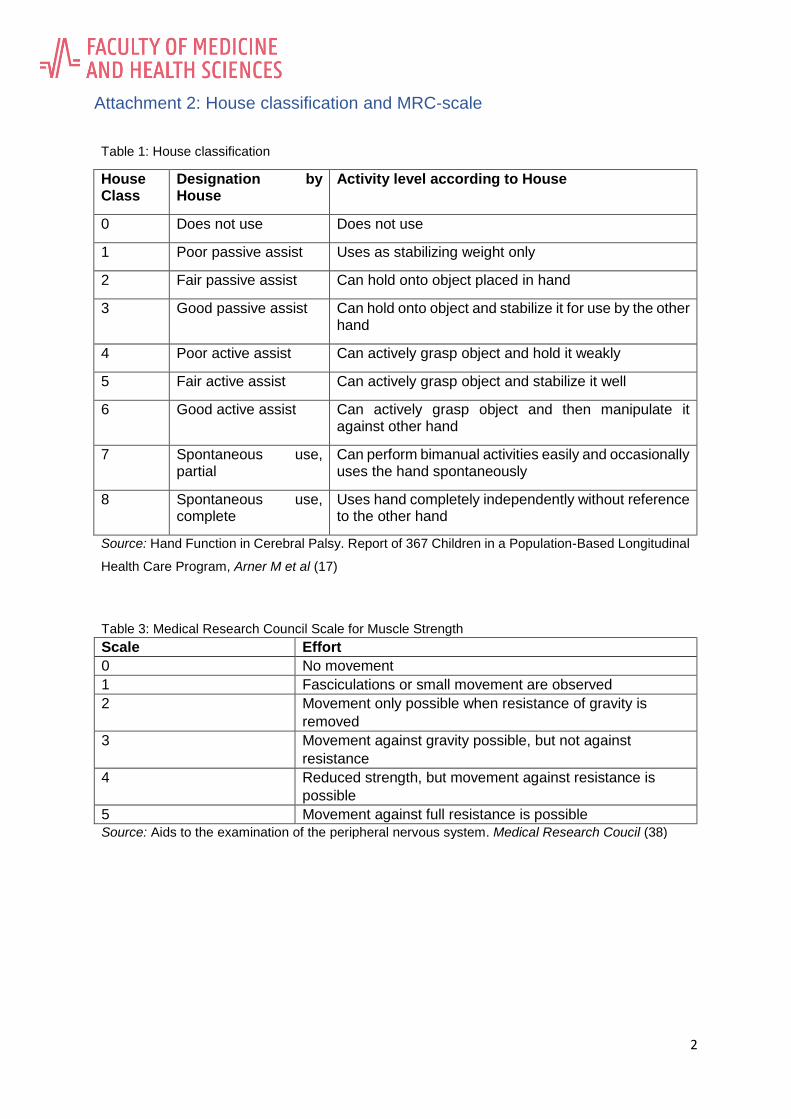
2
Attachment 2: House classification and MRC-scale
Table 1: House classification
House Class
Designation by House
Activity level according to House
0 Does not use Does not use
1 Poor passive assist Uses as stabilizing weight only
2 Fair passive assist Can hold onto object placed in hand
3 Good passive assist Can hold onto object and stabilize it for use by the other hand
4 Poor active assist Can actively grasp object and hold it weakly
5 Fair active assist Can actively grasp object and stabilize it well
6 Good active assist Can actively grasp object and then manipulate it against other hand
7 Spontaneous use, partial
Can perform bimanual activities easily and occasionally uses the hand spontaneously
8 Spontaneous use, complete
Uses hand completely independently without reference to the other hand
Source: Hand Function in Cerebral Palsy. Report of 367 Children in a Population-Based Longitudinal
Health Care Program, Arner M et al (17)
Table 3: Medical Research Council Scale for Muscle Strength
Scale Effort
0 No movement
1 Fasciculations or small movement are observed
2 Movement only possible when resistance of gravity is
removed
3 Movement against gravity possible, but not against
resistance
4 Reduced strength, but movement against resistance is
possible
5 Movement against full resistance is possible
Source: Aids to the examination of the peripheral nervous system. Medical Research Coucil (38)

3
Attachment 3: Informed consent for a child above the age of 12

4

5
Attachment 4: Informed consent for a child younger than 12 years old

6

7
Attachment 5: Approval of the Ethics Committee

8

9
Attachment 6: Tables of the parameters and the results Table 4: Overview analytical parameters
Parameter Measurements L/R
AROM pronation Maximum pronation
In degrees
Symmetry index
(Non-dom/dom)
AROM supination Maximum supination
In degrees
Symmetry index
(Non-dom/dom)
Supination over
pronation index
(max. sup)/(-(max. pron) Symmerty index
(Non-dom/dom)
AROM Mean active range of motion
of 9 cycles, SD
In degrees
Symmetry index
(Non-dom/dom)
Average peak angular
velocity pronation
Average peak angular
velocity of 9 cycles, SD
In rad per second
Symmetry index
(Non-dom/dom)
Average peak angular
velocity supination
Average peak angular
velocity of 9 cycles, SD
In rad per second
Symmetry index
(Non-dom/dom)
Supination over
pronation index
(average peak ang. vel.
sup)/(-(average peak ang.
vel. pron)
Symmerty index
(Non-dom/dom)
Average peak angular
accerleration
pronation
Average peak angular
acceleration of 9 cycles, SD
In rad per second per
second
Symmerty index
(Non-dom/dom)
Average peak angular
acceleration
supination
Average peak angular
acceleration of 9 cycles, SD
In rad per second per
second
Symmerty index
(Non-dom/dom)
Supination over
pronation index
(average peak ang. acc.
sup)/(-(average peak acc.
pron)
Symmerty index
(Non-dom/dom)
Jerk Area under the acceleration
curve, SD
Symmetry index
(Non-dom/dom)
Table 5: Overview of the parameters of the ping-pong exercise
Parameter Measurements L/R
AROM pronation Maximum pronation
In degrees
Symmetry index
(Non-dom/dom)
AROM supination Maximum supination
In degrees
Symmetry index
(Non-dom/dom)
AROM Mean active range of
motion, SD
In degrees
Symmetry index
(Non-dom/dom)
Mean angular velocity
pronation
Mean angular velocity for
pronation, SD
Symmetry index
(Non-dom/dom)

10
In rad per second
Mean angular velocity
supination
Mean angular velocity for
supination, SD
In rad per second
Symmetry index
(Non-dom/dom)
Average peak angular
acceleration
pronation
Average peak angular
acceleration for pronation,
SD
In rad per second per
second
Symmetry index
(Non-dom/dom)
Average peak angular
acceleration
supination
Average peak angular
acceleration for supination,
SD
In rad per second per
second
Symmetry index
(Non-dom/dom)
Number of completed
cycles
Mean # completed cycles
Symmetry index
(Non-dom/dom)
Deadtime Mean time
In ms
Symmetry index
(Non-dom/dom)
Table 6: Overview of the parameters of the hand-to-head exercise
Parameter Measurements L/R
AROM pronation Maximum pronation
In degrees
Symmetry index
(Non-dom/dom)
AROM supination Maximum supination
In degrees
Symmetry index
(Non-dom/dom)
AROM Mean active range of motion,
SD
In degrees
Symmetry index
(Non-dom/dom)
Success Yes or no No symmetry index
Table 7: Overview of the parameters of the clapping exercise
Parameter Measurements L/R
AROM pronation Maximum pronation
In degrees
Symmetry index
(Non-dom/dom)
AROM supination Maximum supination
In degrees
Symmetry index
(Non-dom/dom)
AROM Mean active range of motion,
SD
In degrees
Symmetry index
(Non-dom/dom)
Move to midline Movement of the wrist markers
relative to the shoulder
markers
In mm
Symmetry index
(L and R
separately)

11
Table 8: Results of the bilateral analytical exercises
Patient
Dominant
side
Affected side
Control
Dominant side
Non-dominant
side
AROM Pronation
Max (°)
62.0
55.1
74.9
76.7
Symmetry 0.89 1.02
AROM Supination
Max (°)
-61.3
25.8
-70.0
-60.6
Symmetry -0.42 0.87
Supination over pronation
index
(Max sup)/(-(max pron)
0.99
-0.47
0.94
0.79
symmetry -0.47 0.84
AROM range
Mean range (°)
SD (°)
112.2
4.3
12.4
3.7
133.9
8.1
129.1
6.1
Symmetry 0.11 0.96
Velocity Pronation
Average peak (rad/s)
SD (rad/s)
3.6 0.7
0.2
0.1
4.5
0.7
4.8
0.6
Symmetry 0.06 1.07
Velocity Supination
Average peak (rad/s)
SD (rad/s)
-2.7
0.4
-0.2
0.1
-3.9
1.0
-4.6
0.8
Symmetry 0.07 1.18
Supination over pronation
index
(average peak vel. sup)/(-
(average peak vel. pron)
0.75
1.00
0.87
0.96
symmetry 1.33 1.11
Acceleration Pronation
Average peak (rad/s/s)
SD (rad/s/s)
11.5
3.4
0.5
0.2
17.5
4.6
19.7
3.8
Symmetry 0.04 1.13
Acceleration Supination
Average peak (rad/s/s)
SD (rad/s/s)
-10.5
3.3
-0.5
0.2
-15.0
4.4
-18.1
4.4
Symmetry 0.05 1.21
Supination over pronation
index
(average peak acc. sup)/(-
(average peak acc. pron)
0.91
1.00
0.86
0.92
symmetry 1.10 1.07
Jerk
Mean Jerk
SD
222.9
40.6
15.8
7
287.5
68.8
315.9
73.8
Symmetry 0.07 1.09

12
Table 9: Results of the unilateral analytical exercises
Patient
Dominant
side
Affected side
Control
Dominant side
Non-dominant
side
AROM Pronation
Max (°)
65.6
53.6
69.5
78.1
Symmetry 0.82 1.12
AROM Supination
Max (°)
-64.2
32.8
-78.0
-62.6
Symmetry -0.51 0.80
Supination over pronation
index
(Max sup)/(-(max pron)
0.98
-0.61
1.12
0.80
Symmetry -0.62 0.71
AROM range
Mean range (°)
SD (°)
121.3
5.8
12.4
2.6
137.2
5.8
128.7
5.7
Symmetry 0.11 0.94
Velocity Pronation
Average peak (rad/s)
SD (rad/s)
4.6 0.6
0.2
0.1
5.5
1.0
5.9
0.8
Symmetry 0.04 1.07
Velocity Supination
Average peak (rad/s)
SD (rad/s)
-4.2
0.6
-0.2
0.1
-5.0
0.3
-6.4
1.6
Symmetry 0.05 1.28
Supination over pronation
index
(average peak vel. sup)/(-
(average peak vel. pron)
0.91
1.00
0.91
1.09
Symmetry 1.10 1.20
Acceleration Pronation
Average peak (rad/s/s)
SD (rad/s/s)
16.5
3.4
0.5
0.1
24.4
7.1
34.8
8.4
Symmetry 0.03 1.43
Acceleration Supination
Average peak (rad/s/s)
SD (rad/s/s)
-15.5
3.7
-0.4
0.1
-21.0
4.0
-33.1
11.3
Symmetry 0.03 1.58
Supination over pronation
index
(average peak acc. sup)/(-
(average peak acc. pron)
0.94
0.80
0.86
0.95
Symmetry 0.85 1.11
Jerk
Mean Jerk
SD
424.6
126.7
21.7
5.8
395.0
91.3
573.4
120.8
Symmetry 0.05 1.45

13
Table 10: Reference angles
Patient Control
Dominant side Affected side Dominant side Non-dominant side
Calibration 90°
pronation
45.7°
SD 0.6°
47.5°
SD 1.3°
46.5°
SD 0.9°
48.8°
SD 1.1°
Calibration 60°
pronation
25.2°
SD 1.1°
29.3°
SD 0.3°
29.8°
SD 0.3°
26.2°
SD 0.6°
Calibration
range
20.5° 18.2° 16.7° 22.6°
Ping-pong max
pronation
37.2°
56.6° 53.0°
34.6°
Ping-pong max
supination
ERROR 46.0° -73.8°
-28.4°
Table 11: Results of the ping-pong exercises
Patient
Dominant
side
Affected side
Control
Dominant side
Non-dominant
side
AROM Pronation
Max (°)
90.0
53.1
86.3
77.5
Symmetry 0.59 0.90
AROM Supination
Max (°)
-77.1
43.7
-77.5
-75.6
Symmetry -0.57 0.98
AROM range
Mean range (°)
SD (°)
142.1
11.5
3.0
2.3
134.3
29.7
118.7
15.8
Symmetry 0.02 0.88
Velocity Pronation
Mean ang. vel. (rad/s)
SD (rad/s)
8.5 0.7
0.1
0.0
9.7
2.6
9.1
1.4
Symmetry 0.01 0.94
Velocity Supination
Mean ang. vel.(rad/s)
SD (rad/s)
-8.9
1.1
0.0
0.0
-10.2
2.5
-9.6
1.6
Symmetry 0.00 0.94
Acceleration Pronation
Average peak (rad/s/s)
SD (rad/s/s)
153.1
28.2
0.2
0.4
197.1
42.1
202.7
43.5
Symmetry 0.00 1.03
Acceleration Supination
Average peak (rad/s/s)
SD (rad/s/s)
-144.0
23.6
-0.2
0.3
-242.2
45.1
-253.3
47.3
Symmetry 0.00 1.05
# Completed cycles
Mean #
5.3
4
8.3
9.3
Symmetry 0.76 1.12
Deadtime
Time (ms)
40.0
45.3
40.3
35
Symmetry 1.13 0.87

14
Table 12: Results of the hand-to-head exercises
Patient
Dominant
side
Affected side
Control
Dominant side
Non-dominant
side
AROM Pronation
Max (°)
55.4
94.9
63.1
62.1
Symmetry 1.71 0.98
AROM Supination
Max (°)
-68.4
35.5
-62.2
-43.7
Symmetry -0.52 0.70
AROM range
Mean range (°)
SD (°)
113.7
3.8
40.4
14.9
104.8
8.1
91.8
6.7
Symmetry 0.36 0.88
Success
Yes/No
Yes
No
Yes
Yes
Table 13: Results of the clapping exercises
Patient
Dominant
side
Affected side
Control
Dominant side
Non-dominant
side
AROM Pronation
Max (°)
80.5
48.4
56.6
51.0
Symmetry 0.60 0.90
AROM Supination
Max (°)
-47.2
29.4
-36.9
-40.4
Symmetry -0.62 1.09
AROM range
Mean range (°)
SD (°)
83.6
10.9
9.4
4.4
74.3
6.8
67.4
7.5
Symmetry 0.11 0.91
Distance to the midline
Mean distance (mm)
SD
97.3
33.0
35.6
22.6
20.3
13.1
54.9
15.8
Symmetry 0.37 2.70