
Molecular programs induced in alveolar resident and recruited
119
Molecular programs induced in alveolar resident and recruited mononuclear phagocytes by Toll-like receptor 2/4 agonists Inaugural Dissertation submitted to the Faculty of Medicine in partial fulfillment of the requirements for the PhD-Degree of the Faculties of Veterinary Medicine and Medicine of the Justus Liebig University Giessen by Cabanski Maciej of Inowroclaw, Poland Giessen 2009
Transcript of Molecular programs induced in alveolar resident and recruited
Molecular programs induced in alveolar resident and recruited mononuclear phagocytes by Toll-like receptor
2/4 agonists
Inaugural Dissertation
submitted to the
Faculty of Medicine
in partial fulfillment of the requirements
for the PhD-Degree
of the Faculties of Veterinary Medicine and Medicine
of the Justus Liebig University Giessen
by
Cabanski Maciej
of
Inowroclaw, Poland
Giessen 2009

From the Department of Medicine
Director / Chairman: Prof. Dr. Werner Seeger
of the Faculty of Medicine of the Justus Liebig University Giessen
First Supervisor and Committee Member: Prof. Dr. Jürgen Lohmeyer
Second Supervisor and Committee Member: Prof. Dr. Stefan Hippenstiel
Committee Members: Prof. Dr. Klaus T. Preissner
Prof. Dr. Michael Martin
Date of Doctoral Defense: 30.01.2009
TABLE OF CONTENTS
I. TABLE OF CONTENTS
I. TABLE OF CONTENTS................................................................................. I
II. LIST OF FIGURES ...................................................................................... IV
III. LIST OF TABLES......................................................................................... VI
IV. ABBREVIATIONS ....................................................................................... VII
V. SUMMARY.................................................................................................... X
VI. ZUSAMMENFASSUNG .............................................................................. XII
1 INTRODUCTION .......................................................................................... 1
1.1 INNATE IMMUNITY............................................................................. 1
1.1.1 Recognition of microbial components by pattern recognition
receptors ................................................................................................... 1
1.1.2 Toll-like receptors.......................................................................... 2
1.1.3 TLR-mediated signalling pathways ............................................... 6
1.2 INNATE IMMUNITY IN THE LUNGS..................................................13
1.2.1 Mononuclear phagocytes in lung host defence ............................14
1.2.2 Lung inflammatory response ........................................................20
2 AIM OF THE STUDY ...................................................................................23
3 MATERIALS AND METHODS .....................................................................24
3.1 Materials .............................................................................................24
3.1.1 Animals ........................................................................................24
3.1.2 Equipment ....................................................................................24
3.1.3 Kits ...............................................................................................26
I
TABLE OF CONTENTS
3.1.4 Reagents......................................................................................26
3.1.5 Software .......................................................................................28
3.2 Methods..............................................................................................29
3.2.1 Treatment of animals....................................................................29
3.2.2 Cell culture and stimulation ..........................................................30
3.2.3 RNA analysis................................................................................31
3.2.4 Protein analysis............................................................................34
3.2.5 Statistical analysis ........................................................................37
4 RESULTS ....................................................................................................39
4.1 Gene expression of TLR associated genes induced by inflammatory
stimuli in AMs...............................................................................................39
4.2 Role of PKR in TLR2/4 signalling in murine AMs................................43
4.2.1 PKR protein expression after stimulation with TLR2/4 ligands .....43
4.2.2 Induction of PKR phosphorylation by TLR2/4 ligands ..................43
4.2.3 Involvement of PKR in PAMP-induced release of inflammatory
cytokines ..................................................................................................47
4.2.4 Role of PKR in activation of NF-�B ..............................................48
4.2.5 The role of PKR in MAPKs signalling pathway.............................50
4.3 Genome-wide transcriptional profiling of MPs recruited to mouse lungs
in response to alveolar challenge with the TLR2-agonist Pam3CSK4...........53
4.3.1 Induction of lung inflammation and alveolar trafficking of circulating
MPs by intratracheal deposition of Pam3CSK4 .........................................53
4.3.2 Genome-wide transcriptional profiling of MPs recruited to the
alveolar space after pulmonary challenge with Pam3CSK4 .....................56
4.3.3 Real time PCR validation of microarray results ............................63
5 DISCUSSION...............................................................................................66
5.1 Requirement for PKR in TLR2/4-medaited signalling in murine AMs..66
5.1.1 TLR2 and TLR4 ligand induced phosphorylation of PKR .............69
II
TABLE OF CONTENTS
5.1.2 PKR-dependent secretion of TNF-� and IL-6 cytokines ...............71
5.1.3 Requirement of PKR for LPS induced activation of NF-�B...........72
5.1.4 PKR dependent regulation of JNK signalling pathway .................73
5.2 Transcriptional profiling of MPs recruited to mouse lungs in response to
alveolar challenge with the TLR2-agonist Pam3CSK4 ..................................76
5.2.1 Alveolar trafficking of peripheral blood MPs is associated with global
changes in their gene expression profile ..................................................76
5.2.2 Attenuation of pro-inflammatory gene expression levels in alveolar
MPs during late inflammation resolution phase ........................................79
5.3 Future perspectives ............................................................................81
6 APPENDIX...................................................................................................83
7 REFERENCES ............................................................................................86
8 DECLARATION ...........................................................................................99
9 CURRICULUM VITAE ...............................................................................100
10 ACKNOWLEDGEMENTS ..........................................................................103
III
LIST OF FIGURES
II. LIST OF FIGURES
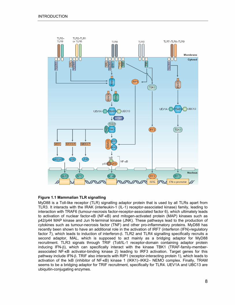
Figure 1.1 Mammalian TLR signalling. .................................................................... 8
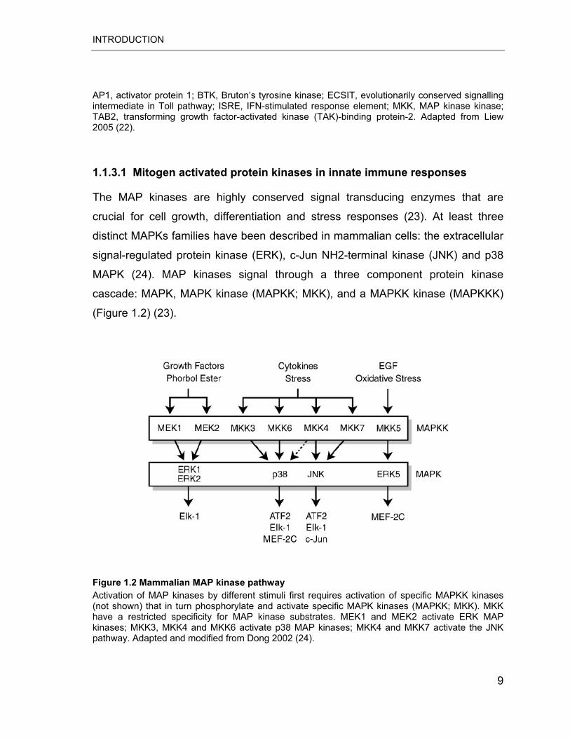
Figure 1.2 Mammalian MAP kinase pathway. ......................................................... 9
Figure 1.3 Monocyte differentiation into DCs and tissue macrophages ................ 17
Figure 1.4 Lung inflammatory response to microbial challenge ............................ 21
Figure 4.1 Flow cytometric identification and separation of AMs of untreated and
LPS treated WT mice ............................................................................................ 41
Figure 4.2 PKR protein accumulation after stimulation with TLR ligands .............. 43
Figure 4.3 Time-course analysis of PKR phosphorylation..................................... 44
Figure 4.4 Activation of PKR by PAMPs is TLR dependent .................................. 45
Figure 4.5 Pharmacological inhibition of PKR phosphorylation by 2-AP ............... 46
Figure 4.6 PAMP-induced inflammatory cytokine secretion requires PKR
phosphorylation..................................................................................................... 47
Figure 4.7 Pam3CSK4-induced activation of NF-�B is PKR-independent .............. 48
Figure 4.8 Immunofluorescence microscopy of p65 nuclear translocation............ 49
Figure 4.9 PKR regulates the TLR2/4 ligand-induced JNK signalling pathway ..... 51
Figure 4.10 PKR regulates JNK signalling pathway via MKK4.............................. 52
Figure 4.11 Inflammatory cell accumulation in the bronchoalveolar compartment in
response to alveolar deposition of Pam3CSK4 ...................................................... 54
Figure 4.12 Lung recruitment of MPs in the absence or presence of the Pam3CSK4
.............................................................................................................................. 55
IV
LIST OF FIGURES
Figure 4.13 Global gene expression differences of alveolar recruited vs. PB MPs
after i.t. application of Pam3CSK4.......................................................................... 56
Figure 4.14 Real-time quantitative PCR validation of microarray data .................. 64
Figure 4.15 Time-dependent changes in the gene expression profile of alveolar
recruited MPs........................................................................................................ 65
Figure 5.1 Functions of PKR in signal transduction and translation ...................... 69
Figure 5.2 Model of PKR transducer function in TLR2/4-mediated signalling in AMs
.............................................................................................................................. 75
Figure 6.1 ELISA for TLR2/4 ligands..................................................................... 84
Figure 6.2 TransAM assay control ........................................................................ 84
Figure 6.3 Secondary antibody control.................................................................. 85
V
LIST OF TABLES
III. LIST OF TABLES
Table 1.1 Toll like receptors and their ligands. ........................................................ 4
Table 1.2 Components of the innate immune system of the lung.......................... 13
Table 1.3 Secretory products of alveolar macrophages ........................................ 16
Table 1.4 Surface antigen expression on murine and human monocyte subsets . 19
Table 4.1 mRNA expression of TLR associated genes in cultured AMs ............... 40
Table 4.2 mRNA expression of TLR associated genes in flow-sorted AMs .......... 42
Table 4.3 Differential gene expression between alveolar recruited and PB MPs .. 58
Table 4.4 Gene ontology (GO) biological processes classification of differentially
regulated genes .................................................................................................... 62
Table 6.1 List of primers used for quantitative real-time PCR ............................... 83
Table 6.2 List of antibodies used for western blotting and immunofluorescence
microscopy. ........................................................................................................... 85
VI
ABBREVIATIONS
IV. ABBREVIATIONS
2-AP 2-aminopurine
AM Alveolar macrophage
AP-1 Activator protein-1
APS Amonium persulfate
ATP Adenosine triphosphate
BAL(F) Bronchoalveolar lavage (fluid)
BLAST Basic local alignment search tool
CCL CC Chemokine ligand
CD Cluster of differentiation
cDNA Complementary DNA
DAPI 4´, 6´-diamidino-2-phenylindole
DC Dendritic cell
dNTP Deoxynucleotide triphosphate
dsRNA Double stranded RNA
DTT Dithiothreitol
ERK Extracellular signal-regulated protein kinase
FCS Fetal calf serum
FSC Forward side scatter
GFP Green fluorescent protein
GPCR G protein-coupled receptor
GPI Glycosylinositol
VII
ABBREVIATIONS
I�B Inhibitor of �B
IFN Interferon
IKK I�B kinase
IL Interleukin
JNK c-Jun NH2-terminal kinase
KO Knock-out
LBP LPS-binding protein
LPS Lipopolysaccharide
LRR Leucine-rich repeat
MAPK Mitogen-activated protein kinase
MCP Monocyte chemoattractant protein
MEK MAP kinase kinase
MIP Macrophage inflammatory protein
MP Mononuclear phagocyte
mRNA Messenger RNA
MyD88 Myeloid differentiation primary-response gene 88
NF-�B Nuclear factor �B
OD Optical density
Pam3CSK4 Pam3-Cys-Ser-Lys-Lys-Lys-Lys-OH
PAMPs Pathogen associated molecular patterns
PB Peripheral blood
PBGD Porphobilinogen deaminase
PCR Polymerase chain reaction
PG Prostaglandin
VIII
ABBREVIATIONS
PKR Protein kinase R
PMN Polymorphonuclear leukocyte
Poly I:C Polyriboinosinic:polyribocytydylic acid
PRR Pattern recognition receptor
RANTES Regulated upon activation normal T cell
RNAsin Ribonuclease inhibitor
SDS Sodium dodecyl sulphate
SPF Specific pathogen free
SSC Side scatter
TGF-� Transforming growth factor beta
TIR Toll/IL-1 receptor domain
TLR Toll-like receptor
TNF-� Tumor necrosis factor alpha
WT Wild type
IX
SUMMARY
V. SUMMARY
Mononuclear phagocytes play a pivotal role in lung host defence to inhaled
pathogens by activation of both innate and adaptive immunity. Resident alveolar
macrophages (AMs) are the primary mononuclear phagocytic cells found in the
lower respiratory tract that play a central role in regulating pulmonary immune
responses. Circulating mononuclear phagocytes (MPs) recruited into inflamed
lungs, however are increasingly implicated as essential players in defence against
a range of inhaled pathogens.
To further elucidate the function of mononuclear phagocytes in regulating
pulmonary immune responses we analysed molecular programs induced by
bacterial ligands that are recognized by different TLRs. First, we discovered that
TLR2 ligand Pam3CSK4 and TLR4 ligand LPS induced in AMs the expression of
PKR, previously identified as an essential component of the innate antiviral
response. More important we found that both TLR2 and TLR4 agonists induced
rapid phosphorylation of PKR strictly dependent on the functionality of the
respective TLR. Pharmacologic inhibition of PKR activity using 2-aminopurine
(2-AP) and PKR gene deletion were found to reduce the TLR2/4-induced activation
of the JNK signalling pathway (MKK4/JNK/c-Jun), but did not affect p38 and
ERK1/2 activation. Moreover, inhibition of PKR phosphorylation severely impaired
TNF-� and IL-6 production by AMs in response to LPS and Pam3CSK4.
Additionally, we found that PKR phosphorylation plays a major role in LPS but not
Pam3CSK4-induced activation of the p65 subunit of NF-�B. Collectively, these
results indicate that functional PKR is critically involved in inflammatory responses
of primary AMs to gram-positive as well as gram-negative bacteria cell wall
components.
In addition, we investigated the induction of lung inflammation and the concomitant
MP recruitment after alveolar deposition of the TLR2 ligand Pam3CSK4. By using
cell sorting, mRNA pre-amplification and whole genome oligonucleotide microarray
X
SUMMARY
techniques we found that alveolar trafficking of MPs was associated with profound
changes of their gene expression profiles post recruitment (~2500 genes
increased). In particular, alveolar recruited MPs showed strong up-regulation for
genes encoding cytokines/chemokines, PRR associated molecules and genes
involved in eicosanoid metabolism. Interestingly, gene expression profiling
revealed that lung recruited MPs displayed simultaneous induction of both pro- and
anti-inflammatory genes. However, we observed a dynamic change of the genetic
program of MPs found in BALF at different time intervals post challenge. Strong
early induction of a subset of pro-inflammatory mediators such as TNF-�, CCL2
and IL-6 was found to decrease during the later resolution phase whereas
increased transcript levels of central anti-inflammatory and pro-resolution
mediators including IL-1RN, IRAK-M, IL-10 and BAX persisted at the same levels.
Collectively, our in vivo study identifies for the first time the global genetic program
activated in MPs at different time points during TLR2 ligand-induced recruitment to
the alveolar space and thus may help to better understand how alveolar recruited
MPS may contribute to the development and termination of pneumonia caused by
gram-positive bacteria.
XI
ZUSAMMENFASSUNG
VI. ZUSAMMENFASSUNG
Mononukleäre Zellen spielen eine entscheidende Rolle bei der angeborenen und
adaptiven Immunabwehr von inhalierten Pathogenen. Alveolarmakrophagen (AM)
stehen dabei an vorderster Front und spielen eine zentrale Rolle bei der
Regulation der pulmonalen Immunantwort. Darüber hinaus wird in der letzten Zeit
zunehmend klar, dass auch Monozyten, die aus dem Gefäßbett in das entzündete
Lungengewebe einwandern, eine wesentliche Rolle in der Wirtsabwehr pulmonaler
Infektionen spielen.
Um die Rolle der residenten und rekrutierten mononukleären Zellpopulationen im
Rahmen der pulmonalen Immunabwehr genauer zu untersuchen, analysierten wir
Signaltransduktions-Kaskaden, die durch Pathogen-assoziierte Gefahrensignale
über verschiedene Toll-like Rezeptoren in diesen Zellen induziert werden. Hierbei
zeigte sich, dass Pam3CSK4 über TLR2 und LPS über TLR4 in AMs die
Expression des Enzyms PKR induziert, das bislang als wichtige Komponente der
angeborenen antiviralen Immunität bekannt war. Außerdem führt die Aktivierung
von TLR2/4 zu einer sehr schnellen Phosphorylierung von PKR, was die
Aktivierung nachgeschalteter Signaltransduktions-Kaskaden entscheidend
mitbestimmt. So reduzierte die Inhibierung der PKR durch 2-Aminopurin (2-AP)
oder die Gendeletion des PKR-Gens die TLR2/4-induzierte Aktivierung des
JNK-Signalweges (MKK4/JNK/c-Jun). Im Gegensatz dazu erfolgte keine Inhibition
der p38 und ERK1/2 Aktivierung. Darüber hinaus führte eine Inhibition der
PKR-Phosphorylierung zu einer starken Hemmung der TNF-� und IL-6-Produktion
der AMs nach LPS- und Pam3CSK4-Stimulation. Außerdem konnten wir zeigen,
dass die PKR-Phosphorylierung eine bedeutende Rolle bei der Aktivierung der
LPS-induzierten, nicht jedoch der Pam3CSK4-induzierten Aktivierung der p65
Untereinheit von NF-�B spielt. Zusammenfassend legen diese Ergebnisse die
Vermutung nahe, dass PKR eine kritische Rolle bei der durch
XII
ZUSAMMENFASSUNG
Zellwandbestandteile gram-positiver und –negativer Bakterien induzierten
inflammatorischen Antwort von Alveolarmakrophagen spielt.
In einem zweiten Ansatz untersuchten wir Mechanismen der Induktion einer
pulmonalen Entzündungs-Reaktion und der nachfolgenden Einwanderung von
Blut-Monozyten nach alveolärer Deposition des TLR2-Liganden Pam3CSK4. Wir
kombinierten Zellsorting, mRNA-Präamplifikation und Gesamtgenom-Microarray
Technologien, um Veränderungen des globalen genetischen Programms von
Monozyten bei Einwanderung in den Alveolarraum zu untersuchen. Hierbei fanden
sich tief greifende Veränderungen der Genexpression in den mononukleären
Phagozyten nach alveolärer Rekrutierung, wobei die Expression von ca. 2500
Genen signifikant erhöht war. Im Einzelnen konnten wir eine Erhöhung der
Expression von Genen der Cytokin-und Chemokin-Familien, Pattern-Recognition-
Rezeptoren sowie Eiconsanoid-metabolisierenden Enzyme nachweisen. Dabei
wurde interessanter Weise sowohl die Transkription pro- als auch
antiinflammatorischer Gene induziert. Allerdings zeigten mononukleäre
Phagozyten, die zu verschiedenen Zeitpunkten nach Pam3CSK4-induzierter
Rekrutierung analysiert wurden, deutliche Unterschiede in ihrem
Genexpressionsprofil: mRNA Spiegel von pro-inflammatorischen Mediatoren wie
TNF-�, CCL2 und IL-6 waren zu frühen Zeitpunkten stark erhöht und sanken dann
rasch ab, während die ebenfalls bereits früh erhöhte Genexpression von zentralen
anti-inflammatorischen Mediatoren wie IL-1RN, IRAK-M, IL-10 und BAX auch zu
späteren Zeitpunkten persistierte. Zusammenfassend erfassen diese
Untersuchungen zum ersten Mal detailliert das globale genetische Programm
aktivierter mononukleärer Phagozyten zu unterschiedlichen Zeitpunkten nach
TLR2-Ligand-induzierter Rekrutierung in den Alveolar-Raum. Dies erlaubt einen
Einblick in das Funktionsrepertoire von mononukleären Phagozyten, mit dem diese
zur pulmonalen Entzündungsinduktion und –terminierung bei durch gram-positive
Bakterien hervorgerufenen Pneumonien beitragen können.
XIII
INTRODUCTION
1 INTRODUCTION
1.1 INNATE IMMUNITY All multi-cellular eukaryotic organisms are constantly exposed to millions of
potential pathogens daily through contact, ingestion and inhalation. To effectively
detect, control and eliminate invading microbial pathogens, higher vertebrates
have developed two interacting protective systems: the innate and adaptive
immune systems. Innate immunity is an evolutionary conserved host defence
armentarium that provides initial protection against invading micro-organisms.
Remarkably, innate immunity has a higher level of specificity in its ability to
distinguish distinct danger signals from different origin than was originally thought.
It employs soluble factors and germline-encoded receptors called pattern
recognition receptors (PRRs) to recognize specific components of microbes called
pathogen-associated molecular patterns (PAMPs). PAMPs are evolutionary
conserved structures that are essential for microbial physiology and survival.
PAMPs include for instance viral double-stranded RNA (dsRNA),
lipopolysaccharide (LPS) derived from gram-negative bacteria, un-methylated CpG
di-nucleotides or lipoproteins of bacteria and fungi (1, 2). Recognition of PAMPs by
PRRs activates different intracellular signalling pathways that result in the
increased production of inflammatory mediators such as cytokines and
chemokines, as well as recruitment of immune cells to inflamed tissues. Among
PRRs, Toll-like receptors (TLRs) were highlighted as key recognition structures of
the innate immune system.
1.1.1 Recognition of microbial components by pattern recognition receptors
The innate immune system utilizes a variety of PRRs that can be expressed on the
cell surface, in intracellular compartments, or secreted into the bloodstream and
1
INTRODUCTION
tissue fluids. The effectors functions of PRRs after ligand binding include
opsonization, activation of complement and coagulation cascades, phagocytosis,
activation of pro-inflammatory signalling pathways, and induction of apoptosis.
Functionally PRRs can be divided into three classes: secreted, endocytic, and
signalling. Mannan-binding lectin (MBL), C-reactive protein (CRP), and serum
amyloid protein (SAP) are secreted pattern recognition molecules, whereas
macrophage mannose receptor (MMR) and macrophage scavenger receptor
(MSR) represent endocytic PRRs. The TLR family is the most investigated class of
signalling PRRs and appears to have a major role in the induction of immune and
inflammatory responses (3).
1.1.2 Toll-like receptors
Following the discovery of the role of Drosophila melanogaster Toll receptor in
antifungal immunity, a mammalian homologue of Toll protein termed Toll-like
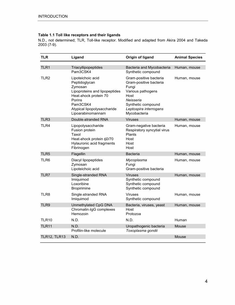
receptor (later named TLR4) was identified (4). The TLR family now consists of 13
mammalian members: 10 human (TLR1–10) and 12 murine (TLR1–9 and
TLR11–13) receptors, of which some are homologues (Table 1.1) (5). Structurally,
the intracellular domain of TLRs shows similarity to that of the interleukin-1
receptor (IL-1R) family and therefore is termed a Toll/IL-1R domain (TIR). The
extracellular portion of TLR, however, is characterized by the presence of
leucine-rich repeat (LRR) motifs, whereas the IL-1R family possesses extracellular
immunoglobulin-like domains. LRR domains are implicated in the recognition of
PAMPs (5, 6). The main PAMP ligands for TLR1 to TLR13 are summarised in
table 1.1.
TLR family members are expressed on various immune cells including monocytes,
macrophages, dendritic cells (DCs), B cells, and even on non-myeloid cells such
as epithelial cells and fibroblasts (7). While some TLRs have been found to be
expressed primarily on the cell surface (TLR1, TLR2, TLR4, TLR5, TLR6 and
TLR11) others are found almost completely in intracellular compartments such as
endosomes (TLR3, TLR7, TLR8 and TLR9) (7). The cellular expression of TLRs is 2

INTRODUCTION
modulated rapidly in response to various stimuli such as pathogens, cytokines and
environmental stresses (7, 8). Of the TLRs identified so far, TLR4 and TLR2 have
been the most extensively studied.
3
INTRODUCTION
Table 1.1 Toll like receptors and their ligands N.D., not determined; TLR, Toll-like receptor. Modified and adapted from Akira 2004 and Takeda 2003 (7-9).
TLR1 Triacyllipopeptides Bacteria and Mycobacteria Human, mousePam3CSK4 Synthetic compound
TLR2 Lipoteichoic acid Gram-positive bacteria Human, mousePeptidoglycan Gram-positive bacteriaZymosan FungiLipoproteins and lipopeptides Various pathogensHeat-shock protein 70 HostPorins NeisseriaPam3CSK4 Synthetic compoundAtypical lipopolysaccharide Leptospira interrogansLipoarabinomannam Mycobacteria
TLR3 Double-stranded RNA Viruses Human, mouse
TLR4 Lipopolysaccharide Gram-negative bacteria Human, mouseFusion protein Respiratory syncytial virus Taxol PlantsHeat-shock protein 60/70 HostHylauronic acid fragments HostFibrinogen Host
TLR5 Flagellin Bacteria Human, mouse
TLR6 Diacyl lipopeptides Mycoplasma Human, mouseZymosan FungiLipoteichoic acid Gram-positive bacteria
TLR7 Single-stranded RNA Viruses Human, mouseImiquimod Synthetic compoundLoxoribine Synthetic compoundBropirimine Synthetic compound
TLR8 Single-stranded RNA Viruses Human, mouseImiquimod Synthetic compound
TLR9 Unmethylated CpG DNA Bacteria, viruses, yeast Human, mouseChromatin-IgG complexes HostHemozoin Protozoa
TLR10 N.D. N.D. Human
TLR11 N.D. Uropathogenic bacteria MouseProfilin-like molecule Toxoplasma gondii
TLR12, TLR13 N.D. Mouse
TLR Ligand Origin of ligand Animal Species
4
INTRODUCTION
1.1.2.1 Toll-like receptor 4 (TLR4)
TLR4 is an essential receptor for the recognition of LPS (also referred as
endotoxin), a major component of the outer membrane of gram-negative bacteria
such as Escherichia coli or Salmonella typhimurium. The link between TLR4 and
LPS appeared in studies showing that genetic mutation responsible for
hypo-responsiveness to LPS in two naturally occurring mouse strains, C3H/HeJ
and C7BL10/ScCr was within the TLR4 gene (10, 11). Similarly, mice with targeted
deletion in the TLR4 gene are hypo-responsive to LPS (12). Later studies
discovered that TLR4 is necessary for the signalling of a range of other pathogen
components and also several endogenous host ligands (Table 1.1) (8).
TLR4 is important for host defence against different types of microbes including
Salmonella, Mycobacterium tuberculosis, Candida albicans or Haemophilus
influenzae (13). However, TLR4 is not the sole receptor involved in LPS
recognition. LPS first interacts with a plasma protein named LPS-binding protein
(LBP) to form stable LBP:LPS complexes that are recognized by the CD14
receptor on the cell surface (14). CD14 is a membrane glycoprotein anchored in
the lipid bilayer by its glycosylinositol (GPI) tail and is preferentially expressed on
monocyte/macrophage and neutrophil (8). CD14 can also circulate in soluble form
in the plasma (15). The major role of CD14 is to concentrate LPS for effective
binding to the constitutively associated TLR4/MD2 complex (16).
1.1.2.2 Toll-like receptor 2 (TLR2)
TLR2 is involved in the recognition of a variety of microbial and synthetic patterns
including bacterial lipoproteins derived from gram-negative as well as gram-
positive bacteria (e.g., Staphylococcus aureus and Streptococcus pneumoniae) or
mycoplasma spp. (Table 1.1) (8). TLR2 has also been reported to recognize
several atypical forms of LPS such as LPS derived from Leptospira interrogans
(17).
5
INTRODUCTION
The wide spectrum of ligand recognition by TLR2 is mostly determined by
heterodimeric interaction with other TLR family members such as TLR1 or TLR6.
For example, TLR2/TLR1 complex recognizes synthetic triacylated lipoproteins
such as Pam3CSK4, whereas TLR2/TLR6 complexes recognize diacylated
lipoproteins including mycoplasmal macrophage-activating lipopeptide 2 (MALP-2)
(8, 18). In addition, TLR2 has been shown to cooperate with distinct types of other
receptors such as dectin-1 to recognize fungal �-glucans, as well as with CD36 to
recognize diacylglycerides (19). TLR2-deficient mice were found to be highly
susceptible to challenge with S. aureus and S. pneumoniae (13, 20).
1.1.3 TLR-mediated signalling pathways
Binding of microbial components to their respective TLR triggers activation of
signalling pathways, which all make use of adapter proteins (Figure 1.1). Four TIR
domain containing adaptors, namely myeloid differentiation primary-response gene
88 (MyD88), TolI/interleukin-1 receptor domain-containing adapter protein
(TIRAP/Mal), TIR-containing adaptor protein inducing interferon (IFN)-�
(TRIF/TICAM1) and TRIF-related adaptor molecule (TRAM/TICAM-2) were shown
to play important roles in the TLR signalling pathway. MyD88 is essential for the
induction of inflammatory responses triggered by all TLRs with the exception of
TLR3 (7). TIRAP is specifically involved in the MyD88-dependent pathway via
TLR2 and TLR4, whereas TRIF is implicated in the TLR3- and TLR4-mediated
MyD88-independent pathways (2, 21).
The MyD88-dependent pathway includes a number of signalling molecules such
as serine/threonine kinase IL-1R-associated kinase (IRAK), tumour necrosis factor
(TNF)-� receptor-associated factor 6 (TRAF6), transforming growth factor (TGF)-�-
activated kinase 1 (TAK1) and TAK1 binding protein-1 (TAB1). Generally, upon
stimulation with PAMPs, MyD88 recruits IRAK4 to ligated TLRs to facilitate IRAK4
mediated phosphorylation of IRAK1 and IRAK2. Activated IRAK1 associates with
TRAF6 and together with TAK1 leads to the activation of two distinct pathways: the
mitogen-activated protein kinases (MAPKs) and the Rel-family transcription factor 6
INTRODUCTION
nuclear factor-�B (NF-�B). In consequence, the MyD88-dependent pathway leads
to the production of inflammatory cytokines such as TNF-�, IL-6, IL-12, and IL-1�
(5). However, subsequent studies have demonstrated that MyD88-independent
activation of NF-�B or interferon regulatory factor (IRF)-3 transcription factors
occurs in TLR downstream signalling as well. This pathway has been implicated in
the expression of IFN-inducible genes and LPS-mediated maturation of DCs (5).
7
INTRODUCTION
Figure 1.1 Mammalian TLR signalling MyD88 is a Toll-like receptor (TLR) signalling adaptor protein that is used by all TLRs apart from TLR3. It interacts with the IRAK (interleukin-1 (IL-1) receptor-associated kinase) family, leading to interaction with TRAF6 (tumour-necrosis factor-receptor-associated factor 6), which ultimately leads to activation of nuclear factor-�B (NF-�B) and mitogen-activated protein (MAP) kinases such as p42/p44 MAP kinase and Jun N-terminal kinase (JNK). These pathways lead to the production of cytokines such as tumour-necrosis factor (TNF) and other pro-inflammatory proteins. MyD88 has recently been shown to have an additional role in the activation of IRF7 (interferon (IFN)-regulatory factor 7), which leads to induction of interferon-�. TLR2 and TLR4 signalling specifically recruits a second adaptor, MAL, which is supposed to act mainly as a bridging adaptor for MyD88 recruitment. TLR3 signals through TRIF (Toll/IL-1 receptor-domain containing adaptor protein inducing IFN-�), which can specifically interact with the kinase TBK1 (TRAF-family-member-associated NF-�B activator-binding kinase 2) leading to IRF3 activation. Target genes for this pathway include IFN-�. TRIF also interacts with RIP1 (receptor-interacting protein 1), which leads to activation of the I�B (inhibitor of NF-�B) kinase 1 (IKK1)–IKK2– NEMO complex. Finally, TRAM seems to be a bridging adaptor for TRIF recruitment, specifically for TLR4. UEV1A and UBC13 are ubiquitin-conjugating enzymes.
8
INTRODUCTION
AP1, activator protein 1; BTK, Bruton’s tyrosine kinase; ECSIT, evolutionarily conserved signalling intermediate in Toll pathway; ISRE, IFN-stimulated response element; MKK, MAP kinase kinase; TAB2, transforming growth factor-activated kinase (TAK)-binding protein-2. Adapted from Liew 2005 (22).
1.1.3.1 Mitogen activated protein kinases in innate immune responses
The MAP kinases are highly conserved signal transducing enzymes that are
crucial for cell growth, differentiation and stress responses (23). At least three
distinct MAPKs families have been described in mammalian cells: the extracellular
signal-regulated protein kinase (ERK), c-Jun NH2-terminal kinase (JNK) and p38
MAPK (24). MAP kinases signal through a three component protein kinase
cascade: MAPK, MAPK kinase (MAPKK; MKK), and a MAPKK kinase (MAPKKK)
(Figure 1.2) (23).
Figure 1.2 Mammalian MAP kinase pathway Activation of MAP kinases by different stimuli first requires activation of specific MAPKK kinases (not shown) that in turn phosphorylate and activate specific MAPK kinases (MAPKK; MKK). MKK have a restricted specificity for MAP kinase substrates. MEK1 and MEK2 activate ERK MAP kinases; MKK3, MKK4 and MKK6 activate p38 MAP kinases; MKK4 and MKK7 activate the JNK pathway. Adapted and modified from Dong 2002 (24).
9
INTRODUCTION
The classic ERK kinases include ERK1 and ERK2 which are often referred as
p44/p42 MAP kinases. Upon activation, ERK1/2 translocate into the nucleus and
phosphorylate numerous targets including transcription factors such as Elk-1.
Many studies have demonstrated the importance of the ERK pathway in LPS
signalling in monocytes and macrophages (25-27). Macrophages from
Tpl2-deficient mice exhibited selective ERK deficits and reduced TNF-� production
upon LPS stimulation (28). Moreover, transport of TNF-� mRNA from the nucleus
to the cytoplasm was inhibited by specific ERK inhibitors (29). Inhibition of the
MEK-ERK pathway in monocytes by U0126 reduced LPS induction of several
inflammatory mediators including IL-1, IL-8, TNF-� and prostaglandin (PG) E2
(30). Similarly, blocking of the ERK pathway by PD98059 inhibited LPS induction
of tissue factor gene expression in human monocytes (27).
The JNK protein kinases, also known as stress-activated MAP kinases (SAPKs),
are encoded by three genes: Jnk1, Jnk2 and Jnk3 (31). JNK kinases are activated
through phosphorylation by MKK4 and MKK7 (31). JNK activation can be strongly
induced in multiple cell types by LPS or inflammatory cytokines such as TNF-� and
IL-1� (24, 31). For instance, LPS stimulation of THP-1 and RAW 264.7 cells rapidly
activates the JNK pathway (32). A critical role for JNK appears to be the regulation
of the activator protein (AP)-1 transcription factor through phosphorylation of the N-
terminal part of c-Jun, a central component of AP-1 (29, 31). Inhibition of JNK
activity results in decreased levels of TNF-� in LPS-exposed macrophages (29).
MKP5 is a negative regulator of JNK activity (33). Consequently, MKP5-deficient
macrophages produced increased levels of inflammatory cytokines upon TLR
ligand stimulation (34).
The p38 MAPK (known as p38 �) was originally described as a 38-kDa polypeptide
that was rapidly tyrosine phosphorylated in response to LPS and osmotic shock
(35). Subsequently, three additional isoforms of p38 have been identified: �, � and
�. The p38 molecules are strongly activated by environmental stresses,
inflammatory cytokines and bacterial ligands. Once activated, p38 MAPKs
10
INTRODUCTION
modulate responses of different target proteins including transcription factors such
as activating transcription factor (ATF)-2, signal transducer and activator of
transcription (STAT)-1 and Elk-1. Lu and co-workers found that disruption of the
MKK3-p38 MAP kinase pathway results in a selective defect in LPS-induced IL-12
production, whereas the production of other cytokines such as TNF-�, IL-6 and IL-
1 was comparable between wild type (WT) and knock-out (KO) mice (36). In
another study, defects in the MK2 gene which is a common p38 substrate resulted
in reduced serum levels of TNF-�. Moreover, MK2-deficient mice displayed
increased stress resistance and survival of endotoxin shock (37).
1.1.3.2 Transcription factors involved in TLR-mediated signalling pathways
Transcription factors tightly regulate the expression of inflammatory mediators.
Stimulation with bacterial components induces activation of various transcription
factors such as the NF-�B, AP-1, STAT-1 and IRF family of transcription factors
(38-40).
NF-�B represents a master transcription factor required for maximal expression of
many genes including cytokines, growth factors, adhesion molecules and acute
phase proteins (41). NF-�B belongs to the Rel family of proteins that includes p50,
p105, p52, p65, Rel B and c-Rel, which function as homo- and heterodimers (42,
43). Under physiological conditions NF-�B dimers are kept in the cytoplasm in an
inactive state by inhibitors of �B (I�B). Upon stimulation, I�B proteins are
phosphorylated by I�B kinases (IKKs) and NF-�B proteins are released and
translocated to the nucleus where they can bind to the specific gene regions (44).
The most ubiquitous activated form of NF-�B involved in LPS signalling is a
heterodimer consisting of p50 and p65 subunits (29). Generally, regulation of NF-
�B activity can be divided into four steps: nuclear translocation, phosphorylation,
interaction with the basal transcription complex and redox regulation (45).
The involvement of NF-�B in the innate immune response has been studied
broadly. Muller et al. have demonstrated for the first time that LPS can activate
11
INTRODUCTION
NF-�B in monocytic cells (46). In later studies NF-�B activation has been linked to
the production of TNF-�, IL-1, IL-6, cyclooxygenase (COX)-2 and CC chemokine
ligand 2 (CCL2, also referred to as monocyte chemoattractant protein (MCP)-1) in
response to LPS (41). Studies using KO mice have shown that each member of
the family plays an important role in LPS signalling. For instance, B cells from mice
deficient in p50, p65, c-Rel, or Rel B displayed an impaired LPS response (43). In
addition, mice deficient in functional subunits of NF-�B were very susceptible to
microbial infections (47-49).
AP-1 represents another important family of transcription factors involved in TLR
downstream signalling (29). This family is composed of homo- and heterodimers of
the c-Jun and c-Fos family (50, 51). Activity of AP-1 is regulated through
phosphorylation by JNK and ERK MAP kinases (52). Stimuli such as LPS,
peptidoglycan and dsRNA enhance the transcriptional activity of AP-1 (29).
12
INTRODUCTION
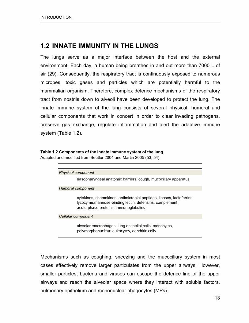
1.2 INNATE IMMUNITY IN THE LUNGS The lungs serve as a major interface between the host and the external
environment. Each day, a human being breathes in and out more than 7000 L of
air (29). Consequently, the respiratory tract is continuously exposed to numerous
microbes, toxic gases and particles which are potentially harmful to the
mammalian organism. Therefore, complex defence mechanisms of the respiratory
tract from nostrils down to alveoli have been developed to protect the lung. The
innate immune system of the lung consists of several physical, humoral and
cellular components that work in concert in order to clear invading pathogens,
preserve gas exchange, regulate inflammation and alert the adaptive immune
system (Table 1.2).
Table 1.2 Components of the innate immune system of the lung Adapted and modified from Beutler 2004 and Martin 2005 (53, 54).
Physical component
nasopharyngeal anatomic barriers, cough, mucociliary apparatus
Humoral component
cytokines, chemokines, antimicrobial peptides, lipases, lactoferrins, lyzozyme,mannose-binding lectin, defensins, complement, acute phase proteins, immunoglobulins
Cellular component
alveolar macrophages, lung epithelial cells, monocytes, polymorphonuclear leukocytes, dendritic cells
Mechanisms such as coughing, sneezing and the mucociliary system in most
cases effectively remove larger particulates from the upper airways. However,
smaller particles, bacteria and viruses can escape the defence line of the upper
airways and reach the alveolar space where they interact with soluble factors,
pulmonary epithelium and mononuclear phagocytes (MPs). 13
INTRODUCTION
1.2.1 Mononuclear phagocytes in lung host defence
The MP system consists of bone marrow (BM) progenitor cells, monocytes, DCs
and tissue macrophages. The major MPs subsets in airways and lung parenchyma
are alveolar macrophages (AMs), with a CD11c+ CD11b- CX3CR1- surface
phenotype, and CD11c+ CD11b+ CX3CR1+ lung DCs. (55, 56). Because of their
phagocytic activity, antigen presentation capacity and secretion of soluble
mediators MPs play a crucial role in lung innate and adaptive immune responses
to pathogens.
1.2.1.1 Resident alveolar macrophages
AMs are specialized mononuclear phagocytic cells that function as a first host
defence line against inhaled particles and pathogens in the lower respiratory tract
(57). These cells are broadly distributed at the air-tissue interface in the alveolar
space in close proximity to alveolar epithelial cells (AECs) and are separated by a
distance of only 0.2–0.5 μm from interstitial DCs (58). AMs originate primarily from
BM derived blood monocytes or may undergo limited local proliferation (59).
Normally, AMs account for approximately 95% of airspace leukocytes of human
lungs and 100% of the pathogen-free mice (54).
AMs have ability to ingest all types of inhaled particulates that reach the alveolar
space. Remarkably, AMs are generally poor initiators of lung immune responses,
thereby keeping the airspace quiet and preserving the ability of the lung to perform
gas exchange. They can remove and digest relatively inert particulates without
triggering inflammatory responses that may damage the structural integrity of
alveolar tissue (54, 58, 60). However, when pathogen exposures are sufficiently
large or when faced with highly virulent pathogens, AMs initiate a localized
inflammatory response by secretion of a wide array of mediators. These include
cytokines (e.g. TNF-� and IL-6), chemokines (e.g. CCL2 and RANTES) and lipid
mediators (arachidonic metabolites) thereby promoting neutrophil, activated
monocyte and lymphocyte sequestration at sites of inflammation in the lungs
14
INTRODUCTION
(Table 1.3). To avoid uncontrolled AM activation, several regulatory mechanisms
have been evolved. One possible inhibitory mechanism involves surfactant
proteins A and D, which bind AM receptors and suppress inflammatory activity in
uninfected lungs. Recently, Snelgrove et al. demonstrated a new mechanism by
which AECs suppress AM activity under baseline conditions involving the negative
regulator CD200 receptor (CD200R) on macrophages and its ligand CD200 on
airway epithelium (60, 61).
The ability of AMs to interact with microbes is mediated by surface receptors
capable to bind specific ligands. The major receptors found on AMs include
scavenger receptors, Fc receptors, G protein-coupled receptors (GPCRs),
cytokines and chemokines receptors and TLRs (29). Among TLRs, function of
TLR2 and TLR4 has been the most extensively studied. Both TLR2 and TLR4
receptors are expressed and functionally active on AMs (62, 63). In AMs,
recognition of LPS by TLR4 results in rapid activation of signalling pathways
including all three major MAP kinases, p38, ERK1/2 and JNK in vitro and in vivo
(29, 64). Likewise, activation of NF-�B is an early event after LPS binding (as early
as 30 min). It has been demonstrated that depletion of AMs attenuated NF-�B
activation in whole lung tissues and decreased the pro-inflammatory content in
BALF. In addition to the initiation and regulation of inflammation, AMs actively
participate in the resolution of inflammatory infiltrates and tissue repair by
phagocytosis of apoptotic cells and elaboration of enzymes, anti-inflammatory
cytokines (e.g. IL-1RN and IL-10) and growth factors such TGF-�1 (Table 1.3).
AMs have an additional role in the regulation of acquired immune responses. They
can effectively suppress adaptive immunity by down-regulation of T- and B-cell
activation and antigen presentation activities of DCs (65, 66). It has been
demonstrated that in vivo depletion of AMs using clodronate-filled liposomes leads
to significant increase in the pulmonary inflammatory response to harmless
particulate and soluble antigens (67).
15
INTRODUCTION
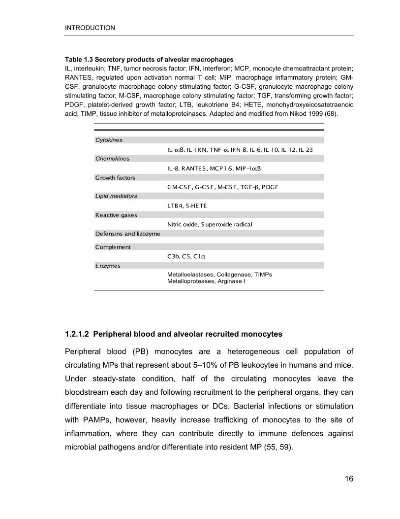
Table 1.3 Secretory products of alveolar macrophages IL, interleukin; TNF, tumor necrosis factor; IFN, interferon; MCP, monocyte chemoattractant protein; RANTES, regulated upon activation normal T cell; MIP, macrophage inflammatory protein; GM-CSF, granulocyte macrophage colony stimulating factor; G-CSF, granulocyte macrophage colony stimulating factor; M-CSF, macrophage colony stimulating factor; TGF, transforming growth factor; PDGF, platelet-derived growth factor; LTB, leukotriene B4; HETE, monohydroxyeicosatetraenoic acid; TIMP, tissue inhibitor of metalloproteinases. Adapted and modified from Nikod 1999 (68).
Cytokines
IL-�/�, IL-1RN, TNF-�, IFN-�, IL-6, IL-10, IL-12, IL-23Chemokines
IL-8, RANTES, MCP1/5, MIP-1�/�Growth factors
GM-CSF, G-CSF, M-CSF, TGF-�, PDGFLipid mediators
LTB4, 5-HETEReactive gases
Nitric oxide, Superoxide radicalDefensins and lizozyme
ComplementC3b, C5, C1q
EnzymesMetalloelastases, Collagenase, TIMPsMetalloproteases, Arginase I
1.2.1.2 Peripheral blood and alveolar recruited monocytes
Peripheral blood (PB) monocytes are a heterogeneous cell population of
circulating MPs that represent about 5–10% of PB leukocytes in humans and mice.
Under steady-state condition, half of the circulating monocytes leave the
bloodstream each day and following recruitment to the peripheral organs, they can
differentiate into tissue macrophages or DCs. Bacterial infections or stimulation
with PAMPs, however, heavily increase trafficking of monocytes to the site of
inflammation, where they can contribute directly to immune defences against
microbial pathogens and/or differentiate into resident MP (55, 59).
16
INTRODUCTION
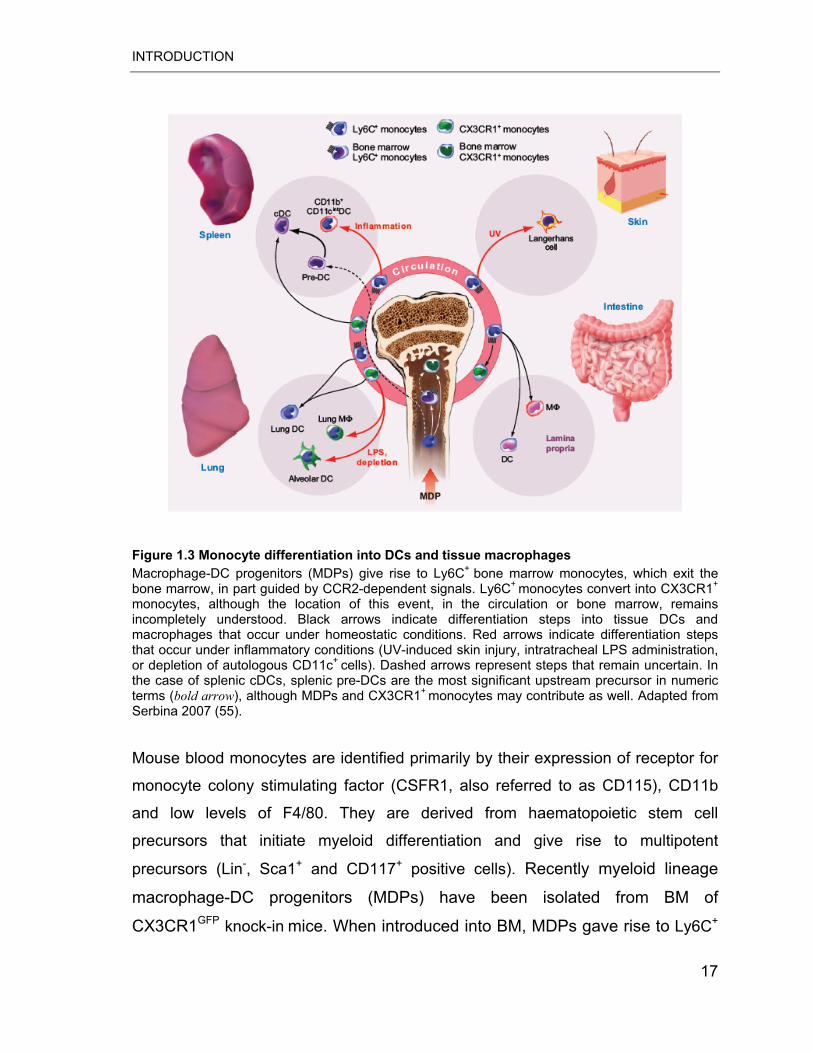
Figure 1.3 Monocyte differentiation into DCs and tissue macrophages Macrophage-DC progenitors (MDPs) give rise to Ly6C+
bone marrow monocytes, which exit the bone marrow, in part guided by CCR2-dependent signals. Ly6C+
monocytes convert into CX3CR1+
monocytes, although the location of this event, in the circulation or bone marrow, remains incompletely understood. Black arrows indicate differentiation steps into tissue DCs and macrophages that occur under homeostatic conditions. Red arrows indicate differentiation steps that occur under inflammatory conditions (UV-induced skin injury, intratracheal LPS administration, or depletion of autologous CD11c+
cells). Dashed arrows represent steps that remain uncertain. In the case of splenic cDCs, splenic pre-DCs are the most significant upstream precursor in numeric terms (bold arrow), although MDPs and CX3CR1+
monocytes may contribute as well. Adapted from Serbina 2007 (55).
Mouse blood monocytes are identified primarily by their expression of receptor for
monocyte colony stimulating factor (CSFR1, also referred to as CD115), CD11b
and low levels of F4/80. They are derived from haematopoietic stem cell
precursors that initiate myeloid differentiation and give rise to multipotent
precursors (Lin-, Sca1+ and CD117+ positive cells). Recently myeloid lineage
macrophage-DC progenitors (MDPs) have been isolated from BM of
CX3CR1GFP knock-in mice. When introduced into BM, MDPs gave rise to Ly6C+
17
INTRODUCTION
and CXCR1+ BM monocytes from which subsequently the two principal circulating
subsets are derived (Figure 1.3). One population (referred to herein as Ly6C+)
expresses low levels of CX3CR1 (CX3CR1low), high levels of Ly6C (Ly6Chigh) and
high levels of CCR2 (CCR2high). It has been demonstrated that Ly6C+ monocytes
home to peripheral tissues in response to inflammatory stimuli, prompting their
designation as inflammatory monocytes. This population corresponds to the main
human monocyte population, which is CD14+ CD16- (so called CD14+). The
second subset of mouse monocytes is similar to human CD14+ CD16+ monocytes
and is defined by high levels of CX3CR1 (CX3CR1high), low levels of Ly6C
(Ly6Clow) and CCR2 (CCR2low) (referred to herein as CX3CR1+). In contrast to
Ly6C+, CX3CR1+ monocytes traffic into peripheral tissues under
non-inflammatory conditions and therefore are referred to as non-inflammatory
monocytes (55, 59). A more detailed description of the surface antigen expression
on human and mouse monocyte subsets is summarized in the table 1.4.
18
INTRODUCTION
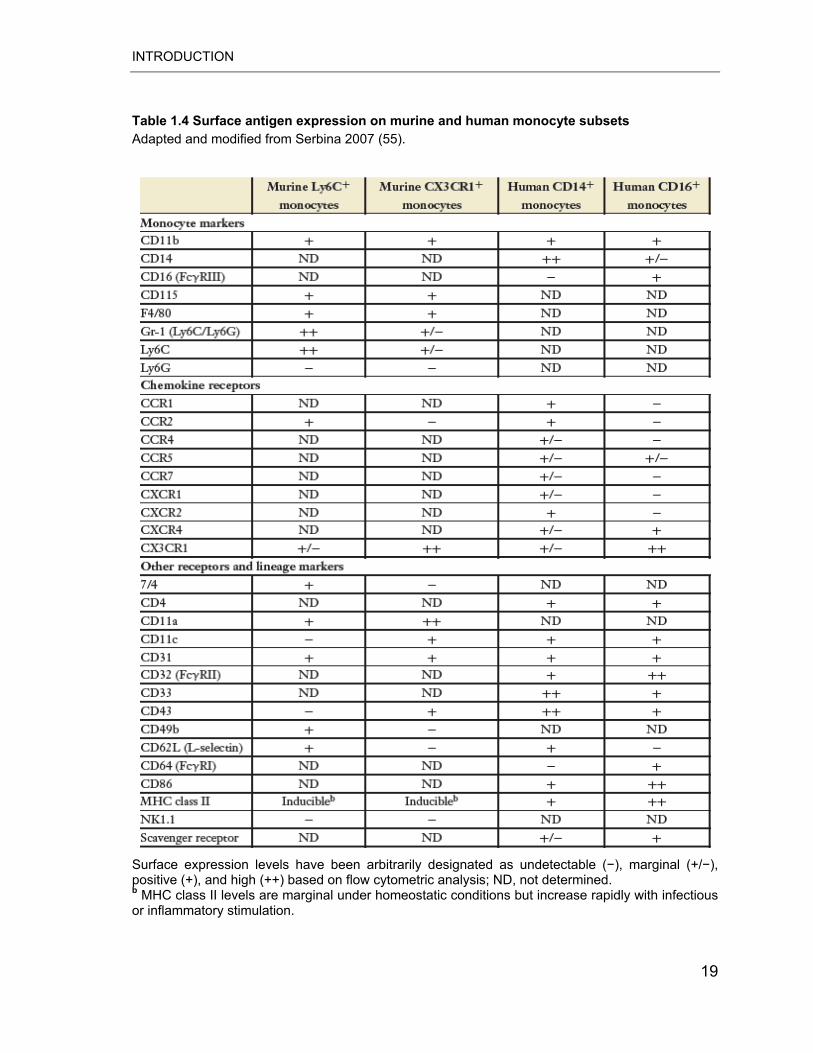
Table 1.4 Surface antigen expression on murine and human monocyte subsets Adapted and modified from Serbina 2007 (55).
Surface expression levels have been arbitrarily designated as undetectable (�), marginal (+/�), positive (+), and high (++) based on flow cytometric analysis; ND, not determined. b MHC class II levels are marginal under homeostatic conditions but increase rapidly with infectious or inflammatory stimulation.
19
INTRODUCTION
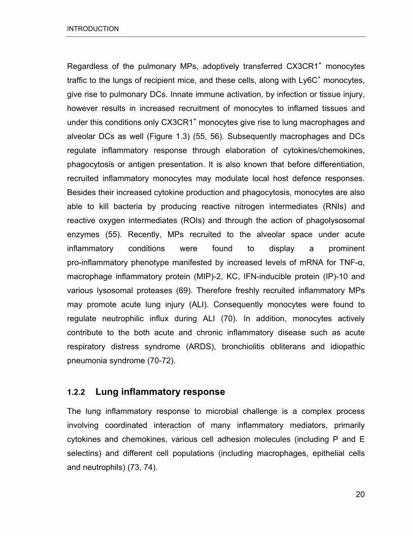
Regardless of the pulmonary MPs, adoptively transferred CX3CR1+ monocytes
traffic to the lungs of recipient mice, and these cells, along with Ly6C+ monocytes,
give rise to pulmonary DCs. Innate immune activation, by infection or tissue injury,
however results in increased recruitment of monocytes to inflamed tissues and
under this conditions only CX3CR1+ monocytes give rise to lung macrophages and
alveolar DCs as well (Figure 1.3) (55, 56). Subsequently macrophages and DCs
regulate inflammatory response through elaboration of cytokines/chemokines,
phagocytosis or antigen presentation. It is also known that before differentiation,
recruited inflammatory monocytes may modulate local host defence responses.
Besides their increased cytokine production and phagocytosis, monocytes are also
able to kill bacteria by producing reactive nitrogen intermediates (RNIs) and
reactive oxygen intermediates (ROIs) and through the action of phagolysosomal
enzymes (55). Recently, MPs recruited to the alveolar space under acute
inflammatory conditions were found to display a prominent
pro-inflammatory phenotype manifested by increased levels of mRNA for TNF-�,
macrophage inflammatory protein (MIP)-2, KC, IFN-inducible protein (IP)-10 and
various lysosomal proteases (69). Therefore freshly recruited inflammatory MPs
may promote acute lung injury (ALI). Consequently monocytes were found to
regulate neutrophilic influx during ALI (70). In addition, monocytes actively
contribute to the both acute and chronic inflammatory disease such as acute
respiratory distress syndrome (ARDS), bronchiolitis obliterans and idiopathic
pneumonia syndrome (70-72).
1.2.2 Lung inflammatory response
The lung inflammatory response to microbial challenge is a complex process
involving coordinated interaction of many inflammatory mediators, primarily
cytokines and chemokines, various cell adhesion molecules (including P and E
selectins) and different cell populations (including macrophages, epithelial cells
and neutrophils) (73, 74).
20
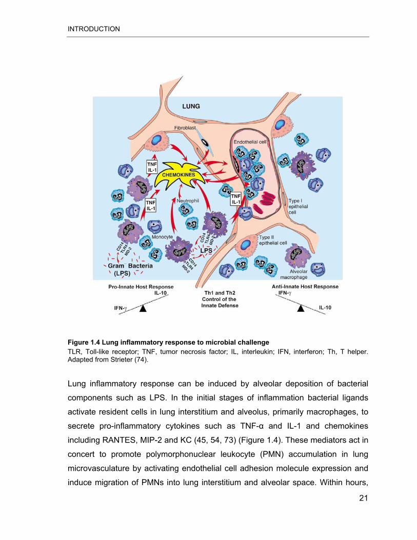
INTRODUCTION
Figure 1.4 Lung inflammatory response to microbial challenge TLR, Toll-like receptor; TNF, tumor necrosis factor; IL, interleukin; IFN, interferon; Th, T helper. Adapted from Strieter (74).
Lung inflammatory response can be induced by alveolar deposition of bacterial
components such as LPS. In the initial stages of inflammation bacterial ligands
activate resident cells in lung interstitium and alveolus, primarily macrophages, to
secrete pro-inflammatory cytokines such as TNF-� and IL-1 and chemokines
including RANTES, MIP-2 and KC (45, 54, 73) (Figure 1.4). These mediators act in
concert to promote polymorphonuclear leukocyte (PMN) accumulation in lung
microvasculature by activating endothelial cell adhesion molecule expression and
induce migration of PMNs into lung interstitium and alveolar space. Within hours,
21
INTRODUCTION
PMNs propagate inflammation through secretion of reactive oxygen species (e.g.,
hypochlorite), antimicrobial proteins (e.g., bactericidal permeability-inducing protein
and lactoferrin), and proteolytic enzymes. However, reactive substances secreted
by PMNs also affect host cells and can damage host tissues contributing to the
lung pathology in pneumonia. Lung inflammatory response is further characterized
by delayed recruitment of MPs, especially monocytes that can actively contribute
to the pathogenesis of both acute and chronic lung inflammatory diseases. To
control inflammatory lung responses, numerous strategies have been evolved
such as elaboration of anti-inflammatory mediators like IL-10 and TGF-�1 (74, 75).
For instance IL-10 blocks expression of IL-2, IFN-�, IL-1, TNF-�, IL-12 and CXC
and CC chemokines (74).
22
AIM OF THE STUDY
2 AIM OF THE STUDY
Resident alveolar macrophages are the major mononuclear phagocytic cells found
in the lower respiratory tract. They act as sentinels for pathogens and their
components such as LPS and bacterial lipoproteins sensed by TLR4 and TLR2,
respectively. The signalling pathways activated in alveolar macrophages by
different TLR agonists have not yet to been fully elucidated. Therefore, in the
present study we aimed to further explore the molecular programs activated by
bacterial TLR ligands. In particular, we sought to identify and functionally
characterize novel mechanisms that regulate alveolar macrophage responses
induced by TLR4 ligand LPS and TLR2 ligand Pam3CSK4.
In addition to activation of lung resident cells, alveolar recognition of inhaled
pathogens promotes increased recruitment of circulating mononuclear phagocytes
to inflamed tissue. Experimental data on their migration pathways, traffic related
cell differentiation and functional role in inflamed lungs in vivo are scarce.
Moreover, compared to the TLR4 ligand LPS a limited number of studies have
addressed the induction of lung inflammation and the concomitant mononuclear
phagocyte recruitment in response to TLR2 agonists. Therefore, this study was
undertaken to analyse the global gene expression profile of mononuclear
phagocytes recruited from the circulation into TLR2 agonist Pam3CSK4-treated
inflamed lungs.
Perspectively, comprehensive insights into the molecular programs induced by
microbial challenge in pulmonary mononuclear phagocytes may help to identify
new molecular target for selective therapeutic intervention.
23
MATERIALS AND METHODS
3 MATERIALS AND METHODS
3.1 Materials
3.1.1 Animals
BALB/c and C57BL/6 WT mice were purchased from Charles River (Sulzfeld,
Germany). CC3TLR/J BALB/c (TLR4-mutated) mice were purchased from Jackson
Laboratories (Bar Harbour, USA) and TLR2-KO C57BL/6 mice from Oriental Yeast
Company (Tagata Shizouka, Japan). PKR-KO C57BL/6 mice were a kind gift of
Jovan Pavlovic (University of Zurich, Switzerland). CX3CR1GFP/GFP mice were
generated on a mixed C57BL/6 x 129/Ola background by targeted disruption of
CX3CR1 gene. Parent CX3CR1GFP/GFP and CX3CR1+/+ mice were bred to yield
heterozygous CX3CR1+/GFP offspring (76). All animals were kept under specific
pathogen-free (SPF) conditions and used throughout the study between 8 and 12
weeks of age. Experimental protocols involving animals were approved by
institutional and local government comities.
3.1.2 Equipment Abbocath Abbott, Germany
ABI PRISM 2400 PCR-thermocycler Applied Biosystems, USA
ABI PRISM 7900HT Sequence detector Applied Biosystems, USA
Agilent Bioanalyser 2100 Agilent Tech., Germany
BioDocAnalyse video system Whatman – Biometra, Germany
Cell culture incubator Heraeus, Germany
Cytospin Cytocentrifuge Thermo Scientific, Germany
Developing machine, Curix 60 Agfa, Germany
24
MATERIALS AND METHODS
Electrophoresis apparatus Keutz, Germany
ELISA reader Molecular Devices, Germany
Eppendorf tubes (1.5ml/2 ml) Eppendorf, Germany
Film cassette Cronex, Germany
Filter tip Greiner bio-one, Germany
Filter units Millipore, USA
Fluorescence 8 chamber glass slides BD, Germany
Fluorescence Activated Cell Sorter BD Biosciences, Germany
Fluorescence microscope Leica, Germany
GenePix 4100A scanner Axon Instruments, USA
Mini Protean 3 cell Bio-Rad, USA
Mini spin centrifuge Heraeus, Germany
Mini Trans Blot Bio-Rad, USA
Mouse Whole Genome Agilent Tech., Germany
Multifuge centrifuge, 1S-R Heraeus, Germany
NanoDrop ND-1000 Nano Technologies, USA
PCR tubes (0.2 ml) Applied Biosystems, USA
Pipetmans: P10, P20, P100, P200, P1000 Gilson, France
Pipette tip BD, Germany
Power supply Biometra, Germany
Serological pipette: 5, 10, 25, 50 ml Falcon, USA
Stereomicroscope Leica, Germany
Test tubes:15, 50 ml Greiner Bio-One, Germany
Test tube thermostat Roth, Germany
Tissue culture plates: 24, 48, 96 well Greiner Bio-One, Germany
25
MATERIALS AND METHODS
3.1.3 Kits BD SMART Probe amplification kit Clontech, Germany
DC protein assay Bio-Rad, Germany
ELISA kits R&D Systems, USA Platinum® SYBR® Green I qPCR SuperMix-UDG
Invitrogen, Germany
RNeasy Micro kit Qiagen, Germany
RNeasy Mini kit Qiagen, Germany
Active Motif, Belgium TransAM p65 NF-�B assay kit
3.1.4 Reagents 2-AP Sigma-Aldrich, Germany
5 X first strand buffer Invitrogen, Germany
Acetic Acid Merck, Germany
Acetone Sigma-Aldrich, Germany
Acrylamide solution, Rotiphorese Gel 30 Roth, Germany
Agarose Peqlab, Germany
Ammonium persulfate (APS) Promega, Germany
�-mercaptoethanol Sigma-Aldrich, Germany
Bovine serum albumin (BSA) Sigma-Aldrich, Germany
Bromophenol blue Sigma-Aldrich, Germany
Calibrate beads BD Biosciences, Germany
DAPI Sigma-Aldrich, Germany
DifcoTM Skim Milk BD, Germany
Dithiothreitol (DTT) Invitrogen, Germany
dNTPs Applied Biosystems, USA
26
MATERIALS AND METHODS
Dublecco’ s phosphate buffered saline (PBS)
PAA Laboratories, Austria
Amersham Biosciences, UK ECL Plus Western Blotting Detection System
E. coli lipopolysaccharide (0111:B4) Calbiochem, Germany
Ethanol Carl Roth, Germany
Ethidium bromide solution Carl Roth, Germany Promega, USA Ethylendinitrilo-N, N, N´, N´, -tetra-acetic
acid (EDTA) Ethylene glycol-bis (2-amino-ethylether)-N,N,N’,N’ –tetra-acetic-acid (EGTA)
Sigma-Aldrich, Germany
Fetal calf serum (FCS) PAA laboratories, Austria
Glycine Sigma-Aldrich, Germany
Hydrochloric acid (HCl) Merck, Germany
Igepal CA-630 Sigma-Aldrich, Germany Isoflurane (1-chloro-2,2,2-trifluoroethly difluoromethyl ether)
Abbott, Germany
Ketavet (Ketamine hydrochloride) Pharmacia & Upjohn, Germany
Methanol Fluka, Germany
M-MLV reverse transcriptase Promega, USA
Mounting medium Dako, Germany N,N,N',N'-tetramethyl-ethane-1.2-diamine (TEMED)
Sigma-Aldrich, Germany
EMC Microcollections, Germany Pam3-Cys-Ser-Lys-Lys-Lys-Lys-OH (Pam3CSK4) Paraformaldehyde Sigma-Aldrich, Germany
Penicillin-streptomycin PAA Laboratories, Austria
Platinum Taq DNA polymerase Invitrogen, Germany
Polyriboinosinic:polyribocytydylic acid (Poly I:C)
Alexis Biochemicals, Germany
Polyvinylidene difluoride (PVDF) membranes
Micron Separations, USA
27
MATERIALS AND METHODS
Precision Plus ProteinTM Standards Bio-Rad, USA
Protease inhibitor cocktail Roche, Germany
Random hexamers Boehringer, Germany
RNase away Molecular Bioproducts, USA
RNase inhibitor Promega, USA
Rompun (Xylazine hydrochloride) Bayer, Germany
RPMI 1640 medium PAA laboratories, Austria
Sodium chloride Braun, Germany
Sodium dodecyl sulphate (SDS) Sigma-Aldrich, Germany
Sodium ortho vanadate Sigma-Aldrich, Germany
Tris Carl Roth, Germany
Tween 20 Sigma-Aldrich, Germany
3.1.5 Software
BioConductor http://www.bioconductor.org
BLAST http://www.ncbi.nlm.nih.gov
FACS DiVa software BD Biosciences, Germany Developed by Dr. Jochen Wilhelm (Giessen University)
GeneBank Analyzer
GENECODIS www.genecodis.com
GenePix Pro 5.0 Axon, USA
MicroWin 3.0 Microtek, Germany
Primer Express 2.0 Applied Biosystems, USA
R software http://www.cran.r-project.org
28
MATERIALS AND METHODS
3.2 Methods
3.2.1 Treatment of animals
3.2.1.1 Mouse model of TLR2/4 ligands-induced lung inflammation
In order to study lung inflammatory responses in vivo, WT or CX3CR1+/GFP mice
received intratracheal (i.t.) application of TLR ligands. Pam3CSK4 was used as a
synthetic TLR2 agonist recognized by the TLR1/TLR2 complex, whereas LPS
served as specific TLR4 ligand. Briefly, mice were anesthetized with tetrazoline
hydrochloride (2.5 mg/Kg) and ketamine (50 mg/Kg) and the trachea was exposed.
Subsequently, a 26-gauge catheter was inserted into trachea and Pam3CSK4
(10 μg or 50 μg/mouse dissolved in a total volume of 60 μl) or LPS (1 μg or 10
μg/mouse dissolved in a total volume of 60 μl) was installed under
stereomicroscopic control. After installation, the catheter was removed, wounds
were closed and mice were allowed to recover with free access to food and water.
3.2.1.2 Collection and analysis of blood and bronchoalveolar lavage (BAL) fluid
Mice were sacrificed with an overdose of isoflurane and blood and BAL fluid
(BALF) collection was done as described previously (69). Briefly, blood was
collected from the vena cava inferior using 23-gauge cannula connected to a 1-ml
insulin syringe that was filled with 100 l of NaCl-EDTA as an anticoagulant. Lysis
of red blood cells was performed using ammonium chloride solution.
For BAL, the trachea was exposed and a small incision was made to insert a
shortened 21-gauge cannula connected to a 1-ml insulin syringe, followed by
repeated i.t. instillations of 0.5 ml aliquots of PBS (pH 7,2, supplemented with 2
mM EDTA). BAL was performed until a total volume of 5 ml was recovered. BALF
29
MATERIALS AND METHODS
was spun for 10 min at 1400 rpm at 4 °C and cells were resuspended in 1 ml of
RPMI 1640 medium containing 10 % FCS and L-glutamine.
To study the inflammatory BALF profile, the cells were counted with a
hemocytometer and quantitation of AMs and neutrophils was done on differential
cell counts of Pappenheim-stained cytocentrifuge preparations as recently
described (77).
3.2.1.3 Fluorescence activated cell sorting (FACS)
Recovered BALF and blood cells were pooled from 10-12 CX3CR1GFP/+ mice to
obtain sufficient cell numbers for flow sorting. Prior to cell sorting, blood and BALF
samples were filtered through a 40 μm cell strainer. The GFP-positive MPs were
counted and sorted using a high-speed FACSVantage SE flow cytometer (BD
Biosciences) as described previously (69). In short, the sorting algorithm for
GFP-positive cells employed (1) forward scatter area (FSC-A) versus side scatter
area (SSC-A), (2) FSC-A versus FL1 (F525 ± 15 nm; FITC/GFP) and (3) FL1
versus FL2 characteristics (F575 ± 25 nm). These settings allowed for the
exclusion of lymphocytes, AMs and neutrophils in analyzed samples. The purity of
sorted GFP-positive cells was 98 %. In addition, Pappenheim staining verified
monocytic cell morphology of isolated GFP-positive cells. Dot plot profiles were
obtained from 104 cells for each sample.
3.2.2 Cell culture and stimulation
3.2.2.1 Culture of murine primary alveolar macrophages
AMs were harvested by BAL from WT BALB/c, CC3TLR/J, TLR2-KO and PKR-KO
mice as described in section 3.2.1.2. Cells were cultured in RPMI 1640 medium
supplemented with 10 % FCS, L-glutamine and penicillin/streptomycin at 37 °C in
a humidified 5 % CO2 environment. After two hours, cells were washed with RPMI
1640 to remove non-adherent cells. Macrophages were seeded at 2.5-3 x 10⁵
30
MATERIALS AND METHODS
cells/well in 24-well tissue culture plates for western blotting and TransAM assay,
at 8 x 10⁴ cells/well in 96-well tissue culture plates for ELISA or at 6 x 10⁴ cells/well
on glass slides for immunofluorescence microscopy.
AMs were left untreated or stimulated with Pam3CSK4 (2 μg/ml), poly I:C (50
μg/ml) or LPS (200 ng/ml) for different periods of time as specified. Were indicated,
AMs were pre-incubated with 4 mM 2-AP for 30 min.
3.2.3 RNA analysis
3.2.3.1 Isolation of total RNA
Total cellular RNA isolation from primary AMs was performed using RNeasy Mini
Kit. Total cellular RNA from freshly sorted GFP-positive MPs was performed using
the RNeasy Micro kit. In both the cases, RNA was isolated following the
instructions of the manufacturer and was quantified spectrophotometrically using
Nanodrop ND-100. For microarray experiments the RNA quality was assessed by
the 18S/28S-rRNA bands in capillary electrophoresis.
3.2.3.2 Microarray analysis
The microarray experiments were performed in the Department of Pathology
(University of Giessen) by Dr. Jochen Wilhelm and Marlene Stein. Purified total
RNA was amplified and Cy-labeled using the SMART™ (switching mechanism at
the 5' end of RNA templates of reverse transcriptase) kit following the kit
instructions. Briefly, this method creates full-length complementary DNA (cDNA)
from all oligo-A RNA with introduced specific primer binding sites at either end.
One priming site is introduced by the oligo-dT primer and the other one is linked by
template-switching of the MMLV RT: the RT adds a stretch of Gs to the end of the
cDNA when the end of the RNA template is reached. A C-rich
oligodesoxyribonucleotide containing the PCR-primer sequence binds to this
stretch; the RT then switches the template and produces a sense cDNA strand that
31
MATERIALS AND METHODS
now contains the binding site for the PCR primer at either side. These full-length
cDNAs are subsequently amplified by PCR. In preliminary experiments, the
number of amplification cycles was determined so that the PCR is not run until the
plateau. The samples were finally amplified for 24 cycles, followed by 4 cycles with
aminoallylated dCTP. After purification, monoreactive Cy-dyes were coupled to the
incorporated amino groups. Cy3- and Cy5-labeled cDNAs were hybridized
overnight to 44K 60mer oligonucleotide spotted microarray slides. Hybridization
and subsequent washing and drying of the slides were performed following the
Agilent hybridization protocol.
Dried slides were scanned using the GenePix 4100A scanner. Image analysis was
performed with GenePix Pro 5.0 software, and calculated values for all spots were
saved as GenePix results files. Stored data were evaluated using the R software
(http://www.R-project.org) and the limma package from BioConductor (78). The
spots were weighted for subsequent analyses according to the spot intensity,
homogeneity, and saturation. The spot intensities were corrected for the local
background using the method of Edwards with an offset of 64 to stabilize the
variance of low-intensity spots (79). The M/A data were LOESS normalized before
averaging (80). Genes were ranked for differential expression using a moderated t-
statistic (81). Candidate lists were created by adjusting the false-discovery rate
(FDR) to 10 %.
Raw data was submitted to NCBI Gene Expression Omnibus (GEO) database
(http://www.ncbi.nlm.nih.gov/geo/query/acc.cgi?token=hpgntswwcowiifq&acc=GSE
11978).
3.2.3.3 Reverse transcription reaction
In order to measure messenger RNA (mRNA), reverse transcriptase (RT) was
used to convert mRNA into cDNA which is then amplified by real-time PCR. To
perform synthesis of cDNA, 50 ng to 500 ng of total RNA was mixed with random
32
MATERIALS AND METHODS
hexamers (10 pmol) in PCR tube, heated at 70 0C for 5 min, placed on ice and the
following RT mix reaction components were added.
RT mix
5x first strand buffer 5 μl dNTPs 10 mM each DTT 250 mM Ribonuclease inhibitor 20 U MMLV 200 U
Subsequently this mixture was incubated for 1 h at 42 0C, heated to 94 0C for 10
min to inactivate the enzyme and used for real-time PCR amplification immediately
or stored at -20 0C till further use.
3.2.3.4 Quantitative real-time PCR
The transcriptional regulation of selected genes was analyzed using real-time
quantitative PCR. Real-time PCR is a method based on the detection and
quantification of a fluorescent reporter signal that increases in direct proportion to
the amount of the PCR product in reaction (82, 83). Quantitative real-time PCR
was performed using SYBR green, a fluorogenic dye that emits a strong
fluorescent signal upon binding to the minor groove of double-stranded DNA
product. PCR reactions were performed in 25 l reactions using the following
master mix.
Real time master mix
cDNA 5 μl Forward primer 10 pmol Reverse primer 10 pmol SYBR® Green I 12 μl ROX reference dye 0.5 μl MgCl2 (50 X) 1 μl
33
MATERIALS AND METHODS
Cycling conditions were as following: 1 cycle of 95 °C for 5 min, followed by 45
cycles of 95 °C for 10 sec, 60 °C for 30 s and 72 °C for 10 sec. After the cycling
process, melting curve analysis and agarose gel electrophoresis (in selected
cases) was performed to confirm the exclusive amplification of the expected PCR
products. Relative gene expression is expressed as Delta Ct ( CT) with mouse
porphobilinogen deaminase (PBGD) serving as reference gene: = Ct [PBGD]
– Ct [target gene] (higher values indicate higer mRNA levels). Differential gene
expression between conditions is expressed as Delta Delta Ct ( ) what
corresponds to the log2 fold-difference in mRNA levels between the conditions
compared (84). The mRNA sequences of the genes of interest were obtained from
NCBI http://www.ncbi.nlm.nih.gov/sites/entrez. The oligonucleotide primer pairs
were designed using Primer Express 2.0. Oligonucleotide primers are listed in
Appendix (Table 6.1.)
3.2.4 Protein analysis
3.2.4.1 Cell extracts and western blotting
After removing the medium, cells were washed twice with cold 1 x PBS and
ice-cold cell-lysis buffer was applied. The cell lysates were kept on ice for 30 min,
followed by centrifugation for 15 min at 13000 rpm at 4 °C. Resulting supernatant
was used as a crude extract.
Cell-lysis buffer
20 mM Tris (pH 7.5) 150 mM NaCl 1 mM EDTA 1 mM EGTA 0.5 % Igepal CA-630 2 mM Na3VO41 X Protease inhibitor mix
34
MATERIALS AND METHODS
Protein concentration was determined using Bio-Rad DC (detergent compatible)
protein assay. This method is a colorimetric reaction similar to the
well-documented Lowry assay (85). Subsequently, proteins were separated
according to their size by SDS polyacrylamide gel electrophoresis (SDS-PAGE)
under reducing and denaturating conditions in 1 x SDS-running buffer (80 V and
40 mA for 2 h). Gels were composed of a 5 % stacking and a gel 10 % separating
gel. Before samples were loaded into gel, proteins were mixed with loading buffer
and heated at 95 °C for 5 min.
10 x Loading buffer
50 mM Tris-HCL 100 mM DTT 2% (w/v) SDS 10% (v/v) Glycerol 0,1% (w/v) Bromophenol Blue
1 x SDS running buffer
25 mM Tris base 250 mM Glycine 0.1% (v/v) SDS
Stacking gel 5 %
125 mM Tris (pH 6.8) 5 % Acrylamid 0.1% (w/v) SDS 0.1%(v/v) Temed 0.1% (w/v) APS
Separating gel 10 %
375 mM Tris (pH 8.8) 10 % Acrylamid 0.1% (w/v) SDS 0.1%(v/v) Temed 0.1% (w/v) APS
35
MATERIALS AND METHODS
After gel electrophoresis, proteins were transferred to a polyvinylidene difluoride
(PVDF) membrane using a Bio-Rad transfer chamber containing transfer buffer.
Transfer was performed at 120 V and 250 mA. After one hour, membranes were
incubated in blocking buffer at room temperature for 1 h. Subsequently primary
antibodies were added and membranes were incubated overnight at 4 °C,
afterwards washed three times and incubated with horse radish peroxidase (HRP)-
conjugated secondary antibody for 1 h. Enhanced Chemiluminescence (ECL)
system was used to visualize immune complexes. Signals were detected using
radiographic films. For reprobing, membranes were incubated at room temperature
for 1 h in stripping buffer. Primary and secondary antibodies used for western
blotting are listed in Appendix (Table 6.2).
Transfer buffer pH 7.4
25 mM Tris base 192 mM Glycine 10 % (v/v) Methanol
Washing buffer (1 L)
100 ml 10 X PBS 900 ml dH2O 0.05 % Tween 20
Blocking Buffer
5 % (w/v) non-fat dry milk 1 X PBS 0.05 % Tween 20
Stripping Buffer (100 ml)
10 ml Glycine (1M) 89 ml dH2O 1 ml 37 % HCl
36
MATERIALS AND METHODS
3.2.4.2 Enzyme-linked immunosorbent assay (ELISA)
The quantification of murine TNF-� and IL-6 proteins in AM culture supernatants
was performed by commercially available ELISA kits following the instructions of
the manufacturer.
3.2.4.3 Immunofluorescence microscopy
Cells were washed twice with 1 x PBS and fixed with cold (-20 °C)
methanol:acethone mixture (1:1) for 10 min. After washing, cells were blocked
overnight with 10 % BSA in PBS, washed twice and then incubated overnight with
rabbit anti-p65 NF-�B antibody (1:50) diluted in 1 % BSA at 4 °C. Immune
complexes were detected with Alexa Fluor 488 goat anti-rabbit secondary antibody
diluted in 1 % BSA (1:1000) at room temperature for 1 h in darkness. After
washing, cells were fixed in 4 % paraformaldehyde for 10 min. Nuclei were stained
with DAPI (1:100) diluted in 1 % BSA. The coverslips were mounted on slides in
mounting medium. Cells were imaged by conventional fluorescence microscopy.
Primary and secondary antibodies used for immunofluorescence microscopy are
listed in Appendix (Table 6.2).
3.2.4.4 TransAM p65 NF-�B assay
Whole cell extract (5 μg) was used to monitor activation of the p65 subunit of
NF-�B by the commercially available TransAM p65 NF-�B assay kit purchased
from Active Motif following the instructions of manufacturer.
3.2.5 Statistical analysis
The data for BALF cell profile are presented as mean ± SD. Differences between
treatments groups were analysed by one-way ANOVA followed by Dunnett post
hoc test. A value of p � 0.05 was considered as significant. �Ct and ��Ct values
are given as mean ± SD or SEM of at least three independent experiments.
37

MATERIALS AND METHODS
Differences in mean �Ct values were tested by two-tailed (paired or unpaired)
t-tests. A value of p � 0.05 was considered significant.
38
RESULTS
4 RESULTS
4.1 Gene expression of TLR associated proteins induced by inflammatory stimuli in AMs
Initial experiments aimed to analyse the transcriptional regulation of genes
involved in TLRs signalling in response to bacterial components that are
differentially recognized by distinct TLR family members. We used synthetic
lipoprotein Pam3CSK4 that is specifically recognized by the TLR1/2 complex,
whereas LPS derived from Escherichia coli was used as specific ligand for TLR4
(86). Genes selected for evaluation belonged to different functional categories:
PRRs (CD14, CD36, TLR2 and TLR4), cytokine regulators (SOCS 1/3),
inflammatory cytokines (TNF-�, IL-6), antiviral defence and negative regulators of
TLRs (IRAK-M, ST2 and SIGIRR). The full names of genes are presented in table
4.1.
AMs isolated from BALF of WT BALB/c mice were left either untreated or
stimulated with the TLR2 ligand Pam3CSK4 or the TLR4 ligand LPS. Using
quantitative real-time PCR, we found that both ligands induced comparable
changes in the expression profile of TLR molecules after 6 h and 24 h in vitro
(Table 4.1). Transcription of two PRRs, TLR2 and CD14 was significantly
increased, whereas expression of TLR4 and CD36 mRNA remained unchanged
after treatment with both ligands. Most drastic changes in mRNA levels were noted
for the pro-inflammatory cytokines IL-6 and TNF-� and for SOCS proteins.
Interestingly, transcription of dsRNA-activated protein kinase R (PKR), the antiviral
defence molecule, was significantly increased in response to Pam3CSK4 and LPS
ligands. Among negative regulators, IRAK-M mRNA was significantly up-regulated
after treatment with both TLR ligands, whereas ST2 was induced only by the TLR2
ligand after 6 h.
39
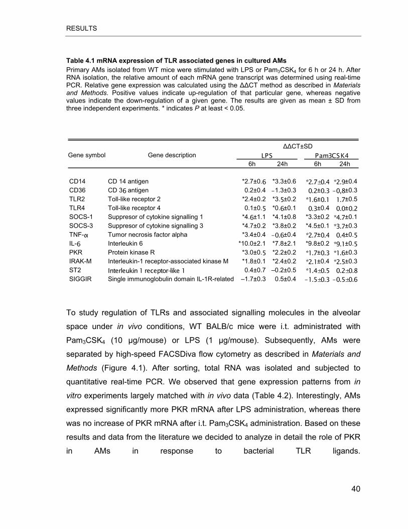
RESULTS
Table 4.1 mRNA expression of TLR associated genes in cultured AMs Primary AMs isolated from WT mice were stimulated with LPS or Pam3CSK4 for 6 h or 24 h. After RNA isolation, the relative amount of each mRNA gene transcript was determined using real-time PCR. Relative gene expression was calculated using the ��CT method as described in Materials and Methods. Positive values indicate up-regulation of that particular gene, whereas negative values indicate the down-regulation of a given gene. The results are given as mean ± SD from three independent experiments. * indicates P at least < 0.05.
Gene symbol Gene description6h 24h 6h 24h
CD14 CD 14 antigen *2.7±0.6 *3.3±0.6 *2.7±0.4 *2.9±0.4CD36 CD 36 antigen 0.2±0.4 –1.3±0.3 0.2±0.3 –0,8±0.3TLR2 Toll-like receptor 2 *2.4±0.2 *3.5±0.2 *1.6±0.1 1.7±0.5TLR4 Toll-like receptor 4 0.1±0.5 *0.6±0.1 0.3±0.4 0.0±0.2SOCS-1 Suppresor of cytokine signalling 1 *4.6±1.1 *4.1±0.8 *3.3±0.2 *4.7±0.1SOCS-3 Suppresor of cytokine signalling 3 *4.7±0.2 *3.8±0.2 *4.5±0.1 *3.7±0.3TNF-� Tumor necrosis factor alpha *3.4±0.4 –0.6±0.4 *2.7±0.4 0.4±0.5IL-6 Interleukin 6 *10.0±2.1 *7.8±2.1 *9.8±0.2 *9.1±0.5PKR Protein kinase R *3.0±0.5 *2.2±0.2 *1.7±0.3 *1.6±0.3IRAK-M Interleukin-1 receptor-associated kinase M *1.8±0.1 *2.4±0.2 *2.1±0.4 *2.5±0.3ST2 Interleukin 1 receptor-like 1 0.4±0.7 –0.2±0.5 *1.4±0.5 0.2±0.8SIGGIR Single immunoglobulin domain IL-1R-related –1.7±0.3 0.5±0.4 –1.5±0.3 –0.5±0.6
��CT±SDPam3CSK4LPS
To study regulation of TLRs and associated signalling molecules in the alveolar
space under in vivo conditions, WT BALB/c mice were i.t. administrated with
Pam3CSK4 (10 μg/mouse) or LPS (1 μg/mouse). Subsequently, AMs were
separated by high-speed FACSDiva flow cytometry as described in Materials and
Methods (Figure 4.1). After sorting, total RNA was isolated and subjected to
quantitative real-time PCR. We observed that gene expression patterns from in
vitro experiments largely matched with in vivo data (Table 4.2). Interestingly, AMs
expressed significantly more PKR mRNA after LPS administration, whereas there
was no increase of PKR mRNA after i.t. Pam3CSK4 administration. Based on these
results and data from the literature we decided to analyze in detail the role of PKR
in AMs in response to bacterial TLR ligands.
40
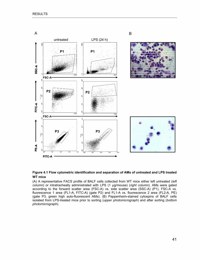
RESULTS
A B
SSC
-AFI
TC-A
PE-A
FSC-A
FSC-A
FITC-A
untreated LPS (24 h)
P1 P1
P2P2
P3P3
SSC
-AFI
TC-A
PE-A
FSC-A
FSC-A
FITC-A
untreated LPS (24 h)
P1 P1
P2P2
P3P3
Figure 4.1 Flow cytometric identification and separation of AMs of untreated and LPS treated WT mice (A) A representative FACS profile of BALF cells collected from WT mice either left untreated (leftcolumn) or intratracheally administrated with LPS (1 μg/mouse) (right column). AMs were gated according to the forward scatter area (FSC-A) vs. side scatter area (SSC-A) (P1), FSC-A vs. fluorescence 1 area (FL1-A, FITC-A) (gate P2) and FL1-A vs. fluorescence 2 area (FL2-A, PE) (gate P3, green high auto-fluorescent AMs). (B) Pappenheim-stained cytospins of BALF cells isolated from LPS-treated mice prior to sorting (upper photomicrograph) and after sorting (bottomphotomicrograph).
41
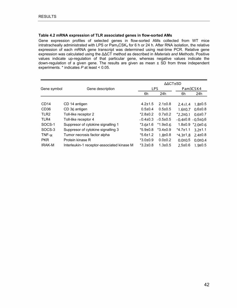
RESULTS
Table 4.2 mRNA expression of TLR associated genes in flow-sorted AMs Gene expression profiles of selected genes in flow-sorted AMs collected from WT mice intratracheally administrated with LPS or Pam3CSK4 for 6 h or 24 h. After RNA isolation, the relative expression of each mRNA gene transcript was determined using real-time PCR. Relative gene expression was calculated using the ��CT method as described in Materials and Methods. Positive values indicate up-regulation of that particular gene, whereas negative values indicate the down-regulation of a given gene. The results are given as mean ± SD from three independent experiments. * indicates P at least < 0.05.
Gene symbol Gene description6h 24h 6h 24h
CD14 CD 14 antigen 4.2±1.5 2.1±0.8 2.4±1.4 1.8±0.5CD36 CD 36 antigen 0.5±0.4 0.5±0.5 1.6±0.7 0.6±0.8TLR2 Toll-like receptor 2 *2.8±0.2 0.7±0.2 *2.2±0.1 0.6±0.7TLR4 Toll-like receptor 4 –0.4±0.3 –0.5±0.5 –0.4±0.8 –0.5±0.6SOCS-1 Suppresor of cytokine signalling 1 *3.6±1.6 *1.9±0.6 1.8±0.9 *2.0±0.6SOCS-3 Suppresor of cytokine signalling 3 *5.9±0.8 *3.4±0.9 *4.7±1.1 3.2±1.1TNF-� Tumor necrosis factor alpha *5.6±1.2 1.8±0.8 *4.3±1.8 2.4±0.8PKR Protein kinase R *3.0±0.9 0.0±0.2 0.0±0.5 0.0±0.4IRAK-M Interleukin-1 receptor-associated kinase M *3.2±0.8 1.3±0.5 2.5±0.6 1.9±0.5
��CT±SDPam3CSK4LPS
42
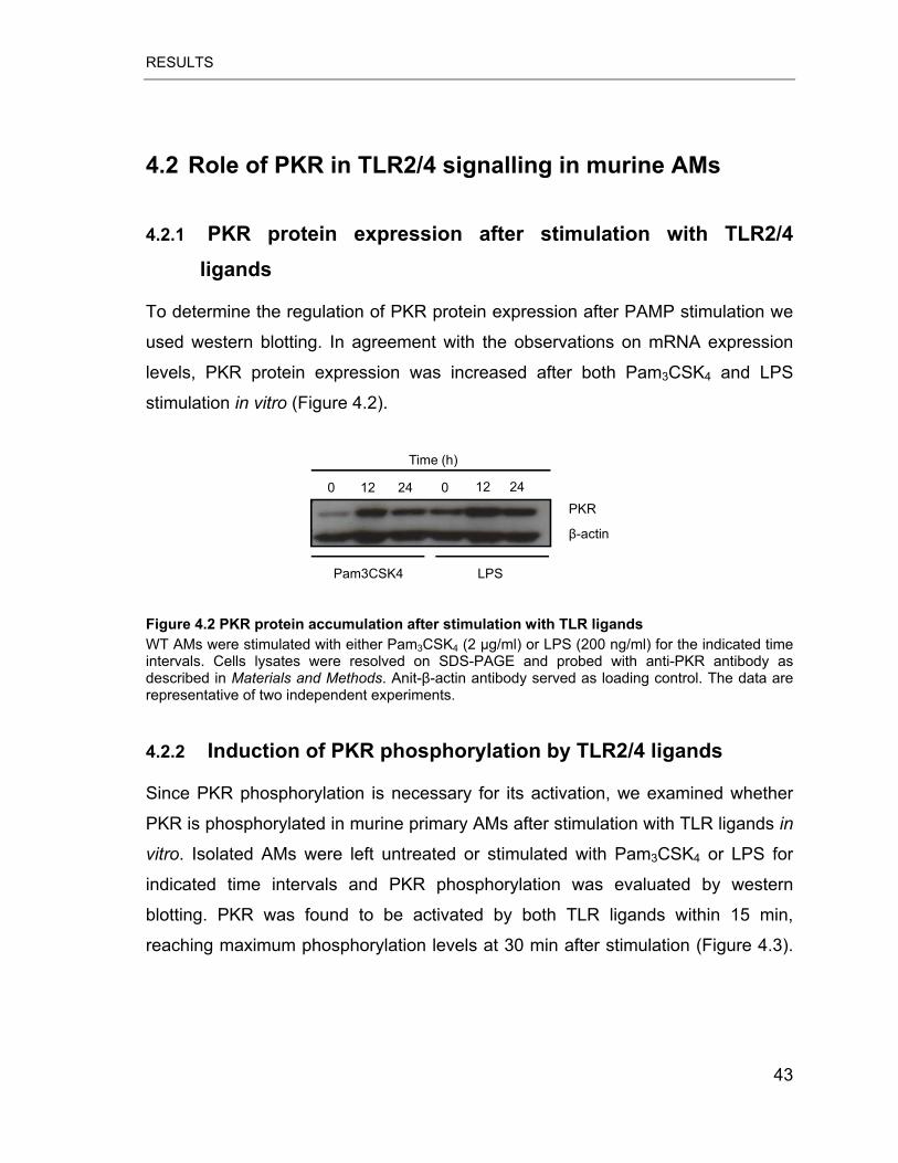
RESULTS
4.2 Role of PKR in TLR2/4 signalling in murine AMs
4.2.1 PKR protein expression after stimulation with TLR2/4 ligands
To determine the regulation of PKR protein expression after PAMP stimulation we
used western blotting. In agreement with the observations on mRNA expression
levels, PKR protein expression was increased after both Pam3CSK4 and LPS
stimulation in vitro (Figure 4.2).
0 12 24 0 12 24
Time (h)
Pam3CSK4 LPS
PKR
�-actin
Figure 4.2 PKR protein accumulation after stimulation with TLR ligands WT AMs were stimulated with either Pam3CSK4 (2 g/ml) or LPS (200 ng/ml) for the indicated time intervals. Cells lysates were resolved on SDS-PAGE and probed with anti-PKR antibody as described in Materials and Methods. Anit-�-actin antibody served as loading control. The data are representative of two independent experiments.
4.2.2 Induction of PKR phosphorylation by TLR2/4 ligands
Since PKR phosphorylation is necessary for its activation, we examined whether
PKR is phosphorylated in murine primary AMs after stimulation with TLR ligands in
vitro. Isolated AMs were left untreated or stimulated with Pam3CSK4 or LPS for
indicated time intervals and PKR phosphorylation was evaluated by western
blotting. PKR was found to be activated by both TLR ligands within 15 min,
reaching maximum phosphorylation levels at 30 min after stimulation (Figure 4.3).
43
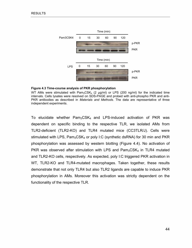
RESULTS
Time (min)
p-PKR
PKR
Pam3CSK4 120906030150
p-PKR
PKR
LPS
Time (min)
120906030150
Figure 4.3 Time-course analysis of PKR phosphorylation WT AMs were stimulated with Pam3CSK4 (2 g/ml) or LPS (200 ng/ml) for the indicated time intervals. Cells lysates were resolved on SDS-PAGE and probed with anti-phospho PKR and anti-PKR antibodies as described in Materials and Methods. The data are representative of three independent experiments.
To elucidate whether Pam3CSK4 and LPS-induced activation of PKR was
dependent on specific binding to the respective TLR, we isolated AMs from
TLR2-deficient (TLR2-KO) and TLR4 mutated mice (CC3TLR/J). Cells were
stimulated with LPS, Pam3CSK4 or poly I:C (synthetic dsRNA) for 30 min and PKR
phosphorylation was assessed by western blotting (Figure 4.4). No activation of
PKR was observed after stimulation with LPS and Pam3CSK4 in TLR4 mutated
and TLR2-KO cells, respectively. As expected, poly I:C triggered PKR activation in
WT, TLR2-KO and TLR4-mutated macrophages. Taken together, these results
demonstrate that not only TLR4 but also TLR2 ligands are capable to induce PKR
phosphorylation in AMs. Moreover this activation was strictly dependent on the
functionality of the respective TLR.
44
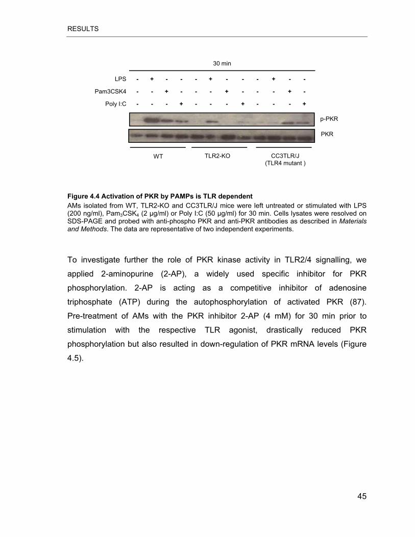
RESULTS
Pam3CSK4
Poly I:C
30 min
p-PKR
PKR
WT CC3TLR/J (TLR4 mutant )
TLR2-KO
+---+---+---
-+---+---+--
--+---+---+-LPS
Figure 4.4 Activation of PKR by PAMPs is TLR dependent AMs isolated from WT, TLR2-KO and CC3TLR/J mice were left untreated or stimulated with LPS (200 ng/ml), Pam3CSK4 (2 g/ml) or Poly I:C (50 g/ml) for 30 min. Cells lysates were resolved on SDS-PAGE and probed with anti-phospho PKR and anti-PKR antibodies as described in Materials and Methods. The data are representative of two independent experiments.
To investigate further the role of PKR kinase activity in TLR2/4 signalling, we
applied 2-aminopurine (2-AP), a widely used specific inhibitor for PKR
phosphorylation. 2-AP is acting as a competitive inhibitor of adenosine
triphosphate (ATP) during the autophosphorylation of activated PKR (87).
Pre-treatment of AMs with the PKR inhibitor 2-AP (4 mM) for 30 min prior to
stimulation with the respective TLR agonist, drastically reduced PKR
phosphorylation but also resulted in down-regulation of PKR mRNA levels (Figure
4.5).
45
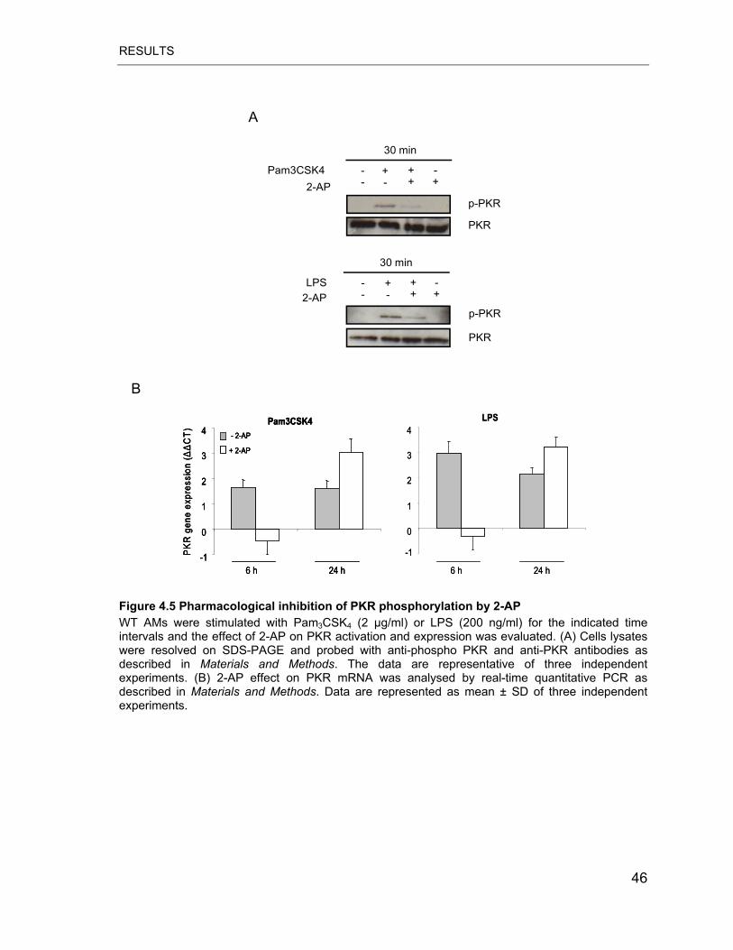
RESULTS
A
30 min
++--Pam3CSK4
PKR
p-PKR 2-AP
-++-
PKR
p-PKR
30 min
++--LPS
2-AP-++-
B
-1
0
1
2
3
4
PK
R g
ene
expr
essi
on (�
�C
T)
Pam3CSK4- 2-AP
+ 2-AP
6 h 24 h
-1
0
1
2
3
4LPS
6 h 24 h-1
0
1
2
3
4
PK
R g
ene
expr
essi
on (�
�C
T)
Pam3CSK4- 2-AP
+ 2-AP
6 h 24 h-1
0
1
2
3
4
PK
R g
ene
expr
essi
on (�
�C
T)
Pam3CSK4- 2-AP
+ 2-AP
6 h 24 h
-1
0
1
2
3
4LPS
6 h 24 h Figure 4.5 Pharmacological inhibition of PKR phosphorylation by 2-AP WT AMs were stimulated with Pam3CSK4 (2 g/ml) or LPS (200 ng/ml) for the indicated time intervals and the effect of 2-AP on PKR activation and expression was evaluated. (A) Cells lysates were resolved on SDS-PAGE and probed with anti-phospho PKR and anti-PKR antibodies as described in Materials and Methods. The data are representative of three independent experiments. (B) 2-AP effect on PKR mRNA was analysed by real-time quantitative PCR as described in Materials and Methods. Data are represented as mean ± SD of three independent experiments.
46
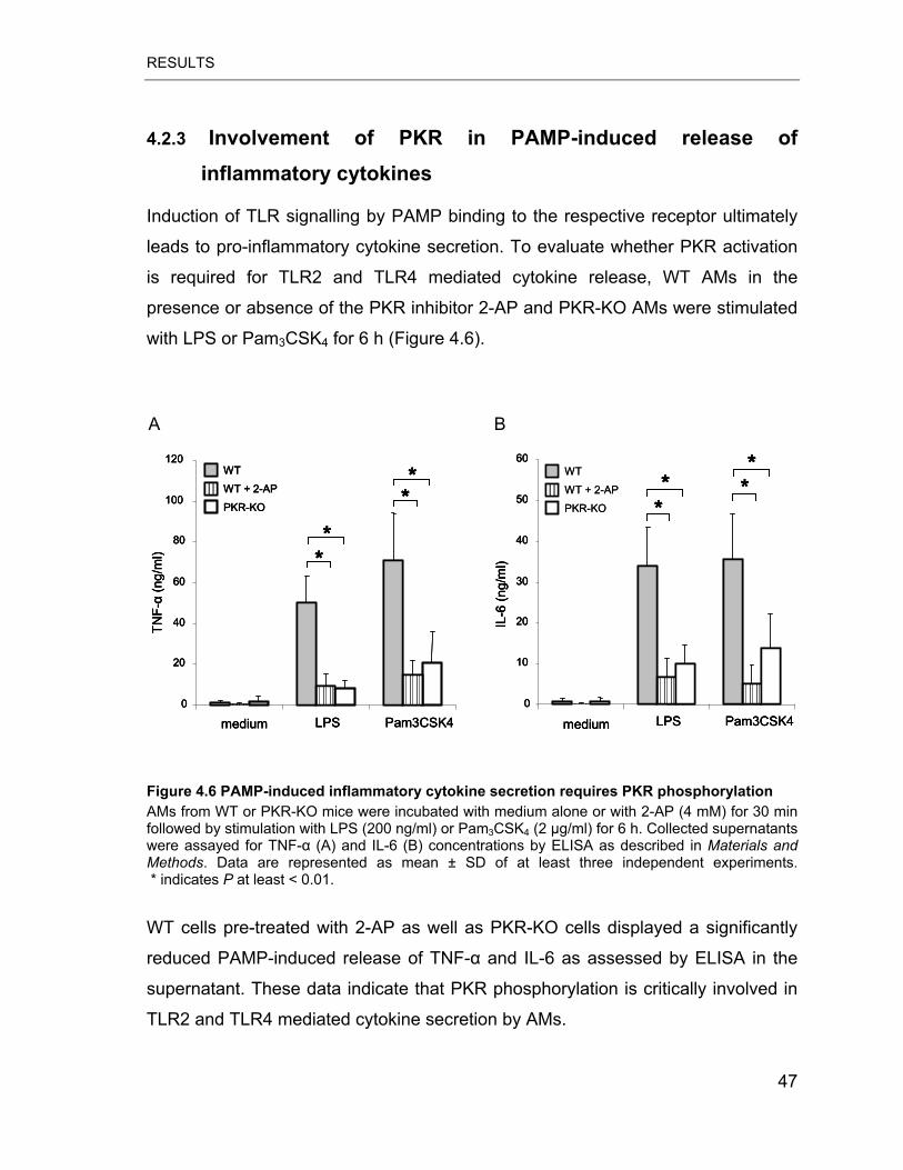
RESULTS
4.2.3 Involvement of PKR in PAMP-induced release of inflammatory cytokines
Induction of TLR signalling by PAMP binding to the respective receptor ultimately
leads to pro-inflammatory cytokine secretion. To evaluate whether PKR activation
is required for TLR2 and TLR4 mediated cytokine release, WT AMs in the
presence or absence of the PKR inhibitor 2-AP and PKR-KO AMs were stimulated
with LPS or Pam3CSK4 for 6 h (Figure 4.6).
A B
0
20
40
60
80
100
120
LPS Pam3CSK4
TNF-
�(n
g/m
l) **
**
WT
WT + 2-AP
PKR-KO
medium0
10
20
30
40
50
60
LPS Pam3CSK4
IL-6
(ng/
ml)
**
**
medium
WT
WT + 2-AP
PKR-KO
0
20
40
60
80
100
120
LPS Pam3CSK4
TNF-
�(n
g/m
l) **
**
WT
WT + 2-AP
PKR-KO
medium0
20
40
60
80
100
120
LPS Pam3CSK4
TNF-
�(n
g/m
l) ****
****
WT
WT + 2-AP
PKR-KO
medium0
10
20
30
40
50
60
LPS Pam3CSK4
IL-6
(ng/
ml)
**
**
medium0
10
20
30
40
50
60
LPS Pam3CSK4
IL-6
(ng/
ml)
****
***
medium
WT
WT + 2-AP
PKR-KO
Figure 4.6 PAMP-induced inflammatory cytokine secretion requires PKR phosphorylation AMs from WT or PKR-KO mice were incubated with medium alone or with 2-AP (4 mM) for 30 min followed by stimulation with LPS (200 ng/ml) or Pam3CSK4 (2 g/ml) for 6 h. Collected supernatants were assayed for TNF-� (A) and IL-6 (B) concentrations by ELISA as described in Materials and Methods. Data are represented as mean ± SD of at least three independent experiments. * indicates P at least < 0.01.
WT cells pre-treated with 2-AP as well as PKR-KO cells displayed a significantly
reduced PAMP-induced release of TNF-� and IL-6 as assessed by ELISA in the
supernatant. These data indicate that PKR phosphorylation is critically involved in
TLR2 and TLR4 mediated cytokine secretion by AMs.
47
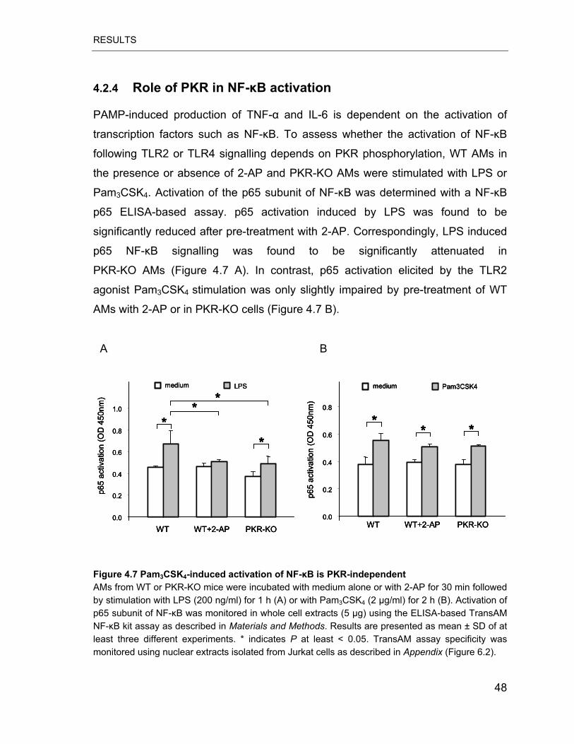
RESULTS
4.2.4 Role of PKR in NF-�B activation
PAMP-induced production of TNF-� and IL-6 is dependent on the activation of
transcription factors such as NF-�B. To assess whether the activation of NF-�B
following TLR2 or TLR4 signalling depends on PKR phosphorylation, WT AMs in
the presence or absence of 2-AP and PKR-KO AMs were stimulated with LPS or
Pam3CSK4. Activation of the p65 subunit of NF-�B was determined with a NF-�B
p65 ELISA-based assay. p65 activation induced by LPS was found to be
significantly reduced after pre-treatment with 2-AP. Correspondingly, LPS induced
p65 NF-�B signalling was found to be significantly attenuated in
PKR-KO AMs (Figure 4.7 A). In contrast, p65 activation elicited by the TLR2
agonist Pam3CSK4 stimulation was only slightly impaired by pre-treatment of WT
AMs with 2-AP or in PKR-KO cells (Figure 4.7 B).
A B
0.0
0.2
0.4
0.6
0.8
1.0
LPS
WT WT+2-AP PKR-KO
**
*
p65
activ
atio
n (O
D 4
50nm
)
medium
*
0.0
0.2
0.4
0.6
WT WT+2-AP PKR-KO
** *
p65
activ
atio
n (O
D 4
50nm
)
0.8
Pam3CSK4medium
0.0
0.2
0.4
0.6
0.8
1.0
LPS
WT WT+2-AP PKR-KO
**
*
p65
activ
atio
n (O
D 4
50nm
)
medium
*
0.0
0.2
0.4
0.6
0.8
1.0
LPS
WT WT+2-AP PKR-KO
***
*
p65
activ
atio
n (O
D 4
50nm
)
medium
**
0.0
0.2
0.4
0.6
WT WT+2-AP PKR-KO
** *
p65
activ
atio
n (O
D 4
50nm
)
0.8
Pam3CSK4medium
0.0
0.2
0.4
0.6
WT WT+2-AP PKR-KO
**** **
p65
activ
atio
n (O
D 4
50nm
)
0.8
Pam3CSK4medium
Figure 4.7 Pam3CSK4-induced activation of NF-�B is PKR-independent AMs from WT or PKR-KO mice were incubated with medium alone or with 2-AP for 30 min followed by stimulation with LPS (200 ng/ml) for 1 h (A) or with Pam3CSK4 (2 g/ml) for 2 h (B). Activation of p65 subunit of NF-�B was monitored in whole cell extracts (5 μg) using the ELISA-based TransAM NF-�B kit assay as described in Materials and Methods. Results are presented as mean ± SD of at least three different experiments. * indicates P at least < 0.05. TransAM assay specificity was monitored using nuclear extracts isolated from Jurkat cells as described in Appendix (Figure 6.2).
48
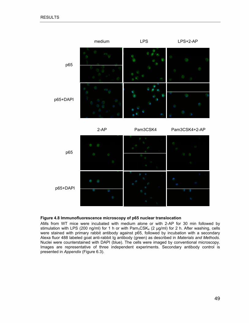
RESULTS
p65
p65+DAPI
p65
p65+DAPI
Pam3CSK4 Pam3CSK4+2-AP2-AP
medium LPS LPS+2-AP
Figure 4.8 Immunofluorescence microscopy of p65 nuclear translocation AMs from WT mice were incubated with medium alone or with 2-AP for 30 min followed by stimulation with LPS (200 ng/ml) for 1 h or with Pam3CSK4 (2 g/ml) for 2 h. After washing, cells were stained with primary rabbit antibody against p65, followed by incubation with a secondary Alexa fluor 488 labeled goat anti-rabbit Ig antibody (green) as described in Materials and Methods. Nuclei were counterstained with DAPI (blue). The cells were imaged by conventional microscopy. Images are representative of three independent experiments. Secondary antibody control is presented in Appendix (Figure 6.3).
49
RESULTS
To confirm this differential effect of PKR phosphorylation inhibition on TLR2 vs.
TLR4 mediated NF-�B activation, we examined PAMP-induced nuclear
translocation of p65 by immunofluorescence microscopy (Figure 4.8). In untreated
AMs, p65 was localized exclusively in the cytoplasm, following stimulation with
LPS or Pam3CSK4, p65 translocated to the nucleus. 2-AP pre-treatment caused a
significant reduction of p65 nuclear translocation after LPS but not after Pam3CSK4
stimulation. Together, these results suggest that PKR phosphorylation plays a
major role in LPS but not Pam3CSK4-induced activation of the p65 NF-�B subunit.
4.2.5 The role of PKR in MAPKs signalling pathway
Previous studies have demonstrated that PKR mediates activation of MAPKs in
response to inflammatory stimuli in various cell types (88-90). Therefore we aimed
to delineate the role of PKR in this process in AMs. PKR inhibitor 2-AP and AMs
deficient in functional PKR were used to describe the role of PKR in MAPKs
activation. We found that only JNK phosphorylation was affected by
pharmacological suppression of PKR activity and in PKR-KO AMs (Figure 4.9).
50

RESULTS
JNK
p-ERK1/2
p-p38
p-JNK
p38
ERK1/2
30 min
++---++-LPS
2-AP
p-ERK1/2
30 min
++---++-
2-APPam3CSK4
JNK
p-p38
p-JNK
p38
ERK1/2
JNK
p-ERK1/2
p-p38
p-JNK
p38
ERK1/2
30 min
++---++-LPS
2-AP
JNK
p-ERK1/2
p-p38
p-JNK
p38
ERK1/2
30 min
++---++-LPS
2-AP
p-ERK1/2
30 min
++---++-
2-APPam3CSK4
JNK
p-p38
p-JNK
p38
ERK1/2
A
PKR-KO WT
LPS
Pam3C
SK4
medium
LPS
Pam3C
SK4
medium
p-JNK
JNK
p-p38
p38
p-ERK
ERK
a
PKR-KO WT
LPS
Pam3C
SK4
medium
LPS
Pam3C
SK4
medium
p-JNK
JNK
p-p38
p38
p-ERK
ERK
PKR-KO WT
LPS
Pam3C
SK4
medium
LPS
Pam3C
SK4
medium
p-JNK
JNK
p-p38
p38
p-ERK
ERK
a
B
Figure 4.9 PKR regulates the TLR2/4 ligand-induced JNK signalling pathway MAP kinases activation was evaluated in WT AMs incubated with medium alone or with 2-AP for 30 min (A) or in AMs derived from PKR-KO mice (B). Cells were stimulated with Pam3CSK4 (2 g/ml) or LPS (200 ng/ml) for 30 min. Subsequently, cells lysates were resolved on SDS-PAGE and analyzed for MAP kinases phosphorylation using antibodies against phospho-JNK, phospho-p38, phospho-ERK1/2, total JNK, total 38 and total ERK as described in Materials and Methods.
To further confirm the role of PKR in the JNK signalling pathway, we tested the
effect of 2-AP on the regulation of MKK4, a kinase upstream of JNK, and on the
downstream JNK target c-Jun. Inhibition of PKR markedly reduced
phosphorylation of MKK4 and strongly decreased c-Jun phosphorylation as
assessed by mobility shift (Figure 4.10). Collectively, our data indicate that PKR
51
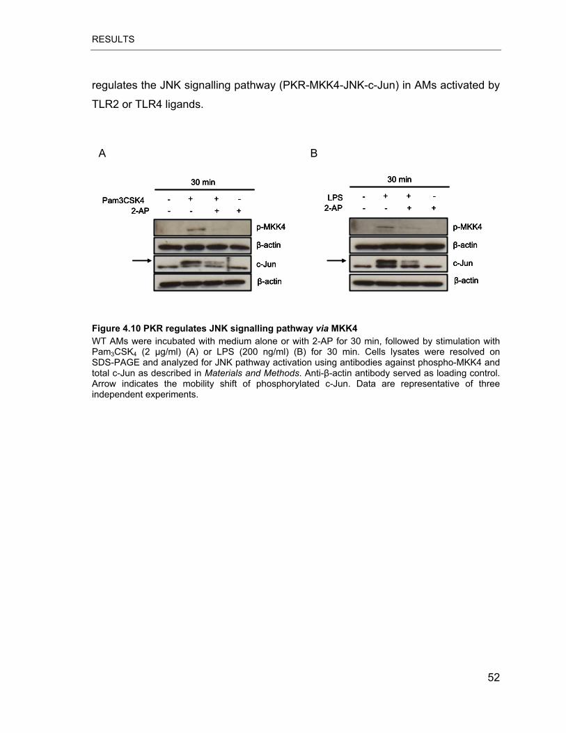
RESULTS
regulates the JNK signalling pathway (PKR-MKK4-JNK-c-Jun) in AMs activated by
TLR2 or TLR4 ligands.
A B
p-MKK4
�-actin
c-Jun
�-actin
30 min
++---++-LPS
2-AP
p-MKK4
�-actin
c-Jun
�-actin
30 min
++---++-
2-APPam3CSK4
p-MKK4
�-actin
c-Jun
�-actin
30 min
++---++-LPS
2-AP
p-MKK4
�-actin
c-Jun
�-actin
30 min
++---++-LPS
2-AP
p-MKK4
�-actin
c-Jun
�-actin
30 min
++---++-
2-APPam3CSK4
p-MKK4
�-actin
c-Jun
�-actin
30 min
++---++-
2-APPam3CSK4
Figure 4.10 PKR regulates JNK signalling pathway via MKK4 WT AMs were incubated with medium alone or with 2-AP for 30 min, followed by stimulation with Pam3CSK4 (2 g/ml) (A) or LPS (200 ng/ml) (B) for 30 min. Cells lysates were resolved on SDS-PAGE and analyzed for JNK pathway activation using antibodies against phospho-MKK4 and total c-Jun as described in Materials and Methods. Anti-�-actin antibody served as loading control. Arrow indicates the mobility shift of phosphorylated c-Jun. Data are representative of three independent experiments.
52
RESULTS
4.3 Genome-wide transcriptional profiling of MPs recruited to mouse lungs in response to alveolar challenge with the TLR2 agonist Pam3CSK4
4.3.1 Induction of lung inflammation and alveolar trafficking of circulating MPs by intratracheal deposition of Pam3CSK4
To determine the capacity of the synthetic TLR2 agonist Pam3CSK4 to induce lung
inflammation in vivo, WT C57Bl/6 mice received i.t. application of Pam3CSK4 (50
μg/mouse) and BALF cellular constituents were monitored at various time points
post challenge. Pulmonary administration of Pam3CSK4 provoked rapid recruitment
of neutrophils into the alveolar compartment that was detectable as early as at 6 h,
peaked at approximately 24 h after challenge and significantly declined within the
next 24 h. The number of BALF macrophages was found to be slightly increased at
24 h post challenge without reaching statistical significance (Figure 4.11).
To further assess alveolar trafficking of MPs we employed transgenic CX3CR1+/GFP
mice expressing green fluorescence protein (GFP) under control of the CX3CR1
promoter as described before (69, 76). This approach allowed identifying and
further high purity sorting of GFP-positive MPs from the peripheral blood and of
freshly recruited MPs from BALF as described in Materials and Methods.
Heterozygous CX3CR1+/GFP mice were either left untreated or received i.t.
application of the TLR2 ligand Pam3CSK4 (50 μg/mouse) and recruited MPs
numbers were assessed in BALF after 24 h or 48 h. Very few GFP-positive cells
(0.3 %-0.5 %) were detected in BALF collected from untreated mice (Figure. 4.12).
In contrast, at 24 h and even more pronounced at 48 h after Pam3CSK4 i.t.
application we observed significantly increased proportions of GFP-positive cells in
BALF samples (1.4-1.7 % and 2.5-3.5 %, respectively). These data demonstrate
that i.t. application of the TLR2 ligand Pam3CSK4 induced enhanced trafficking of
circulating MPs into the alveolar space.
53

RESULTS
BA
LF c
ell n
umbe
r (x
10*5
)
0
5
10
15
20
25
Total cell number Macrophages Neutrophils
0 h
6 h
12 h
24 h
48 h
n.d
**
*
BA
LF c
ell n
umbe
r (x
10*5
)
0
5
10
15
20
25
Total cell number Macrophages Neutrophils
0 h
6 h
12 h
24 h
48 h
n.d
***
*
Figure 4.11 Inflammatory cell accumulation in the bronchoalveolar compartment in response to alveolar deposition of Pam3CSK4
C57BL/6 WT mice were either left untreated (0 h) or were challenged intratracheally with Pam3CSK4 (50 μg/mouse) for 6h, 12h, 24h, or 48 h. After indicated times, BALF cells were isolated and microscopic differentiation and quantification of macrophages and alveolar recruited neutrophils were performed as described in Materials and Methods. Means of different time points (five mice per treatment group) were compared using the ANOVA test as described in Materials and Methods. * indicates P at least < 0.01.
54
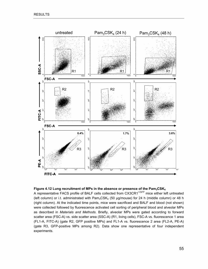
RESULTS
FSC-A
SSC
-A
FSC-A
FITC
-A
FITC-A
PE-A
untreated
0.4%
Pam3CSK4 (24 h)
1.7% 3.6%
R2
R3 R3 R3
R2 R2
R1R1R1
Pam3CSK4 (48 h)
FSC-A
SSC
-A
FSC-A
FITC
-A
FITC-A
PE-A
untreated
0.4%
Pam3CSK4 (24 h)
1.7% 3.6%
R2
R3 R3 R3
R2 R2
R1R1R1
Pam3CSK4 (48 h)
Figure 4.12 Lung recruitment of MPs in the absence or presence of the Pam3CSK4
A representative FACS profile of BALF cells collected from CX3CR1+/GFP mice either left untreated (left column) or i.t. administrated with Pam3CSK4 (50 μg/mouse) for 24 h (middle column) or 48 h (right column). At the indicated time points, mice were sacrificed and BALF and blood (not shown) were collected followed by fluorescence activated cell sorting of peripheral blood and alveolar MPs as described in Materials and Methods. Briefly, alveolar MPs were gated according to forward scatter area (FSC-A) vs. side scatter area (SSC-A) (R1, living cells), FSC-A vs. fluorescence 1 area (FL1-A, FITC-A) (gate R2, GFP positive MPs) and FL1-A vs. fluorescence 2 area (FL2-A, PE-A) (gate R3, GFP-positive MPs among R2). Data show one representative of four independent experiments.
55
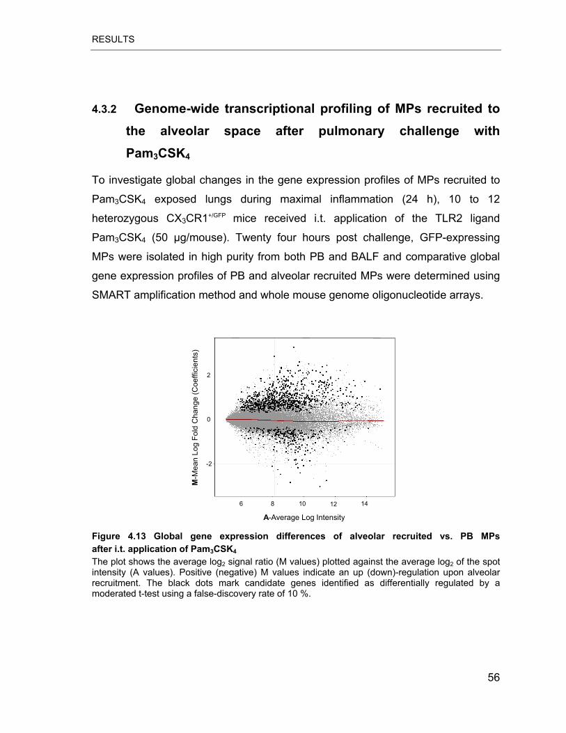
RESULTS
4.3.2 Genome-wide transcriptional profiling of MPs recruited to the alveolar space after pulmonary challenge with Pam3CSK4
To investigate global changes in the gene expression profiles of MPs recruited to
Pam3CSK4 exposed lungs during maximal inflammation (24 h), 10 to 12
heterozygous CX3CR1+/GFP mice received i.t. application of the TLR2 ligand
Pam3CSK4 (50 μg/mouse). Twenty four hours post challenge, GFP-expressing
MPs were isolated in high purity from both PB and BALF and comparative global
gene expression profiles of PB and alveolar recruited MPs were determined using
SMART amplification method and whole mouse genome oligonucleotide arrays.
A-Average Log Intensity
6 8 10 12 14
M-M
ean
Log
Fold
Cha
nge
(Coe
ffici
ents
)
0
-2
2
Figure 4.13 Global gene expression differences of alveolar recruited vs. PB MPs after i.t. application of Pam3CSK4 The plot shows the average log2 signal ratio (M values) plotted against the average log2 of the spot intensity (A values). Positive (negative) M values indicate an up (down)-regulation upon alveolar recruitment. The black dots mark candidate genes identified as differentially regulated by a moderated t-test using a false-discovery rate of 10 %.
56
RESULTS
We found that the global transcriptome of MPs recruited to the alveolar
compartment at 24 h in response to Pam3CSK4 was dramatically changed in
comparison to circulating MPs obtained at the same time point (Figure 4.13).
Approximately 3700 genes differentially expressed between PB and alveolar
recruited MPs were identified (10 % FDR) and the majority of which (~2500) were
up-regulated in the alveolar MP cell population. This group contains a broad panel
of genes encoding pro-inflammatory cytokines previously described to be robustly
up-regulated by different microbial components in various cell types such as
interleukin IL-1�, TNF-�, IL-6, and IL-12A (Table 4.3). We additionally observed
up-regulated mRNA levels of chemokines such as KC, CXCL11, CXCL12,
CXCL13, CXCL16, CCL2 and CCL7, whereas the gene expression of the
neutrophil and monocyte chemoattractant CXCL7 was significantly down-regulated
in alveolar mononuclear phagocytes.
57
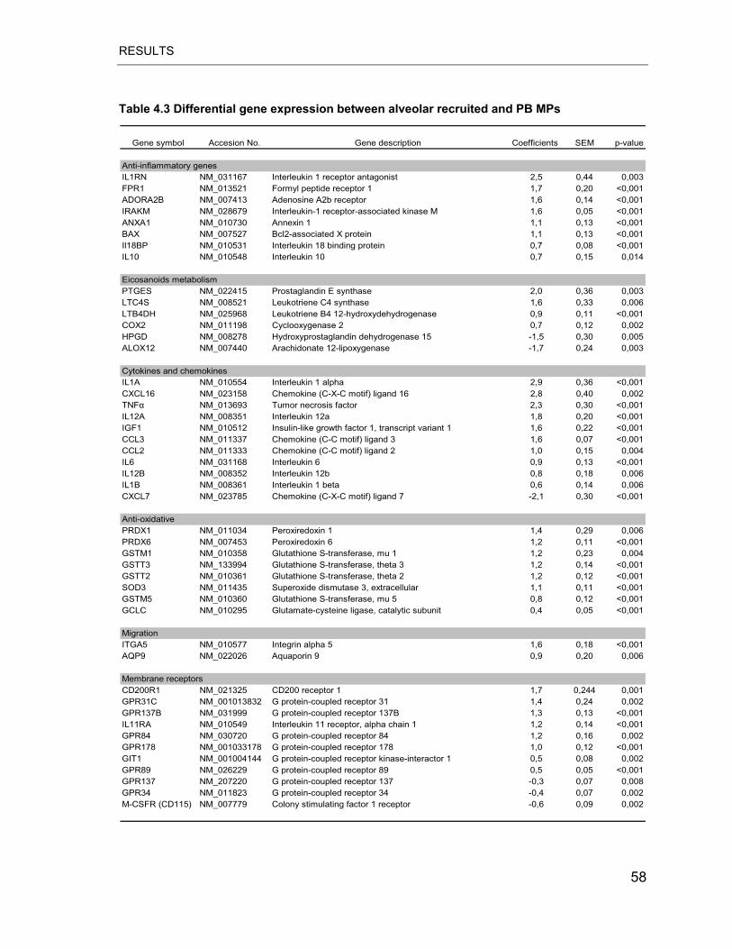
RESULTS
Table 4.3 Differential gene expression between alveolar recruited and PB MPs
Gene symbol Accesion No. Gene description Coefficients SEM p-value
Anti-inflammatory genes IL1RN NM_031167 Interleukin 1 receptor antagonist 2,5 0,44 0,003FPR1 NM_013521 Formyl peptide receptor 1 1,7 0,20 <0,001ADORA2B NM_007413 Adenosine A2b receptor 1,6 0,14 <0,001IRAKM NM_028679 Interleukin-1 receptor-associated kinase M 1,6 0,05 <0,001ANXA1 NM_010730 Annexin 1 1,1 0,13 <0,001BAX NM_007527 Bcl2-associated X protein 1,1 0,13 <0,001Il18BP NM_010531 Interleukin 18 binding protein 0,7 0,08 <0,001IL10 NM_010548 Interleukin 10 0,7 0,15 0,014
Eicosanoids metabolismPTGES NM_022415 Prostaglandin E synthase 2,0 0,36 0,003LTC4S NM_008521 Leukotriene C4 synthase 1,6 0,33 0,006LTB4DH NM_025968 Leukotriene B4 12-hydroxydehydrogenase 0,9 0,11 <0,001COX2 NM_011198 Cyclooxygenase 2 0,7 0,12 0,002HPGD NM_008278 Hydroxyprostaglandin dehydrogenase 15 -1,5 0,30 0,005ALOX12 NM_007440 Arachidonate 12-lipoxygenase -1,7 0,24 0,003
Cytokines and chemokinesIL1A NM_010554 Interleukin 1 alpha 2,9 0,36 <0,001CXCL16 NM_023158 Chemokine (C-X-C motif) ligand 16 2,8 0,40 0,002TNF� NM_013693 Tumor necrosis factor 2,3 0,30 <0,001IL12A NM_008351 Interleukin 12a 1,8 0,20 <0,001IGF1 NM_010512 Insulin-like growth factor 1, transcript variant 1 1,6 0,22 <0,001CCL3 NM_011337 Chemokine (C-C motif) ligand 3 1,6 0,07 <0,001CCL2 NM_011333 Chemokine (C-C motif) ligand 2 1,0 0,15 0,004IL6 NM_031168 Interleukin 6 0,9 0,13 <0,001IL12B NM_008352 Interleukin 12b 0,8 0,18 0,006IL1B NM_008361 Interleukin 1 beta 0,6 0,14 0,006CXCL7 NM_023785 Chemokine (C-X-C motif) ligand 7 -2,1 0,30 <0,001
Anti-oxidativePRDX1 NM_011034 Peroxiredoxin 1 1,4 0,29 0,006PRDX6 NM_007453 Peroxiredoxin 6 1,2 0,11 <0,001GSTM1 NM_010358 Glutathione S-transferase, mu 1 1,2 0,23 0,004GSTT3 NM_133994 Glutathione S-transferase, theta 3 1,2 0,14 <0,001GSTT2 NM_010361 Glutathione S-transferase, theta 2 1,2 0,12 <0,001SOD3 NM_011435 Superoxide dismutase 3, extracellular 1,1 0,11 <0,001GSTM5 NM_010360 Glutathione S-transferase, mu 5 0,8 0,12 <0,001GCLC NM_010295 Glutamate-cysteine ligase, catalytic subunit 0,4 0,05 <0,001
MigrationITGA5 NM_010577 Integrin alpha 5 1,6 0,18 <0,001AQP9 NM_022026 Aquaporin 9 0,9 0,20 0,006
Membrane receptorsCD200R1 NM_021325 CD200 receptor 1 1,7 0,244 0,001GPR31C NM_001013832 G protein-coupled receptor 31 1,4 0,24 0,002GPR137B NM_031999 G protein-coupled receptor 137B 1,3 0,13 <0,001IL11RA NM_010549 Interleukin 11 receptor, alpha chain 1 1,2 0,14 <0,001GPR84 NM_030720 G protein-coupled receptor 84 1,2 0,16 0,002GPR178 NM_001033178 G protein-coupled receptor 178 1,0 0,12 <0,001GIT1 NM_001004144 G protein-coupled receptor kinase-interactor 1 0,5 0,08 0,002GPR89 NM_026229 G protein-coupled receptor 89 0,5 0,05 <0,001GPR137 NM_207220 G protein-coupled receptor 137 -0,3 0,07 0,008GPR34 NM_011823 G protein-coupled receptor 34 -0,4 0,07 0,002M-CSFR (CD115) NM_007779 Colony stimulating factor 1 receptor -0,6 0,09 0,002
58
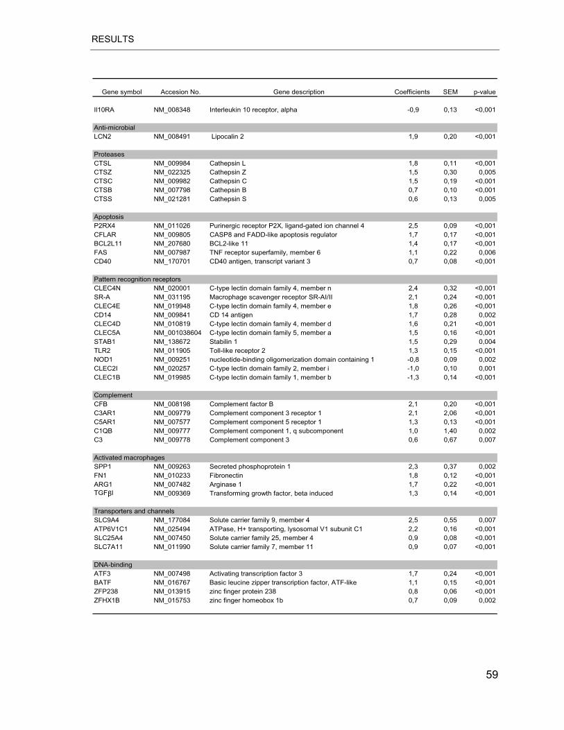
RESULTS
Gene symbol Accesion No. Gene description Coefficients SEM p-value
Il10RA NM_008348 Interleukin 10 receptor, alpha -0,9 0,13 <0,001
Anti-microbialLCN2 NM_008491 Lipocalin 2 1,9 0,20 <0,001
ProteasesCTSL NM_009984 Cathepsin L 1,8 0,11 <0,001CTSZ NM_022325 Cathepsin Z 1,5 0,30 0,005CTSC NM_009982 Cathepsin C 1,5 0,19 <0,001CTSB NM_007798 Cathepsin B 0,7 0,10 <0,001CTSS NM_021281 Cathepsin S 0,6 0,13 0,005
ApoptosisP2RX4 NM_011026 Purinergic receptor P2X, ligand-gated ion channel 4 2,5 0,09 <0,001CFLAR NM_009805 CASP8 and FADD-like apoptosis regulator 1,7 0,17 <0,001BCL2L11 NM_207680 BCL2-like 11 1,4 0,17 <0,001FAS NM_007987 TNF receptor superfamily, member 6 1,1 0,22 0,006CD40 NM_170701 CD40 antigen, transcript variant 3 0,7 0,08 <0,001
Pattern recognition receptorsCLEC4N NM_020001 C-type lectin domain family 4, member n 2,4 0,32 <0,001SR-A NM_031195 Macrophage scavenger receptor SR-AI/II 2,1 0,24 <0,001CLEC4E NM_019948 C-type lectin domain family 4, member e 1,8 0,26 <0,001CD14 NM_009841 CD 14 antigen 1,7 0,28 0,002CLEC4D NM_010819 C-type lectin domain family 4, member d 1,6 0,21 <0,001CLEC5A NM_001038604 C-type lectin domain family 5, member a 1,5 0,16 <0,001STAB1 NM_138672 Stabilin 1 1,5 0,29 0,004TLR2 NM_011905 Toll-like receptor 2 1,3 0,15 <0,001NOD1 NM_009251 nucleotide-binding oligomerization domain containing 1 -0,8 0,09 0,002CLEC2I NM_020257 C-type lectin domain family 2, member i -1,0 0,10 0,001CLEC1B NM_019985 C-type lectin domain family 1, member b -1,3 0,14 <0,001
ComplementCFB NM_008198 Complement factor B 2,1 0,20 <0,001C3AR1 NM_009779 Complement component 3 receptor 1 2,1 2,06 <0,001C5AR1 NM_007577 Complement component 5 receptor 1 1,3 0,13 <0,001C1QB NM_009777 Complement component 1, q subcomponent 1,0 1,40 0,002C3 NM_009778 Complement component 3 0,6 0,67 0,007
Activated macrophages SPP1 NM_009263 Secreted phosphoprotein 1 2,3 0,37 0,002FN1 NM_010233 Fibronectin 1,8 0,12 <0,001ARG1 NM_007482 Arginase 1 1,7 0,22 <0,001TGF�I NM_009369 Transforming growth factor, beta induced 1,3 0,14 <0,001
Transporters and channelsSLC9A4 NM_177084 Solute carrier family 9, member 4 2,5 0,55 0,007ATP6V1C1 NM_025494 ATPase, H+ transporting, lysosomal V1 subunit C1 2,2 0,16 <0,001SLC25A4 NM_007450 Solute carrier family 25, member 4 0,9 0,08 <0,001SLC7A11 NM_011990 Solute carrier family 7, member 11 0,9 0,07 <0,001
DNA-bindingATF3 NM_007498 Activating transcription factor 3 1,7 0,24 <0,001BATF NM_016767 Basic leucine zipper transcription factor, ATF-like 1,1 0,15 <0,001ZFP238 NM_013915 zinc finger protein 238 0,8 0,06 <0,001ZFHX1B NM_015753 zinc finger homeobox 1b 0,7 0,09 0,002
59
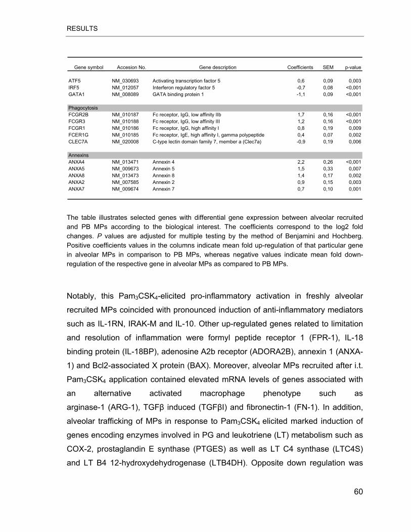
RESULTS
Gene symbol Accesion No. Gene description Coefficients SEM p-value
ATF5 NM_030693 Activating transcription factor 5 0,6 0,09 0,003IRF5 NM_012057 Interferon regulatory factor 5 -0,7 0,08 <0,001GATA1 NM_008089 GATA binding protein 1 -1,1 0,09 <0,001
PhagocytosisFCGR2B NM_010187 Fc receptor, IgG, low affinity IIb 1,7 0,16 <0,001FCGR3 NM_010188 Fc receptor, IgG, low affinity III 1,2 0,16 <0,001FCGR1 NM_010186 Fc receptor, IgG, high affinity I 0,8 0,19 0,009FCER1G NM_010185 Fc receptor, IgE, high affinity I, gamma polypeptide 0,4 0,07 0,002CLEC7A NM_020008 C-type lectin domain family 7, member a (Clec7a) -0,9 0,19 0,006
AnnexinsANXA4 NM_013471 Annexin 4 2,2 0,26 <0,001ANXA5 NM_009673 Annexin 5 1,5 0,33 0,007ANXA8 NM_013473 Annexin 8 1,4 0,17 0,002ANXA2 NM_007585 Annexin 2 0,9 0,15 0,003ANXA7 NM_009674 Annexin 7 0,7 0,10 0,001
The table illustrates selected genes with differential gene expression between alveolar recruited and PB MPs according to the biological interest. The coefficients correspond to the log2 fold changes. P values are adjusted for multiple testing by the method of Benjamini and Hochberg. Positive coefficients values in the columns indicate mean fold up-regulation of that particular gene in alveolar MPs in comparison to PB MPs, whereas negative values indicate mean fold down-regulation of the respective gene in alveolar MPs as compared to PB MPs.
Notably, this Pam3CSK4-elicited pro-inflammatory activation in freshly alveolar
recruited MPs coincided with pronounced induction of anti-inflammatory mediators
such as IL-1RN, IRAK-M and IL-10. Other up-regulated genes related to limitation
and resolution of inflammation were formyl peptide receptor 1 (FPR-1), IL-18
binding protein (IL-18BP), adenosine A2b receptor (ADORA2B), annexin 1 (ANXA-
1) and Bcl2-associated X protein (BAX). Moreover, alveolar MPs recruited after i.t.
Pam3CSK4 application contained elevated mRNA levels of genes associated with
an alternative activated macrophage phenotype such as
arginase-1 (ARG-1), TGF� induced (TGF�I) and fibronectin-1 (FN-1). In addition,
alveolar trafficking of MPs in response to Pam3CSK4 elicited marked induction of
genes encoding enzymes involved in PG and leukotriene (LT) metabolism such as
COX-2, prostaglandin E synthase (PTGES) as well as LT C4 synthase (LTC4S)
and LT B4 12-hydroxydehydrogenase (LTB4DH). Opposite down regulation was
60
RESULTS
observed for arachidonate 12-lipoxygenase (ALOX12) and hydroxyprostaglandin
dehydrogenase 15 (HPDG) transcripts.
Finally, a large number of genes encoding surface receptors were differentially
regulated in MPs recruited to the lung by alveolar exposure to Pam3CSK4. Thus we
observed elevated transcript levels in alveolar MPs for PRR genes such as CD14,
TLR2, CLEC4N and scavenger receptor (SR)-A and reduced levels for IL-10
receptor and M-CSFR (known as CD115). Moreover, alveolar trafficking of
mononuclear phagocytes was found to be associated with marked up-regulation of
some of G-protein coupled receptors (GPCRs) such as GPR31C, GPR137B,
GPR84 and GPR178.
To determine which gene ontology (GO) categories were statistically
over-represented among the genes found to be the differentially regulated in
alveolar recruited vs. circulating MPs we used the web-based tool GENECODIS
(91). When grouped according to the biological processes, GO analysis highlighted
a significant over-representation of molecules associated with
inflammatory/immune responses including genes involved in chemotaxis (e.g.
C3AR1 and FPR-1), apoptosis (e.g. FAS and CD40) and phagocytosis (such as
CD23 and CD64). In addition, genes related to protein and ATP biosynthesis and
lipid storage were significantly over-represented (Table 4.4).
61
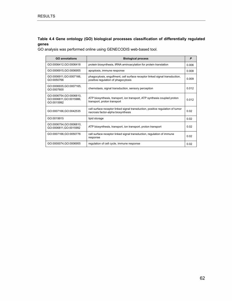
RESULTS
Table 4.4 Gene ontology (GO) biological processes classification of differentially regulated genes GO analysis was performed online using GENECODIS web-based tool.
GO annotations Biological process P
GO:0006412,GO:0006418 protein biosynthesis, tRNA aminoacylation for protein translation 0.006
GO:0006915,GO:0006955 apoptosis, immune response 0.008
GO:0006911,GO:0007166,GO:0050766
phagocytosis, engulfment, cell surface receptor linked signal transduction, positive regulation of phagocytosis 0.009
GO:0006935,GO:0007165,GO:0007600 chemotaxis, signal transduction, sensory perception 0.012
GO:0006754,GO:0006810,GO:0006811,GO:0015986,GO:0015992
ATP biosynthesis, transport, ion transport, ATP synthesis coupled proton transport, proton transport 0.012
GO:0007166,GO:0042535cell surface receptor linked signal transduction, positive regulation of tumor necrosis factor-alpha biosynthesis 0.02
GO:0019915 lipid storage 0.02
GO:0006754,GO:0006810,GO:0006811,GO:0015992 ATP biosynthesis, transport, ion transport, proton transport 0.02
GO:0007166,GO:0050776 cell surface receptor linked signal transduction, regulation of immune response 0.02
GO:0000074,GO:0006955 regulation of cell cycle, immune response 0.02
62
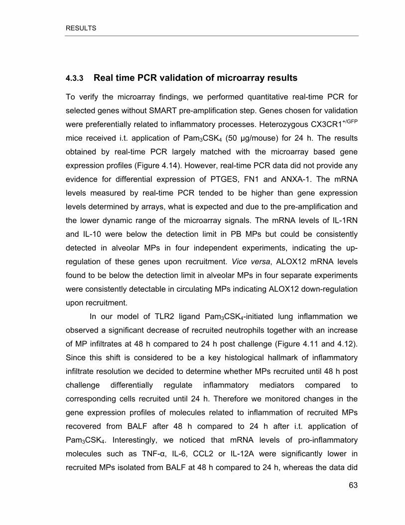
RESULTS
4.3.3 Real time PCR validation of microarray results
To verify the microarray findings, we performed quantitative real-time PCR for
selected genes without SMART pre-amplification step. Genes chosen for validation
were preferentially related to inflammatory processes. Heterozygous CX3CR1+/GFP
mice received i.t. application of Pam3CSK4 (50 μg/mouse) for 24 h. The results
obtained by real-time PCR largely matched with the microarray based gene
expression profiles (Figure 4.14). However, real-time PCR data did not provide any
evidence for differential expression of PTGES, FN1 and ANXA-1. The mRNA
levels measured by real-time PCR tended to be higher than gene expression
levels determined by arrays, what is expected and due to the pre-amplification and
the lower dynamic range of the microarray signals. The mRNA levels of IL-1RN
and IL-10 were below the detection limit in PB MPs but could be consistently
detected in alveolar MPs in four independent experiments, indicating the up-
regulation of these genes upon recruitment. Vice versa, ALOX12 mRNA levels
found to be below the detection limit in alveolar MPs in four separate experiments
were consistently detectable in circulating MPs indicating ALOX12 down-regulation
upon recruitment.
In our model of TLR2 ligand Pam3CSK4-initiated lung inflammation we
observed a significant decrease of recruited neutrophils together with an increase
of MP infiltrates at 48 h compared to 24 h post challenge (Figure 4.11 and 4.12).
Since this shift is considered to be a key histological hallmark of inflammatory
infiltrate resolution we decided to determine whether MPs recruited until 48 h post
challenge differentially regulate inflammatory mediators compared to
corresponding cells recruited until 24 h. Therefore we monitored changes in the
gene expression profiles of molecules related to inflammation of recruited MPs
recovered from BALF after 48 h compared to 24 h after i.t. application of
Pam3CSK4. Interestingly, we noticed that mRNA levels of pro-inflammatory
molecules such as TNF-�, IL-6, CCL2 or IL-12A were significantly lower in
recruited MPs isolated from BALF at 48 h compared to 24 h, whereas the data did
63
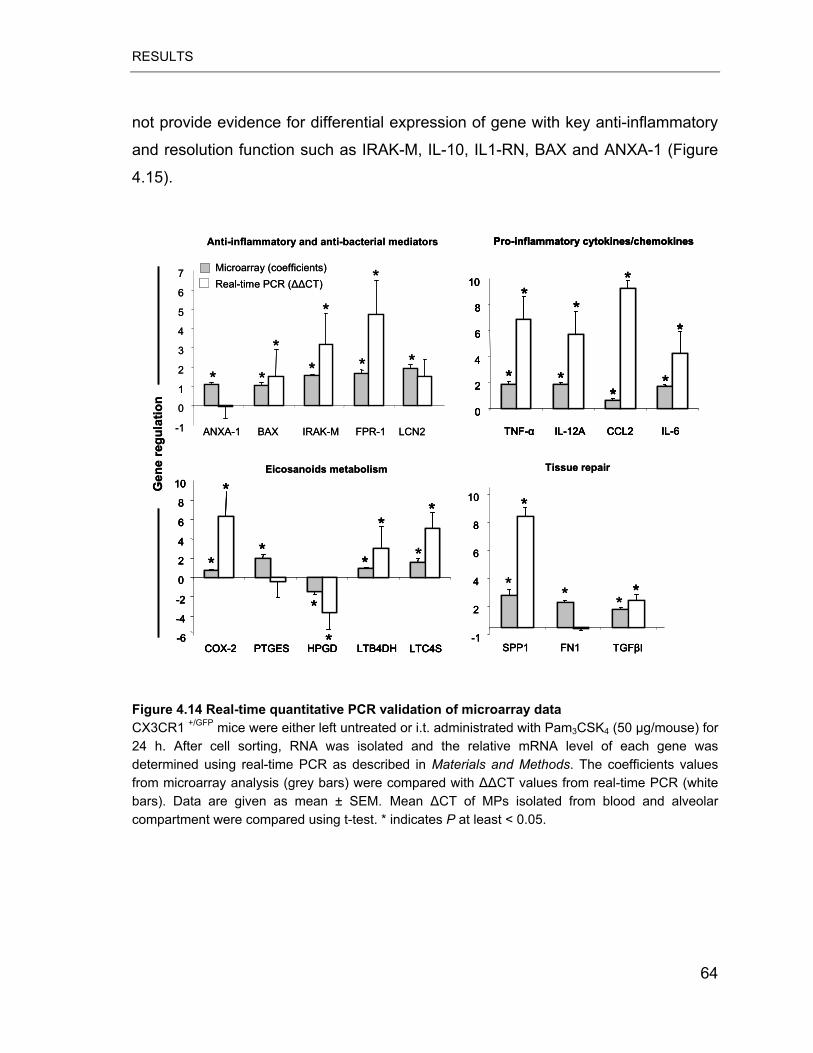
RESULTS
not provide evidence for differential expression of gene with key anti-inflammatory
and resolution function such as IRAK-M, IL-10, IL1-RN, BAX and ANXA-1 (Figure
4.15).
0
2
4
6
8
10
TNF-� IL-12A CCL2 IL-6
**
*
* **
*
*
Pro-inflammatory cytokines/chemokines
-6
-4
-2
0
2
4
6
8
10
COX-2 PTGES HPGD LTB4DH LTC4S
*
*
**
**
*
* *
Eicosanoids metabolism
-1
2
4
6
8
10
SPP1 FN1 TGF�I
*
** * *
Tissue repair
Microarray (coefficients)Real-time PCR (��CT)
-1
0
1
2
3
4
5
6
7
ANXA-1 BAX IRAK-M FPR-1
* * *
*
*
*
Anti-inflammatory and anti-bacterial mediators
Gen
e re
gula
tion
*
LCN2
*
0
2
4
6
8
10
TNF-� IL-12A CCL2 IL-6
**
*
* **
*
*
Pro-inflammatory cytokines/chemokines
0
2
4
6
8
10
TNF-� IL-12A CCL2 IL-6
**
*
* **
*
*
0
2
4
6
8
10
TNF-� IL-12A CCL2 IL-6
**
*
* **
*
*
Pro-inflammatory cytokines/chemokines
-6
-4
-2
0
2
4
6
8
10
COX-2 PTGES HPGD LTB4DH LTC4S
*
*
**
**
*
* *
-6
-4
-2
0
2
4
6
8
10
COX-2 PTGES HPGD LTB4DH LTC4S
*
*
**
-6
-4
-2
0
2
4
6
8
10
COX-2 PTGES HPGD LTB4DH LTC4S
*
*
**
**
*
* *
Eicosanoids metabolism
-1
2
4
6
8
10
SPP1 FN1 TGF�I
*
*
-1
2
4
6
8
10
SPP1 FN1 TGF�I
*
** * *
Tissue repair
Microarray (coefficients)Real-time PCR (��CT)
-1
0
1
2
3
4
5
6
7
ANXA-1 BAX IRAK-M FPR-1
* * *
*
*
*
Anti-inflammatory and anti-bacterial mediators
Gen
e re
gula
tion
*
LCN2
*
Figure 4.14 Real-time quantitative PCR validation of microarray data CX3CR1 +/GFP mice were either left untreated or i.t. administrated with Pam3CSK4 (50 μg/mouse) for 24 h. After cell sorting, RNA was isolated and the relative mRNA level of each gene was determined using real-time PCR as described in Materials and Methods. The coefficients values from microarray analysis (grey bars) were compared with ��CT values from real-time PCR (white bars). Data are given as mean ± SEM. Mean �CT of MPs isolated from blood and alveolar compartment were compared using t-test. * indicates P at least < 0.05.
64
RESULTS
-6
-4
-2
0
2
4
6
8
IL1-RN IL-10 IRAK-M FPR-1 ANXA-1 BAX LCN2
Rel
ativ
e ge
ne e
xpre
ssio
n (�
CT)
Anti-inflammatory and anti-bacterial mediators
24 h post challenge48 h post challenge
*
Pro-inflammatory cytokines/chemokines
-2
0
2
4
6
8
TNF-� IL-6 CCL2 IL-12A
*
*
*
Rel
ativ
e ge
ne e
xpre
ssio
n (�
CT)
*
Pro-inflammatory cytokines/chemokines
-2
0
2
4
6
8
TNF-� IL-6 CCL2 IL-12A
*
*
*
Rel
ativ
e ge
ne e
xpre
ssio
n (�
CT)
Figure 4.15 Time-dependent changes in the gene expression profile of alveolar recruited MPs CX3CR1 +/GFP mice were either left untreated or i.t. administrated with Pam3CSK4 (50 μg/mouse) for 24 h or 48 h. After cell sorting, RNA was isolated and the relative mRNA level of each gene was determined using real-time PCR as described in Materials and Methods. Relative gene expression was calculated using the ��CT method. The results are given as mean ± SEM from three independent experiments. Mean �CT of samples collected at different time points post-treatment were compared using t-test. * indicates P at least < 0.05.
65
DISCUSSION
5 DISCUSSION
The aim of the current study was to analyse the molecular programs activated by
bacterial ligands in alveolar resident as well as alveolar recruited MPs. We have
found that key antiviral defence molecule named PKR is critically involved in an
inflammatory response of AMs to gram-positive as well as gram-negative bacteria
cell wall components in vitro. We have also analyzed, by microarray technology,
the whole genome response of MPs recruited from circulation to the alveolar space
following pulmonary challenge with the TLR2 ligand Pam3CSK4. We found that
lung recruited MPs displayed simultaneous induction of both pro- and anti-
inflammatory genes. In addition, we observed a dynamic change of the genetic
program in MPs obtained from BALF at different time points post alveolar TLR2
ligand deposition.
5.1 Requirement for PKR in TLR2/4-medaited signalling in murine AMs
To identify novel molecular pathways activated in resident AMs by the E. coli LPS
(TLR4 agonist) and synthetic lipoprotein Pam3CSK4 (TLR2 agonist) we performed
a gene expression screen of selected TLR associated molecules. We found that
both TLR2 and TLR4 ligand induced a similar expression pattern of TLR molecules
in vitro and in a murine model of lung inflammation. The screen confirmed that TLR
ligands induced activation of previously reported genes such as TNF-�, IL-6,
SOCS proteins and IRAK-M. Interestingly, our gene expression study identified an
antiviral defence gene named PKR that was significantly up-regulated by bacterial
ligands in vitro and in murine model of lung inflammation. It has been previously
reported that LPS induced PKR mRNA in peritoneal and
ANA-1 macrophages, but the regulation of PKR gene expression in AMs was
unknown (92). Furthermore, our experiments demonstrated for the first time that
also TLR2 ligand (synthetic lipoprotein Pam3CSK4) is a potent inducer of PKR
mRNA and protein in vitro.
66
DISCUSSION
In contrast to the in vitro observations, we noticed that agonists for distinct TLRs
differentially regulated PKR transcription in vivo. The expression of PKR in AMs
was significantly increased after alveolar deposition of LPS at 6 h post challenge
and decreased at later time points (24 h post challenge). In contrast, alveolar
administration of Pam3CSK4 did not affect PKR mRNA levels at any of the
analysed time points. Altogether, our findings suggest that PKR could be involved
in TLR downstream signalling in AM during pulmonary bacterial infections.
PKR is a serine/threonine protein kinase ubiquitously expressed in the cytoplasm
that can be induced by IFN and dsRNA. This enzyme was initially characterized as
translational inhibitor in IFN-induced antiviral response (93). It contains two
functionally distinct domains: an N-terminal ds-RNA binding domain (DBD) and a
C-terminal kinase domain (KD). Binding of dsRNA induces PKR dimerization,
trans-autophosphorylation and subsequent phosphorylation of substrate targets.
The most intensively characterised cellular substrate of PKR kinase activity is the
alpha subunit of the eukaryotic translational initiation factor 2 (eIF-2), a
heterotrimeric protein (composed of: �, � and � subunits), that binds guanosine
triphosphate (GTP) and the initiator tRNAimet to form a ternary complex. This
ternary complex eIF2–GTP–Met-tRNAimet binds to the 40S ribosomal subunit to
form a 43S preinitiation structure, which binds mRNA and 60S ribosomal subunit
with concomitant hydrolysis of GTP. To promote another round of translation
initiation, guanosine diphosphate (GDP) associated with free eIF-2 must be
exchanged for GTP by the activity of eIF-2B complex. Phosphorylation of eIF-2�
on Ser51 leads to the inhibition of protein synthesis by preventing the recycling of
eIF-2. The phosphorylated form of eIF-2 cannot form a ternary complex with the
initiator tRNAmet and the 40S ribosomal subunit. As a consequence,
phosphorylation of eIF-2 by activated PKR leads to the shutdown of protein
synthesis, resulting in a block of virus replication and selective apoptosis of the
infected cells. Translation is highly sensitive to inhibition by activated PKR and
phosphorylation of only 10-20% of eIF-2� suffices to block translation. In addition
to its role in antiviral defence, PKR is involved in many aspects of cellular function
67
DISCUSSION
including, protein synthesis, cell growth, differentiation, tumor suppression and
apoptosis.
Recently, a growing number of evidence implicated PKR in several signal
transduction pathways activated by a wide range of pro-inflammatory factors
including LPS, dsRNA, TNF-� and IL-1 (93). Cytokines and bacterial products
activate indirectly PKR via upstream activators that could be kinases or other
protein activators (Figure 5.1) (93). PKR has been shown to mediate activation of
important transcription factors such as NF-kB (by poly I:C) (94), ATF-2 (by poly I:C
and TNF-�), STAT1 (by IFN-� and LPS) (95) or IRF-1 (by dsRNA) (96). It has been
also suggested that PKR is involved in TLR signalling in various cell types such as
glial cells, fibroblasts or mast cells. However, there are no reports addressing PKR
function in lung innate immunity in response to bacterial PAMPs.
68
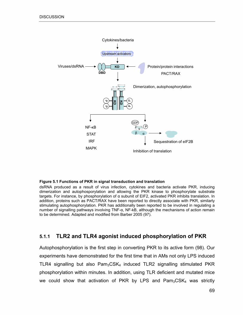
DISCUSSION
Protein/protein interactions
PACT/RAX
KDViruses/dsRNA
Dimerization, autophosphorylation
PP
KD
PPK
DP
GDP
��
Cytokines/bacteria
DBD
NF-�B
STAT
IRF
MAPKInhibition of translation
Sequestration of eIF2B
Upstream activators
Figure 5.1 Functions of PKR in signal transduction and translation dsRNA produced as a result of virus infection, cytokines and bacteria activate PKR, inducing dimerization and autophosporylation and allowing the PKR kinase to phosphorylate substrate targets. For instance, by phosphorylation of � subunit of EIF2, activated PKR inhibits translation. In addition, proteins such as PACT/RAX have been reported to directly associate with PKR, similarly stimulating autophosphorylation. PKR has additionally been reported to be involved in regulating a number of signalling pathways involving TNF-�, NF-kB, although the mechanisms of action remain to be determined. Adapted and modified from Barber 2005 (97).
5.1.1 TLR2 and TLR4 agonist induced phosphorylation of PKR
Autophosphorylation is the first step in converting PKR to its active form (98). Our
experiments have demonstrated for the first time that in AMs not only LPS induced
TLR4 signalling but also Pam3CSK4 induced TLR2 signalling stimulated PKR
phosphorylation within minutes. In addition, using TLR deficient and mutated mice
we could show that activation of PKR by LPS and Pam3CSK4 was strictly
69
DISCUSSION
dependent on the cellular expression of the respective PRR molecules. These data
indicate that PKR can act as an early step in the TLR2/4 induced inflammatory
response. However, the mechanism of PKR activation by TLR ligands remains an
important issue to be explored. The properties of dsRNA as a negatively charged
polysaccharide resemble LPS, but it is rather unlikely that LPS and Pam3CSK4 can
directly bind to intracellular located PKR. Recently, Horng and colleges
demonstrated that PKR is a downstream target of the TIRAP/Mal signalling
pathway that is essential for TLR2 and TLR4 (99). Besides TIRAP, PKR has been
shown to physically interact with other proteins such as MKK6 or PACT (100, 101).
Thus, activation of PKR by TLR ligands is likely to require complex but rapid
signalling pathways involving protein-protein interactions.
To delineate the role of PKR in TLR2/4-mediated activation of AMs, we applied
nucleoside analogue 2-AP. Previously, 2-AP has been shown to posses a relative
specificity for the inhibition of PKR phosphorylation by competing with ATP (92). In
earlier experiments, 2-AP selectively blocked the expression of various genes in
response to dsRNA, IFNs and virus infection (102, 103). In our in vitro assays, we
observed a strong inhibition of PKR phosphorylation by 2-AP, most probably by
competitive inhibition of ATP at the PKR-ATP binding site. However, 2-AP also
blocked PKR induction by TLR ligands, thereby suggesting that inhibitor may act
on the kinase activity and gene expression of PKR. Our results are in contradiction
to previous study demonstrating that 2-AP affects only phosphorylation, but not
gene expression of PKR (54, 92). The reasons for such differences in the 2-AP
effect on PKR gene expression may be a result of (a) cell type (ANA-1
macrophages vs. primary AMs); (b) different concentrations of LPS and 2-AP used
in the studies; and finally (c) the method and time point for evaluation of PKR
gene-expression. It is also possible that 2-AP blocked the expression of genes
directly involved in the regulation of PKR transcription but on the other hand PKR
may regulate the transcription of it is own mRNA. Nevertheless, in our
experimental system, we used 2-AP to specifically blocked PKR phosphorylation.
70
DISCUSSION
5.1.2 PKR-dependent secretion of TNF-� and IL-6 cytokines
Pro-inflammatory cytokines are known to be important in the host response to
pulmonary bacterial infections. They contribute to the recognition of pathogens,
recruitment of immune cells from bloodstream to inflamed tissues and the removal
of the invading pathogens (54, 104). TNF-� is a typical pro-inflammatory cytokine
primarily produced by MPs. Production of TNF-� is increased in patients with
pneumonia and in animal models of pulmonary infection caused by
S. pneumoniae, Klebsiella pneumoniae or Pseudomonas aeruginosa (54). One
consequence of elevated levels of TNF-� is increased IL-6 production. IL-6
induces production of acute phase response proteins, reduces albumin and
transferrin levels and directs differentiation of B cells to immunoglobulin-producing
plasma cells (75).
AMs play a primary role in inflammation, principally through elaboration of variety
of cytokines including TNF-� and IL-6. To delineate the role of PKR in AM cytokine
secretion, 2-AP and PKR KO mice were used. Our data clearly demonstrated that
PKR is closely involved in inflammatory cytokine secretion. In addition, using PKR-
KO AMs we provided data that confirm the specificity of 2-AP as a valuable PKR
inhibitor in our system. The profound decrease of PAMP-induced TNF-� and IL-6
production after PKR inhibition suggests that PKR activity may also determine the
extent of alveolar inflammation in vivo. However, inhibition of PKR did not
completely abolish the cytokine production. It will be interesting to evaluate
whether remaining cytokines are still potent inducers of inflammatory response.
Our results support the pro-inflammatory properties and immune defence activities
of PKR, proposed in various cell types. Goh and co-workers demonstrated that
PKR-deficient mouse embryonic fibroblasts (MEFs) were unable to cytokine
production in response to bacterial LPS. Moreover, IL-6 and IL-12 p40 serum
levels after intra peritoneal (i.p.) LPS challenge were diminished in PKR-KO mice,
suggesting requirement of PKR in TLR4-mediated inflammatory responses (88).
Therefore, the requirement for functional PKR in animal model of lung
inflammation/infection needs to be evaluated. PKR was also found to play a crucial
71
DISCUSSION
role in the regulation of inflammatory cytokine expression in human blood
monocytes stimulated with bacillus Calmette-Guerin (BCG) (89).
5.1.3 Requirement of PKR for LPS induced activation of NF-�B
Binding of bacterial PAMPs to their respective TLRs leads to increased production
of inflammatory cytokines and chemokines. Expression of pro-inflammatory genes
is regulated by transcriptional mechanisms. NF-�B represents one of the major
transcription factors linked to the production of variety of inflammatory mediators
including TNF-�, IL-6, IL-12 and CCL2 (45). The main regulatory mechanism of
NF-�B activation is mediated by interaction with the I�B proteins that retain NF-�B
proteins in the cytoplasm in an inactive state. On exposure of the cell to activation
signals such as LPS, I�B proteins are phosphorylated by I�B kinases (IKKs). The
phospho-I�B proteins become targets for ubiquitination and degradation, whereas
NF-�B proteins are translocated to the nucleus to activate the transcription of
cognate genes. There have been a number of proteins identified as possible IKKs
(42, 44, 45).
The NF-�B link to PKR was suggested by a series of experiments showing that
dsRNA could activate NF-�B in various cell lines, however the mechanism still
remained controversial (105). PKR has been proposed to be a component of the
IKK complex and directly phosphorylates I�B proteins (106). For example,
induction of E-selectin, an important for leukocyte rolling adhesion molecule, by
TNF-� and poly I:C is impaired in PKR-KO endothelial cells due to the defective
activation of NF-�B (107). Similarly Gon and colleges observed that pre-treatment
of cells with 2-AP attenuated LPS-induced NF-�B activation in RBL-2H3 mast cells
(90). Other groups, however, reported that activation of NF-�B by inflammatory
stimuli did not require PKR kinase activity (108). Der and co-workers showed that
PKR gene deletion had no effect on TNF-� or LPS-induced NF-�B activation (96).
Our work supports requirement for PKR phosphorylation in NF-�B activation in
response to LPS. Using both pharmacological inhibition and gene KO approaches
we have demonstrated that LPS-induced activation and translocation of p65 72
DISCUSSION
depends on PKR phosphorylation. However, we could observe that NF-�B remains
still active in PKR-KO AMs, suggesting that PKR-independent activation of NF-�B
exists. Our results differ with those of Hsu and colleges, who found that PKR-KO
BM derived macrophages (BMDMs) exhibited normal IKK activation and normal
induction of numerous NF-�B target genes in response to LPS (109). These
different observations may be due to distinct mechanisms of NF-�B activation in
different macrophages subsets in mice.
In contrast to LPS, TLR2-mediated activation of NF-�B was found to be largely
PKR phosphorylation-independent. This finding suggests that the molecular steps
regulating NF-�B activity differ in TLR2 and TLR4 mediated signalling pathways of
AMs.
5.1.4 PKR dependent regulation of the JNK signalling pathway
The MAP kinases are a family of evolutionary conserved enzymes that connect
cell surface receptors to regulatory targets and coordinate activation of gene
transcription, protein synthesis, cell cycle machinery, cell death, and differentiation
(23, 24). They represent the second major signalling pathway activated by TLRs in
AMs involved in cytokine production (29). The connection between MAPK and
PKR has been recognized just recently, through only a few studies. Goh and co-
workers demonstrated for the first time that PKR-deficient MEFs were unable to
activate p38 and JNK MAP kinases in response to bacterial LPS or poly I:C (88).
Moreover, authors observed that activation of the specific MAPK kinases, MKK3
and MKK6 activating p38 MAPK and MKK4 activating JNK MAPK, were
correspondingly reduced in PKR-deficient MEFs after stimulation with LPS or poly
I:C. Interestingly, JNK activation was no longer dependent on PKR activity in
immortalized fibroblast cell lines generated from the MEFs (88). Similarly, Cheung
and colleagues revealed that the early signal transduction events of mycobacterial
activation of human monocytes requires PKR dependent ERK phosphorylation
(89). In our study, we observed phosphorylation of three major MAP kinases, p38,
JNK and ERK1/2 in response to Pam3CSK4 and LPS in AMs. Inhibition of PKR 73
DISCUSSION
phosphorylation by 2-AP and gene deletion in PKR-KO AMs however abolished
PAMP-induced JNK but not ERK1/2 MAPK and p38 MAPK activation. Our results
are in line with those of Goh and colleagues, who found that PKR was required for
efficient activation of JNK by LPS in primary low-passage MEFs (88). However,
our results differ from those of Hsu and colleagues, who found that BMDMs
derived from PKR-KO exhibited normal p38, JNK and ERK MAP kinases activation
in response to LPS (109).
JNK have been reported to be an important mediator of the production of
inflammatory cytokines induced by LPS. A critical role for JNK appears to be the
regulation of the AP-1 transcription factor through phosphorylation of
the N-terminal part of c-Jun, a central component of AP-1 (29, 31).
Correspondingly, we observed reduced phosphorylation of c-Jun/AP-1 when PKR
was inhibited. In addition, phosphorylation of MKK4, a direct activator of JNK, was
found to be dependent on PKR activity. Collectively, these data indicate that PKR
specifically regulates the JNK signalling pathway (PKR-MKK4-JNK-c-Jun) in AMs
activated by TLR2 and TLR4 agonists. Our results suggest that there are multiple
pathways for JNK, p38 and ERK1/2 MAPK activation in AMs. Moreover our
observations support the idea that PKR can be one of the MAPKKKs (93).
In summary, our study shows that the secretion of TNF-� and IL-6 by primary AMs
in response to cell wall components of gram-positive as well as gram-negative
bacteria in vitro is critically dependent on PKR phosphorylation. Activated PKR
was found to act upstream of the Pam3CSK4 and LPS-activated JNK pathway.
PKR also regulates TLR4- but not TLR2-induced activation of NF-�B in AMs.
These data identify PKR phosphorylation as important signalling step in the
TLR2/4-mediated inflammatory response of AMs to bacterial PAMPs (Figure 5.2).
74
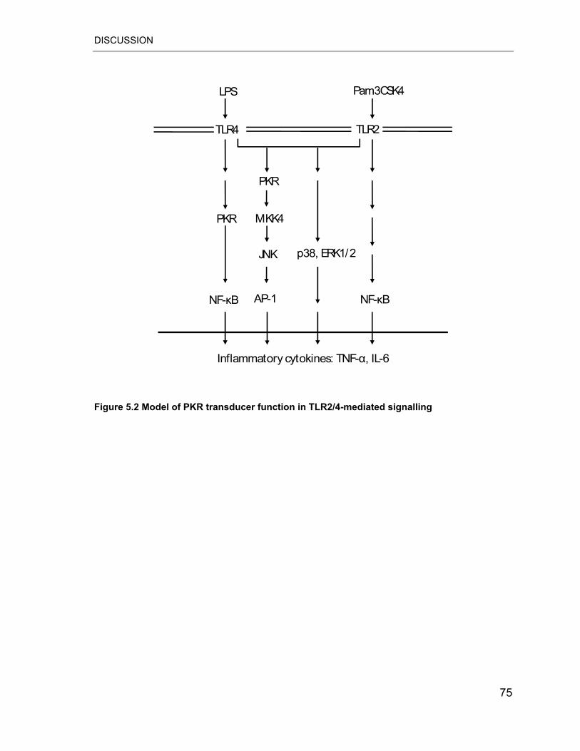
DISCUSSION
AP-1
TLR2
p38, ERK1/2
PKR
JNK
NF-�B
LPS
MKK4
Inflammatory cytokines: TNF-�, IL-6
Pam3CSK4
NF-�B
PKR
TLR4
Figure 5.2 Model of PKR transducer function in TLR2/4-mediated signalling
75
DISCUSSION
5.2 Transcriptional profiling of MPs recruited to mouse lungs in response to alveolar challenge with the TLR2 agonist Pam3CSK4
5.2.1 Alveolar trafficking of PB MPs is associated with global changes in their gene expression profile
MPs represent a central part of both the innate and adaptive immune response to
pathogens. Under steady-state condition, half of the circulating MPs leave the
bloodstream each day to enter peripheral tissues (59). Inflammatory stimuli such
as invading pathogens or TLR ligands, however, elicit increased MP traffic to the
sites of inflammation where MPs can modulate local host responses by e.g.
secreting inflammatory mediators such as cytokines, chemokines and lipids (55).
Compared to the TLR4 ligand LPS restricted to gram-negative bacteria, a limited
number of studies have addressed the induction of lung inflammation and the
concomitant MP recruitment in response to TLR2 ligands. Consequently, in vivo
experimental data on migration and function of MPs recruited to the lungs in
response to TLR2 agonists are rare.
This study is the first report showing that selective TLR2 ligand stimulation within
the alveolar compartment likewise promoted lung inflammation in mice and
induced the migration of circulatory immune cells including MPs into the inflamed
alveolar space. This model seems of considerable interest because most cases of
community acquired pneumonia (CAP) are caused by gram-positive bacteria
lacking LPS but expressing TLR2 ligands. Our study also represents the first
global expression profiling of MPs recruited from the circulation to the alveolar
compartment in response to the TLR2 agonist Pam3CSK4. By combination of
FACS cell sorting, mRNA amplification methods and whole genome microarrays
we were able to profile MP transcriptome from small amounts of BALF and blood
of Pam3CSK4-treated mice. We found that expression of approximately 2500
76
DISCUSSION
genes was significantly changed in alveolar recruited MPs compared to circulating
MPs. In particular, trafficking of MPs was associated with drastic up-regulation of a
broad array of pro-inflammatory molecules including TNF-�, IL-1�/�, IL-6,
CXCL16, KC or CCL2 indicating that inflammatory elicited MPs bear the potential
to promote lung inflammation by increasing alveolar levels of cytokines and
chemotactic mediators for neutrophils, monocytes and lymphocytes. This
observation supports previous reports in which MPs were identified as active
contributors to the development of acute lung inflammation in patients with ALI and
were shown to promote neutrophil recruitment in response to alveolar challenge
with LPS in mice (70, 110). Interestingly our transcriptome analysis revealed that
Pam3CSK4 challenge induced not only pro-inflammatory but also pronounced
anti-inflammatory MP responses. Thus we noticed strong up-regulation of IL-1RN,
IRAK-M, IL-10, ANXA-1, BAX, IL-18BP and FPR-1 genes in alveolar recruited
MPs.
IL-1RN acts as specific inhibitor of IL-1�/� signalling by competitive binding to the
IL-1 receptor. Previous reports already demonstrated that IL-1RN is mainly
produced by monocytes/macrophages in response to TLR4 ligands such as LPS
(75). Thus, the strong increase of alveolar MP IL-1RN transcripts observed after
Pam3CSK4 administration suggests a potential role for IL-1RN in blocking the
action of IL-1�/� and in limiting of TLR2 driven lung inflammatory responses.
IRAK-M represents another host strategy evolved to dampen inflammatory TLR
signalling. IRAK-M negatively regulates MyD88 dependent TLR2/4/9 signalling by
inhibiting the dissociation of IRAK1 and IRAK4 from the TLR complex (22). In vivo,
IRAK-M-deficient mice exhibited increased inflammatory response during bacterial
infections (111). Moreover, it has been demonstrated that lung macrophages from
IRAK-M–deficient septic mice produced higher levels of pro-inflammatory
cytokines such as IL-12 and TNF-� when stimulated with LPS ex vivo (112). After
Pam3CSK4 challenge augmented IRAK-M transcript levels were found in alveolar
recruited MPs both at 24 and 48 h. These data suggest that activation of IRAK-M
may represent a potential mechanism to limit TLR2-medaited pulmonary
77
DISCUSSION
inflammation. Our study revealed that Pam3CSK4 induced high up-regulation of
IL-10 mRNA levels in alveolar MPs. IL-10 is an important immunomodulatory
cytokine with known anti-inflammatory and immunosuppressive properties (75). It
has been shown that LPS-induced secretion of IL-10 limits production of
pro-inflammatory cytokines including TNF-�, IL-1 and IL-12 thereby promoting the
termination of inflammation (113). Consequently increase of anti-inflammatory and
pro-resolving transcripts observed after Pam3CSK4 administration suggests a
potential role for alveolar recruited MPs in limiting TLR2 driven lung inflammatory
responses.
Lipids such as arachidonic acid derivatives are potent inflammatory mediators
(114). Our gene array data revealed that inflammatory trafficking of MPs was
associated with profound regulation of genes involved in PG and LT metabolism
such as COX-2 and PTGES. COX-2 catalyzes PG synthesis including PGE2
commonly considered as central pro-inflammatory lipid mediator involved in the
pathogenesis of several inflammatory diseases such as periodontitis and
rheumatoid arthritis (115, 116). In lungs, however, it has been reported to display
anti-inflammatory and pro-resolution activity as well (117). For example PGE2 has
been shown to down-regulate the production of pro-inflammatory cytokines such
as IL-12 or CCL2 thus inhibiting inflammatory cell recruitment (118). In addition,
the expression of the genes encoding the enzymes LTB4DH involved in
deactivation of the PMN chemoattractant LTB4 and HPGD promoting the
deactivation of PGs and lipoxins were found to be up-regulated in MPs recruited to
the alveolar air space. Thus by alveolar recruitment, MPs may acquire the potential
to actively contribute to lowering the local levels of lung lipid inflammatory
mediators.
In vivo studies indicated the potential of circulating MPs to differentiate into lung
macrophages under both inflammatory and non-inflammatory conditions (56).
Recently macrophages have been assigned to two groups: classically activated
(pro-inflammatory activities) and alternatively activated macrophages (anti-
inflammatory activities) (119). In our study we found that i.t. administration of
78
DISCUSSION
Pam3CSK4 elevated mRNA levels of genes such as SPP-1, ARG-1, TGF�I, FN-1
and IL-10 that are preferentially associated with an alternatively activated
macrophages phenotype. Alternatively activated macrophages are known to play a
major role in tissue repair during the healing phase by secretion of extracellular
matrix (ECM) components and release of anti-inflammatory mediators (119).
Based on these results freshly recruited MPs may represent important factors for
the resolution of pulmonary inflammation.
The present work also revealed differential expression of GPCRs with unknown
function such as GPR31C, GPR137B.and GPR178 in lung recruited compared to
circulating MPs. Since different stages of mononuclear phagocyte differentiation
are characterized by different repertoires of GPCR, these differentially regulated
receptors may reflect MP changes during migration to the lungs. However, the
potential role of these molecules during lung inflammatory processes remains to
be further elucidated in detailed functional studies.
5.2.2 Attenuation of pro-inflammatory gene expression levels in alveolar MPs during late inflammatory response phases
As unbalanced production of pro-inflammatory mediators may lead to inflammatory
damage of the highly sensitive respiratory structures, the activation of host defence
responses in the lung must be tightly regulated and later terminated (120).
Numerous control strategies have been evolved including inhibition of
pro-inflammatory mediators and secretion of anti-inflammatory and pro-resolving
mediators. In our model of TLR2 ligand Pam3CSK4-initiated lung inflammation,
intense inflammatory injury peaks at 24 h post challenge and then slowly recedes.
At the cellular level, these events are characterized by increased trafficking of MPs
and reduced neutrophil accumulation within the lung. This observation is
consistent with the current concept of cellular inflammation resolution. Elucidation
of molecular programs activated in MPs during this late phase may provide
79
DISCUSSION
important insights into how MPs contribute to the termination of TLR2 mediated
lung inflammatory diseases.
In this respect, we monitored temporal changes in the gene expression of pro- and
anti-inflammatory mediators of alveolar recruited MPs at 24 h and 48 h post
Pam3CSK4 treatment. We found that mRNA levels of pro-inflammatory molecules
such as TNF-�, IL-6, CCL2 and IL-12A were significantly lower in BALF MPs at 48
h compared to BALF MPs at 24 h, whereas gene expression of molecules with key
anti-inflammatory and pro-resolution function such as IRAK-M, IL-10, IL1-RN, BAX
and ANXA-1 persisted at the same level at 48 h and 24 h. Thus MPs recruited
during later phase of inflammation, activate regulatory mechanisms to switch off
the pro-inflammatory phenotype overrepresented during maximal inflammation.
Moreover, by persistent high induction of IL-10 and IL1-RN genes MPs may
actively contribute to the termination of inflammation.
Recruitment of MPs from the circulation to the alveolar space is a multistep
process involving multiple cellular interactions. In addition, after trafficking to the
airspace MPs become exposed the specialized alveolar microenvironment
stimulated by PAMP induced inflammation with the potential to further modulate
the phenotype of the recruited cell population. Our global gene profiling approach
does not allow to assign the observed changes in the genetic program of lung
recruited MPs to any of these migratory steps. We also cannot discriminate
whether the changes in gene activation found in alveolar recruited MPs at 48 vs.
24 h are due to phenotype changes occurring in MPs within the alveolar
compartment or reflect the recruitment of functionally different MP during later
stages that have acquired altered functional programs either within the vascular
compartment or during the trafficking process. The fact, however, that the gene
expression profiles of circulating MPs obtained at 48 vs. 24 h also display a shifted
profile suggests that indeed the extravasation of functionally modified MPs may
contribute to the altered gene expression profile of BALF MPs at later time points
after inflammatory challenge.
80
DISCUSSION
In conclusion, our data demonstrates the feasibility to evaluate cell specific
transcriptome profiles in highly purified flow sorted MPs from both PB and BALF in
a mouse model of TLR2 ligand induced lung inflammation. We identified more than
3500 genes differentially expressed, mostly up-regulated in alveolar recruited
compared to PB MPs demonstrating that inflammatory MP trafficking into lung
tissue was associated with large scale genetic program activation. Notably, the MP
gene clusters undergoing activation in response to i.t. Pam3CSK4 application
encoded both pro- and anti-inflammatory activities, however with a profound profile
change over time. MPs recruited within 24 h post challenge showed simultaneous
induction of both pro- and anti-inflammatory genes, whereas in MPs isolated from
BALF after 48 h post challenge mainly genes encoding anti-inflammatory
mediators were found to persist in an up-regulated. Collectively, our results provide
important new insights into how MPs contribute to the promotion and
termination/resolution of TLR2 mediated inflammation.
5.3 Future perspectives Cell specific profiles of the molecular programs induced by gram-positive as well
as gram-negative bacteria ligands in pulmonary MPs provide important insights
into the host defence molecular network in infectious lung diseases such as
pneumonia and ARDS. In the present study we aimed to analyse the molecular
programs activated by TLR2/4 ligands in alveolar resident as well as alveolar
recruited MPs to further delineate their role in pulmonary defence. This knowledge
may help to identify new molecular targets for selective therapeutic intervention in
pneumonia.
We discovered that key antiviral defence molecule PKR was activated and critically
involved in inflammatory response induced by bacterial TLR agonists in AMs in
vitro. Our data clearly demonstrate that treatment with specific pharmacological
inhibitor 2-AP and PKR gene deletion reduced acute inflammatory cytokine
response. Therefore modulation of PKR activity may have a therapeutic potential
in treatment of patients during pulmonary bacterial infections.
81
DISCUSSION
Since S. pneumoniae and K. pneumoniae are frequently isolated causative
pathogens of pneumonia it is reasonable to determine the role of PKR in host
defence in gram-positive and gram-negative pneumonia in mice. Nevertheless,
studies with human samples are required to clarify the role of PKR during bacterial
lung infections in patients. Since several cancer drugs have been shown to
modulate PKR activity; their potential in the treatment bacterial lung infections
should be evaluated.
In addition to activation of lung resident cells, alveolar recognition of inhaled
pathogens promotes increased recruitment of circulating MPs to inflamed tissue.
Therefore we examined the global gene expression profiles of MPs recruited from
peripheral blood to the alveolar space following alveolar deposition of the TLR2
ligand Pam3CSK4. We found that alveolar trafficking of MPs was associated with
profound changes of their gene expression profiles post recruitment. Importantly,
whereas the induced mRNA levels of selected pro-inflammatory mediators were
found to decrease during later inflammation phases, the increased transcript levels
of central anti-inflammatory mediators persisted. To further study the role of MPs in
TLR2-driven inflammatory response it would be interesting to evaluate whether the
transcriptome level changes are also reflected at the proteome and metabolome
level. These studies should be also complemented by whole genome expression
profiling of MPs recruited to the lungs in response to infection with TLR2 ligands
bearing gram positive bacteria such as S. pneumoniae. It is known that MPs are
not a homogenous population. In our study using CX3CR1+/GFP mice we were not
able to specifically monitor MP subset specific changes in gene expression. In
addition, we could not discriminate whether the changes in gene expression found
in alveolar recruited MPs at 48 h vs. 24 h are due to phenotype changes occurring
during migratory processes. Therefore it will be of interest to isolate distinct
alveolar and vascular populations of recruited MPs and perform gene expression
analysis. In addition using specific KO mouse models or gene silencing strategies
would be helpful to further define the role of alveolar recruited MPs and their
relative contribution to the lung inflammatory response.
82
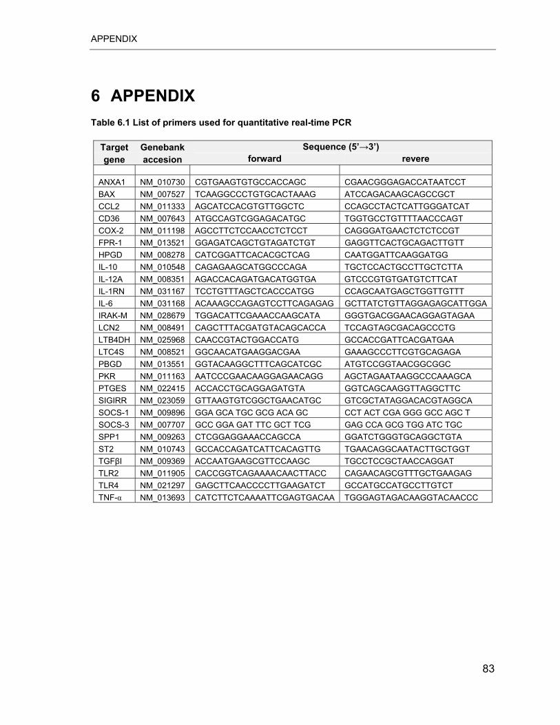
APPENDIX
6 APPENDIX Table 6.1 List of primers used for quantitative real-time PCR
Target Genebank Sequence (5’�3’) accesion forward revere gene
ANXA1 NM_010730 CGTGAAGTGTGCCACCAGC CGAACGGGAGACCATAATCCT BAX NM_007527 TCAAGGCCCTGTGCACTAAAG ATCCAGACAAGCAGCCGCT CCL2 NM_011333 AGCATCCACGTGTTGGCTC CCAGCCTACTCATTGGGATCAT CD36 NM_007643 ATGCCAGTCGGAGACATGC TGGTGCCTGTTTTAACCCAGT COX-2 NM_011198 AGCCTTCTCCAACCTCTCCT CAGGGATGAACTCTCTCCGT FPR-1 NM_013521 GGAGATCAGCTGTAGATCTGT GAGGTTCACTGCAGACTTGTT HPGD NM_008278 CATCGGATTCACACGCTCAG CAATGGATTCAAGGATGG IL-10 NM_010548 CAGAGAAGCATGGCCCAGA TGCTCCACTGCCTTGCTCTTA IL-12A NM_008351 AGACCACAGATGACATGGTGA GTCCCGTGTGATGTCTTCAT IL-1RN NM_031167 TCCTGTTTAGCTCACCCATGG CCAGCAATGAGCTGGTTGTTT IL-6 NM_031168 ACAAAGCCAGAGTCCTTCAGAGAG GCTTATCTGTTAGGAGAGCATTGGA IRAK-M NM_028679 TGGACATTCGAAACCAAGCATA GGGTGACGGAACAGGAGTAGAA LCN2 NM_008491 CAGCTTTACGATGTACAGCACCA TCCAGTAGCGACAGCCCTG LTB4DH NM_025968 CAACCGTACTGGACCATG GCCACCGATTCACGATGAA LTC4S NM_008521 GGCAACATGAAGGACGAA GAAAGCCCTTCGTGCAGAGA PBGD NM_013551 GGTACAAGGCTTTCAGCATCGC ATGTCCGGTAACGGCGGC PKR NM_011163 AATCCCGAACAAGGAGAACAGG AGCTAGAATAAGGCCCAAAGCA PTGES NM_022415 ACCACCTGCAGGAGATGTA GGTCAGCAAGGTTAGGCTTC SIGIRR NM_023059 GTTAAGTGTCGGCTGAACATGC GTCGCTATAGGACACGTAGGCA SOCS-1 NM_009896 GGA GCA TGC GCG ACA GC CCT ACT CGA GGG GCC AGC T SOCS-3 NM_007707 GCC GGA GAT TTC GCT TCG GAG CCA GCG TGG ATC TGC SPP1 NM_009263 CTCGGAGGAAACCAGCCA GGATCTGGGTGCAGGCTGTA ST2 NM_010743 GCCACCAGATCATTCACAGTTG TGAACAGGCAATACTTGCTGGT TGF�I NM_009369 ACCAATGAAGCGTTCCAAGC TGCCTCCGCTAACCAGGAT TLR2 NM_011905 CACCGGTCAGAAAACAACTTACC CAGAACAGCGTTTGCTGAAGAG TLR4 NM_021297 GAGCTTCAACCCCTTGAAGATCT GCCATGCCATGCCTTGTCT TNF-� NM_013693 CATCTTCTCAAAATTCGAGTGACAA TGGGAGTAGACAAGGTACAACCC
83
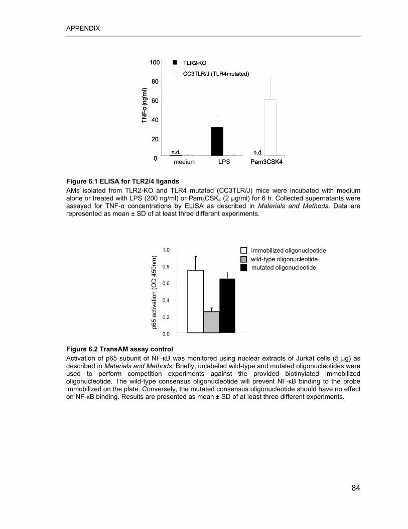
APPENDIX
0
20
40
60
80
100
- LPS Pam3CSK4
TNF-
�( n
g/m
l)
TLR2-KO
CC3TLR/J (TLR4-mutated)
n.d.n.d.0
20
40
60
80
100
medium Pam3CSK4
TNF-
�( n
g/m
l)
TLR2-KO
CC3TLR/J (TLR4-mutated)
n.d.n.d.
Figure 6.1 ELISA for TLR2/4 ligands AMs isolated from TLR2-KO and TLR4 mutated (CC3TLR/J) mice were incubated with medium alone or treated with LPS (200 ng/ml) or Pam3CSK4 (2 g/ml) for 6 h. Collected supernatants were assayed for TNF-� concentrations by ELISA as described in Materials and Methods. Data are represented as mean ± SD of at least three different experiments.
0,0
0,2
0,4
0,6
0,8
1,0 immobilized oligonucleotide
p65
activ
atio
n (O
D 4
50nm
) wild-type oligonucleotidemutated oligonucleotide
Figure 6.2 TransAM assay control Activation of p65 subunit of NF-�B was monitored using nuclear extracts of Jurkat cells (5 μg) as described in Materials and Methods. Briefly, unlabeled wild-type and mutated oligonucleotides were used to perform competition experiments against the provided biotinylated immobilized oligonucleotide. The wild-type consensus oligonucleotide will prevent NF-�B binding to the probe immobilized on the plate. Conversely, the mutated consensus oligonucleotide should have no effect on NF-�B binding. Results are presented as mean ± SD of at least three different experiments.
84
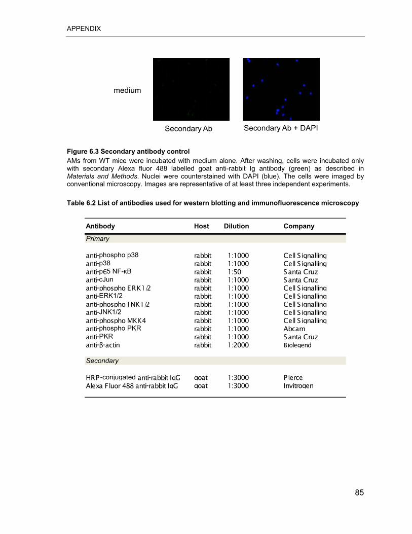
APPENDIX
medium
Secondary Ab Secondary Ab + DAPI
Figure 6.3 Secondary antibody control AMs from WT mice were incubated with medium alone. After washing, cells were incubated only with secondary Alexa fluor 488 labelled goat anti-rabbit Ig antibody (green) as described in Materials and Methods. Nuclei were counterstained with DAPI (blue). The cells were imaged by conventional microscopy. Images are representative of at least three independent experiments. Table 6.2 List of antibodies used for western blotting and immunofluorescence microscopy
Primary
anti-phospho p38 rabbit 1:1000 Cell S ignallinganti-p38 rabbit 1:1000 Cell S ignallinganti-p65 NF-�B rabbit 1:50 Santa Cruzanti-cJun rabbit 1:1000 Santa Cruzanti-phospho ERK1/2 rabbit 1:1000 Cell S ignallinganti-ERK1/2 rabbit 1:1000 Cell S ignallinganti-phospho J NK1/2 rabbit 1:1000 Cell S ignallinganti-JNK1/2 rabbit 1:1000 Cell S ignallinganti-phospho MKK4 rabbit 1:1000 Cell S ignallinganti-phospho PKR rabbit 1:1000 Abcamanti-PKR rabbit 1:1000 Santa Cruzanti-�-actin rabbit 1:2000 Biolegend
Secondary
HRP -conjugated anti-rabbit IgG goat 1:3000 PierceAlexa Fluor 488 anti-rabbit IgG goat 1:3000 Invitrogen
Antibody Host Dilution Company
85
REFERENCES
7 REFERENCES
1. Medzhitov, R., and C. Janeway, Jr. 2000. Innate immunity. N Engl J Med
343(5):338-44.
2. Medzhitov, R., and C. Janeway, Jr. 2000. Innate immune recognition:
mechanisms and pathways. Immunol Rev 173:89-97.
3. Janeway, C. A., Jr., and R. Medzhitov. 2002. Innate immune recognition.
Annu Rev Immunol 20:197-216.
4. Medzhitov, R., P. Preston-Hurlburt, and C. A. Janeway, Jr. 1997. A human
homologue of the Drosophila Toll protein signals activation of adaptive immunity.
Nature 388(6640):394-7.
5. Uematsu, S., and S. Akira. 2006. Toll-like receptors and innate immunity. J
Mol Med 84(9):712-25.
6. Bowie, A., and L. A. O'Neill. 2000. The interleukin-1 receptor/Toll-like
receptor superfamily: signal generators for pro-inflammatory interleukins and
microbial products. J Leukoc Biol 67(4):508-14.
7. Akira, S., S. Uematsu, and O. Takeuchi. 2006. Pathogen recognition and
innate immunity. Cell 124(4):783-801.
8. Takeda, K., T. Kaisho, and S. Akira. 2003. Toll-like receptors. Annu Rev
Immunol 21:335-76.
9. Akira, S., and K. Takeda. 2004. Toll-like receptor signalling. Nat Rev
Immunol 4(7):499-511.
10. Poltorak, A., X. He, I. Smirnova, M. Y. Liu, C. Van Huffel, X. Du, D. Birdwell,
E. Alejos, M. Silva, C. Galanos, M. Freudenberg, P. Ricciardi-Castagnoli, B.
Layton, and B. Beutler. 1998. Defective LPS signaling in C3H/HeJ and
C57BL/10ScCr mice: mutations in Tlr4 gene. Science 282(5396):2085-8.
86
REFERENCES
11. Qureshi, S. T., L. Lariviere, G. Leveque, S. Clermont, K. J. Moore, P. Gros,
and D. Malo. 1999. Endotoxin-tolerant mice have mutations in Toll-like receptor 4
(Tlr4). J Exp Med 189(4):615-25.
12. Hoshino, K., O. Takeuchi, T. Kawai, H. Sanjo, T. Ogawa, Y. Takeda, K.
Takeda, and S. Akira. 1999. Cutting edge: Toll-like receptor 4 (TLR4)-deficient
mice are hyporesponsive to lipopolysaccharide: evidence for TLR4 as the Lps
gene product. J Immunol 162(7):3749-52.
13. Kopp, E., and R. Medzhitov. 2003. Recognition of microbial infection by
Toll-like receptors. Curr Opin Immunol 15(4):396-401.
14. Wright, S. D., R. A. Ramos, P. S. Tobias, R. J. Ulevitch, and J. C. Mathison.
1990. CD14, a receptor for complexes of lipopolysaccharide (LPS) and LPS
binding protein. Science 249(4975):1431-3.
15. Wurfel, M. M., E. Hailman, and S. D. Wright. 1995. Soluble CD14 acts as a
shuttle in the neutralization of lipopolysaccharide (LPS) by LPS-binding protein and
reconstituted high density lipoprotein. J Exp Med 181(5):1743-54.
16. Shimazu, R., S. Akashi, H. Ogata, Y. Nagai, K. Fukudome, K. Miyake, and
M. Kimoto. 1999. MD-2, a molecule that confers lipopolysaccharide
responsiveness on Toll-like receptor 4. J Exp Med 189(11):1777-82.
17. Werts, C., R. I. Tapping, J. C. Mathison, T. H. Chuang, V. Kravchenko, I.
Saint Girons, D. A. Haake, P. J. Godowski, F. Hayashi, A. Ozinsky, D. M.
Underhill, C. J. Kirschning, H. Wagner, A. Aderem, P. S. Tobias, and R. J.
Ulevitch. 2001. Leptospiral lipopolysaccharide activates cells through a TLR2-
dependent mechanism. Nat Immunol 2(4):346-52.
18. Ozinsky, A., D. M. Underhill, J. D. Fontenot, A. M. Hajjar, K. D. Smith, C. B.
Wilson, L. Schroeder, and A. Aderem. 2000. The repertoire for pattern recognition
of pathogens by the innate immune system is defined by cooperation between toll-
like receptors. Proc Natl Acad Sci U S A 97(25):13766-71.
87
REFERENCES
19. Gantner, B. N., R. M. Simmons, S. J. Canavera, S. Akira, and D. M.
Underhill. 2003. Collaborative induction of inflammatory responses by dectin-1 and
Toll-like receptor 2. J Exp Med 197(9):1107-17.
20. Echchannaoui, H., K. Frei, C. Schnell, S. L. Leib, W. Zimmerli, and R.
Landmann. 2002. Toll-like receptor 2-deficient mice are highly susceptible to
Streptococcus pneumoniae meningitis because of reduced bacterial clearing and
enhanced inflammation. J Infect Dis 186(6):798-806.
21. Takeda, K., and S. Akira. 2004. TLR signaling pathways. Semin Immunol
16(1):3-9.
22. Liew, F. Y., D. Xu, E. K. Brint, and L. A. O'Neill. 2005. Negative regulation of
toll-like receptor-mediated immune responses. Nat Rev Immunol 5(6):446-58.
23. Kyriakis, J. M., and J. Avruch. 2001. Mammalian mitogen-activated protein
kinase signal transduction pathways activated by stress and inflammation. Physiol
Rev 81(2):807-69.
24. Dong, C., R. J. Davis, and R. A. Flavell. 2002. MAP kinases in the immune
response. Annu Rev Immunol 20:55-72.
25. Swantek, J. L., M. H. Cobb, and T. D. Geppert. 1997. Jun N-terminal
kinase/stress-activated protein kinase (JNK/SAPK) is required for
lipopolysaccharide stimulation of tumor necrosis factor alpha (TNF-alpha)
translation: glucocorticoids inhibit TNF-alpha translation by blocking JNK/SAPK.
Mol Cell Biol 17(11):6274-82.
26. Liu, M. K., P. Herrera-Velit, R. W. Brownsey, and N. E. Reiner. 1994. CD14-
dependent activation of protein kinase C and mitogen-activated protein kinases
(p42 and p44) in human monocytes treated with bacterial lipopolysaccharide. J
Immunol 153(6):2642-52.
27. Guha, M., and N. Mackman. 2001. LPS induction of gene expression in
human monocytes. Cell Signal 13(2):85-94.
88
REFERENCES
28. Eliopoulos, A. G., C. D. Dumitru, C. C. Wang, J. Cho, and P. N. Tsichlis.
2002. Induction of COX-2 by LPS in macrophages is regulated by Tpl2-dependent
CREB activation signals. Embo J 21(18):4831-40.
29. Monick, M. M., and G. W. Hunninghake. 2003. Second messenger
pathways in pulmonary host defense. Annu Rev Physiol 65:643-67.
30. Scherle, P. A., E. A. Jones, M. F. Favata, A. J. Daulerio, M. B. Covington, S.
A. Nurnberg, R. L. Magolda, and J. M. Trzaskos. 1998. Inhibition of MAP kinase
kinase prevents cytokine and prostaglandin E2 production in lipopolysaccharide-
stimulated monocytes. J Immunol 161(10):5681-6.
31. Davis, R. J. 2000. Signal transduction by the JNK group of MAP kinases.
Cell 103(2):239-52.
32. Hambleton, J., S. L. Weinstein, L. Lem, and A. L. DeFranco. 1996.
Activation of c-Jun N-terminal kinase in bacterial lipopolysaccharide-stimulated
macrophages. Proc Natl Acad Sci U S A 93(7):2774-8.
33. Zhang, Y., J. N. Blattman, N. J. Kennedy, J. Duong, T. Nguyen, Y. Wang, R.
J. Davis, P. D. Greenberg, R. A. Flavell, and C. Dong. 2004. Regulation of innate
and adaptive immune responses by MAP kinase phosphatase 5. Nature
430(7001):793-7.
34. Zhang, Y. L., and C. Dong. 2005. MAP kinases in immune responses. Cell
Mol Immunol 2(1):20-7.
35. Raingeaud, J., S. Gupta, J. S. Rogers, M. Dickens, J. Han, R. J. Ulevitch,
and R. J. Davis. 1995. Pro-inflammatory cytokines and environmental stress cause
p38 mitogen-activated protein kinase activation by dual phosphorylation on
tyrosine and threonine. J Biol Chem 270(13):7420-6.
36. Lu, H. T., D. D. Yang, M. Wysk, E. Gatti, I. Mellman, R. J. Davis, and R. A.
Flavell. 1999. Defective IL-12 production in mitogen-activated protein (MAP)
kinase kinase 3 (Mkk3)-deficient mice. Embo J 18(7):1845-57.
89
REFERENCES
37. Kotlyarov, A., A. Neininger, C. Schubert, R. Eckert, C. Birchmeier, H. D.
Volk, and M. Gaestel. 1999. MAPKAP kinase 2 is essential for LPS-induced TNF-
alpha biosynthesis. Nat Cell Biol 1(2):94-7.
38. Kovarik, P., D. Stoiber, M. Novy, and T. Decker. 1998. Stat1 combines
signals derived from IFN-gamma and LPS receptors during macrophage
activation. Embo J 17(13):3660-8.
39. Tsukada, J., W. R. Waterman, Y. Koyama, A. C. Webb, and P. E. Auron.
1996. A novel STAT-like factor mediates lipopolysaccharide, interleukin 1 (IL-1),
and IL-6 signaling and recognizes a gamma interferon activation site-like element
in the IL1B gene. Mol Cell Biol 16(5):2183-94.
40. Ohmori, Y., and T. A. Hamilton. 2001. Requirement for STAT1 in LPS-
induced gene expression in macrophages. J Leukoc Biol 69(4):598-604.
41. Ghosh, S., and M. Karin. 2002. Missing pieces in the NF-kappaB puzzle.
Cell 109 Suppl:S81-96.
42. Karin, M., and F. R. Greten. 2005. NF-kappaB: linking inflammation and
immunity to cancer development and progression. Nat Rev Immunol 5(10):749-59.
43. Ghosh, S., M. J. May, and E. B. Kopp. 1998. NF-kappa B and Rel proteins:
evolutionarily conserved mediators of immune responses. Annu Rev Immunol
16:225-60.
44. Li, Q., and I. M. Verma. 2002. NF-kappaB regulation in the immune system.
Nat Rev Immunol 2(10):725-34.
45. Fan, J., R. D. Ye, and A. B. Malik. 2001. Transcriptional mechanisms of
acute lung injury. Am J Physiol Lung Cell Mol Physiol 281(5):L1037-50.
46. Muller, J. M., H. W. Ziegler-Heitbrock, and P. A. Baeuerle. 1993. Nuclear
factor kappa B, a mediator of lipopolysaccharide effects. Immunobiology 187(3-
5):233-56.
90
REFERENCES
47. Sha, W. C., H. C. Liou, E. I. Tuomanen, and D. Baltimore. 1995. Targeted
disruption of the p50 subunit of NF-kappa B leads to multifocal defects in immune
responses. Cell 80(2):321-30.
48. Caamano, J., J. Alexander, L. Craig, R. Bravo, and C. A. Hunter. 1999. The
NF-kappa B family member RelB is required for innate and adaptive immunity to
Toxoplasma gondii. J Immunol 163(8):4453-61.
49. Franzoso, G., L. Carlson, L. Poljak, E. W. Shores, S. Epstein, A. Leonardi,
A. Grinberg, T. Tran, T. Scharton-Kersten, M. Anver, P. Love, K. Brown, and U.
Siebenlist. 1998. Mice deficient in nuclear factor (NF)-kappa B/p52 present with
defects in humoral responses, germinal center reactions, and splenic
microarchitecture. J Exp Med 187(2):147-59.
50. Karin, M., Z. Liu, and E. Zandi. 1997. AP-1 function and regulation. Curr
Opin Cell Biol 9(2):240-6.
51. Shaulian, E., and M. Karin. 2002. AP-1 as a regulator of cell life and death.
Nat Cell Biol 4(5):E131-6.
52. Karin, M. 1995. The regulation of AP-1 activity by mitogen-activated protein
kinases. J Biol Chem 270(28):16483-6.
53. Beutler, B. 2004. Innate immunity: an overview. Mol Immunol 40(12):845-
59.
54. Martin, T. R., and C. W. Frevert. 2005. Innate immunity in the lungs. Proc
Am Thorac Soc 2(5):403-11.
55. Serbina, N. V., T. Jia, T. M. Hohl, and E. G. Pamer. 2007. Monocyte-
Mediated Defense Against Microbial Pathogens. Annu Rev Immunol.
56. Landsman, L., C. Varol, and S. Jung. 2007. Distinct differentiation potential
of blood monocyte subsets in the lung. J Immunol 178(4):2000-7.
57. Sibille, Y., and H. Y. Reynolds. 1990. Macrophages and polymorphonuclear
neutrophils in lung defense and injury. Am Rev Respir Dis 141(2):471-501.
91
REFERENCES
58. Lambrecht, B. N. 2006. Alveolar macrophage in the driver's seat. Immunity
24(4):366-8.
59. Geissmann, F., S. Jung, and D. R. Littman. 2003. Blood monocytes consist
of two principal subsets with distinct migratory properties. Immunity 19(1):71-82.
60. Mizgerd, J. P. 2008. Acute lower respiratory tract infection. N Engl J Med
358(7):716-27.
61. Snelgrove, R. J., J. Goulding, A. M. Didierlaurent, D. Lyonga, S. Vekaria, L.
Edwards, E. Gwyer, J. D. Sedgwick, A. N. Barclay, and T. Hussell. 2008. A critical
function for CD200 in lung immune homeostasis and the severity of influenza
infection. Nat Immunol 9(9):1074-83.
62. Oshikawa, K., and Y. Sugiyama. 2003. Regulation of toll-like receptor 2 and
4 gene expression in murine alveolar macrophages. Exp Lung Res 29(6):401-12.
63. Oshikawa, K., and Y. Sugiyama. 2003. Gene expression of Toll-like
receptors and associated molecules induced by inflammatory stimuli in the primary
alveolar macrophage. Biochem Biophys Res Commun 305(3):649-55.
64. Shepherd, E. G., Q. Zhao, S. E. Welty, T. N. Hansen, C. V. Smith, and Y.
Liu. 2004. The function of mitogen-activated protein kinase phosphatase-1 in
peptidoglycan-stimulated macrophages. J Biol Chem 279(52):54023-31.
65. Holt, P. G., J. Oliver, N. Bilyk, C. McMenamin, P. G. McMenamin, G. Kraal,
and T. Thepen. 1993. Downregulation of the antigen presenting cell function(s) of
pulmonary dendritic cells in vivo by resident alveolar macrophages. J Exp Med
177(2):397-407.
66. Strickland, D. H., T. Thepen, U. R. Kees, G. Kraal, and P. G. Holt. 1993.
Regulation of T-cell function in lung tissue by pulmonary alveolar macrophages.
Immunology 80(2):266-72.
67. Thepen, T., N. Van Rooijen, and G. Kraal. 1989. Alveolar macrophage
elimination in vivo is associated with an increase in pulmonary immune response
in mice. J Exp Med 170(2):499-509. 92
REFERENCES
68. Nicod, L. P. 1999. Pulmonary defence mechanisms. Respiration 66(1):2-11.
69. Srivastava, M., S. Jung, J. Wilhelm, L. Fink, F. Buhling, T. Welte, R. M.
Bohle, W. Seeger, J. Lohmeyer, and U. A. Maus. 2005. The inflammatory versus
constitutive trafficking of mononuclear phagocytes into the alveolar space of mice
is associated with drastic changes in their gene expression profiles. J Immunol
175(3):1884-93.
70. Rosseau, S., P. Hammerl, U. Maus, H. D. Walmrath, H. Schutte, F.
Grimminger, W. Seeger, and J. Lohmeyer. 2000. Phenotypic characterization of
alveolar monocyte recruitment in acute respiratory distress syndrome. Am J
Physiol Lung Cell Mol Physiol 279(1):L25-35.
71. Belperio, J. A., M. P. Keane, M. D. Burdick, J. P. Lynch, 3rd, Y. Y. Xue, A.
Berlin, D. J. Ross, S. L. Kunkel, I. F. Charo, and R. M. Strieter. 2001. Critical role
for the chemokine MCP-1/CCR2 in the pathogenesis of bronchiolitis obliterans
syndrome. J Clin Invest 108(4):547-56.
72. Bhalla, K. S., and R. J. Folz. 2002. Idiopathic pneumonia syndrome after
syngeneic bone marrow transplant in mice. Am J Respir Crit Care Med 166(12 Pt
1):1579-89.
73. Thacker, E. L. 2006. Lung inflammatory responses. Vet Res 37(3):469-86.
74. Strieter, R. M., J. A. Belperio, and M. P. Keane. 2002. Cytokines in innate
host defense in the lung. J Clin Invest 109(6):699-705.
75. Opal, S. M., and V. A. DePalo. 2000. Anti-inflammatory cytokines. Chest
117(4):1162-72.
76. Jung, S., J. Aliberti, P. Graemmel, M. J. Sunshine, G. W. Kreutzberg, A.
Sher, and D. R. Littman. 2000. Analysis of fractalkine receptor CX(3)CR1 function
by targeted deletion and green fluorescent protein reporter gene insertion. Mol Cell
Biol 20(11):4106-14.
77. Steinmuller, M., M. Srivastava, W. A. Kuziel, J. W. Christman, W. Seeger, T.
Welte, J. Lohmeyer, and U. A. Maus. 2006. Endotoxin induced peritonitis elicits 93
REFERENCES
monocyte immigration into the lung: implications on alveolar space inflammatory
responsiveness. Respir Res 7:30.
78. Gentleman, R. C., V. J. Carey, D. M. Bates, B. Bolstad, M. Dettling, S.
Dudoit, B. Ellis, L. Gautier, Y. Ge, J. Gentry, K. Hornik, T. Hothorn, W. Huber, S.
Iacus, R. Irizarry, F. Leisch, C. Li, M. Maechler, A. J. Rossini, G. Sawitzki, C.
Smith, G. Smyth, L. Tierney, J. Y. Yang, and J. Zhang. 2004. Bioconductor: open
software development for computational biology and bioinformatics. Genome Biol
5(10):R80.
79. Edwards, D. 2003. Non-linear normalization and background correction in
one-channel cDNA microarray studies. Bioinformatics 19(7):825-33.
80. Smyth, G. K., and T. Speed. 2003. Normalization of cDNA microarray data.
Methods 31(4):265-73.
81. Smyth, G. K. 2004. Linear models and empirical bayes methods for
assessing differential expression in microarray experiments. Stat Appl Genet Mol
Biol 3:Article3.
82. Livak, K. J., S. J. Flood, J. Marmaro, W. Giusti, and K. Deetz. 1995.
Oligonucleotides with fluorescent dyes at opposite ends provide a quenched probe
system useful for detecting PCR product and nucleic acid hybridization. PCR
Methods Appl 4(6):357-62.
83. Lee, L. G., C. R. Connell, and W. Bloch. 1993. Allelic discrimination by nick-
translation PCR with fluorogenic probes. Nucleic Acids Res 21(16):3761-6.
84. Livak, K. J., and T. D. Schmittgen. 2001. Analysis of relative gene
expression data using real-time quantitative PCR and the 2(-Delta Delta C(T))
Method. Methods 25(4):402-8.
85. Lowry, O. H., N. J. Rosebrough, A. L. Farr, and R. J. Randall. 1951. Protein
measurement with the Folin phenol reagent. J Biol Chem 193(1):265-75.
86. Takeuchi, O., K. Hoshino, T. Kawai, H. Sanjo, H. Takada, T. Ogawa, K.
Takeda, and S. Akira. 1999. Differential roles of TLR2 and TLR4 in recognition of 94
REFERENCES
gram-negative and gram-positive bacterial cell wall components. Immunity
11(4):443-51.
87. Hu, Y., and T. W. Conway. 1993. 2-Aminopurine inhibits the double-
stranded RNA-dependent protein kinase both in vitro and in vivo. J Interferon Res
13(5):323-8.
88. Goh, K. C., M. J. deVeer, and B. R. Williams. 2000. The protein kinase PKR
is required for p38 MAPK activation and the innate immune response to bacterial
endotoxin. Embo J 19(16):4292-7.
89. Cheung, B. K., D. C. Lee, J. C. Li, Y. L. Lau, and A. S. Lau. 2005. A role for
double-stranded RNA-activated protein kinase PKR in Mycobacterium-induced
cytokine expression. J Immunol 175(11):7218-25.
90. Gon, Y., S. Nunomura, and C. Ra. 2005. Common and distinct signalling
cascades in the production of tumour necrosis factor-alpha and interleukin-13
induced by lipopolysaccharide in RBL-2H3 cells. Clin Exp Allergy 35(5):635-42.
91. Carmona-Saez, P., M. Chagoyen, F. Tirado, J. M. Carazo, and A. Pascual-
Montano. 2007. GENECODIS: a web-based tool for finding significant concurrent
annotations in gene lists. Genome Biol 8(1):R3.
92. Gusella, G. L., T. Musso, S. E. Rottschafer, K. Pulkki, and L. Varesio. 1995.
Potential requirement of a functional double-stranded RNA-dependent protein
kinase (PKR) for the tumoricidal activation of macrophages by lipopolysaccharide
or IFN-alpha beta, but not IFN-gamma. J Immunol 154(1):345-54.
93. Williams, B. R. 2001. Signal integration via PKR. Sci STKE 2001(89):RE2.
94. Zamanian-Daryoush, M., T. H. Mogensen, J. A. DiDonato, and B. R.
Williams. 2000. NF-kappaB activation by double-stranded-RNA-activated protein
kinase (PKR) is mediated through NF-kappaB-inducing kinase and IkappaB
kinase. Mol Cell Biol 20(4):1278-90.
95
REFERENCES
95. Ramana, C. V., N. Grammatikakis, M. Chernov, H. Nguyen, K. C. Goh, B.
R. Williams, and G. R. Stark. 2000. Regulation of c-myc expression by IFN-gamma
through Stat1-dependent and -independent pathways. Embo J 19(2):263-72.
96. Der, S. D., Y. L. Yang, C. Weissmann, and B. R. Williams. 1997. A double-
stranded RNA-activated protein kinase-dependent pathway mediating stress-
induced apoptosis. Proc Natl Acad Sci U S A 94(7):3279-83.
97. Barber, G. N. 2005. The dsRNA-dependent protein kinase, PKR and cell
death. Cell Death Differ 12(6):563-70.
98. Thomis, D. C., and C. E. Samuel. 1993. Mechanism of interferon action:
evidence for intermolecular autophosphorylation and autoactivation of the
interferon-induced, RNA-dependent protein kinase PKR. J Virol 67(12):7695-700.
99. Horng, T., G. M. Barton, and R. Medzhitov. 2001. TIRAP: an adapter
molecule in the Toll signaling pathway. Nat Immunol 2(9):835-41.
100. Silva, A. M., M. Whitmore, Z. Xu, Z. Jiang, X. Li, and B. R. Williams. 2004.
Protein kinase R (PKR) interacts with and activates mitogen-activated protein
kinase kinase 6 (MKK6) in response to double-stranded RNA stimulation. J Biol
Chem 279(36):37670-6.
101. Peters, G. A., R. Hartmann, J. Qin, and G. C. Sen. 2001. Modular structure
of PACT: distinct domains for binding and activating PKR. Mol Cell Biol
21(6):1908-20.
102. Tiwari, R. K., J. Kusari, R. Kumar, and G. C. Sen. 1988. Gene induction by
interferons and double-stranded RNA: selective inhibition by 2-aminopurine. Mol
Cell Biol 8(10):4289-94.
103. Tiwari, R. K., J. Kusari, and G. C. Sen. 1987. Functional equivalents of
interferon-mediated signals needed for induction of an mRNA can be generated by
double-stranded RNA and growth factors. Embo J 6(11):3373-8.
104. Strieter, R. M., J. A. Belperio, and M. P. Keane. 2003. Host innate defenses
in the lung: the role of cytokines. Curr Opin Infect Dis 16(3):193-8. 96
REFERENCES
105. Visvanathan, K. V., and S. Goodbourn. 1989. Double-stranded RNA
activates binding of NF-kappa B to an inducible element in the human beta-
interferon promoter. Embo J 8(4):1129-38.
106. Kumar, A., J. Haque, J. Lacoste, J. Hiscott, and B. R. Williams. 1994.
Double-stranded RNA-dependent protein kinase activates transcription factor NF-
kappa B by phosphorylating I kappa B. Proc Natl Acad Sci U S A 91(14):6288-92.
107. Bandyopadhyay, S. K., C. A. de La Motte, and B. R. Williams. 2000.
Induction of E-selectin expression by double-stranded RNA and TNF-alpha is
attenuated in murine aortic endothelial cells derived from double-stranded RNA-
activated kinase (PKR)-null mice. J Immunol 164(4):2077-83.
108. Iordanov, M. S., J. Wong, J. C. Bell, and B. E. Magun. 2001. Activation of
NF-kappaB by double-stranded RNA (dsRNA) in the absence of protein kinase R
and RNase L demonstrates the existence of two separate dsRNA-triggered
antiviral programs. Mol Cell Biol 21(1):61-72.
109. Hsu, L. C., J. M. Park, K. Zhang, J. L. Luo, S. Maeda, R. J. Kaufman, L.
Eckmann, D. G. Guiney, and M. Karin. 2004. The protein kinase PKR is required
for macrophage apoptosis after activation of Toll-like receptor 4. Nature
428(6980):341-5.
110. Maus, U. A., K. Waelsch, W. A. Kuziel, T. Delbeck, M. Mack, T. S.
Blackwell, J. W. Christman, D. Schlondorff, W. Seeger, and J. Lohmeyer. 2003.
Monocytes are potent facilitators of alveolar neutrophil emigration during lung
inflammation: role of the CCL2-CCR2 axis. J Immunol 170(6):3273-8.
111. Kobayashi, K., L. D. Hernandez, J. E. Galan, C. A. Janeway, Jr., R.
Medzhitov, and R. A. Flavell. 2002. IRAK-M is a negative regulator of Toll-like
receptor signaling. Cell 110(2):191-202.
112. Deng, J. C., G. Cheng, M. W. Newstead, X. Zeng, K. Kobayashi, R. A.
Flavell, and T. J. Standiford. 2006. Sepsis-induced suppression of lung innate
immunity is mediated by IRAK-M. J Clin Invest 116(9):2532-42.
97
REFERENCES
113. Marchant, A., C. Bruyns, P. Vandenabeele, M. Ducarme, C. Gerard, A.
Delvaux, D. De Groote, D. Abramowicz, T. Velu, and M. Goldman. 1994.
Interleukin-10 controls interferon-gamma and tumor necrosis factor production
during experimental endotoxemia. Eur J Immunol 24(5):1167-71.
114. Lawrence, T., D. A. Willoughby, and D. W. Gilroy. 2002. Anti-inflammatory
lipid mediators and insights into the resolution of inflammation. Nat Rev Immunol
2(10):787-95.
115. McCoy, J. M., J. R. Wicks, and L. P. Audoly. 2002. The role of prostaglandin
E2 receptors in the pathogenesis of rheumatoid arthritis. J Clin Invest 110(5):651-
8.
116. Wilgus, T. A., M. L. Parrett, M. S. Ross, K. L. Tober, F. M. Robertson, and
T. M. Oberyszyn. 2002. Inhibition of ultraviolet light B-induced cutaneous
inflammation by a specific cyclooxygenase-2 inhibitor. Adv Exp Med Biol 507:85-
92.
117. Vancheri, C., C. Mastruzzo, M. A. Sortino, and N. Crimi. 2004. The lung as
a privileged site for the beneficial actions of PGE2. Trends Immunol 25(1):40-6.
118. Rolfe, M. W., S. L. Kunkel, T. J. Standiford, M. B. Orringer, S. H. Phan, H. L.
Evanoff, M. D. Burdick, and R. M. Strieter. 1992. Expression and regulation of
human pulmonary fibroblast-derived monocyte chemotactic peptide-1. Am J
Physiol 263(5 Pt 1):L536-45.
119. Gordon, S. 2003. Alternative activation of macrophages. Nat Rev Immunol
3(1):23-35.
120. Holt, P. G., D. H. Strickland, M. E. Wikstrom, and F. L. Jahnsen. 2008.
Regulation of immunological homeostasis in the respiratory tract. Nat Rev Immunol
8(2):142-52.
98
DECLARATION
8 DECLARATION “I declare that I have completed this dissertation single-handedly without the unauthorized help of a second party and only with the assistance acknowledged therein. I have appropriately acknowledged and referenced all text passages that are derived literally from or are based on the content of published or unpublished work of others, and all information that relates to verbal communications. I have abided by the principles of good scientific conduct laid down in the charter of the Justus Liebig University of Giessen in carrying out the investigations described in the dissertation.” Place and Date Maciej Cabanski
99

Der Lebenslauf wurde aus der elektronischen Version der Arbeit entfernt.
The curriculum vitae was removed from the electronic version of the paper.
CURRICULUM VITAE
2001-2002 Study in the field of Biotechnology and Molecular Biology
at Adam Mickiewicz University, Poznan, Poland. BSc thesis: “Gene expression analysis by DNA microarrays”.
1999-2001 Study in the field of Biotechnology at University of Technology and Agriculture, Bydgoszcz, Poland. Secondary School of General Education in the class with an extended syllabus in mathematics and computer science.
1995-1999
Publications: Maciej Cabanski, Mirko Steinmueller, Leigh Marsh, Ewa Surdziel, Werner Seeger, Juergen Lohmeyer. “PKR regulates TLR2/TLR4-dependent signaling in murine alveolar macrophages“. Am J Respir Cell Mol Biol (2008)
Maciej Cabanski, Jochen Wilhelm, Mirko Steinmueller, Werner Seeger, Juergen Lohmeyer. ”Genome-wide transcriptional profiling of mononuclear phagocytes recruited to mouse lungs in response to alveolar challenge with the TLR2 agonist Pam3CSK4”. Am J Physiol Lung Cell Mol Physiol. In revision (2008) Poster presentations: M. Cabanski, M. Steinmueller, L. Marsh, E. Surdziel, W. Seeger, J. Lohmeyer. “Double-Stranded RNA-Activated Protein Kinase (PKR) regulates TLR-dependent signalling in murine alveolar macrophages”. ATS 2007, 18-23 May, San Francisco, California T. Shafiei Tabar, M. Steinmueller, M. Cabanski, W. Kuziel, W. Seeger, J. Lohmeyer. “Alveolär rekrutierte CCR2-positive Monozyten regulieren die LPS-induzierte pulmonale Inflammation in vivo“. Kongress der Deutschen Gesellschaft für Innere Medizin 2007, Wiesbaden, Germany M. Cabanski, M. Steinmueller, L. Marsh, E. Surdziel, W. Seeger, J. Lohmeyer. “Double-Stranded RNA-Activated Protein Kinase (PKR) is involved in TLR4-mediated signaling in murine alveolar macrophages”. The 39th Annual
101
CURRICULUM VITAE
Meeting of Society for Leukocyte Biology (SLB), 9-11 November 2006, San Antonio, Texas Scientific interests: Signal transduction; innate and adaptive immunity; bacterial infections, inflammatory response of the lungs; allergic asthma; transcriptional profiling and regulation; mononuclear phagocytes, alveolar macrophages Skills: Basic techniques used in molecular biology: isolation of DNA, RNA, proteins; electrophoresis (DNA, RNA, PAGE, SDS-PAGE); PCR; qualitative and quantitative RT-PCR; transfection and siRNA, flow cytometry (FACS Canto) and cell sorting (FACS Vantage); mice models of TLR-mediated lung inflammation (LPS and Pam3CSK4 models); mouse model of allergic asthma (OVA model): maintaining primary cell cultures (alveolar macrophages) and cell lines (MHS alveolar macrophages); immunofluorescence; ELISA and ELISA-based assays (e.g. TransAM assay); Microsoft Office; knowledge of biological databases (e.g. NCBI, Ensembl, KEGG, Onto-Tools, GENECODIS)
102

ACKNOWLEDGEMENTS
10 ACKNOWLEDGEMENTS During three years of my work and living in Giessen I have been accompanied and
supported by many people. However, there are a few friends that I would like to
specially acknowledge.
Firstly I would like to gratefully acknowledge the enthusiastic supervision of Prof.
Jürgen Lohmeyer who gave me the opportunity to do PhD in his group and
provided me with many helpful suggestions, important advices and constant
encouragement during the course of this work. I am also indebted to Prof. Werner
Seeger for many helpful suggestions and important advices during preparations of
manuscripts. Here, I also would like to acknowledge my laboratory colleague Mirko
Steinmüller who introduced me to the world of Toll-like receptors and took care
after me during initial period of my PhD.
I am grateful to my dear laboratory colleagues who also became my friends in
private life: Lidija Cakarova, Susanne Herold, Leigh Marsh, Werner von Wulffen
and Zbigniew Zaslona for constant support, stimulating suggestions and having fun
during our work. Special acknowledgments to the charming technicians: Gudrun
Biemer-Mansouri, Emma Braun, Petra Janssen, and Margret Lohmeyer for their
smile day by day and invaluable help in many experiments.
It was a particular pleasure to work together with people from Department of
Pathology. I am indebted to Prof. Ludgar Fink for his helpful suggestions and
important advices during preparations of manuscript; true acknowledgement to
Maria M. Stein for her excellent technical help during microarray experiments;
Grazyna Kwapiszewska-Marsh for stimulating discussions. Special thanks I would
like to express to (Sir) Jochen Wilhelm for his helpful suggestions and unlimited
patient to my person. Moreover all of them became my real friends.
103

ACKNOWLEDGEMENTS
I am grateful to all my friends from International MBML College for vivid and helpful
discussions.
I would like to thank my fantastic colleagues from Giessen-Unterhof: Aparna
Jayachandran, Ruth Pinto, Anna and Dariusz Zakrzewicz for being the surrogate
family during three years.
I would like to acknowledge Jovan Pavlovic (University of Zurich, Switzerland) for
providing us with PKR-KO mice.
To my new laboratory colleagues from Hannover Medical School. Especially I
would like to acknowledge Prof. Gesine Hansen and Christian Hennig for giving
me the opportunity to work in their team and invaluable help during finalization of
my PhD thesis.
I gratefully acknowledge the funding sources that made my PhD work possible.
Financial support has been provided by the German Research Foundation, grant
547 "Cardiopulmonary Vascular System", the National Network on Community-
Acquired Pneumonia (CAPNETZ) and the BMBF funded clinical research group in
clinical infectiology BMBF 01 KI 0770.
I would like to thank my family for all their love and encouragement. My deepest
gratitude goes to my mother Grazyna, father Zygmunt and departed soul of my
grandmother Danuta.
Finally, I am very grateful to Ewa Surdziel, for her support and understanding
during the PhD period and after. Thank you also for being my best friend.
104