Molecular Mechanisms Underlying the Cardiovascular ...
18
cells Review Molecular Mechanisms Underlying the Cardiovascular Toxicity of Specific Uremic Solutes Jonathan D. Ravid 1 and Vipul C. Chitalia 1,2,3, * 1 Renal Section, Department of Medicine, Boston University School of Medicine, Boston, MA 02118, USA; [email protected] 2 Boston Veterans Affairs Healthcare System, Boston, MA 02130, USA 3 Global Cocreation Labs, Institute of Medical Engineering and Science, Massachusetts Institute of Technology, Cambridge, MA 02139, USA * Correspondence: [email protected] Received: 3 August 2020; Accepted: 27 August 2020; Published: 2 September 2020 Abstract: Mounting evidence strongly suggests a causal link between chronic kidney disease (CKD) and cardiovascular disease (CVD). Compared with non-CKD patients, patients with CKD suffer disproportionately from CVD and derive suboptimal benefits from interventions targeting conventional CVD risk factors. Uremic toxins (UTs), whose plasma levels rapidly rise as CKD progresses, represent a unique risk factor in CKD, which has protean manifestations on CVD. Among the known UTs, tryptophan metabolites and trimethylamine N-oxide are well-established cardiovascular toxins. Their molecular mechanisms of effect warrant special consideration to draw translational value. This review surveys current knowledge on the effects of specific UTs on different pathways and cell functions that influence the integrity of cardiovascular health, with implication for CVD progression. The effect of UTs on cardiovascular health is an example of a paradigm in which a cascade of molecular and metabolic events induced by pathology in one organ in turn induces dysfunction in another organ. Deciphering the molecular mechanisms underlying such cross-organ pathologies will help uncover therapeutic targets to improve the management of CVD in patients with CKD. Keywords: uremic toxins; chronic kidney disease; cardiovascular disease 1. Introduction 1.1. Overview The prevalence of CKD might be approaching pandemic levels in the United States, with about 30 million people suffering from the disease, and with significant associated rates of morbidity and mortality [1]. According to the Center for Disease Control, 15% of US adults are predicted to develop CKD in their lifetime. Morbidity and mortality in CKD patients are largely related to CVD [2]. This includes heart failure, atherothrombotic disorders driving myocardial infarction and stroke, and arrhythmias and sudden cardiac death in adult and pediatric populations [3]. Given the profound public health implications of CVD in CKD, it is imperative to target CKD for primary and secondary prevention of cardiovascular complications. For this translation to the human realm, an in-depth understanding of the mechanisms underlying the toxicity of certain UTs is needed. 1.2. Uremic Solutes A uremic milieu is characterized by retention of a host of chemical compounds, which results in injury to various organs. These compounds are termed uremic solutes or uremic toxins. Of several Cells 2020, 9, 2024; doi:10.3390/cells9092024 www.mdpi.com/journal/cells
Transcript of Molecular Mechanisms Underlying the Cardiovascular ...

cells
Review
Molecular Mechanisms Underlying theCardiovascular Toxicity of Specific Uremic Solutes
Jonathan D. Ravid 1 and Vipul C. Chitalia 1,2,3,*1 Renal Section, Department of Medicine, Boston University School of Medicine, Boston, MA 02118, USA;
[email protected] Boston Veterans Affairs Healthcare System, Boston, MA 02130, USA3 Global Cocreation Labs, Institute of Medical Engineering and Science, Massachusetts Institute of Technology,
Cambridge, MA 02139, USA* Correspondence: [email protected]
Received: 3 August 2020; Accepted: 27 August 2020; Published: 2 September 2020�����������������
Abstract: Mounting evidence strongly suggests a causal link between chronic kidney disease(CKD) and cardiovascular disease (CVD). Compared with non-CKD patients, patients with CKDsuffer disproportionately from CVD and derive suboptimal benefits from interventions targetingconventional CVD risk factors. Uremic toxins (UTs), whose plasma levels rapidly rise as CKDprogresses, represent a unique risk factor in CKD, which has protean manifestations on CVD.Among the known UTs, tryptophan metabolites and trimethylamine N-oxide are well-establishedcardiovascular toxins. Their molecular mechanisms of effect warrant special consideration to drawtranslational value. This review surveys current knowledge on the effects of specific UTs on differentpathways and cell functions that influence the integrity of cardiovascular health, with implication forCVD progression. The effect of UTs on cardiovascular health is an example of a paradigm in whicha cascade of molecular and metabolic events induced by pathology in one organ in turn inducesdysfunction in another organ. Deciphering the molecular mechanisms underlying such cross-organpathologies will help uncover therapeutic targets to improve the management of CVD in patientswith CKD.
Keywords: uremic toxins; chronic kidney disease; cardiovascular disease
1. Introduction
1.1. Overview
The prevalence of CKD might be approaching pandemic levels in the United States, with about30 million people suffering from the disease, and with significant associated rates of morbidityand mortality [1]. According to the Center for Disease Control, 15% of US adults are predicted todevelop CKD in their lifetime. Morbidity and mortality in CKD patients are largely related to CVD [2].This includes heart failure, atherothrombotic disorders driving myocardial infarction and stroke,and arrhythmias and sudden cardiac death in adult and pediatric populations [3]. Given the profoundpublic health implications of CVD in CKD, it is imperative to target CKD for primary and secondaryprevention of cardiovascular complications. For this translation to the human realm, an in-depthunderstanding of the mechanisms underlying the toxicity of certain UTs is needed.
1.2. Uremic Solutes
A uremic milieu is characterized by retention of a host of chemical compounds, which results ininjury to various organs. These compounds are termed uremic solutes or uremic toxins. Of several
Cells 2020, 9, 2024; doi:10.3390/cells9092024 www.mdpi.com/journal/cells

Cells 2020, 9, 2024 2 of 18
varieties of UTs, protein bound uremic toxins (PBUTs) are particularly toxic to the cardiovascularsystem [4–6]. Specifically, indoxyl sulfate (IS) [7,8], p-cresol/p-cresyl sulfate (PCS) [9,10], trimethylamineN-oxide (TMAO) [11], kynurenine (Kyn) [12], and indole-3-acetic acid (IAA) [13] have been wellcharacterized for their cardiovascular toxicity. These uremic solutes are generated by mammalianprocessing of metabolites of gut microbial catabolism, and are excreted by renal tubular secretion [14,15].The source of PBUTs includes tryptophan metabolites such as IS, Kyn, and kynurenic acid (KA).After absorption, the majority of tryptophan is metabolized to bioactive molecules through theactivities of tryptophan dioxygenase (TDO) or hepatic indoleamine dioxygenase (IDO) to formKyn, which is then further catabolized to compounds, such as kynurenic acid and quinolinic acid,as reviewed in [16]. The remaining tryptophan is metabolized by intestinal microbes expressingtryptophanase, an enzyme which generates indols. Indols, in turn, are converted to IS by hepaticcytochrome P450 family 2 subfamily E member 1 (CYP2E1) and then microsomal sulfotransferasefamily A1 member 1 (SULT1A1). While these PBUTs are the products of amino acids, uremictoxins like trimethylamine N-oxide (TMAO) are derived from a combination of choline, L-carnitine,and phosphatidylcholine [17,18]. The plasma level of TMAO can be elevated after ingestion througha fish diet, or can originate from liver metabolism of trimethylamine (TMA), a gut bacteria product,to TMAO [19]. A five-year follow-up study of 521 CKD patients found that a higher level of plasmaTMAO is a predictor of shorter survival, and, in accordance, chronically elevated TMAO levels in ratsinduced renal fibrosis and renal pathology [18].
In this review, we will focus on the effects of a set of UTs on various cell types and organs withinthe cardiovascular system, as well as survey mechanisms by which specific molecules impact suchcells. UTs contribute to several cardiovascular pathologies, including accelerated atherosclerosis andneointimal hyperplasia, hyperthrombotic state, abnormal vascular calcification, and microvasculaturerarefaction and suppressed angiogenesis (Figure 1) [20–28]. Because these pathological processesinvolve a large array of cells, the following account summarizes the effect of a set of UTs (tryptophanmetabolites and TMAO) on various cell types, and how these specific perturbations are integral toCVD pathogenesis in CKD patients.
Cells 2020, 9, x FOR PEER REVIEW 2 of 18
varieties of UTs, protein bound uremic toxins (PBUTs) are particularly toxic to the cardiovascular system [4–6]. Specifically, indoxyl sulfate (IS) [7,8], p-cresol/p-cresyl sulfate (PCS) [9,10], trimethylamine N-oxide (TMAO) [11], kynurenine (Kyn) [12], and indole-3-acetic acid (IAA) [13] have been well characterized for their cardiovascular toxicity. These uremic solutes are generated by mammalian processing of metabolites of gut microbial catabolism, and are excreted by renal tubular secretion [14,15]. The source of PBUTs includes tryptophan metabolites such as IS, Kyn, and kynurenic acid (KA). After absorption, the majority of tryptophan is metabolized to bioactive molecules through the activities of tryptophan dioxygenase (TDO) or hepatic indoleamine dioxygenase (IDO) to form Kyn, which is then further catabolized to compounds, such as kynurenic acid and quinolinic acid, as reviewed in [16]. The remaining tryptophan is metabolized by intestinal microbes expressing tryptophanase, an enzyme which generates indols. Indols, in turn, are converted to IS by hepatic cytochrome P450 family 2 subfamily E member 1 (CYP2E1) and then microsomal sulfotransferase family A1 member 1 (SULT1A1). While these PBUTs are the products of amino acids, uremic toxins like trimethylamine N-oxide (TMAO) are derived from a combination of choline, L-carnitine, and phosphatidylcholine [17,18]. The plasma level of TMAO can be elevated after ingestion through a fish diet, or can originate from liver metabolism of trimethylamine (TMA), a gut bacteria product, to TMAO [19]. A five-year follow-up study of 521 CKD patients found that a higher level of plasma TMAO is a predictor of shorter survival, and, in accordance, chronically elevated TMAO levels in rats induced renal fibrosis and renal pathology [18].
In this review, we will focus on the effects of a set of UTs on various cell types and organs within the cardiovascular system, as well as survey mechanisms by which specific molecules impact such cells. UTs contribute to several cardiovascular pathologies, including accelerated atherosclerosis and neointimal hyperplasia, hyperthrombotic state, abnormal vascular calcification, and microvasculature rarefaction and suppressed angiogenesis (Figure 1) [20–28]. Because these pathological processes involve a large array of cells, the following account summarizes the effect of a set of UTs (tryptophan metabolites and TMAO) on various cell types, and how these specific perturbations are integral to CVD pathogenesis in CKD patients.
Figure 1. Pleotropic effects of uremic toxins on various cells involved in CKD-associated cardiovascular disease.
Figure 1. Pleotropic effects of uremic toxins on various cells involved in CKD-associated cardiovasculardisease.

Cells 2020, 9, 2024 3 of 18
2. Effects of UTs on Endothelial and Endothelial Progenitor Cells
The vasculature is the largest organ system in the body, and UTs come in contact first withendothelial cells. UTs have been linked to endothelial dysfunction, which is observed starting even inthe early stages of CKD, and is almost universal in later stage CKD patients [29]. Endothelial cell (EC)dysfunction manifests with a myriad of pathophysiological disturbances compromising EC survival,migration, autophagy, and alterations in the permeability and coagulation properties of the vasculature.
2.1. Alteration in Mitogenic Signaling in Endothelial Cells and Endothelial Progenitor Cells
EC proliferation, migration and survival are critical for several biological processes such asangiogenesis and recovery after vascular injury characterized by an endothelial wound. Indolic solutesat plasma levels seen in CKD suppressed EC survival and migration, as well as chemotactic motility ofendothelial cell progenitors (EPCs) [30–32]. The mitogen-induced pro-proliferation and pro-survivalsignaling pathways are important for EC proliferation and survival. PBUTs modulate different pathwaysto suppress fundamental EC functions. The mitogen-activated protein kinase (MAPK) pathway is awell-characterized mitogenic pathway, known to regulate the cell cycle, cell survival and proliferationthrough various mechanisms. Proteins in this pathway are members of a family of serine/threoninekinases, and are traditionally grouped into three main sets: extracellular signal-regulated kinases(ERK), C-Jun N-terminal kinases (JNK), and p38 MAPKs [33]. Similar to any mitogenic pathway,the MAPK signaling pathway is initiated with the engagement of the ligand with the receptor, which,in turn, triggers a signaling cascade in the cells, resulting in cellular response. The MAPK signalingconsists of a cascade of kinases that propagate the activation of kinases through phosphorylation statusof pathway members. Buendia et al. treated human umbilical vein endothelial cells with sera obtainedfrom CKD patients and observed hyperphosphorylation of ERK1/2 in these cells as compared to cellstreated with normal serum [34]. The MAPK pathway is activated within 30 min of treatment withIS, as reflected by phosphorylation of downstream signaling molecules, such as p38 and ERK1/2 [34].Taken together, these studies in different systems collectively point to the MAPK pathway as a mediatorof the effect of UTs on vascular cells.
The MAPK signaling pathway also experiences cross-talk with other signaling events. It is knownthat ERK1/2 (a component of the MAPK pathway) regulates components of the NF-κB pathway,namely p65 a/p50. The nuclear translocation of p65/p50 signifies the activation of the NF-κB pathway,and regulates the expression of several chemokines, such as of monocyte chemoattractant protein-1(MCP-1). This event may suggest a cross-talk between the mitogenic pathway and the pro-inflammatorysignaling regulated by the NF-κB pathway. This model is supported by the observation that treatment ofcells with specific inhibitors of ERK1/2 and p38 MAPK suppressed p65 phosphorylation and inductionof MCP-1 induced by IS [34]. It is thus possible that IS activates the MAPK pathway indirectly. Indolicsolutes (IS and indoxyl acetate) are known ligands for the aryl hydrocarbon receptor signaling (AHR)pathway. Indoxyl acetate or indolic acetic acid (IAA) binds to AHR and activates the AHR pathway(more details in the following section). Addi et al. noted that AHR signaling regulates tissue factor(TF), a procoagulant protein highly expressed in vascular cell types such as endothelial cells, vascularsmooth muscle cells, and pericytes. Stimulation of AHR induced TF through activation of p38 MAPKand binding to NF-κB to the TF promoter. The later event was mitigated by specific inhibitors of p38MAPK [35] (Figure 2). Taken together, the above studies showed stimulation of mitogenic pathwayswith the treatment of cells with PBUTs to regulate various cellular functions in the uremic milieu.

Cells 2020, 9, 2024 4 of 18Cells 2020, 9, x FOR PEER REVIEW 4 of 18
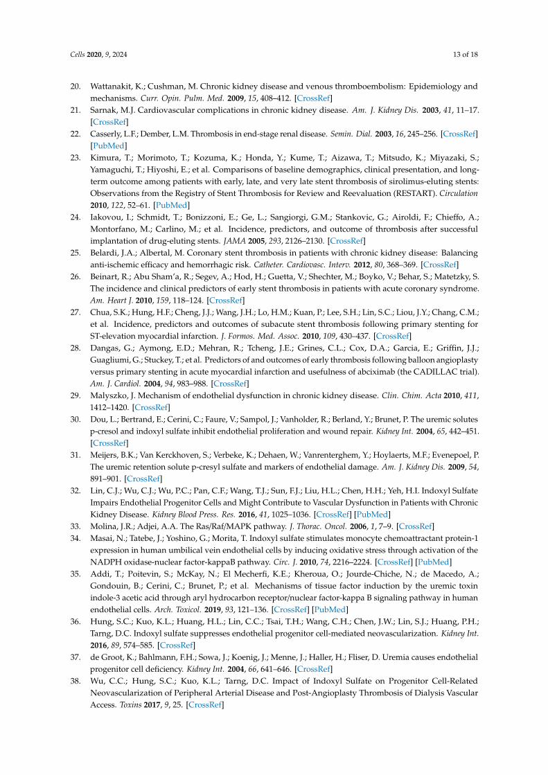
Figure 2. Examples of molecular mechanisms underlying the effects of uremic toxins on various cells, with focus on the aryl hydrocarbon receptor signaling (AHR) pathway. Indolic uremic solutes and kynurenine, all tryptophan metabolites, are agonists of the AHR pathway. These solutes enter cells through the organic anion transport channel (OAT) in target cells. Upon binding to the cytosolic AHR protein, the ligand-bound AHR (represented as a red circle on AHR) elicits distinct signaling cascades in various cell types. In endothelial cells, AHR activation regulates cell permeability through Src and prothrombotic propensity through tissue factor (TF) activation or through p38 mitogen-activated protein kinase (MAPK) signaling. AHR activation in vSMCs induces thrombosis through TF stabilization and by activating the NF-kB pathway. In macrophages, AHR induces a proinflammatory state by increasing reactive oxygen species, and through the NF-kB-MAPK signaling cascade.
Uremic solutes also affect EPCs, which together with ECs are critical in angiogenesis. In this process, along with ECs, EPCs are recruited to form angiogenic buds for new vessel formation. IS diminishes hypoxia-induced EPC migration and capillary tube formation [36]. Hung et al. showed that mice that underwent subtotal nephrectomy had significantly increased plasma levels of IS, and had reduced reperfusion and impaired neovascularization in ischemic hindlimbs compared with control mice that underwent sham operation. Increased IS levels were also associated with diminished levels of phospho-eNOS, phospho-signal transducer and activation of transcription (STAT)3, and vascular endothelial growth factor (VEGF). The collective phenotype was reversed by treating the mice with an oral adsorbent of UTs, AST-120. Concordant cell culture studies using human EPCs showed a suppressive effect of IS on VEGF expression through interleukin-10/STAT3 signaling [36].
2.2. Alteration of the Procoagulant Function of Endothelial Cells
EC proliferation is critical for the rapid reendothelialization that is warranted during repair of endothelial wounds inflicted spontaneously during rupture of an unstable plaque, or caused by endovascular procedures. Poor endothelial wound repair is likely to expose the highly prothrombotic subendothelial layer to form a reactive vascular bed for thrombogenesis. In general, healthy endothelium is considered anti-coagulant, preventing aberrant activation of the coagulation cascade and fibrin formation and/or platelet activation. Delayed reendothelialization secondary to decreased endothelial cell proliferation and migration is related to increased risk for delayed thrombosis observed after endovascular therapy in CKD patients [37,38]. This is especially relevant to CKD
Figure 2. Examples of molecular mechanisms underlying the effects of uremic toxins on various cells,with focus on the aryl hydrocarbon receptor signaling (AHR) pathway. Indolic uremic solutes andkynurenine, all tryptophan metabolites, are agonists of the AHR pathway. These solutes enter cellsthrough the organic anion transport channel (OAT) in target cells. Upon binding to the cytosolic AHRprotein, the ligand-bound AHR (represented as a red circle on AHR) elicits distinct signaling cascadesin various cell types. In endothelial cells, AHR activation regulates cell permeability through Src andprothrombotic propensity through tissue factor (TF) activation or through p38 mitogen-activated proteinkinase (MAPK) signaling. AHR activation in vSMCs induces thrombosis through TF stabilization andby activating the NF-κB pathway. In macrophages, AHR induces a proinflammatory state by increasingreactive oxygen species, and through the NF-κB-MAPK signaling cascade.
Uremic solutes also affect EPCs, which together with ECs are critical in angiogenesis. In thisprocess, along with ECs, EPCs are recruited to form angiogenic buds for new vessel formation.IS diminishes hypoxia-induced EPC migration and capillary tube formation [36]. Hung et al. showedthat mice that underwent subtotal nephrectomy had significantly increased plasma levels of IS, and hadreduced reperfusion and impaired neovascularization in ischemic hindlimbs compared with controlmice that underwent sham operation. Increased IS levels were also associated with diminished levelsof phospho-eNOS, phospho-signal transducer and activation of transcription (STAT)3, and vascularendothelial growth factor (VEGF). The collective phenotype was reversed by treating the mice withan oral adsorbent of UTs, AST-120. Concordant cell culture studies using human EPCs showed asuppressive effect of IS on VEGF expression through interleukin-10/STAT3 signaling [36].
2.2. Alteration of the Procoagulant Function of Endothelial Cells
EC proliferation is critical for the rapid reendothelialization that is warranted during repairof endothelial wounds inflicted spontaneously during rupture of an unstable plaque, or caused byendovascular procedures. Poor endothelial wound repair is likely to expose the highly prothromboticsubendothelial layer to form a reactive vascular bed for thrombogenesis. In general, healthy endotheliumis considered anti-coagulant, preventing aberrant activation of the coagulation cascade and fibrinformation and/or platelet activation. Delayed reendothelialization secondary to decreased endothelialcell proliferation and migration is related to increased risk for delayed thrombosis observed afterendovascular therapy in CKD patients [37,38]. This is especially relevant to CKD patients, as adequate

Cells 2020, 9, 2024 5 of 18
vascular access sites for hemodialysis (HD) often serve as their lifeline, and these sites are especiallysusceptible to thrombosis in the setting of repeated endothelial injury [39–41].
Healthy endothelial monolayers exhibit anti-inflammatory and anti-coagulant properties [42].Endothelial injury or endothelial dysfunction activates the extrinsic coagulation system, in which TFserves as a primary trigger for fibrin generation, creating a nidus for thrombus formation [43,44]. Indolicsolutes such as IS or IAA activate the aryl hydrocarbon receptor (AHR) pathway in ECs to upregulateTF [45]. AHR is a member of the family of basic helix–loop–helix transcription factors, which was firstidentified over three decades ago, and described then as a regulator of metabolism of xenobiotics andtoxicity [46]. Since then, various ligands have been found to activate AHR. The cytosolic non-activatedform of this receptor appears as a complex with several chaperone proteins such heat shock protein 90and a co-chaperone p23 [47]. In this folded configuration, AHR recognizes its ligand to allow nuclearlocalization. In the nucleus, it binds to the promoter of its target genes [48]. AHR upregulates TFmRNA to increases its surface expression [39] (Figure 2). IS and IAA binding and activation of AHRalso result in increased release of microparticles loaded with TF. In support of this, the blood levels ofIAA, IS, and PCS in CKD patients are associated with a reduced number of circulating endothelialprogenitor cells, signs of vascular injury, and increased plasma levels of endothelial microparticles(EMPs) [39]. Addi et al. examined a wide array of endothelial cells such as human umbilical veins,human aortic endothelial cells, and cardiac-derived microvascular cells [35]. They observed thattreatment of cells with IAA resulted in AHR activation and TF upregulation. However, AHR wasnot found to bind to the TF gene promoter, suggesting a possibility of an alternative mechanism orcross-talk among signaling pathways. Interestingly, their analysis of the TF promoter showed thatNF-κB was essential in TF induction by IAA. In support of these findings, an inhibitor of the NF-κBpathway reduced the effect IAA on TF (Figure 2). Taken together, these studies showed that differentUTs regulate TF and thrombosis through various pathways and cross-talks.
ECs are also responsible for the production of prostaglandins, which are important in hemostasis.In detail, ECs produce prostaglandin E2 (PGE2) from arachidonic acid via cyclooxygenase 2 (COX-2).In turn, PGE2 has been shown to upregulate platelet aggregation and lead to hypercoagulable statethrough binding to the platelet EP3 receptor. In a study of cultured human ECs, IAA led to COX-2upregulation via an AHR/p38 MAPK/ NF-κB pathway, leading to increased endothelial production ofPGE2, suggesting a potential mechanism of the hypercoagulable state in uremia.
2.3. Inflammation, Oxidative Stress and Atherosclerosis
UTs are pro-inflammatory, which is also known to activate ECs to expresses TF, thereby augmentingcoagulation and thrombosis [49,50]. Inflammation is the fundamental driver of atherosclerosis. Of allUTs, tryptophan metabolites such as IS, IAA, and indoxyl-β-d-glucuronide and p-cresyl sulfate (pCS) areespecially implicated in inflammation [13,51]. In specific, CKD patients stages 3–5 followed for 5.2 yearsshowed a strong correlation between the level of IS, IAA, and pCS and various markers of vascularinflammation, such as IL-6, C-reactive protein (CRP), monocyte chemoattractant protein-1 (MCP-1),soluble vascular adhesion molecule-1 (sVCAM-1), and soluble intercellular adhesion molecule-1(sICAM-1) [52]. A correlation between IAA levels and CRP in CKD patients was also confirmed inanother report [13]. In a cohort of 2399 CKD patients followed for 7.3 years as well as in a cohort ofHD patients, increased inflammation and IS levels were independently associated with atheroscleroticvascular disease [53,54].
The direct effect of uremic toxins on inflammatory molecules and oxidative stress was evaluatedin different EC cultures. A human EC line treated with normal or uremic sera from end-stage renaldisease (ESRD) patients with diabetes and/or hypertension showed an upregulated expression ofknown inflammation markers such as MCP-1 and stromal cell-derived factor 1 (SDF-1), as comparedto cells incubated with healthy sera [55]. The role of AHR in regulating some of these inflammatoryprocesses was established using mice in which AHR was specifically knocked out in endothelialcells [56]. IS treatment induced expression of E-selectin on ECs and increased adhesion of inflammatory

Cells 2020, 9, 2024 6 of 18
leukocytes to the endothelium. EC inflammation and adhesion of leukocytes are key processes inthe progress of atherosclerosis. This effect was diminished in AHR knockout mice as comparedto controls [56]. In accordance, other studies showed that activation of AHR by dioxins inducesmacrophage-mediated inflammation, appearance of cholesterol laden foam cells, and increasedatherosclerotic lesions in ApoE deleted mice [57].
IAA upregulates the AHR/p38 MAPK/NF-κB pathway, and augments the generation of reactiveoxygen species (ROS) in endothelial cells [13] (Figure 2). pCS and IAA incubated with human ECseach inhibit autophagic processes and cause accumulation of carbonylated proteins, again indicativeof oxidative stress [58]. Further, in human umbilical vein ECs, IS increased the level of reactiveoxygen species generated by the cells, as well as mitochondrial depolarization, while decreasingmitochondrial mass and function [59]. In the same cell system, IS also induced senescence byincreasing the level of reactive oxygen species (ROS) [60]. Oxidative stress is also linked to endothelialdysfunction characterized by derangement in EC-dependent flow-mediated vessel relaxation. In astudy using human umbilical vein endothelial cells (HUVECs), IS upregulated expression of NADPHoxidases (Nox4, Nox2) and production of ROS, with consequent downregulation of nitric oxide(NO) expression, the primary mediator of this phenomenon [61]. Accordingly, acetylcholine-inducedendothelium-dependent relaxation of a rat superior mesenteric artery was reduced by IS, owing to itsnegative effect on NO production [62]. In conclusion, the above studies strongly suggest the ability ofprotein bound uremic solutes to induce pro-inflammatory and oxidative stress within a variety of celltypes augmenting atherosclerotic cardiovascular disease [56,57,60].
2.4. Endothelial Cells and the Permeability Barrier
The endothelial cell layer regulates permeability using two distinct pathways—transcellular andparacellular pathways to tightly modulate the movement of circulating cells from the blood into thevessel walls, and then into the interstitium. This layer also regulates the movement of nutrients andelectrolytes. Alterations in the endothelial barrier are reflected by disrupted endothelial morphologyand reduced expression of the cell-to-cell junction proteins vascular endothelial (VE)-cadherin andzonula occludens-1 in the endothelium. Studies have shown augmented endothelial permeabilityin the aorta of experimental models of CKD [63,64]. In cultured ECs, both VE-cadherin and ZO-1protein expression decreased after exposure to uremic serum [65]. VE-cadherin mRNA expression wasreduced after exposure to PCS, IS, and uremic serum. Assefa et al. exposed bovine aorta endothelialcells to IS, subsequently noting increased cell permeability [63]. As noted earlier, in this case too,IS treatment resulted in the activation of AHR signaling as well as Src, a non-receptor tyrosine kinaselinked to remodeling of actin cytokine machinery. The AHR inhibitors CH223191 and resveratrol,a dietary polyphenol, inhibited IS-induced enhanced cell permeability. Similarly, treatment with a Srcinhibitor abolished Src activation and endothelial cell permeability (Figure 2). These results supportan IS-AHR/Src/VE-cadherin axis regulating endothelial hyperpermeability. While other factors suchas vitamin D deficiency in CKD patients are also shown to disrupt VE-cadherin interactions andF-actin reorganization [64], the direct effect of UTs on critical components of endothelial permeability isillustrated by the above studies. Irrespective of mechanism, a compromised endothelial barrier affectsthe traffic of molecules and solutes between the vessel lumen and the vessel wall, and these processesare mechanistically related to the development of atherosclerosis [66].
3. Effects of UTs on Vascular Smooth Muscle Cells
3.1. Effects of Uremic Toxins on Mitogenic Signaling in Vascular Smooth Muscle Cells
Derangement of vascular smooth muscle cells (vSMCs) is a quintessential component of uremicvascular disease. vSMC proliferation and hyperplasia are hallmarks of atherosclerosis and intimal stenosis,and constitute a major contributor to thrombosis in arterial diseases. Several UTs such as inorganicphosphate or IS have been shown to induce vSMC proliferation, migration, and calcification [67], in part

Cells 2020, 9, 2024 7 of 18
through activation of the MAPK pathway [68]. Using both CKD rats and IS-administrated rats,Yisireyili et al. showed that IS activated prorenin receptors in vSMCs through oxidative stress,AHR stimulation, and NF-κB activation [69]. Administration of N-acetylcysteine (an antioxidant)and diphenyleneiodonium (an inhibitor of nicotinamide adenine dinucleotide phosphate oxidase),or knockdown of AHR and NF-κB, inhibited IS-induced expression of prorenin in vSMCs [69]. Moreover,silencing of prorenin abrogated IS-induced vSMCS proliferation and TF expression. Other mediatorsof IS-induced vSMC proliferation include reactive oxygen species, mitogenic pathways such asplatelet-derived growth factor-ß (PDGFβ), and regulation of cell cycle proteins such as cyclin D1and p21, p53 and Glut1. Muteliefu et al. showed that IS significantly promoted the proliferation ofhuman aortic vSMCs in a concentration dependent manner, but which was significantly suppressed byantioxidants such as vitamin E, vitamin C, and N-acetylcysteine [70]. PDGF is a mitogenic ligand thatbinds to PDGF receptor (PDGFR), a tyrosine kinase receptor pathway receptor, to activate downstreamsignaling. IS increased the sensitivity of vSMCs to PDGF and augmented their proliferation [71].PDGF stimulation also increased reactive oxygen species production, thereby upregulating vSMCproliferation through this mechanism. The effect of IS was specifically observed through the PDGFβreceptor. In addition, IS-induced vSMC proliferation was suppressed by inhibitors of ERK and p38MAPK pathways [71].
IS also exploits pathways mediated through the stimulation of the MAS1 receptor, a G protein-coupled receptor which binds the angiotensin II (Ang II) metabolite angiotensin-(1-7) (Ang-(1-7)) [72].Ang-(1-7), by acting via the Mas receptor, exerts inhibitory effects on Ang-II and suppresses inflammationand vascular and cellular growth mechanisms [73]. IS downregulated the Mas receptor in the aorta ofnormotensive and hypertensive rats [72]. This effect was mitigated by silencing of AHR and NF-κB.Ang (1-7) attenuated IS-induced cell proliferation and TF expression in vSMCs, and exerted this effectthrough by inhibiting mitogenic signaling cascades such as ERK1/2 and NF-κB [72].
IS also regulates vSMCs proliferation through the induction of glucose transporter-1 (GLUT1)expression [74]. Lin et al. noted that GLUT1 facilitates the transport of glucose into vSMCs, and GLUT1overexpression increases vSMC proliferation [74]. IS induced a significant increase in expression ofGLUT1 protein as well as of pro-proliferative cyclin D1 and p21 mRNA, in addition to a modest increasein expression of antiapoptotic p53 mRNA in vSMCs. In this system, IS also significantly suppressedAkt phosphorylation after 6 h and 12 h treatments of vSMCs, and increased S6K phosphorylation(involved in protein translation) after a 3 h treatment. Treatment of vSMCs with rapamycin, a well-established inhibitor of mTOR signaling, mitigated the effect of IS on GLUT1 expression and vSMCproliferation [74].
In addition to the generation of atherosclerotic plaque, vSMC proliferation is central to vascularremodeling following injury. Treatment of ECs or vSMCs with pCS at concentrations typically found inCKD patients induced oxidative stress in them, and incubation of aortas with pCS for two days causedvascular remodeling characterized by luminal narrowing [75]. A study involving atherosclerosis proneApoE(-/-) mice showed that 5/6 nephrectomy followed by eight weeks of pCS administration resultedin greater development of atherosclerosis and vSMC proliferation as compared with control mice [76].
3.2. Effect of Uremic Toxins on Neointimal Hyperplasia and Calcification
Neointimal hyperplasia (NH) is characterized by increased number of vSMCs and deposition ofproteoglycans in the intima. NH is enhanced in the uremic milieu [77], and is frequently observed incontext of arteriovenous fistulae (AVFs) [78]. Using a murine model of CKD, the mechanism leadingto NH was probed after AV fistula surgery. An increase in NH at the fistula site in CKD animals ascompared with controls was not associated with increased vSMC proliferation, but rather augmentedcell migration, as examined in aortic explants from CKD mice. Interestingly, a uremic milieu inducedexpression of osteopontin in vSMCs, suggesting their programming to osteoblasts. Accordingly,enhancing vSMC differentiation with bone morphogenic protein-7 (BMP7) prior to AV anastomosisprevented the development of NH [77]. Taken together, the above studies indicate that a uremic

Cells 2020, 9, 2024 8 of 18
milieu and IS induce vSMC proliferation as well as migration through a host of mechanisms, in turnpromoting the development of pathologies such as NH and atherosclerosis.
Calcification of the vascular media layers is another common complication in CKD patientsowing to vSMCs shifting to osteoblastic programming. Cultured primary human umbilical veinsmooth muscle cells exposed to different concentrations of IS increased calcium deposition andaugmented the expression of osteoblast-specific proteins [79,80]. Several pathways have been studiedto deconvolute the mechanisms underlying the effects of IS on vSMC calcification. One study reportedan effect through upregulation of 1α-hydroxylase, which is responsible for hydroxylation of calcifediolto calcitriol (the bioactive form of Vitamin D) [81], and other studies have suggested a role of thePI3K/Akt/NK-κB axis [82] or regulation of Wnt/β-catenin signaling mediated through microRN-29b [83].Zhang et al. demonstrated that vascular miR-29b was down-regulated in radial arteries of patientswith end-stage renal disease (ESRD). IS also decreased miR-29b expression in human aortic vSMCsand potentiated their calcification [83]. They observed increased expression of Wnt7b/β-catenin inradial arteries of ESRD compared with a control group. IS increased Wnt7b/β-catenin expression invSMCS within three days of exposure. They further demonstrated that miR-29b indeed downregulatedWnt7b/β-catenin signaling. These results showed that IS induces expression of miR-29b, in turnsuppressing Wnt//β-catenin signaling to induce vascular calcification in CKD a milieu. Similar to IS,pCS augmented osteoblastic markers such as alkaline phosphatase and osteopontin in human aorticvSMCs, partially mediated through pCS-induced expression of NOX4 [84].
3.3. Prothrombotic Effects of Specific Uremic Toxins
An important mediator of vSMC-driven cardiovascular pathologies is TF, as it controls severalprocesses [50]. The circulating levels of TF in CKD patients positively correlate with plasma levels of IS,IAA [45], and Kyn, another tryptophan-based uremic solute [85]. IS level is also positively correlatedwith increased levels of von-Willebrand factor (vWF). As noted earlier, TF is a cell-surface protein;the dysregulation of which contributes to the development of thrombotic events. Our studies showedincreased TF activity on the surfaces of primary human aortic vSMCs and endothelial cells in responseto sera from CKD patients with varying stages of disease [86–89]. Human vSMCs pretreated withuremic serum obtained from ESRD patients, or with indolic solutes, and subsequently exposed to acoronary-like blood flow system, demonstrated a significantly greater clot formation. This was inturn reduced with an anti-TF neutralizing antibody [88]. Further mechanistic probing revealed that ISinhibited TF protein ubiquitination and subsequent proteasomal degradation [86,90]. As proposedearlier [91], recent studies showed that IS-induced TF upregulation leading to thrombosis is mediatedthrough AHR signaling [88]. Upon activation by IS, the cytosolic AHR bound to TF and protectedit from ubiquitination and proteasomal degradation by an E3 ubiquitin ligase STIP1 homology andU-box-containing E3 ubiquitin ligase protein 1 (STUB1). AHR inhibitor reduced TF-AHR interactionand augmented TF ubiquitination by STUB1 in vSMCs. In fact, a STUB1 activator, YL-109, augmentedTF degradation. Both AHR inhibitor and STUB-1 activator significantly mitigated IS and uremicserum-mediated thrombosis in flow loops coated with vSMCs, in a FeCl3-induced carotid arteryinjury model in the setting of CKD, and in an IS-specific solute model [88]. Importantly, in thisstudy, AHR inhibitor and STUB-1 activator ameliorated the effects of IS on a thrombotic phenotypewithout affecting bleeding time, suggesting that targeting CKD-specific mechanisms can result in saferantithrombotics [88]. In accordance with these molecular studies, analysis of sera from a CKD cohortfrom the Dialysis Access Consortium Clopidogrel Prevention of Early AV Fistula Thrombosis trial orthe Thrombolysis in Myocardial Infarction II trial showed that patients with subsequent arteriovenousthrombosis had significantly higher serum levels of IS and Kyn, and they induced greater AHR andTF activity in endothelial cells and vSMCs as compared to those without thrombosis. Using machinelearning techniques, these parameters sufficiently segregated patients who subsequently developedpost-angioplasty and AVF thrombosis, both of which are examples of post vascular injury-inducedthrombosis. Furthermore, administration of Kyn to a mouse model of vascular injury increased

Cells 2020, 9, 2024 9 of 18
thrombosis, which was subsequently attenuated by an AHR inhibitor [89]. This series of experimentsdelineated CKD-specific mechanisms driving thrombosis and also uncovered therapeutic targets thatcan be used for further development of safer antithrombotic agents for CKD-associated thrombosis.Taken together, all these studies strongly suggest that indolic metabolites and Kyn activate AHRsignaling to regulate TF, the primary trigger of extrinsic coagulation. This event culminates in thrombusformation at the site of reactive vascular bed, created after vascular injury (rupture of atheroscleroticplaque or intervention [86–89].
4. Effect of Uremic Toxins on Cardiomyocytes
CKD patients often develop congestive heart failure (both preserved and reduced ejection fraction),and their illness is often pathologically characterized by cardiac fibrosis and remodeling [6]. In aprospective study of 433 ESRD patients, 74% were reported to develop ventricular hypertrophy [92].Several molecular mechanisms have been proposed to account for effects of UT on the heart. IS wasfound to increase ROS levels within cardiomyocytes and inhibited AMP-activated protein kinase(AMPK). These events resulted in increased protein synthesis and cell volume of cultured neonatalrat cardiomyocytes, consistent with the cardiac hypertrophy observed in CKD patients [93]. Similarfindings including IS-induced heart fibrosis were reported in rats treated with IS [94]. In anotherset of studies, IS affected endoplasmic reticulum stress modulators and induced apoptosis in thecardiomyocyte cell line H9C2 [95]. pCS administered to mice induced apoptosis in cardiomyocytes,resulting in diastolic dysfunction, partially attributed to augmented ROS [96]. pCS also reducedspontaneous contraction of cultured cardiomyocytes, attributed to a damaging effect on cellular gapjunctions [97]. While cardiomyopathy is common in CKD patients and is the driver of frequenthospitalization and mortality in these patients, little is known about how these UTs induce thesepathological changes.
5. The Effects of Uremic Toxins on Monocytes/Macrophages, Polymorphonuclear Cells and Platelets
5.1. The Influence of Uremic Toxins on Macrophages and Monocytes
Macrophages are key contributors to vascular inflammation and progression of atherosclerosis,and the uremic milieu has been shown to enhance their activation and pro-inflammatory effect [98–102].Nakano et al. showed that IS is transported into macrophages by organic anion transporter (OAT)P2B1 [103]. Once inside the cell, IS increases the expression of ubiquitin-specific peptidase 5, leadingto inhibition of the ubiquitin–proteasome pathway, and consequent accumulation of δ-like ligand4 (DLL4). DLL4 is responsible for the cleavage of the intracellular domain of Notch1, a member ofthe Notch transmembrane receptor family that has been implicated in proinflammatory activationof macrophages and development of CVD. Augmented levels of DLL4 enhance cleavage of theintracellular domain of Notch1, which undergoes nuclear translocation to serve as a transcriptionalregulator and induce proinflammatory genes. In 5/6 nephrectomy atherosclerotic mice, DLL4 andNotch1 intracellular domain expression were found to be increased in macrophages residing theatherosclerotic plaque, supporting an in vivo activation of Notch signaling in CKD milieu. Treatmentwith an anti-DLL4 antibody targeted to macrophages through lipid coated nanoparticles inhibitedproduction of inflammatory cytokines, decreased macrophage accumulation, and decreased progressionof atherosclerosis in CKD mice. Similarly, mice receiving 4 weeks of intraperitoneal IS and subsequentlysilenced for OAT P2B1 and DLL4 in macrophages showed decreased vascular inflammation andreduced atherosclerotic plaque burden as compared to controls [103]. This interesting study showedthe role of IS and uremic milieu in manipulating macrophages in the atherosclerotic plaque in a CKDmilieu. In a separate study, THP-1-derived macrophages exposed to IS in vitro were shown to haveincreased production of inflammatory cytokines such as TNF-α and IL-1β. Notably, these macrophageswere shown to have significantly decreased activity of ATP-binding cassette transporter A1 (ABCA1),

Cells 2020, 9, 2024 10 of 18
a lipid transporter essential for cholesterol efflux, thus potentially leading to accelerated developmentof foam cells and progression of atherosclerosis [104].
Monocytes express elevated levels of angiotensin converting enzyme (ACE) in ESRD patients,which has been hypothesized to contribute to accelerated atherosclerosis [101,105]. In an in vitrostudy, both human primary monocytes and THP-1 cells treated with uremic serum showed higherACE expression. The authors surmised that this ACE overexpression might promote M2 macrophagedifferentiation, with associated pro-inflammatory and pro-atherosclerotic properties. The monocyteswith higher ACE2 expressions showed increased levels of inflammatory cytokines TNF-α and IL-6,as well as increased levels of arginase-1 (Arg1), which is a marker of M2 cells present in plaques,and may itself also promote plaque stabilization and proliferation of vSMCs. ACE overexpressingmonocytes were also shown to have greater expression of MCP-1 and its ligand CCR2, potentiallyleading to enhanced transmigration through the endothelium. Additionally, ACE overexpression insuch monocytes led to upregulation of adhesion molecules such as ICAM-1 and VCAM-1, moleculesconsidered essential to the initial steps of atherosclerotic plaque formation [101,102,105]. One of thehallmarks of CVD secondary to kidney disease is intravascular macrophage inflammation. An in vitrostudy showed that IS activated AHR/NFκB/MAPK cascades in macrophages, causing an inflammatoryresponse [106]. A study involving 268 HD patients followed for 36 months showed that a highneutrophil-to-lymphocyte ratio was an independent predictor of cardiovascular mortality [107].Another uremic toxic, phenylacetic acid (PAA) [108], increases oxidative burst and inflammatorycytokine production in polymorphonuclear cells, which could contribute to augmented cardiovascularrisk [109]. Collectively, these studies demonstrated UTs inducing substantial alterations in monocytesand macrophages, associated with vascular inflammation and cardiovascular pathology.
5.2. Effect of Uremic Toxins on Platelets
The CKD milieu profoundly alters platelet functions, which contributes to atherothrombosisin these patients [38]. Indeed, earlier studies showed that renal dysfunction is associated withinappropriate platelet activation [110,111]. Mouse platelets treated with IS displayed increased activityin response to collagen and thrombin [112]. IS also augmented platelet-derived microparticles andplatelet-monocyte aggregates, which are also contributors to vascular dysfunction [112]. In accordance,IS enhanced thrombus formation as measured in a flow chamber assay and carotid artery thrombosismodel that improved with AST-120 administration (an oral adsorbent of uremic toxins) or klotho [112].Interestingly, ROS induction of p38 MAPK signaling in platelets is a driver in IS-mediated plateletactivation, both in vivo and in vitro [112].
6. Uremic Toxin-Induced Microparticles and Cardiovascular Disease
Microparticles (MPs) are fragments derived from cell membranes, and contain various membrane-associated receptors and ligands [113]. MPs can originate from endothelial cells, platelets, erythrocytes,polymorphonuclear cells, monocytes, and lymphocytes, and bear cell-type-specific markers. On the onehand, MPs may help facilitate intracellular communications and maintenance of homeostasis, but onthe other, they might trigger a deleterious pro-inflammatory response [114–117]. MPs bind to and entertarget cells by various endocytic pathways, and inside the cell may activate inflammatory pathways,thereby causing functional impairment [116,118]. MPs are released secondary to plasma membraneremodeling in the setting of programmed cell death, injury, or cellular activation [117].
Endothelial injury secondary to UTs induces release of endothelial microparticles (EMPs), oftenbeing specifically mediated by IS, pCS, IAA, and inorganic phosphate (accumulating secondaryto hyperphosphatemia) [119–123]. pCS promotes EMP formation through an effect on Rho kinaseand resultant cytoskeletal reorganization [75,124]. Likewise, IS induces the release of EMPs fromendothelial cells in a dose-dependent manner via upregulation of the p38 MAPK signaling pathway [125].Erythrocytes treated with IS or IAA showed increased phosphatidylserine (PS) exposure along withrelease of PS-laden MPs, in turn leading to a hypercoagulable state [126].

Cells 2020, 9, 2024 11 of 18
MPs from different origins can regulate distinct phenotypes in the vascular system. For example,a study analyzed plasma from ESRD patients and found an increase in MPs of platelet, erythrocyte,and endothelial origin [127]. However, only EMPs correlated with loss of flow-mediated relaxation,whereas MPs of other origins did not. The same study also showed that in vitro, only EMPs impairedendothelial cGMP generation and NO release, leading to a compromised endothelium-dependentrelaxation [127]. Furthermore, EMPs isolated from CKD patients have been shown to induce osteocalcinexpression in EPCs, vSMCs, and fibroblasts, leading to vascular calcification [128]. Concordantly,the number of circulating EMPs is greater in CKD patients with vascular calcification than withoutit, and the increase in EMPs in CKD patients is associated with a decrease in the number of EPCs,suggesting a role for EMPs in impaired tissue renewal [128]. EMPs can also regulate immune cells,which in turn can influence inflammation and thrombosis [129].
IS-induced EMPs (IsEMPs) in particular have been shown to contain miRNAs that reduce theangiogenic capacity of EPCs, as well as upregulate expression of NF-κB and p53 in them, all ofwhich culminate in an inflammatory response and EPC apoptosis [130]. Accordingly, in vitro studieshave shown that ECs treated with TNF-α produced EMPs that can increase monocytic TF-dependentprocoagulant activity [131]. A separate study explored the role of IsEMPs in NH, one of the main causesof vascular access malfunction in patients on hemodialysis. It showed that vessels exposed to EMPsex vivo developed significantly greater NH and vSMC proliferation via heightened activation of theTGF-β signaling pathway [121]. In accordance with this study, EMPs collected from the culture mediaof human umbilical vascular endothelial cells treated with IS augmented NH and vSMC proliferationin cultured porcine internal jugular veins, concomitant with activation of the MAPK pathway, and in asimilar manner to TGF-β [121].
IS-induced EMP release can be modulated by pharmacological approaches. Ryu et al. demonstratedthat IS induced generation of EMPs in culture media of cells [132]. Such EMPs stimulated vSMCproliferation in a concentration-dependent manner, upregulated TGF-β, and promoted the TGF-βsignaling pathway, as illustrated by phosphorylation of downstream targets, including Akt, ERK1/2,p38 MAPK, and Smad3, all of which being significantly inhibited by an anti-TGF-β antibody andinhibitors of ERK and Smad. Several compounds have been investigated to suppress IS-inducedEMP production, including losartan, lovastatin, clopidogrel, and mesoglycan [125]. All of these drugsinhibited EMP generation induced by IS at concentrations between 10–50 µM. However, the suppressiveeffect was highest with clopidogrel, which significantly suppressed MAPK signaling activated by IS inendothelial cells.
7. Conclusions
It has become clear that pathology in one organ can induce systemic effects resulting in pathologyin other organs. The case of CKD-associated accumulation of UTs and their untoward effects on differentcomponents of the cardiovascular system is an example of this paradigm. While epidemiologicalstudies pointed to CKD as an independent risk factor for various forms of cardiovascular disease,the molecular mechanisms by which this occurs have only recently been gradually elucidated. To thisend, the current review provides a comprehensive overview of cellular signaling pathways triggered bya set of well-established cardiovascular uremic toxins. As illustrated in Figure 1, different types of UTshave the ability to derange molecular pathways in various cell types, whether vSMCs, ECs, platelets,or cardiomyocytes, all of which are central to a healthy cardiovascular system. Understanding whichbiological processes are impacted in these cells could lead to more targeted treatments for differentforms of CVD in CKD.
Author Contributions: J.D.R. and V.C.C. participated in reviewing the literature, writing, and editing themanuscript. All authors have read and agreed to the published version of the manuscript.
Funding: This work was funded by R01 HL132325 (V.C.C.).

Cells 2020, 9, 2024 12 of 18
Acknowledgments: We regret potential omission of published studies in light of the space limit. We thank theEvans Center for Interdisciplinary Biomedical Research and the Department of Medicine at Boston UniversitySchool of Medicine for their ongoing support of the Affinity Research Collaborative on Thrombosis and Hemostasis.
Conflicts of Interest: The authors have no conflict of interest to declare.
References
1. Temgoua, M.N.; Danwang, C.; Agbor, V.N.; Noubiap, J.J. Prevalence, incidence and associated mortalityof cardiovascular disease in patients with chronic kidney disease in low- and middle-income countries:A protocol for a systematic review and meta-analysis. BMJ Open 2017, 7, e016412. [CrossRef] [PubMed]
2. Weaver, D.J.; Mitsnefes, M. Cardiovascular Disease in Children and Adolescents with Chronic KidneyDisease. Semin. Nephrol. 2018, 38, 559–569. [CrossRef]
3. Charytan, D.M. Introduction: Cardiovascular Disease in Chronic Kidney Disease. Semin. Nephrol. 2018,38, 541. [CrossRef] [PubMed]
4. Duranton, F.; Cohen, G.; De Smet, R.; Rodriguez, M.; Jankowski, J.; Vanholder, R.; Argiles, A.; EuropeanUremic Toxin Work Group. Normal and pathologic concentrations of uremic toxins. J. Am. Soc. Nephrol.2012, 23, 1258–1270. [CrossRef] [PubMed]
5. Fujii, H.; Goto, S.; Fukagawa, M. Role of Uremic Toxins for Kidney, Cardiovascular, and Bone Dysfunction.Toxins 2018, 10, 202. [CrossRef]
6. Lekawanvijit, S. Cardiotoxicity of Uremic Toxins: A Driver of Cardiorenal Syndrome. Toxins 2018, 10, 352.[CrossRef] [PubMed]
7. Lin, C.J.; Liu, H.L.; Pan, C.F.; Chuang, C.K.; Jayakumar, T.; Wang, T.J.; Chen, H.H.; Wu, C.J. Indoxylsulfate predicts cardiovascular disease and renal function deterioration in advanced chronic kidney disease.Arch. Med. Res. 2012, 43, 451–456. [CrossRef] [PubMed]
8. Fan, P.C.; Chang, J.C.; Lin, C.N.; Lee, C.C.; Chen, Y.T.; Chu, P.H.; Kou, G.; Lu, Y.A.; Yang, C.W.; Chen, Y.C.Serum indoxyl sulfate predicts adverse cardiovascular events in patients with chronic kidney disease.J. Formos. Med. Assoc. 2019, 118, 1099–1106. [CrossRef] [PubMed]
9. Meijers, B.K.; Bammens, B.; De Moor, B.; Verbeke, K.; Vanrenterghem, Y.; Evenepoel, P. Free p-cresol isassociated with cardiovascular disease in hemodialysis patients. Kidney Int. 2008, 73, 1174–1180. [CrossRef]
10. Liabeuf, S.; Barreto, D.V.; Barreto, F.C.; Meert, N.; Glorieux, G.; Schepers, E.; Temmar, M.; Choukroun, G.;Vanholder, R.; Massy, Z.A.; et al. Free p-cresylsulphate is a predictor of mortality in patients at differentstages of chronic kidney disease. Nephrol. Dial. Transplant. 2010, 25, 1183–1191. [CrossRef]
11. Stubbs, J.R.; House, J.A.; Ocque, A.J.; Zhang, S.; Johnson, C.; Kimber, C.; Schmidt, K.; Gupta, A.; Wetmore, J.B.;Nolin, T.D.; et al. Serum Trimethylamine-N-Oxide is Elevated in CKD and Correlates with CoronaryAtherosclerosis Burden. J. Am. Soc. Nephrol. 2016, 27, 305–313. [CrossRef] [PubMed]
12. Pedersen, E.R.; Tuseth, N.; Eussen, S.J.; Ueland, P.M.; Strand, E.; Svingen, G.F.; Midttun, O.; Meyer, K.;Mellgren, G.; Ulvik, A.; et al. Associations of plasma kynurenines with risk of acute myocardial infarction inpatients with stable angina pectoris. Arterioscler. Thromb. Vasc. Biol. 2015, 35, 455–462. [CrossRef] [PubMed]
13. Dou, L.; Sallee, M.; Cerini, C.; Poitevin, S.; Gondouin, B.; Jourde-Chiche, N.; Fallague, K.; Brunet, P.; Calaf, R.;Dussol, B.; et al. The cardiovascular effect of the uremic solute indole-3 acetic acid. J. Am. Soc. Nephrol. 2015,26, 876–887. [CrossRef] [PubMed]
14. Leong, S.C.; Sirich, T.L. Indoxyl Sulfate-Review of Toxicity and Therapeutic Strategies. Toxins 2016, 8, 358.[CrossRef] [PubMed]
15. Gryp, T.; Vanholder, R.; Vaneechoutte, M.; Glorieux, G. p-Cresyl Sulfate. Toxins 2017, 9, 52. [CrossRef]16. Le Floc’h, N.; Otten, W.; Merlot, E. Tryptophan metabolism, from nutrition to potential therapeutic
applications. Amino Acids 2011, 41, 1195–1205. [CrossRef]17. Abbasi, J. TMAO and Heart Disease: The New Red Meat Risk? JAMA 2019, 321, 2149–2151. [CrossRef]18. Tang, W.H.; Wang, Z.; Kennedy, D.J.; Wu, Y.; Buffa, J.A.; Agatisa-Boyle, B.; Li, X.S.; Levison, B.S.; Hazen, S.L.
Gut microbiota-dependent trimethylamine N-oxide (TMAO) pathway contributes to both development ofrenal insufficiency and mortality risk in chronic kidney disease. Circ. Res. 2015, 116, 448–455. [CrossRef]
19. Cheung, W.; Keski-Rahkonen, P.; Assi, N.; Ferrari, P.; Freisling, H.; Rinaldi, S.; Slimani, N.; Zamora-Ros, R.;Rundle, M.; Frost, G.; et al. A metabolomic study of biomarkers of meat and fish intake. Am. J. Clin. Nutr.2017, 105, 600–608. [CrossRef]
Cells 2020, 9, 2024 13 of 18
20. Wattanakit, K.; Cushman, M. Chronic kidney disease and venous thromboembolism: Epidemiology andmechanisms. Curr. Opin. Pulm. Med. 2009, 15, 408–412. [CrossRef]
21. Sarnak, M.J. Cardiovascular complications in chronic kidney disease. Am. J. Kidney Dis. 2003, 41, 11–17.[CrossRef]
22. Casserly, L.F.; Dember, L.M. Thrombosis in end-stage renal disease. Semin. Dial. 2003, 16, 245–256. [CrossRef][PubMed]
23. Kimura, T.; Morimoto, T.; Kozuma, K.; Honda, Y.; Kume, T.; Aizawa, T.; Mitsudo, K.; Miyazaki, S.;Yamaguchi, T.; Hiyoshi, E.; et al. Comparisons of baseline demographics, clinical presentation, and long-term outcome among patients with early, late, and very late stent thrombosis of sirolimus-eluting stents:Observations from the Registry of Stent Thrombosis for Review and Reevaluation (RESTART). Circulation2010, 122, 52–61. [PubMed]
24. Iakovou, I.; Schmidt, T.; Bonizzoni, E.; Ge, L.; Sangiorgi, G.M.; Stankovic, G.; Airoldi, F.; Chieffo, A.;Montorfano, M.; Carlino, M.; et al. Incidence, predictors, and outcome of thrombosis after successfulimplantation of drug-eluting stents. JAMA 2005, 293, 2126–2130. [CrossRef]
25. Belardi, J.A.; Albertal, M. Coronary stent thrombosis in patients with chronic kidney disease: Balancinganti-ischemic efficacy and hemorrhagic risk. Catheter. Cardiovasc. Interv. 2012, 80, 368–369. [CrossRef]
26. Beinart, R.; Abu Sham’a, R.; Segev, A.; Hod, H.; Guetta, V.; Shechter, M.; Boyko, V.; Behar, S.; Matetzky, S.The incidence and clinical predictors of early stent thrombosis in patients with acute coronary syndrome.Am. Heart J. 2010, 159, 118–124. [CrossRef]
27. Chua, S.K.; Hung, H.F.; Cheng, J.J.; Wang, J.H.; Lo, H.M.; Kuan, P.; Lee, S.H.; Lin, S.C.; Liou, J.Y.; Chang, C.M.;et al. Incidence, predictors and outcomes of subacute stent thrombosis following primary stenting forST-elevation myocardial infarction. J. Formos. Med. Assoc. 2010, 109, 430–437. [CrossRef]
28. Dangas, G.; Aymong, E.D.; Mehran, R.; Tcheng, J.E.; Grines, C.L.; Cox, D.A.; Garcia, E.; Griffin, J.J.;Guagliumi, G.; Stuckey, T.; et al. Predictors of and outcomes of early thrombosis following balloon angioplastyversus primary stenting in acute myocardial infarction and usefulness of abciximab (the CADILLAC trial).Am. J. Cardiol. 2004, 94, 983–988. [CrossRef]
29. Malyszko, J. Mechanism of endothelial dysfunction in chronic kidney disease. Clin. Chim. Acta 2010, 411,1412–1420. [CrossRef]
30. Dou, L.; Bertrand, E.; Cerini, C.; Faure, V.; Sampol, J.; Vanholder, R.; Berland, Y.; Brunet, P. The uremic solutesp-cresol and indoxyl sulfate inhibit endothelial proliferation and wound repair. Kidney Int. 2004, 65, 442–451.[CrossRef]
31. Meijers, B.K.; Van Kerckhoven, S.; Verbeke, K.; Dehaen, W.; Vanrenterghem, Y.; Hoylaerts, M.F.; Evenepoel, P.The uremic retention solute p-cresyl sulfate and markers of endothelial damage. Am. J. Kidney Dis. 2009, 54,891–901. [CrossRef]
32. Lin, C.J.; Wu, C.J.; Wu, P.C.; Pan, C.F.; Wang, T.J.; Sun, F.J.; Liu, H.L.; Chen, H.H.; Yeh, H.I. Indoxyl SulfateImpairs Endothelial Progenitor Cells and Might Contribute to Vascular Dysfunction in Patients with ChronicKidney Disease. Kidney Blood Press. Res. 2016, 41, 1025–1036. [CrossRef] [PubMed]
33. Molina, J.R.; Adjei, A.A. The Ras/Raf/MAPK pathway. J. Thorac. Oncol. 2006, 1, 7–9. [CrossRef]34. Masai, N.; Tatebe, J.; Yoshino, G.; Morita, T. Indoxyl sulfate stimulates monocyte chemoattractant protein-1
expression in human umbilical vein endothelial cells by inducing oxidative stress through activation of theNADPH oxidase-nuclear factor-kappaB pathway. Circ. J. 2010, 74, 2216–2224. [CrossRef] [PubMed]
35. Addi, T.; Poitevin, S.; McKay, N.; El Mecherfi, K.E.; Kheroua, O.; Jourde-Chiche, N.; de Macedo, A.;Gondouin, B.; Cerini, C.; Brunet, P.; et al. Mechanisms of tissue factor induction by the uremic toxinindole-3 acetic acid through aryl hydrocarbon receptor/nuclear factor-kappa B signaling pathway in humanendothelial cells. Arch. Toxicol. 2019, 93, 121–136. [CrossRef] [PubMed]
36. Hung, S.C.; Kuo, K.L.; Huang, H.L.; Lin, C.C.; Tsai, T.H.; Wang, C.H.; Chen, J.W.; Lin, S.J.; Huang, P.H.;Tarng, D.C. Indoxyl sulfate suppresses endothelial progenitor cell-mediated neovascularization. Kidney Int.2016, 89, 574–585. [CrossRef]
37. de Groot, K.; Bahlmann, F.H.; Sowa, J.; Koenig, J.; Menne, J.; Haller, H.; Fliser, D. Uremia causes endothelialprogenitor cell deficiency. Kidney Int. 2004, 66, 641–646. [CrossRef]
38. Wu, C.C.; Hung, S.C.; Kuo, K.L.; Tarng, D.C. Impact of Indoxyl Sulfate on Progenitor Cell-RelatedNeovascularization of Peripheral Arterial Disease and Post-Angioplasty Thrombosis of Dialysis VascularAccess. Toxins 2017, 9, 25. [CrossRef]

Cells 2020, 9, 2024 14 of 18
39. Jourde-Chiche, N.; Dou, L.; Sabatier, F.; Calaf, R.; Cerini, C.; Robert, S.; Camoin-Jau, L.; Charpiot, P.;Argiles, A.; Dignat-George, F.; et al. Levels of circulating endothelial progenitor cells are related to uremictoxins and vascular injury in hemodialysis patients. J. Thromb. Haemost. 2009, 7, 1576–1584. [CrossRef]
40. Roy-Chaudhury, P.; Sukhatme, V.P.; Cheung, A.K. Hemodialysis vascular access dysfunction: A cellular andmolecular viewpoint. J. Am. Soc. Nephrol. 2006, 17, 1112–1127. [CrossRef]
41. Chen, T.Y.; Lin, T.T.; Hsieh, M.Y.; Lin, L.; Yang, C.W.; Chuang, S.Y.; Huang, P.H.; Wu, C.C. CirculatingProgenitor Cells Affect Thrombosis of Dialysis Arteriovenous Fistulas. Am. J. Nephrol. 2016, 44, 428–438.[CrossRef] [PubMed]
42. Endemann, D.H.; Schiffrin, E.L. Endothelial dysfunction. J. Am. Soc. Nephrol. 2004, 15, 1983–1992. [CrossRef][PubMed]
43. Mackman, N.; Tilley, R.E.; Key, N.S. Role of the extrinsic pathway of blood coagulation in hemostasis andthrombosis. Arterioscler. Thromb. Vasc. Biol. 2007, 27, 1687–1693. [CrossRef]
44. Mackman, N. Triggers, targets and treatments for thrombosis. Nature 2008, 451, 914–918. [CrossRef]45. Gondouin, B.; Cerini, C.; Dou, L.; Sallee, M.; Duval-Sabatier, A.; Pletinck, A.; Calaf, R.; Lacroix, R.;
Jourde-Chiche, N.; Poitevin, S.; et al. Indolic uremic solutes increase tissue factor production in endothelialcells by the aryl hydrocarbon receptor pathway. Kidney Int. 2013, 84, 733–744. [CrossRef] [PubMed]
46. Sherr, D.H.; Monti, S. The role of the aryl hydrocarbon receptor in normal and malignant B cell development.Semin. Immunopathol. 2013, 35, 705–716. [CrossRef]
47. Petrulis, J.R.; Perdew, G.H. The role of chaperone proteins in the aryl hydrocarbon receptor core complex.Chem. Biol. Interact. 2002, 141, 25–40. [CrossRef]
48. Guyot, E.; Chevallier, A.; Barouki, R.; Coumoul, X. The AhR twist: Ligand-dependent AhR signaling andpharmaco-toxicological implications. Drug Discov. Today 2013, 18, 479–486. [CrossRef]
49. Yau, J.W.; Teoh, H.; Verma, S. Endothelial cell control of thrombosis. BMC Cardiovasc. Disord. 2015, 15, 130.[CrossRef]
50. Mackman, N. Role of tissue factor in hemostasis, thrombosis, and vascular development. Arterioscler. Thromb.Vasc. Biol. 2004, 24, 1015–1022. [CrossRef]
51. Sallee, M.; Dou, L.; Cerini, C.; Poitevin, S.; Brunet, P.; Burtey, S. The aryl hydrocarbon receptor-activating effectof uremic toxins from tryptophan metabolism: A new concept to understand cardiovascular complicationsof chronic kidney disease. Toxins 2014, 6, 934–949. [CrossRef] [PubMed]
52. Claro, L.M.; Moreno-Amaral, A.N.; Gadotti, A.C.; Dolenga, C.J.; Nakao, L.S.; Azevedo, M.L.V.; de Noronha, L.;Olandoski, M.; de Moraes, T.P.; Stinghen, A.E.M.; et al. The Impact of Uremic Toxicity Induced InflammatoryResponse on the Cardiovascular Burden in Chronic Kidney Disease. Toxins 2018, 10, 384. [CrossRef][PubMed]
53. Amdur, R.L.; Feldman, H.I.; Dominic, E.A.; Anderson, A.H.; Beddhu, S.; Rahman, M.; Wolf, M.; Reilly, M.;Ojo, A.; Townsend, R.R.; et al. Use of Measures of Inflammation and Kidney Function for Prediction ofAtherosclerotic Vascular Disease Events and Death in Patients With CKD: Findings From the CRIC Study.Am. J. Kidney Dis. 2019, 73, 344–353. [CrossRef] [PubMed]
54. Taki, K.; Tsuruta, Y.; Niwa, T. Indoxyl sulfate and atherosclerotic risk factors in hemodialysis patients. Am. J.Nephrol. 2007, 27, 30–35. [CrossRef]
55. Eloueyk, A.; Osta, B.; Alameldinne, R.; Awad, D. Uremic Serum Induces Inflammation in Cultured HumanEndothelial Cells and Triggers Vascular Repair Mechanisms. Inflammation 2019, 42, 2003–2010. [CrossRef]
56. Ito, S.; Osaka, M.; Edamatsu, T.; Itoh, Y.; Yoshida, M. Crucial Role of the Aryl Hydrocarbon Receptor (AhR)in Indoxyl Sulfate-Induced Vascular Inflammation. J. Atheroscler. Thromb. 2016, 23, 960–975. [CrossRef]
57. Wu, D.; Nishimura, N.; Kuo, V.; Fiehn, O.; Shahbaz, S.; Van Winkle, L.; Matsumura, F.; Vogel, C.F. Activationof aryl hydrocarbon receptor induces vascular inflammation and promotes atherosclerosis in apolipoproteinE-/- mice. Arterioscler. Thromb. Vasc. Biol. 2011, 31, 1260–1267. [CrossRef]
58. Rodrigues, S.D.; Santos, S.S.; Meireles, T.; Romero, N.; Glorieux, G.; Pecoits-Filho, R.; Zhang, D.D.; Nakao, L.S.Uremic toxins promote accumulation of oxidized protein and increased sensitivity to hydrogen peroxidein endothelial cells by impairing the autophagic flux. Biochem. Biophys. Res. Commun. 2019, 523, 123–129.[CrossRef]
59. Lee, W.C.; Li, L.C.; Chen, J.B.; Chang, H.W. Indoxyl sulfate-induced oxidative stress, mitochondrialdysfunction, and impaired biogenesis are partly protected by vitamin C and N-acetylcysteine. Sci. World J.2015, 2015, 620826. [CrossRef]

Cells 2020, 9, 2024 15 of 18
60. Adelibieke, Y.; Shimizu, H.; Muteliefu, G.; Bolati, D.; Niwa, T. Indoxyl sulfate induces endothelial cellsenescence by increasing reactive oxygen species production and p53 activity. J. Ren. Nutr. 2012, 22, 86–89.[CrossRef]
61. Tumur, Z.; Niwa, T. Indoxyl sulfate inhibits nitric oxide production and cell viability by inducing oxidativestress in vascular endothelial cells. Am. J. Nephrol. 2009, 29, 551–557. [CrossRef] [PubMed]
62. Matsumoto, T.; Takayanagi, K.; Kojima, M.; Katome, T.; Taguchi, K.; Kobayashi, T. Direct Impairment of theEndothelial Function by Acute Indoxyl Sulfate through Declined Nitric Oxide and Not Endothelium-DerivedHyperpolarizing Factor or Vasodilator Prostaglandins in the Rat Superior Mesenteric Artery. Biol. Pharm. Bull.2019, 42, 1236–1242. [CrossRef] [PubMed]
63. Assefa, E.G.; Yan, Q.; Gezahegn, S.B.; Salissou, M.T.M.; He, S.; Wu, N.; Zuo, X.; Ying, C. Role ofResveratrol on Indoxyl Sulfate-Induced Endothelial Hyperpermeability via Aryl Hydrocarbon Receptor(AHR)/Src-Dependent Pathway. Oxid. Med. Cell. Longev. 2019, 2019, 5847040. [CrossRef] [PubMed]
64. Vila Cuenca, M.; van Bezu, J.; Beelen, R.H.J.; Vervloet, M.G.; Hordijk, P.L. Stabilization of cell-cell junctionsby active vitamin D ameliorates uraemia-induced loss of human endothelial barrier function. Nephrol. Dial.Transplant. 2019, 34, 252–264. [CrossRef]
65. Maciel, R.A.P.; Cunha, R.S.; Busato, V.; Franco, C.R.C.; Gregorio, P.C.; Dolenga, C.J.R.; Nakao, L.S.; Massy, Z.A.;Boullier, A.; Pecoits-Filho, R.; et al. Uremia Impacts VE-Cadherin and ZO-1 Expression in Human EndothelialCell-to-Cell Junctions. Toxins 2018, 10, 404. [CrossRef] [PubMed]
66. Mundi, S.; Massaro, M.; Scoditti, E.; Carluccio, M.A.; van Hinsbergh, V.W.M.; Iruela-Arispe, M.L.;De Caterina, R. Endothelial permeability, LDL deposition, and cardiovascular risk factors—A review.Cardiovasc. Res. 2018, 114, 35–52. [CrossRef] [PubMed]
67. Henaut, L.; Mary, A.; Chillon, J.M.; Kamel, S.; Massy, Z.A. The Impact of Uremic Toxins on Vascular SmoothMuscle Cell Function. Toxins 2018, 10, 218. [CrossRef]
68. Yamamoto, H.; Tsuruoka, S.; Ioka, T.; Ando, H.; Ito, C.; Akimoto, T.; Fujimura, A.; Asano, Y.; Kusano, E.Indoxyl sulfate stimulates proliferation of rat vascular smooth muscle cells. Kidney Int. 2006, 69, 1780–1785.[CrossRef]
69. Yisireyili, M.; Saito, S.; Abudureyimu, S.; Adelibieke, Y.; Ng, H.Y.; Nishijima, F.; Takeshita, K.; Murohara, T.;Niwa, T. Indoxyl sulfate-induced activation of (pro)renin receptor promotes cell proliferation and tissuefactor expression in vascular smooth muscle cells. PLoS ONE 2014, 9, e109268. [CrossRef]
70. Muteliefu, G.; Enomoto, A.; Niwa, T. Indoxyl sulfate promotes proliferation of human aortic smooth musclecells by inducing oxidative stress. J. Ren. Nutr. 2009, 19, 29–32. [CrossRef]
71. Shimizu, H.; Hirose, Y.; Nishijima, F.; Tsubakihara, Y.; Miyazaki, H. ROS and PDGF-beta [corrected] receptorsare critically involved in indoxyl sulfate actions that promote vascular smooth muscle cell proliferation andmigration. Am. J. Physiol. Cell Physiol. 2009, 297, C389–C396. [CrossRef] [PubMed]
72. Ng, H.Y.; Bolati, W.; Lee, C.T.; Chien, Y.S.; Yisireyili, M.; Saito, S.; Pei, S.N.; Nishijima, F.; Niwa, T. IndoxylSulfate Downregulates Mas Receptor via Aryl Hydrocarbon Receptor/Nuclear Factor-kappa B, and InducesCell Proliferation and Tissue Factor Expression in Vascular Smooth Muscle Cells. Nephron 2016, 133, 205–212.[CrossRef] [PubMed]
73. Simoes e Silva, A.C.; Silveira, K.D.; Ferreira, A.J.; Teixeira, M.M. ACE2, angiotensin-(1-7) and Mas receptoraxis in inflammation and fibrosis. Br. J. Pharmacol. 2013, 169, 477–492. [CrossRef] [PubMed]
74. Lin, C.Y.; Hsu, S.C.; Lee, H.S.; Lin, S.H.; Tsai, C.S.; Huang, S.M.; Shih, C.C.; Hsu, Y.J. Enhanced expressionof glucose transporter-1 in vascular smooth muscle cells via the Akt/tuberous sclerosis complex subunit 2(TSC2)/mammalian target of rapamycin (mTOR)/ribosomal S6 protein kinase (S6K) pathway in experimentalrenal failure. J. Vasc. Surg. 2013, 57, 475–485. [CrossRef]
75. Gross, P.; Massy, Z.A.; Henaut, L.; Boudot, C.; Cagnard, J.; March, C.; Kamel, S.; Drueke, T.B.; Six, I. Para-cresylsulfate acutely impairs vascular reactivity and induces vascular remodeling. J. Cell. Physiol. 2015, 230,2927–2935. [CrossRef]
76. Han, H.; Chen, Y.; Zhu, Z.; Su, X.; Ni, J.; Du, R.; Zhang, R.; Jin, W. p-Cresyl sulfate promotes the formation ofatherosclerotic lesions and induces plaque instability by targeting vascular smooth muscle cells. Front. Med.2016, 10, 320–329. [CrossRef]
77. Kokubo, T.; Ishikawa, N.; Uchida, H.; Chasnoff, S.E.; Xie, X.; Mathew, S.; Hruska, K.A.; Choi, E.T. CKDaccelerates development of neointimal hyperplasia in arteriovenous fistulas. J. Am. Soc. Nephrol. 2009, 20,1236–1245. [CrossRef]

Cells 2020, 9, 2024 16 of 18
78. Rotmans, J.I.; Pasterkamp, G.; Verhagen, H.J.; Pattynama, P.M.; Blankestijn, P.J.; Stroes, E.S. Hemodialysisaccess graft failure: Time to revisit an unmet clinical need? J. Nephrol. 2005, 18, 9–20.
79. Wu, Y.; Han, X.; Wang, L.; Diao, Z.; Liu, W. Indoxyl sulfate promotes vascular smooth muscle cell calcificationvia the JNK/Pit-1 pathway. Ren. Fail. 2016, 38, 1702–1710. [CrossRef]
80. Muteliefu, G.; Enomoto, A.; Jiang, P.; Takahashi, M.; Niwa, T. Indoxyl sulphate induces oxidative stress andthe expression of osteoblast-specific proteins in vascular smooth muscle cells. Nephrol. Dial. Transplant. 2009,24, 2051–2058. [CrossRef]
81. Torremade, N.; Bozic, M.; Panizo, S.; Barrio-Vazquez, S.; Fernandez-Martin, J.L.; Encinas, M.; Goltzman, D.;Arcidiacono, M.V.; Fernandez, E.; Valdivielso, J.M. Vascular Calcification Induced by Chronic Kidney DiseaseIs Mediated by an Increase of 1alpha-Hydroxylase Expression in Vascular Smooth Muscle Cells. J. BoneMiner. Res. 2016, 31, 1865–1876. [CrossRef] [PubMed]
82. He, X.; Jiang, H.; Gao, F.; Liang, S.; Wei, M.; Chen, L. Indoxyl sulfate-induced calcification of vascularsmooth muscle cells via the PI3K/Akt/NF-kappaB signaling pathway. Microsc. Res. Tech. 2019, 82, 2000–2006.[CrossRef] [PubMed]
83. Zhang, H.; Chen, J.; Shen, Z.; Gu, Y.; Xu, L.; Hu, J.; Zhang, X.; Ding, X. Indoxyl sulfate accelerates vascularsmooth muscle cell calcification via microRNA-29b dependent regulation of Wnt/beta-catenin signaling.Toxicol. Lett. 2018, 284, 29–36. [CrossRef] [PubMed]
84. Watanabe, H.; Miyamoto, Y.; Enoki, Y.; Ishima, Y.; Kadowaki, D.; Kotani, S.; Nakajima, M.; Tanaka, M.;Matsushita, K.; Mori, Y.; et al. p-Cresyl sulfate, a uremic toxin, causes vascular endothelial and smoothmuscle cell damages by inducing oxidative stress. Pharmacol. Res. Perspect. 2015, 3, e00092. [CrossRef]
85. Pawlak, K.; Tankiewicz, J.; Mysliwiec, M.; Pawlak, D. Tissue factor/its pathway inhibitor system andkynurenines in chronic kidney disease patients on conservative treatment. Blood Coagul. Fibrinolysis 2009, 20,590–594. [CrossRef]
86. Shivanna, S.; Kolandaivelu, K.; Shashar, M.; Belghasim, M.; Al-Rabadi, L.; Balcells, M.; Zhang, A.; Weinberg, J.;Francis, J.; Pollastri, M.P.; et al. The Aryl Hydrocarbon Receptor is a Critical Regulator of Tissue FactorStability and an Antithrombotic Target in Uremia. J. Am. Soc. Nephrol. 2016, 27, 189–201. [CrossRef]
87. Kaminski, T.W.; Pawlak, K.; Karbowska, M.; Mysliwiec, M.; Pawlak, D. Indoxyl sulfate—The uremic toxinlinking hemostatic system disturbances with the prevalence of cardiovascular disease in patients with chronickidney disease. BMC Nephrol. 2017, 18, 35. [CrossRef]
88. Shashar, M.; Belghasem, M.E.; Matsuura, S.; Walker, J.; Richards, S.; Alousi, F.; Rijal, K.; Kolachalama, V.B.;Balcells, M.; Odagi, M.; et al. Targeting STUB1-tissue factor axis normalizes hyperthrombotic uremicphenotype without increasing bleeding risk. Sci. Transl. Med. 2017, 9, 1–11. [CrossRef]
89. Kolachalama, V.B.; Shashar, M.; Alousi, F.; Shivanna, S.; Rijal, K.; Belghasem, M.E.; Walker, J.; Matsuura, S.;Chang, G.H.; Gibson, C.M.; et al. Uremic Solute-Aryl Hydrocarbon Receptor-Tissue Factor Axis Associateswith Thrombosis after Vascular Injury in Humans. J. Am. Soc. Nephrol. 2018, 29, 1063–1072. [CrossRef]
90. Chitalia, V.C.; Shivanna, S.; Martorell, J.; Balcells, M.; Bosch, I.; Kolandaivelu, K.; Edelman, E.R. Uremicserum and solutes increase post-vascular interventional thrombotic risk through altered stability of smoothmuscle cell tissue factor. Circulation 2013, 127, 365–376. [CrossRef]
91. Shashar, M.; Francis, J.; Chitalia, C.V. Thrombosis in the uremic milieu–emerging role of “thrombolome”.Semin. Dial. 2015, 28, 198–205. [CrossRef] [PubMed]
92. Foley, R.N.; Parfrey, P.S.; Harnett, J.D.; Kent, G.M.; Martin, C.J.; Murray, D.C.; Barre, P.E. Clinical andechocardiographic disease in patients starting end-stage renal disease therapy. Kidney Int. 1995, 47, 186–192.[CrossRef] [PubMed]
93. Yang, K.; Xu, X.; Nie, L.; Xiao, T.; Guan, X.; He, T.; Yu, Y.; Liu, L.; Huang, Y.; Zhang, J.; et al. Indoxylsulfate induces oxidative stress and hypertrophy in cardiomyocytes by inhibiting the AMPK/UCP2 signalingpathway. Toxicol. Lett. 2015, 234, 110–119. [CrossRef]
94. Yisireyili, M.; Shimizu, H.; Saito, S.; Enomoto, A.; Nishijima, F.; Niwa, T. Indoxyl sulfate promotes cardiacfibrosis with enhanced oxidative stress in hypertensive rats. Life Sci. 2013, 92, 1180–1185. [CrossRef][PubMed]
95. Tan, X.; Cao, X.S.; Zhang, P.; Xiang, F.F.; Teng, J.; Zou, J.Z.; Ding, X.Q. Endoplasmic reticulum stress associatedapoptosis as a novel mechanism in indoxyl sulfateinduced cardiomyocyte toxicity. Mol. Med. Rep. 2018, 18,5117–5122. [PubMed]

Cells 2020, 9, 2024 17 of 18
96. Han, H.; Zhu, J.; Zhu, Z.; Ni, J.; Du, R.; Dai, Y.; Chen, Y.; Wu, Z.; Lu, L.; Zhang, R. p-Cresyl sulfate aggravatescardiac dysfunction associated with chronic kidney disease by enhancing apoptosis of cardiomyocytes. J. Am.Heart Assoc. 2015, 4, e001852. [CrossRef]
97. Peng, Y.S.; Ding, H.C.; Lin, Y.T.; Syu, J.P.; Chen, Y.; Wang, S.M. Uremic toxin p-cresol induces disassembly ofgap junctions of cardiomyocytes. Toxicology 2012, 302, 11–17. [CrossRef]
98. Nahrendorf, M. Myeloid cell contributions to cardiovascular health and disease. Nat. Med. 2018, 24, 711–720.[CrossRef]
99. Cochain, C.; Zernecke, A. Macrophages in vascular inflammation and atherosclerosis. Pflugers Arch. 2017,469, 485–499. [CrossRef]
100. Decano, J.L.; Mattson, P.C.; Aikawa, M. Macrophages in Vascular Inflammation: Origins and Functions.Curr. Atheroscler. Rep. 2016, 18, 34. [CrossRef]
101. Blankenberg, S.; Barbaux, S.; Tiret, L. Adhesion molecules and atherosclerosis. Atherosclerosis 2003, 170,191–203. [CrossRef]
102. Tuttolomondo, A.; Di Raimondo, D.; Pecoraro, R.; Arnao, V.; Pinto, A.; Licata, G. Atherosclerosis as aninflammatory disease. Curr. Pharm. Des. 2012, 18, 4266–4288. [CrossRef] [PubMed]
103. Nakano, T.; Katsuki, S.; Chen, M.; Decano, J.L.; Halu, A.; Lee, L.H.; Pestana, D.V.S.; Kum, A.S.T.;Kuromoto, R.K.; Golden, W.S.; et al. Uremic Toxin Indoxyl Sulfate Promotes Proinflammatory MacrophageActivation Via the Interplay of OATP2B1 and Dll4-Notch Signaling. Circulation 2019, 139, 78–96. [CrossRef][PubMed]
104. Matsuo, K.; Yamamoto, S.; Wakamatsu, T.; Takahashi, Y.; Kawamura, K.; Kaneko, Y.; Goto, S.; Kazama, J.J.;Narita, I. Increased Proinflammatory Cytokine Production and Decreased Cholesterol Efflux Due toDownregulation of ABCG1 in Macrophages Exposed to Indoxyl Sulfate. Toxins 2015, 7, 3155–3166. [CrossRef]
105. Trojanowicz, B.; Ulrich, C.; Seibert, E.; Fiedler, R.; Girndt, M. Uremic conditions drive human monocytesto pro-atherogenic differentiation via an angiotensin-dependent mechanism. PLoS ONE 2014, 9, e102137.[CrossRef]
106. Wakamatsu, T.; Yamamoto, S.; Ito, T.; Sato, Y.; Matsuo, K.; Takahashi, Y.; Kaneko, Y.; Goto, S.; Kazama, J.J.;Gejyo, F.; et al. Indoxyl Sulfate Promotes Macrophage IL-1beta Production by Activating Aryl HydrocarbonReceptor/NF-kappa/MAPK Cascades, but the NLRP3 inflammasome Was Not Activated. Toxins 2018, 10, 124.[CrossRef]
107. Li, H.; Lu, X.; Xiong, R.; Wang, S. High Neutrophil-to-Lymphocyte Ratio Predicts Cardiovascular Mortalityin Chronic Hemodialysis Patients. Mediators Inflamm. 2017, 2017, 9327136. [CrossRef]
108. Schmidt, S.; Westhoff, T.H.; Krauser, P.; Ignatius, R.; Jankowski, J.; Jankowski, V.; Zidek, W.; van der Giet, M.The uraemic toxin phenylacetic acid impairs macrophage function. Nephrol. Dial. Transplant. 2008, 23,3485–3493. [CrossRef]
109. Cohen, G.; Raupachova, J.; Horl, W.H. The uraemic toxin phenylacetic acid contributes to inflammation bypriming polymorphonuclear leucocytes. Nephrol. Dial. Transplant. 2013, 28, 421–429. [CrossRef]
110. Thijs, A.; Nanayakkara, P.W.; Ter Wee, P.M.; Huijgens, P.C.; van Guldener, C.; Stehouwer, C.D. Mild-to-moderaterenal impairment is associated with platelet activation: A cross-sectional study. Clin. Nephrol. 2008, 70, 325–331.
111. Boccardo, P.; Remuzzi, G.; Galbusera, M. Platelet dysfunction in renal failure. Semin. Thromb. Hemost. 2004,30, 579–589. [CrossRef]
112. Yang, K.; Du, C.; Wang, X.; Li, F.; Xu, Y.; Wang, S.; Chen, S.; Chen, F.; Shen, M.; Chen, M.; et al. Indoxylsulfate induces platelet hyperactivity and contributes to chronic kidney disease-associated thrombosis inmice. Blood 2017, 129, 2667–2679. [CrossRef] [PubMed]
113. Morel, O.; Jesel, L.; Freyssinet, J.M.; Toti, F. Cellular mechanisms underlying the formation of circulatingmicroparticles. Arterioscler. Thromb. Vasc. Biol. 2011, 31, 15–26. [CrossRef] [PubMed]
114. Tesse, A.; Martinez, M.C.; Hugel, B.; Chalupsky, K.; Muller, C.D.; Meziani, F.; Mitolo-Chieppa, D.;Freyssinet, J.M.; Andriantsitohaina, R. Upregulation of proinflammatory proteins through NF-kappaBpathway by shed membrane microparticles results in vascular hyporeactivity. Arterioscler. Thromb. Vasc. Biol.2005, 25, 2522–2527. [CrossRef] [PubMed]
115. Hugel, B.; Martinez, M.C.; Kunzelmann, C.; Freyssinet, J.M. Membrane microparticles: Two sides of the coin.Physiology (Bethesda) 2005, 20, 22–27. [CrossRef] [PubMed]
116. Chironi, G.N.; Boulanger, C.M.; Simon, A.; Dignat-George, F.; Freyssinet, J.M.; Tedgui, A. Endothelialmicroparticles in diseases. Cell Tissue Res. 2009, 335, 143–151. [CrossRef]

Cells 2020, 9, 2024 18 of 18
117. Burger, D.; Schock, S.; Thompson, C.S.; Montezano, A.C.; Hakim, A.M.; Touyz, R.M. Microparticles:Biomarkers and beyond. Clin. Sci. (Lond.) 2013, 124, 423–441. [CrossRef]
118. Lemoinne, S.; Thabut, D.; Housset, C.; Moreau, R.; Valla, D.; Boulanger, C.M.; Rautou, P.E. The emergingroles of microvesicles in liver diseases. Nat. Rev. Gastroenterol. Hepatol. 2014, 11, 350–361. [CrossRef]
119. Faure, V.; Dou, L.; Sabatier, F.; Cerini, C.; Sampol, J.; Berland, Y.; Brunet, P.; Dignat-George, F. Elevation ofcirculating endothelial microparticles in patients with chronic renal failure. J. Thromb. Haemost. 2006, 4,566–573. [CrossRef]
120. Gao, C.; Xie, R.; Yu, C.; Ma, R.; Dong, W.; Meng, H.; Zhang, Y.; Si, Y.; Zhang, Z.; Novakovic, V.; et al.Thrombotic Role of Blood and Endothelial Cells in Uremia through Phosphatidylserine Exposure andMicroparticle Release. PLoS ONE 2015, 10, e0142835. [CrossRef]
121. Ryu, J.H.; Park, H.; Kim, S.J. The effects of indoxyl sulfate-induced endothelial microparticles on neointimalhyperplasia formation in an ex vivo model. Ann. Surg. Treat. Res. 2017, 93, 11–17. [CrossRef] [PubMed]
122. Sekula, M.; Janawa, G.; Stankiewicz, E.; Stepien, E. Endothelial microparticle formation in moderateconcentrations of homocysteine and methionine in vitro. Cell. Mol. Biol. Lett. 2011, 16, 69–78. [CrossRef][PubMed]
123. Abbasian, N.; Burton, J.O.; Herbert, K.E.; Tregunna, B.E.; Brown, J.R.; Ghaderi-Najafabadi, M.; Brunskill, N.J.;Goodall, A.H.; Bevington, A. Hyperphosphatemia, Phosphoprotein Phosphatases, and Microparticle Releasein Vascular Endothelial Cells. J. Am. Soc. Nephrol. 2015, 26, 2152–2162. [CrossRef] [PubMed]
124. Meijers, B.K.; De Loor, H.; Bammens, B.; Verbeke, K.; Vanrenterghem, Y.; Evenepoel, P. p-Cresyl sulfate andindoxyl sulfate in hemodialysis patients. Clin. J. Am. Soc. Nephrol. 2009, 4, 1932–1938. [CrossRef]
125. Ryu, J.H.; Kim, S.J. Clopidogrel effectively suppresses endothelial microparticle generation induced byindoxyl sulfate via inhibition of the p38 mitogen-activated protein kinase pathway. Blood Purif. 2011, 32,186–194. [CrossRef]
126. Gao, C.; Ji, S.; Dong, W.; Qi, Y.; Song, W.; Cui, D.; Shi, J. Indolic uremic solutes enhance procoagulant activityof red blood cells through phosphatidylserine exposure and microparticle release. Toxins 2015, 7, 4390–4403.[CrossRef]
127. Amabile, N.; Guerin, A.P.; Leroyer, A.; Mallat, Z.; Nguyen, C.; Boddaert, J.; London, G.M.; Tedgui, A.;Boulanger, C.M. Circulating endothelial microparticles are associated with vascular dysfunction in patientswith end-stage renal failure. J. Am. Soc. Nephrol. 2005, 16, 3381–3388. [CrossRef]
128. Soriano, S.; Carmona, A.; Trivino, F.; Rodriguez, M.; Alvarez-Benito, M.; Martin-Malo, A.; Alvarez-Lara, M.A.;Ramirez, R.; Aljama, P.; Carracedo, J. Endothelial damage and vascular calcification in patients with chronickidney disease. Am. J. Physiol. Renal Physiol. 2014, 307, F1302–F1311. [CrossRef]
129. Morel, O.; Morel, N.; Jesel, L.; Freyssinet, J.M.; Toti, F. Microparticles: A critical component in the nexusbetween inflammation, immunity, and thrombosis. Semin. Immunopathol. 2011, 33, 469–486. [CrossRef]
130. Carmona, A.; Guerrero, F.; Buendia, P.; Obrero, T.; Aljama, P.; Carracedo, J. Microvesicles Derived fromIndoxyl Sulfate Treated Endothelial Cells Induce Endothelial Progenitor Cells Dysfunction. Front. Physiol.2017, 8, 666. [CrossRef]
131. Sabatier, F.; Roux, V.; Anfosso, F.; Camoin, L.; Sampol, J.; Dignat-George, F. Interaction of endothelialmicroparticles with monocytic cells in vitro induces tissue factor-dependent procoagulant activity. Blood2002, 99, 3962–3970. [CrossRef] [PubMed]
132. Ryu, J.H.; Jeon, E.Y.; Kim, S.J. Indoxyl Sulfate-Induced Extracellular Vesicles Released from EndothelialCells Stimulate Vascular Smooth Muscle Cell Proliferation by Inducing Transforming Growth Factor-BetaProduction. J. Vasc. Res. 2019, 56, 129–138. [CrossRef] [PubMed]
© 2020 by the authors. Licensee MDPI, Basel, Switzerland. This article is an open accessarticle distributed under the terms and conditions of the Creative Commons Attribution(CC BY) license (http://creativecommons.org/licenses/by/4.0/).