Molecular Mechanisms of Action of Antimutagens -...
28
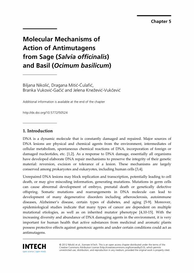
Chapter 5 © 2012 Nikolić et al., licensee InTech. This is an open access chapter distributed under the terms of the Creative Commons Attribution License (http://creativecommons.org/licenses/by/3.0), which permits unrestricted use, distribution, and reproduction in any medium, provided the original work is properly cited. Molecular Mechanisms of Action of Antimutagens from Sage (Salvia officinalis) and Basil (Ocimum basilicum) Biljana Nikolić, Dragana Mitić-Ćulafić, Branka Vuković-Gačić and Jelena Knežević-Vukčević Additional information is available at the end of the chapter http://dx.doi.org/10.5772/50524 1. Introduction DNA is a dynamic molecule that is constantly damaged and repaired. Major sources of DNA lesions are physical and chemical agents from the environment, intermediates of cellular metabolism, spontaneous chemical reactions of DNA, incorporation of foreign or damaged nucleotides, etc. [1,2]. As a response to DNA damage, essentially all organisms have developed elaborate DNA repair mechanisms to preserve the integrity of their genetic material: reversion, excision or tolerance of a lesion. These mechanisms are largely conserved among prokaryotes and eukaryotes, including human cells [3,4]. Unrepaired DNA lesions may block replication and transcription, potentially leading to cell death, or may give miscoding information, generating mutations. Mutations in germ cells can cause abnormal development of embryo, prenatal death or genetically defective offspring. Somatic mutations and rearrangements in DNA molecule can lead to development of many degenerative disorders including atherosclerosis, autoimmune diseases, Alzheimer’s disease, certain types of diabetes, and aging [5-9]. Moreover, epidemiological studies indicate that many types of cancer are dependent on multiple mutational etiologies, as well as on inherited mutator phenotype [4,10-15]. With the increasing diversity and abundance of DNA damaging agents in the environment, it is very important for human health that active substances from medicinal and aromatic plants possess protective effects against genotoxic agents and under certain conditions could act as antimutagens.
Transcript of Molecular Mechanisms of Action of Antimutagens -...
Chapter 5
© 2012 Nikolić et al., licensee InTech. This is an open access chapter distributed under the terms of the Creative Commons Attribution License (http://creativecommons.org/licenses/by/3.0), which permits unrestricted use, distribution, and reproduction in any medium, provided the original work is properly cited.
Molecular Mechanisms of Action of Antimutagens from Sage (Salvia officinalis) and Basil (Ocimum basilicum)
Biljana Nikolić, Dragana Mitić-Ćulafić, Branka Vuković-Gačić and Jelena Knežević-Vukčević
Additional information is available at the end of the chapter
http://dx.doi.org/10.5772/50524
1. Introduction
DNA is a dynamic molecule that is constantly damaged and repaired. Major sources of DNA lesions are physical and chemical agents from the environment, intermediates of cellular metabolism, spontaneous chemical reactions of DNA, incorporation of foreign or damaged nucleotides, etc. [1,2]. As a response to DNA damage, essentially all organisms have developed elaborate DNA repair mechanisms to preserve the integrity of their genetic material: reversion, excision or tolerance of a lesion. These mechanisms are largely conserved among prokaryotes and eukaryotes, including human cells [3,4].
Unrepaired DNA lesions may block replication and transcription, potentially leading to cell death, or may give miscoding information, generating mutations. Mutations in germ cells can cause abnormal development of embryo, prenatal death or genetically defective offspring. Somatic mutations and rearrangements in DNA molecule can lead to development of many degenerative disorders including atherosclerosis, autoimmune diseases, Alzheimer’s disease, certain types of diabetes, and aging [5-9]. Moreover, epidemiological studies indicate that many types of cancer are dependent on multiple mutational etiologies, as well as on inherited mutator phenotype [4,10-15]. With the increasing diversity and abundance of DNA damaging agents in the environment, it is very important for human health that active substances from medicinal and aromatic plants possess protective effects against genotoxic agents and under certain conditions could act as antimutagens.
Mutagenesis 86
2. Antimutagens
In order to protect human health, a relatively new area of research, designated as antimutagenesis and anticarcinogenesis, is continuously developing. The aim of antimutagenesis studies is to identify natural substances with antigenotoxic and antimutagenic potential and to determine the cellular and molecular mechanisms of their action. Possible application of plant antimutagens is in development of dietary and pharmaceutical supplements useful in primary prevention of mutation related diseases, including cancer.
Different prokaryotic and eukaryotic tests, routinely used to detect environmental mutagens and carcinogens, are suitably adapted for identifying agents with antigenotoxic, antimutagenic and anticarcinogenic potential, as well as for elucidating the mechanisms of their action. Due to rapidity and low costs, bacterial short-term tests are recommended to provide preliminary, but considerable information about cellular mechanisms of antimutagenesis. In combination with mammalian enzymes, they can provide information about the kind of metabolic activation or detoxification that an agent may undergo in vivo. However, for obvious reasons, bacterial short-term tests can not replace the antimutagenicity/antigenotoxicity studies in mammalian cells and in vivo, in order to identify mechanisms possibly relevant for human protection [16-19].
After several decades of research, antimutagenic effect of many naturally occurring compounds extracted from plants has been well established in bacteria and mammalian cells [20,21]. However, due to diversity of DNA lesions and the complexity of DNA repair pathways it is difficult to identify the processes involved in antimutagenesis. Antimutagens may be effective against single mutagen or a class of mutagens, may act by multiple, sometimes strictly interconnected or partially overlapping mechanisms, may be even mutagenic at certain concentrations or in certain test systems, which implies a discriminative approach in antimutagenesis studies, as well as careful interpretation of the results [22].
According to Kada et al. [23] antimutagens are placed in two major groups: desmutagens and bioantimutagens. Desmutagens are agents which prevent the formation of premutagenic lesions, while bioantimutagens prevent processing of premutagenic lesions into mutations by modulating DNA replication and repair. A revised and updated classification of antimutagens and anticarcinogenesis was given several times by different authors [18,19,24]. The classification took into consideration the multiple phases involved in the pathogenesis of cancer and other mutation related diseases. It analyzed first the inhibition of mutations and of cancer initiation, either extracellularly or inside the cells, and then the mechanisms interfering with promotion, progression, invasion and metastasis. A modified scheme incorporated possible points for intervention in primary, secondary and tertiary prevention.
Extensive search for natural compounds with antimutagenic effect often pointed at terpenes, a class of substances abundantly found in fruits, vegetables, and aromatic and medicinal plants. They are biosynthetically derived from isoprene units (C5H8) which may be linked to form monoterpenes (C10), sesquiterpenes (C15), diterpenes (C20), triterpenes (C30), tetraterpenes (C40), and polyterpenes. Terpenes exist as hydrocarbons or have oxygen-
Molecular Mechanisms of Action of Antimutagens from Sage (Salvia officinalis) and Basil (Ocimum basilicum) 87
containing substituents, such as hydroxyl, carbonyl, ketone, or aldehide groups; the latter usually are referred to as terpenoids. Both in vitro tests and epidemiological studies suggest that many dietary monoterpenes (including monoterpenoids) exert antimutagenic properties and could be helpful in the prevention and therapy of cancers [25-27].
The research efforts of our group have been focused on detection of antimutagenic properties of medicinal and aromatic plants of our region. In our initial search we screened crude extracts obtained from plants frequently used in our traditional medicine: sage (Salvia officinalis L.), lime-tree (Tilia chordata Mill.), mint (Mentha piperita L.), nettle (Urtica dioica L.), camomile (Matricaria chammomilla L.), aloe (Aloe arborescens L.), thyme (Thymus serpyllum L.), St. John’s wort (Hypericum perforatum L.) and sweet basil (Ocimum basilicum L.). Analysis of the obtained data showed heterogeneous responses, depending on the extract, concentration applied, genetic background and end-point monitored. Comparison of obtained data promoted St. John’s wort, mint, sweet basil and sage as potential source of antimutagens [28]. In further study, we focused our attention on antimutagenic effect of sage and sweet basil.
3. Medical properties of sage and basil
Salvia and Ocimum are genera of the family Lamiaceae consisting of about 900 and 35 species, respectively. S. officinalis and O. basilicum are employed as folklore remedy for a wide spectrum of ailments in many traditional medicines, including ours. Furthermore, the latter is irreplaceable spice of many national cuisines. Numerous biological activities of different extracts of Salvia and Ocimum species have been described, including antimicrobial, anti-inflammatory, antioxidative, antidiarrheal, blood-sugar lowering, immunomodulatory, a nervous system stimulatory, spasmolytic, and cholinergic binding [29-40]. Several reports also indicate antigenotoxic and chemopreventive activities of different extracts from Salvia and Ocimum species [41-45].
4. The strategy and assays for antimutagenesis study
In order to investigate the antimutagenic potential of plant extracts, we constructed and validated a new Escherichia coli K12 assay system, specially designed for detection of antimutagens and elucidation of molecular mechanisms of antimutagenesis [46,47]. We used this assay along with appropriatelly modified standard mutagenicity tests (Salmonella/microsome, E. coli WP2 and S. cerevisiae D7), to determine the antimutagenic potential, and applied comet assay for measuring the effect of antimutagen on mutagen induced DNA damage and repair. In all tests antimutagenic potential was determined in the range of non-toxic concentrations.
4.1. E. coli assay for bioantimutagens
The bacterial assay is composed of four tests measuring different end-points at the DNA level: spontaneous and induced mutagenesis in different genetic backgrounds, SOS induction and homologous recombination. To evaluate the effect on spontaneous and

Mutagenesis 88
induced mutagenesis we first use reversion test on repair proficient strain SY252, constructed in our laboratory (Table 1). The strain contains an ochre mutation in the argE3 gene, which can revert to prototrophy by base substitutions at the site of mutation or at specific suppressor loci [48]. We initially chose UV-irradiation (254 nm) to induce mutations for several reasons: (i) it mainly induces base substitutions [49] which can be detected in SY252; (ii) it shares cellular mechanisms of mutation avoidance (nucleotide excision and post-replication recombination repair) and mutation fixation (translesion error-prone replication mediated by SOS regulated UmuD’C complex) with many chemical mutagens and carcinogens [50,51] (iii) possible chemical interaction between mutagen and antimutagen is prevented, which is essential for detection of bioantimutagens. UV-mimetic mutagen 4-nitroquinoline-1-oxide (4NQO) [52,53] was recently used to provide comparison with the results obtained with UV. The possibility for chemical interaction between 4NQO and antimutagen was avoided in experimental procedure.
Since nucleotide excision repair (NER) is the major error-free pathway involved in repair of pyrimidine dimers and bulky DNA lesions such as 4NQO-DNA adducts [54], we also analyze potential of antimutagen to reduce mutagenesis in NER deficient uvrA counterpart of SY252. Comparison of results obtained in repair proficient and NER deficient strains indicates if observed antimutagenic effect involves increased capacity for NER.
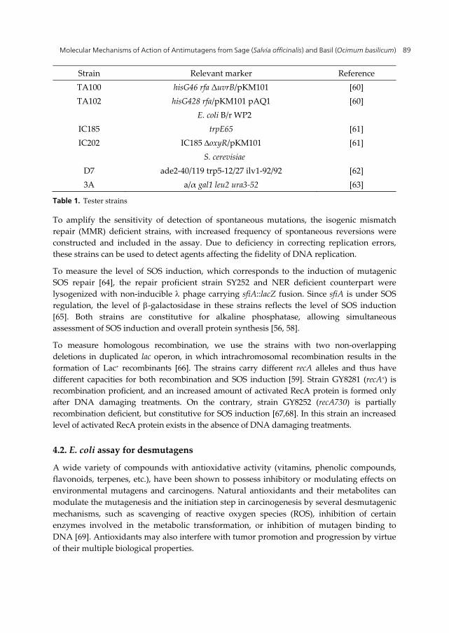
Strain Relevant marker Reference
E. coli K12 SY252 argE3 [55] IB101 SY252 mutH471::Tn5 [46] IB103 SY252 mutS215::Tn10 [46] IB105 SY252 uvrA::Tn10 [56] IB106 SY252 mutT::Tn5 [57]
IB111 SY252 [p(sfiA::lacZ)cIind1] PHOC [56] IB127 IB111 uvrA::Tn10 [58]
IB122 SY252/pAJ47 [57] IB123 IB101/pAJ47 [57]
GY7066 lacMS286 80dIIlacBK1 recA306 srl::Tn10 [59] GY8281 GY7066/miniFrecA+ [59] GY8252 GY7066/miniFrecA730 [59]
S. typhimurium TA98 hisD3052 rfa ΔuvrB/pKM101 [60]
Molecular Mechanisms of Action of Antimutagens from Sage (Salvia officinalis) and Basil (Ocimum basilicum) 89
Strain Relevant marker Reference TA100 hisG46 rfa ΔuvrB/pKM101 [60] TA102 hisG428 rfa/pKM101 pAQ1 [60]
E. coli B/r WP2 IC185 trpE65 [61] IC202 IC185 oxyR/pKM101 [61]
S. cerevisiae D7 ade2-40/119 trp5-12/27 ilv1-92/92 [62] 3A a/α gal1 leu2 ura3-52 [63]
Table 1. Tester strains
To amplify the sensitivity of detection of spontaneous mutations, the isogenic mismatch repair (MMR) deficient strains, with increased frequency of spontaneous reversions were constructed and included in the assay. Due to deficiency in correcting replication errors, these strains can be used to detect agents affecting the fidelity of DNA replication.
To measure the level of SOS induction, which corresponds to the induction of mutagenic SOS repair [64], the repair proficient strain SY252 and NER deficient counterpart were lysogenized with non-inducible phage carrying sfiA::lacZ fusion. Since sfiA is under SOS regulation, the level of -galactosidase in these strains reflects the level of SOS induction [65]. Both strains are constitutive for alkaline phosphatase, allowing simultaneous assessment of SOS induction and overall protein synthesis [56, 58].
To measure homologous recombination, we use the strains with two non-overlapping deletions in duplicated lac operon, in which intrachromosomal recombination results in the formation of Lac+ recombinants [66]. The strains carry different recA alleles and thus have different capacities for both recombination and SOS induction [59]. Strain GY8281 (recA+) is recombination proficient, and an increased amount of activated RecA protein is formed only after DNA damaging treatments. On the contrary, strain GY8252 (recA730) is partially recombination deficient, but constitutive for SOS induction [67,68]. In this strain an increased level of activated RecA protein exists in the absence of DNA damaging treatments.
4.2. E. coli assay for desmutagens
A wide variety of compounds with antioxidative activity (vitamins, phenolic compounds, flavonoids, terpenes, etc.), have been shown to possess inhibitory or modulating effects on environmental mutagens and carcinogens. Natural antioxidants and their metabolites can modulate the mutagenesis and the initiation step in carcinogenesis by several desmutagenic mechanisms, such as scavenging of reactive oxygen species (ROS), inhibition of certain enzymes involved in the metabolic transformation, or inhibition of mutagen binding to DNA [69]. Antioxidants may also interfere with tumor promotion and progression by virtue of their multiple biological properties.
Mutagenesis 90
In order to identify antimutagens with antioxidative properties, we modified our E. coli K12 assay for bioantimutagens. In repair proficient strain SY252 mutations are induced by t-butyl hydroperoxide (t-BOOH), a latent donor of ROS, which promotes oxidative damage of DNA [70]. Since DNA damage induced by t-BOOH cause both transitions and transversions of AT base pairs, it can be used to increase argE3 → Arg+ reversions. The mutT strain was constructed for the assay in order to evaluate protective capacity of antioxidants against formation of oxidatively damaged bases in the cell pool [57]. Due to deficiency in removing 8-oxo-G, mutT strains have high frequency of A:8-oxo-G mispairs and show increased level of spontaneous AT→CG transversions [71]. During validation of the test we determined that the frequency of argE3 → Arg+ reversions is significantly increased in IB106 strain [47]. Since MMR is additionally involved in the repair of mispairs between normal and oxidized bases [72], MMR deficient strains from the assay for bioantimutagens are also included.
Considering that microsatellite instability (MSI) could be induced by oxidative DNA damage, by MMR deficiency or in many forms of cancer [73-75], we also designed the test for detection of MSI. The repair proficient and MMR deficient strains were transformed with the low copy number plasmid pAJ47 (Table 1). This plasmid contains dinucleotide repeats (CA)11 placed out-of-frame within the coding region of β–lactamase gene. Cells harbouring plasmid are sensitive to β–lactam antibiotics, such as carbenicillin. Microsatellite sequence is a +2 frame construct and the mutation that restores the reading frame and provides resistance to carbenicillin is a 2 bp deletion. Repair-proficient strain is used for screening of t-BOOH-induced MSI, while mutH strain is used for monitoring of spontaneous MSI [57].
4.3. Other reversion tests
Preliminary screening of plant extracts included evaluation of possible mutagenic effects by standard Salmonella/microsome (Ames) test, recommended by OECD [76]. The mutagenicity was determined in strains TA98, TA100 and TA102 (Table 1) in plate incorporation assay [60]. For evaluation of antimutagenic effect, the tester strain and the mutagen were selected according to the mutational event monitored.
WP2 mutagenicity test, especially recommended for monitoring of oxidative mutagenesis [76], is used along with E. coli K12 assay to detect antimutagenic potential of plant extracts based on antioxidative properties. Test is performed on both OxyR proficient IC185 and OxyR deficient IC202 strains [61]. The OxyR protein is a redox-sensitive transcriptional activator of genes encoding antioxidative enzymes: catalase-hydroperoxidase I, alkyl hydroperoxide reductase and glutathione reductase [77]. Mutants in oxyR are deficient in inducible expression of antioxidant enzymes and thus very sensitive for detection of oxidative mutagens and antimutagenic effect of antioxidants. Comparison of results obtained in OxyR+ and OxyR- strains indicates if observed antimutagenic effect is based on antioxidative properties.
To obtain preliminary information about mutagenic and antimutagenic potential of plant extracts in eukaryotic cells we used the S. cerevisiae diploid strain D7 [62], which permits simultaneous evaluation of point mutations (ilv1-92→Ilv+), mitotic crossing over (ade2→Ade+) and mitotic gene conversion (trp5→Trp+).
Molecular Mechanisms of Action of Antimutagens from Sage (Salvia officinalis) and Basil (Ocimum basilicum) 91
4.4. Comet assay – direct monitoring of DNA damage
The alkaline comet assay was used in order to monitor the effect of plant extracts on formation and repair of DNA lesions induced by a mutagen. The comet assay or single-cell gel electrophoresis (SCGE) is a simple method for measuring DNA strand breaks, mostly in eukaryotic cells. It has become one of the standard methods for assessing DNA damage and found applications in different fields including genotoxicity/antigenotoxicity testing, human biomonitoring and molecular epidemiology, ecogenotoxicology, as well in fundamental research of DNA repair [78]. The assay was performed on repair proficient Vero cells, originated from the kidney of African green monkey (ECACC No: 88020401), and on two human cell lines: hepatoma HepG2 (ATCC HB-8065) and B lymphoid NC-NC cells (DSMZ ACC120). We also used the modified version of alkaline comet assay on S. cerevisiae 3A strain, designed by Miloshev et al. [63].
5. Antimutagenic potential of sage
Given the possibility to obtain large quantities of chemically characterized extracts from different varieties of sage, we focused our research on this plant. We screened the fractionated extracts of two varieties of sage. Wild sage originated from Pelješac, Croatia, while cultivated sage (variety D-70) was selected and grown at the Institute for Hop, Sorghum and Medicinal Plants, Bački Petrovac, Serbia. The most striking difference between the two plants is the composition of essential oils (EO). While both plants contain α+β Thujone (Thu), Camphor (Cam) is present only in traces in the wild sage, whereas it represents 1/5 of the monoterpenes in variety D-70 [79]. Extract 1 (E1) was prepared from the cultivated sage, collected during the flowering period, dried and subjected to ethanolic extraction as the whole herb. Extract 2 (E2) was prepared from the same herb as Extracts 1, but it was steam distilled prior to ethanolic extraction to remove EO. Extract 3 (E3) was prepared from the wild sage, treated in the same way as Extract 1. All extracts (E1-E3) were re-extracted by CO2 at different pressure (200, 300, 400, 500 bar), resulting in the extracts (E/2-E/5) with high content of terpenes. The extracts obtained at low CO2 pressure (200, 300 bar) contained mainly monoterpenes, while extraction at higher CO2 pressure resulted in the increase of relative proportion of high molecular weight terpenes. Preliminary determination of antioxidative properties, performed with lipid peroxidation test, indicated significant antioxidative activity of the extracts obtained at high CO2 pressure, which was attributed to diterpenes, such as 6-methyl-ether-γ-lactone carnosic acid and rosmanol-9-ethyl ether [79,80].
5.1. Desmutagenic potential of sage extracts
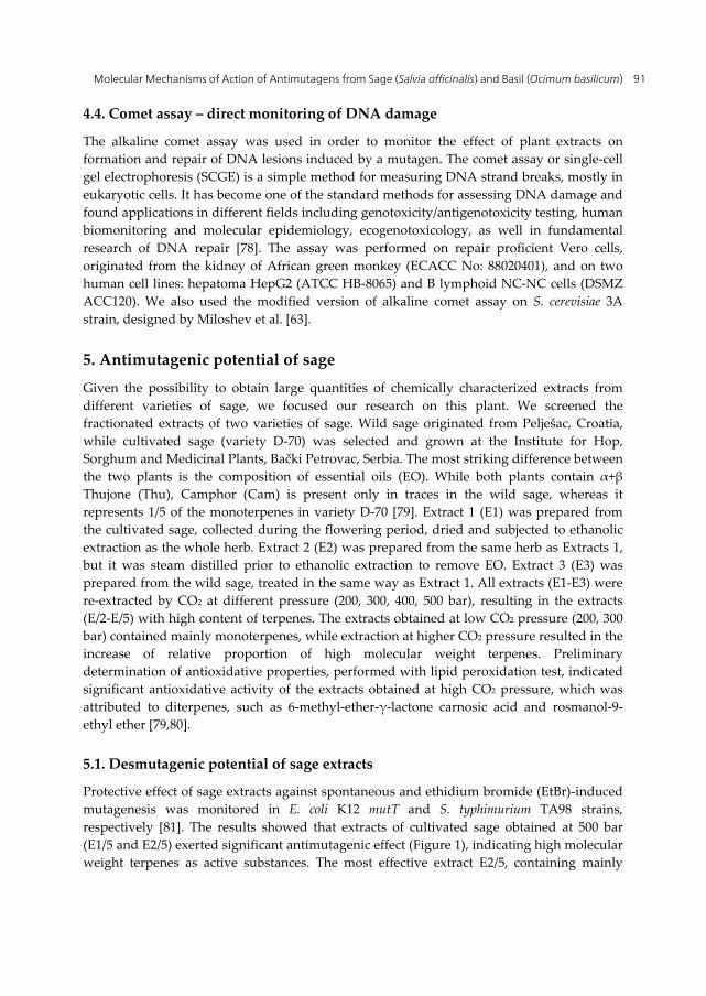
Protective effect of sage extracts against spontaneous and ethidium bromide (EtBr)-induced mutagenesis was monitored in E. coli K12 mutT and S. typhimurium TA98 strains, respectively [81]. The results showed that extracts of cultivated sage obtained at 500 bar (E1/5 and E2/5) exerted significant antimutagenic effect (Figure 1), indicating high molecular weight terpenes as active substances. The most effective extract E2/5, containing mainly
Mutagenesis 92
rosmanol-9-ethyl ether (40%), was further investigated in order to elucidate the molecular mechanism of antimutagenicity. Different experimental procedures were applied: A – co-incubation of mutagen, extract and S9 fraction, followed by addition of bacteria and plating; B – pre-incubation of mutagen and S9, followed by addition of the extract, incubation, and final addition of bacteria and plating; C- pre-incubation of mutagen, S9 and bacteria, followed by removal of mutagen and S9, addition of the extract, and plating. The strongest inhibition was obtained when the mutagen and E2/5 were pre-incubated with S9 (procedure A), indicating that the main antimutagenic mechanism was inhibition of metabolic activation of EtBr. Extract E2/5 also moderately reduced spontaneous mutations (37%) in mutT strain.
Figure 1. Effect of sage extracts against EtBr –induced mutagenesis in TA98 strain
5.2. Bioantimutagenic potential of sage
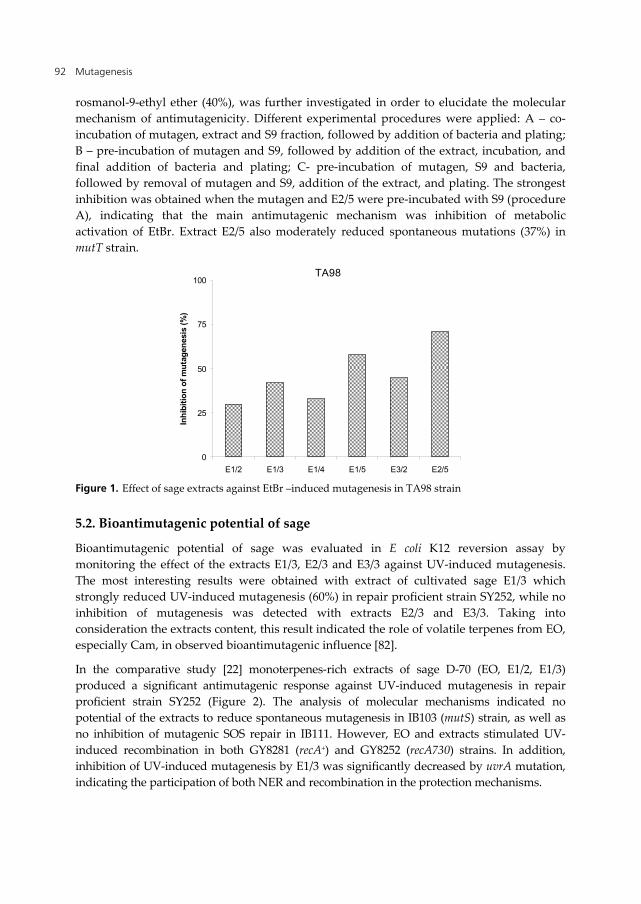
Bioantimutagenic potential of sage was evaluated in E coli K12 reversion assay by monitoring the effect of the extracts E1/3, E2/3 and E3/3 against UV-induced mutagenesis. The most interesting results were obtained with extract of cultivated sage E1/3 which strongly reduced UV-induced mutagenesis (60%) in repair proficient strain SY252, while no inhibition of mutagenesis was detected with extracts E2/3 and E3/3. Taking into consideration the extracts content, this result indicated the role of volatile terpenes from EO, especially Cam, in observed bioantimutagenic influence [82].
In the comparative study [22] monoterpenes-rich extracts of sage D-70 (EO, E1/2, E1/3) produced a significant antimutagenic response against UV-induced mutagenesis in repair proficient strain SY252 (Figure 2). The analysis of molecular mechanisms indicated no potential of the extracts to reduce spontaneous mutagenesis in IB103 (mutS) strain, as well as no inhibition of mutagenic SOS repair in IB111. However, EO and extracts stimulated UV-induced recombination in both GY8281 (recA+) and GY8252 (recA730) strains. In addition, inhibition of UV-induced mutagenesis by E1/3 was significantly decreased by uvrA mutation, indicating the participation of both NER and recombination in the protection mechanisms.
0
25
50
75
100
E1/2 E1/3 E1/4 E1/5 E3/2 E2/5
Inh
ibit
ion
of
mu
tag
enes
is (
%)
TA98
Molecular Mechanisms of Action of Antimutagens from Sage (Salvia officinalis) and Basil (Ocimum basilicum) 93
Figure 2. Bioantimutagenic effect of extracts of sage D-70
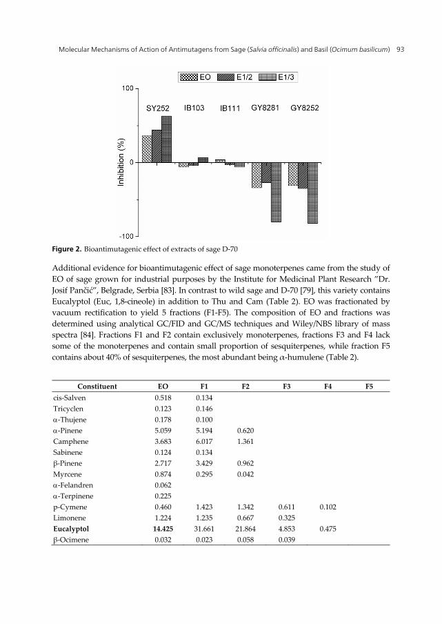
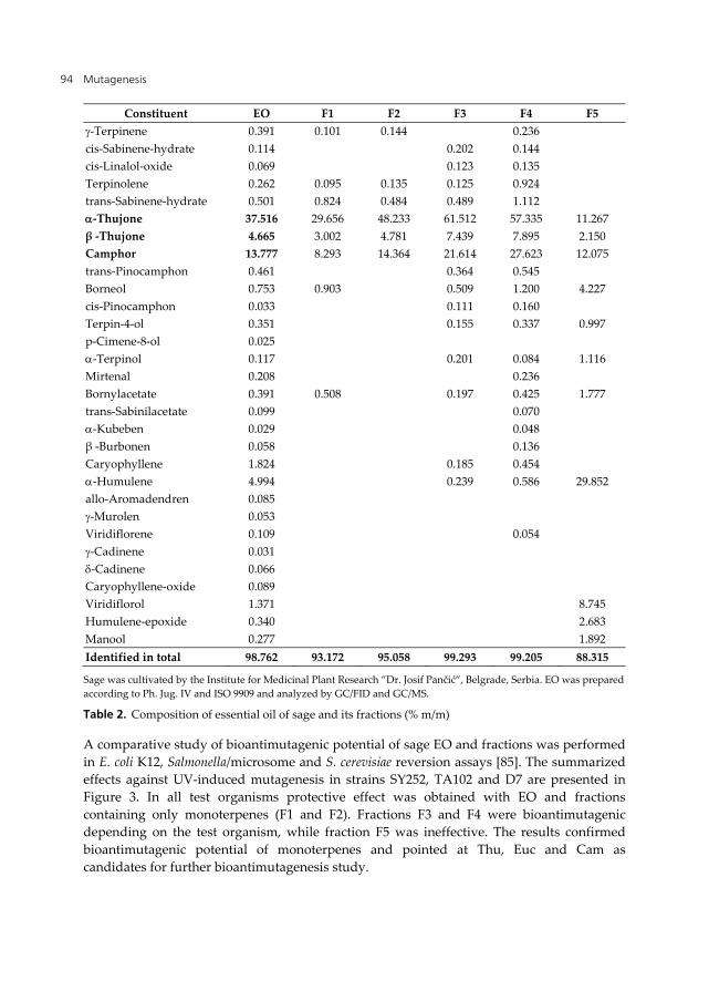
Additional evidence for bioantimutagenic effect of sage monoterpenes came from the study of EO of sage grown for industrial purposes by the Institute for Medicinal Plant Research ”Dr. Josif Pančić”, Belgrade, Serbia [83]. In contrast to wild sage and D-70 [79], this variety contains Eucalyptol (Euc, 1,8-cineole) in addition to Thu and Cam (Table 2). EO was fractionated by vacuum rectification to yield 5 fractions (F1-F5). The composition of EO and fractions was determined using analytical GC/FID and GC/MS techniques and Wiley/NBS library of mass spectra [84]. Fractions F1 and F2 contain exclusively monoterpenes, fractions F3 and F4 lack some of the monoterpenes and contain small proportion of sesquiterpenes, while fraction F5 contains about 40% of sesquiterpenes, the most abundant being α-humulene (Table 2).
Constituent EO F1 F2 F3 F4 F5
cis-Salven 0.518 0.134 Tricyclen 0.123 0.146 -Thujene 0.178 0.100 -Pinene 5.059 5.194 0.620 Camphene 3.683 6.017 1.361 Sabinene 0.124 0.134 β-Pinene 2.717 3.429 0.962 Myrcene 0.874 0.295 0.042 -Felandren 0.062 -Terpinene 0.225 p-Cymene 0.460 1.423 1.342 0.611 0.102 Limonene 1.224 1.235 0.667 0.325 Eucalyptol 14.425 31.661 21.864 4.853 0.475 β-Ocimene 0.032 0.023 0.058 0.039
Mutagenesis 94
Constituent EO F1 F2 F3 F4 F5 -Terpinene 0.391 0.101 0.144 0.236 cis-Sabinene-hydrate 0.114 0.202 0.144 cis-Linalol-oxide 0.069 0.123 0.135 Terpinolene 0.262 0.095 0.135 0.125 0.924 trans-Sabinene-hydrate 0.501 0.824 0.484 0.489 1.112 -Thujone 37.516 29.656 48.233 61.512 57.335 11.267 β -Thujone 4.665 3.002 4.781 7.439 7.895 2.150 Camphor 13.777 8.293 14.364 21.614 27.623 12.075 trans-Pinocamphon 0.461 0.364 0.545 Borneol 0.753 0.903 0.509 1.200 4.227 cis-Pinocamphon 0.033 0.111 0.160 Terpin-4-ol 0.351 0.155 0.337 0.997 p-Cimene-8-ol 0.025 -Terpinol 0.117 0.201 0.084 1.116 Mirtenal 0.208 0.236 Bornylacetate 0.391 0.508 0.197 0.425 1.777 trans-Sabinilacetate 0.099 0.070 -Kubeben 0.029 0.048 β -Burbonen 0.058 0.136 Caryophyllene 1.824 0.185 0.454 -Humulene 4.994 0.239 0.586 29.852 allo-Aromadendren 0.085 -Murolen 0.053 Viridiflorene 0.109 0.054 -Cadinene 0.031 -Cadinene 0.066 Caryophyllene-oxide 0.089 Viridiflorol 1.371 8.745 Humulene-epoxide 0.340 2.683 Manool 0.277 1.892 Identified in total 98.762 93.172 95.058 99.293 99.205 88.315
Sage was cultivated by the Institute for Medicinal Plant Research ”Dr. Josif Pančić”, Belgrade, Serbia. EO was prepared according to Ph. Jug. IV and ISO 9909 and analyzed by GC/FID and GC/MS.
Table 2. Composition of essential oil of sage and its fractions (% m/m)
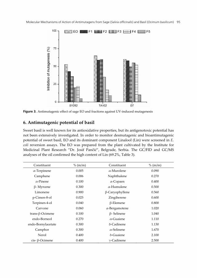
A comparative study of bioantimutagenic potential of sage EO and fractions was performed in E. coli K12, Salmonella/microsome and S. cerevisiae reversion assays [85]. The summarized effects against UV-induced mutagenesis in strains SY252, TA102 and D7 are presented in Figure 3. In all test organisms protective effect was obtained with EO and fractions containing only monoterpenes (F1 and F2). Fractions F3 and F4 were bioantimutagenic depending on the test organism, while fraction F5 was ineffective. The results confirmed bioantimutagenic potential of monoterpenes and pointed at Thu, Euc and Cam as candidates for further bioantimutagenesis study.
Molecular Mechanisms of Action of Antimutagens from Sage (Salvia officinalis) and Basil (Ocimum basilicum) 95
Figure 3. Antimutagenic effect of sage EO and fractions against UV-induced mutagenesis
6. Antimutagenic potential of basil
Sweet basil is well known for its antioxidative properties, but its antigenotoxic potential has not been extensively investigated. In order to monitor desmutagenic and bioantimutagenic potential of sweet basil, EO and its dominant component Linalool (Lin) were screened in E. coli reversion assays. The EO was prepared from the plant cultivated by the Institute for Medicinal Plant Research ”Dr. Josif Pančić”, Belgrade, Serbia. The GC/FID and GC/MS analyses of the oil confirmed the high content of Lin (69.2%, Table 3).
Constituent % (m/m) Constituent % (m/m)
α-Terpinene 0.005 α-Murolene 0.090 Camphene 0.006 Naphthalene 0.270 α-Pinene 0.100 α-Copaen 0.400 β- Myrcene 0.300 α-Humulene 0.500 Limonene 0.900 β-Caryophyllene 0.560
p-Cimen-8-ol 0.025 Zingiberene 0.600 Terpinen-4-ol 0.040 β-Elemene 0.800
Carvone 0.060 α-Bergamotene 1.020 trans-β-Ocimene 0.100 β- Selinene 1.040
endo-Borneol 0.270 α-Guaiene 1.110 endo-Bornylacetate 0.300 δ-Cadinene 1.130
Camphor 0.300 α-Selinene 1.670 Nerol 0.400 δ-Guaiene 2.100
cis- β-Ocimene 0.400 γ-Cadinene 2.500
Mutagenesis 96
Constituent % (m/m) Constituent % (m/m) α-Terpinolene 0.400 Nerodiol 0.110 Thiogeraniol 0.560 cis-Farnesol 0.180 α-Terpineol 0.700 trans-Murolol 0.430 Eucalyptol 0.800 α-Cadinol 2.560 Geraniol 1.900 Linalool 69.200 Eugenol 1.400 Estragole 2.400
β-Burbonene 0.080 Identified in total 97.716%
Basil was cultivated by the Institute for Medicinal Plant Research ”Dr. Josif Pančić” Belgrade, Serbia. EO was prepared according to Ph. Jug. IV and ISO 9909, and analyzed by GC/FID and GC/MS.
Table 3. Composition of essential oil of basil (Ocimum basilicum L.)
6.1. Desmutagenic potential of basil
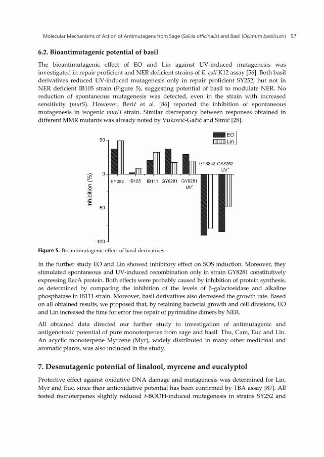
The desmutagenic effect of EO of basil and Lin was monitored in E. coli K12 assay system with t-BOOH as a mutagen. Strong reduction was observed in repair proficient SY252 for both EO and Lin (Figure 4). Moreover, the spontaneous base substitutions in MMR deficient strain IB101 (mutH) were slightly decreased by EO, and moderately by Lin. Both basil derivatives also moderately decreased t-BOOH-induced MSI in repair proficient strain IB122 and spontaneous MSI in its MMR deficient counterpart IB123 (mutH). Antimutagenic potential determined in all tests was tentatively attributed to antioxidative properties and indicated Lin as principal active substance. The confirmation of proposed mechanism was obtained in oxyR deficient IC202 strain, where reduction of t-BOOH-induced mutagenesis was 72% and 70%, for EO and Lin, respectively [86].
Figure 4. Antimutagenic effect of basil derivatives against t-BOOH
Molecular Mechanisms of Action of Antimutagens from Sage (Salvia officinalis) and Basil (Ocimum basilicum) 97
6.2. Bioantimutagenic potential of basil
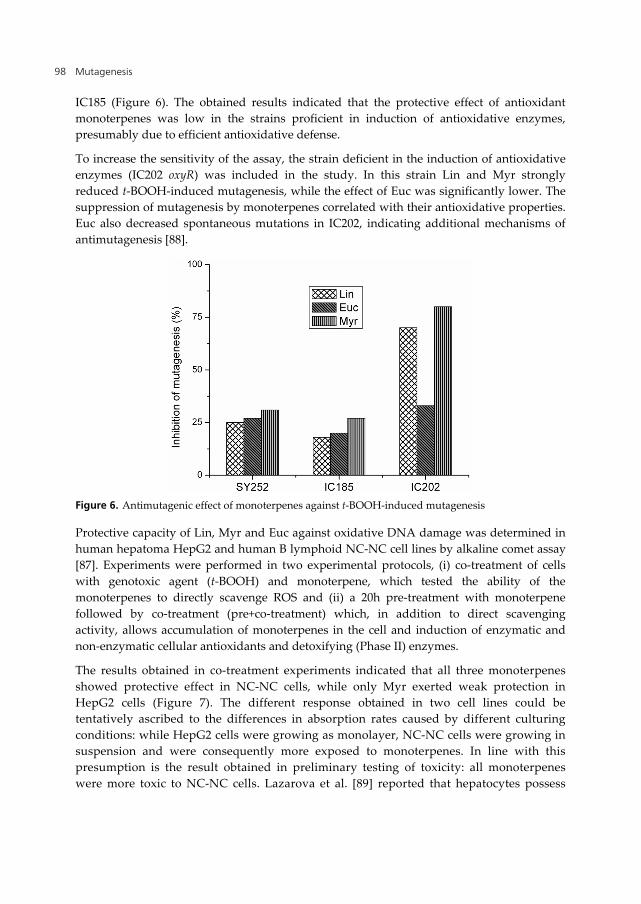
The bioantimutagenic effect of EO and Lin against UV-induced mutagenesis was investigated in repair proficient and NER deficient strains of E. coli K12 assay [56]. Both basil derivatives reduced UV-induced mutagenesis only in repair proficient SY252, but not in NER deficient IB105 strain (Figure 5), suggesting potential of basil to modulate NER. No reduction of spontaneous mutagenesis was detected, even in the strain with increased sensitivity (mutS). However, Berić et al. [86] reported the inhibition of spontaneous mutagenesis in isogenic mutH strain. Similar discrepancy between responses obtained in different MMR mutants was already noted by Vuković-Gačić and Simić [28].
Figure 5. Bioantimutagenic effect of basil derivatives
In the further study EO and Lin showed inhibitory effect on SOS induction. Moreover, they stimulated spontaneous and UV-induced recombination only in strain GY8281 constitutively expressing RecA protein. Both effects were probably caused by inhibition of protein synthesis, as determined by comparing the inhibition of the levels of -galactosidase and alkaline phosphatase in IB111 strain. Moreover, basil derivatives also decreased the growth rate. Based on all obtained results, we proposed that, by retaining bacterial growth and cell divisions, EO and Lin increased the time for error free repair of pyrimidine dimers by NER.
All obtained data directed our further study to investigation of antimutagenic and antigenotoxic potential of pure monoterpenes from sage and basil: Thu, Cam, Euc and Lin. An acyclic monoterpene Myrcene (Myr), widely distributed in many other medicinal and aromatic plants, was also included in the study.
7. Desmutagenic potential of linalool, myrcene and eucalyptol
Protective effect against oxidative DNA damage and mutagenesis was determined for Lin, Myr and Euc, since their antioxidative potential has been confirmed by TBA assay [87]. All tested monoterpenes slightly reduced t-BOOH-induced mutagenesis in strains SY252 and
Mutagenesis 98
IC185 (Figure 6). The obtained results indicated that the protective effect of antioxidant monoterpenes was low in the strains proficient in induction of antioxidative enzymes, presumably due to efficient antioxidative defense.
To increase the sensitivity of the assay, the strain deficient in the induction of antioxidative enzymes (IC202 oxyR) was included in the study. In this strain Lin and Myr strongly reduced t-BOOH-induced mutagenesis, while the effect of Euc was significantly lower. The suppression of mutagenesis by monoterpenes correlated with their antioxidative properties. Euc also decreased spontaneous mutations in IC202, indicating additional mechanisms of antimutagenesis [88].
Figure 6. Antimutagenic effect of monoterpenes against t-BOOH-induced mutagenesis
Protective capacity of Lin, Myr and Euc against oxidative DNA damage was determined in human hepatoma HepG2 and human B lymphoid NC-NC cell lines by alkaline comet assay [87]. Experiments were performed in two experimental protocols, (i) co-treatment of cells with genotoxic agent (t-BOOH) and monoterpene, which tested the ability of the monoterpenes to directly scavenge ROS and (ii) a 20h pre-treatment with monoterpene followed by co-treatment (pre+co-treatment) which, in addition to direct scavenging activity, allows accumulation of monoterpenes in the cell and induction of enzymatic and non-enzymatic cellular antioxidants and detoxifying (Phase II) enzymes.
The results obtained in co-treatment experiments indicated that all three monoterpenes showed protective effect in NC-NC cells, while only Myr exerted weak protection in HepG2 cells (Figure 7). The different response obtained in two cell lines could be tentatively ascribed to the differences in absorption rates caused by different culturing conditions: while HepG2 cells were growing as monolayer, NC-NC cells were growing in suspension and were consequently more exposed to monoterpenes. In line with this presumption is the result obtained in preliminary testing of toxicity: all monoterpenes were more toxic to NC-NC cells. Lazarova et al. [89] reported that hepatocytes possess
Molecular Mechanisms of Action of Antimutagens from Sage (Salvia officinalis) and Basil (Ocimum basilicum) 99
better antioxidative defense than lymphocytes. Therefore, we could conclude that stronger protective effect was obtained in cells with reduced antioxidative capacity, similarly as in bacteria.
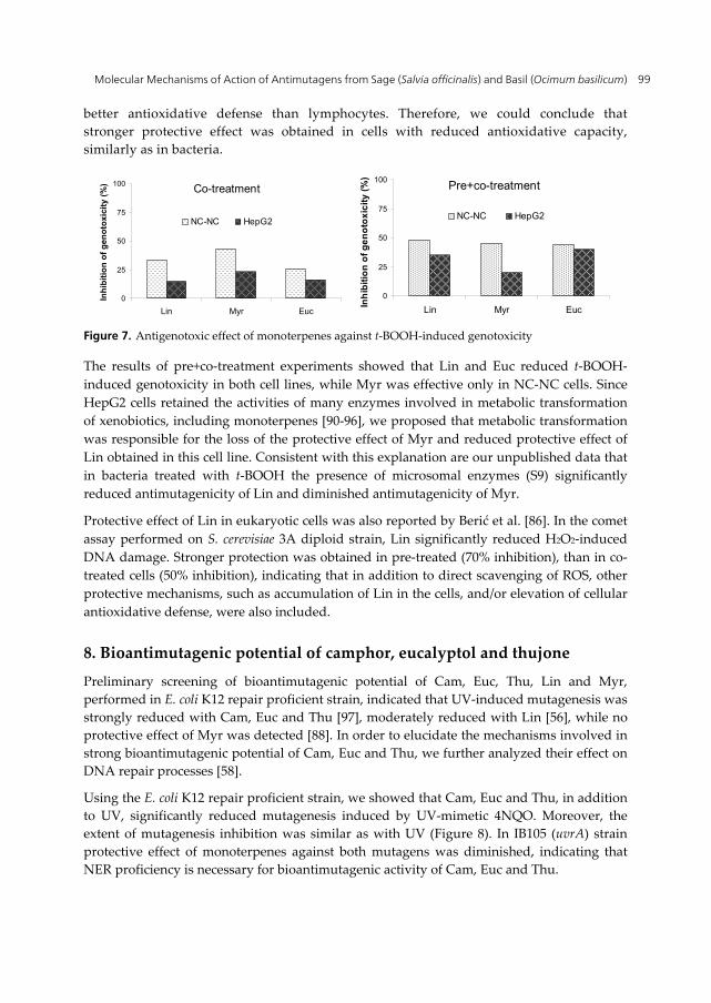
Figure 7. Antigenotoxic effect of monoterpenes against t-BOOH-induced genotoxicity
The results of pre+co-treatment experiments showed that Lin and Euc reduced t-BOOH-induced genotoxicity in both cell lines, while Myr was effective only in NC-NC cells. Since HepG2 cells retained the activities of many enzymes involved in metabolic transformation of xenobiotics, including monoterpenes [90-96], we proposed that metabolic transformation was responsible for the loss of the protective effect of Myr and reduced protective effect of Lin obtained in this cell line. Consistent with this explanation are our unpublished data that in bacteria treated with t-BOOH the presence of microsomal enzymes (S9) significantly reduced antimutagenicity of Lin and diminished antimutagenicity of Myr.
Protective effect of Lin in eukaryotic cells was also reported by Berić et al. [86]. In the comet assay performed on S. cerevisiae 3A diploid strain, Lin significantly reduced H2O2-induced DNA damage. Stronger protection was obtained in pre-treated (70% inhibition), than in co-treated cells (50% inhibition), indicating that in addition to direct scavenging of ROS, other protective mechanisms, such as accumulation of Lin in the cells, and/or elevation of cellular antioxidative defense, were also included.
8. Bioantimutagenic potential of camphor, eucalyptol and thujone
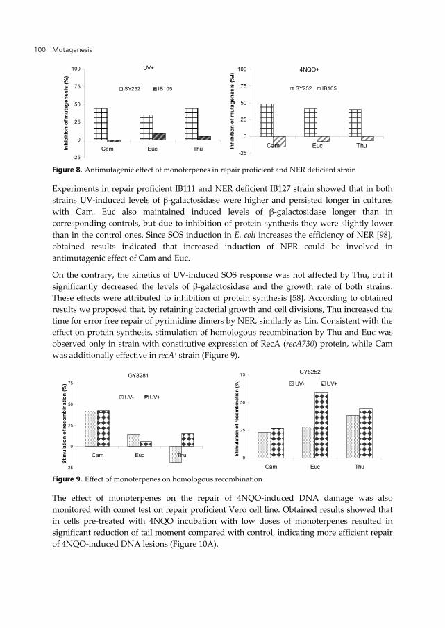
Preliminary screening of bioantimutagenic potential of Cam, Euc, Thu, Lin and Myr, performed in E. coli K12 repair proficient strain, indicated that UV-induced mutagenesis was strongly reduced with Cam, Euc and Thu [97], moderately reduced with Lin [56], while no protective effect of Myr was detected [88]. In order to elucidate the mechanisms involved in strong bioantimutagenic potential of Cam, Euc and Thu, we further analyzed their effect on DNA repair processes [58].
Using the E. coli K12 repair proficient strain, we showed that Cam, Euc and Thu, in addition to UV, significantly reduced mutagenesis induced by UV-mimetic 4NQO. Moreover, the extent of mutagenesis inhibition was similar as with UV (Figure 8). In IB105 (uvrA) strain protective effect of monoterpenes against both mutagens was diminished, indicating that NER proficiency is necessary for bioantimutagenic activity of Cam, Euc and Thu.
0
25
50
75
100
Lin Myr Euc
Inh
ibit
ion
of
gen
oto
xici
ty (
%)
NC-NC HepG2
Co-treatment
0
25
50
75
100
Lin Myr Euc
Inh
ibit
ion
of
ge
no
tox
icit
y (
%)
NC-NC HepG2
Pre+co-treatment
Mutagenesis 100
Figure 8. Antimutagenic effect of monoterpenes in repair proficient and NER deficient strain
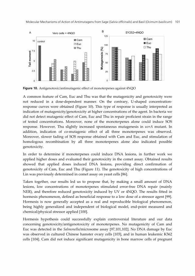
Experiments in repair proficient IB111 and NER deficient IB127 strain showed that in both strains UV-induced levels of β-galactosidase were higher and persisted longer in cultures with Cam. Euc also maintained induced levels of β-galactosidase longer than in corresponding controls, but due to inhibition of protein synthesis they were slightly lower than in the control ones. Since SOS induction in E. coli increases the efficiency of NER [98], obtained results indicated that increased induction of NER could be involved in antimutagenic effect of Cam and Euc.
On the contrary, the kinetics of UV-induced SOS response was not affected by Thu, but it significantly decreased the levels of β-galactosidase and the growth rate of both strains. These effects were attributed to inhibition of protein synthesis [58]. According to obtained results we proposed that, by retaining bacterial growth and cell divisions, Thu increased the time for error free repair of pyrimidine dimers by NER, similarly as Lin. Consistent with the effect on protein synthesis, stimulation of homologous recombination by Thu and Euc was observed only in strain with constitutive expression of RecA (recA730) protein, while Cam was additionally effective in recA+ strain (Figure 9).
Figure 9. Effect of monoterpenes on homologous recombination
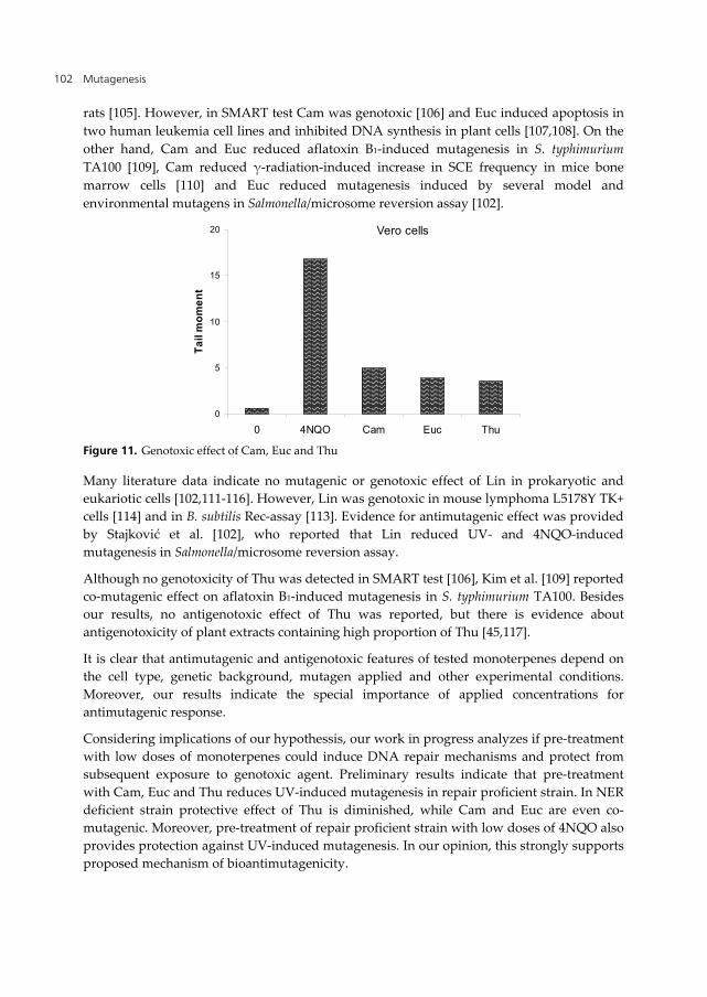
The effect of monoterpenes on the repair of 4NQO-induced DNA damage was also monitored with comet test on repair proficient Vero cell line. Obtained results showed that in cells pre-treated with 4NQO incubation with low doses of monoterpenes resulted in significant reduction of tail moment compared with control, indicating more efficient repair of 4NQO-induced DNA lesions (Figure 10A).
-25
0
25
50
75
100
Cam Euc ThuInh
ibit
ion
of
mu
tag
en
es
is (
%)
SY252 IB105
UV+
-25
0
25
50
75
100
Cam Euc ThuInh
ibit
ion
of
mu
tag
en
es
is (
%I)
SY252 IB105
4NQO+
-25
0
25
50
75
Cam Euc Thu
Sti
mu
lati
on
of
rec
om
bin
ati
on
(%
)
UV- UV+
GY8281
0
25
50
75
Cam Euc Thu
Sti
mu
lati
on
of
reco
mb
ina
tio
n (
%)
UV- UV+
GY8252
Molecular Mechanisms of Action of Antimutagens from Sage (Salvia officinalis) and Basil (Ocimum basilicum) 101
Figure 10. Antigenotoxic/antimutagenic effect of monoterpenes against 4NQO
A common feature of Cam, Euc and Thu was that the mutagenicity and genotoxicity were not reduced in a dose-dependent manner. On the contrary, U-shaped concentration-response curves were obtained (Figure 10). This type of response is usually interpreted as indication of mutagenicity/genotoxicity at higher concentrations of the agent. In bacteria we did not detect mutagenic effect of Cam, Euc and Thu in repair proficient strain in the range of tested concentrations. Moreover, none of the monoterpenes alone could induce SOS response. However, Thu slightly increased spontaneous mutagenesis in uvrA mutant. In addition, indication of co-mutagenic effect of all three monoterpenes was observed. Moreover, slower fading of SOS response obtained with Cam and Euc, and stimulation of homologous recombination by all three monoterpenes alone also indicated possible genotoxicity.
In order to determine if monoterpenes could induce DNA lesions, in further work we applied higher doses and evaluated their genotoxicity in the comet assay. Obtained results showed that applied doses induced DNA lesions, providing direct confirmation of genotoxicity of Cam, Euc and Thu (Figure 11). The genotoxicity of high concentrations of Lin was previously determined in comet assay on yeast cells [86].
Taken together, our results led us to propose that, by making a small amount of DNA lesions, low concentrations of monoterpenes stimulated error-free DNA repair (mainly NER), and therefore reduced genotoxicity induced by UV or 4NQO. The results fitted in hormesis phenomenon, defined as beneficial response to a low dose of a stressor agent [99]. Hormesis is now generally accepted as a real and reproducible biological phenomenon, being highly generalized and independent of biological model, end-point measured and chemical/physical stressor applied [100].
Hormesis hypothesis could successfully explain controversial literature and our data concerning genotoxicity/antigenotoxicity of monoterpenes. No mutagenicity of Cam and Euc was detected in the Salmonella/microsome assay [97,101,102]. No DNA damage by Euc was observed in cultured Chinese hamster ovary cells [103], and in human leukemic K562 cells [104]. Cam did not induce significant mutagenicity in bone marrow cells of pregnant
0
2.5
5
7.5
10
Ta
il m
om
en
t
Cam
Euc
Thu
Vero cells + 4NQO
control
A
0
25
50
75
100
125
Mu
tag
en
es
is (
%)
Cam
Euc
Thu
SY252+4NQOB
Mutagenesis 102
rats [105]. However, in SMART test Cam was genotoxic [106] and Euc induced apoptosis in two human leukemia cell lines and inhibited DNA synthesis in plant cells [107,108]. On the other hand, Cam and Euc reduced aflatoxin B1-induced mutagenesis in S. typhimurium TA100 [109], Cam reduced γ-radiation-induced increase in SCE frequency in mice bone marrow cells [110] and Euc reduced mutagenesis induced by several model and environmental mutagens in Salmonella/microsome reversion assay [102].
Figure 11. Genotoxic effect of Cam, Euc and Thu
Many literature data indicate no mutagenic or genotoxic effect of Lin in prokaryotic and eukariotic cells [102,111-116]. However, Lin was genotoxic in mouse lymphoma L5178Y TK+ cells [114] and in B. subtilis Rec-assay [113]. Evidence for antimutagenic effect was provided by Stajković et al. [102], who reported that Lin reduced UV- and 4NQO-induced mutagenesis in Salmonella/microsome reversion assay.
Although no genotoxicity of Thu was detected in SMART test [106], Kim et al. [109] reported co-mutagenic effect on aflatoxin B1-induced mutagenesis in S. typhimurium TA100. Besides our results, no antigenotoxic effect of Thu was reported, but there is evidence about antigenotoxicity of plant extracts containing high proportion of Thu [45,117].
It is clear that antimutagenic and antigenotoxic features of tested monoterpenes depend on the cell type, genetic background, mutagen applied and other experimental conditions. Moreover, our results indicate the special importance of applied concentrations for antimutagenic response.
Considering implications of our hypothessis, our work in progress analyzes if pre-treatment with low doses of monoterpenes could induce DNA repair mechanisms and protect from subsequent exposure to genotoxic agent. Preliminary results indicate that pre-treatment with Cam, Euc and Thu reduces UV-induced mutagenesis in repair proficient strain. In NER deficient strain protective effect of Thu is diminished, while Cam and Euc are even co-mutagenic. Moreover, pre-treatment of repair proficient strain with low doses of 4NQO also provides protection against UV-induced mutagenesis. In our opinion, this strongly supports proposed mechanism of bioantimutagenicity.
0
5
10
15
20
0 4NQO Cam Euc Thu
Ta
il m
om
en
t
Vero cells
Molecular Mechanisms of Action of Antimutagens from Sage (Salvia officinalis) and Basil (Ocimum basilicum) 103
9. Conclusions
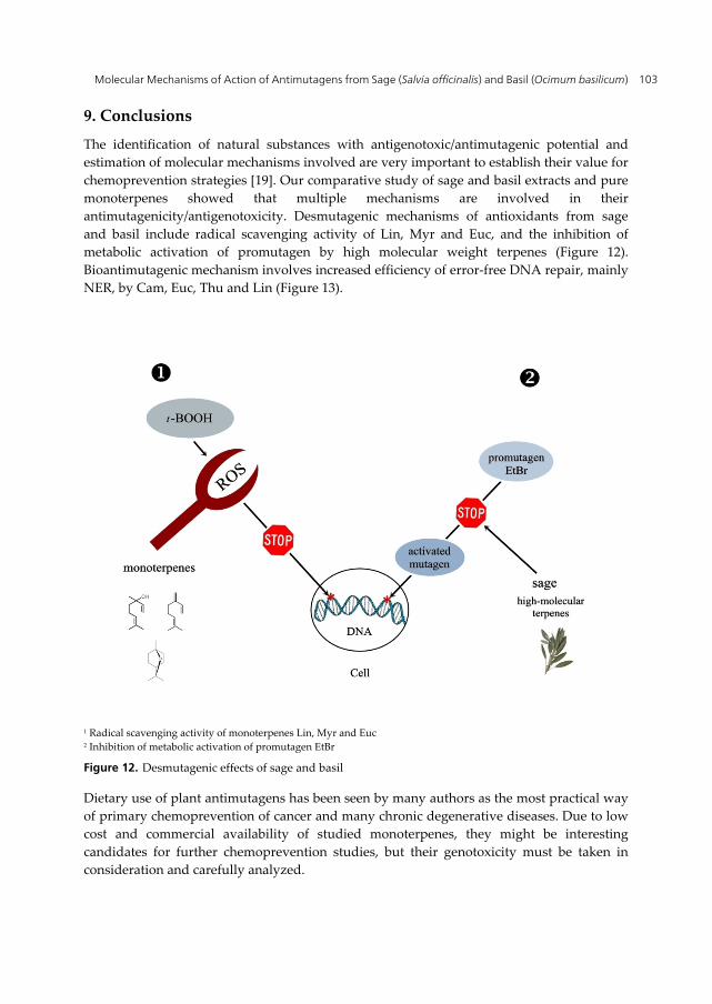
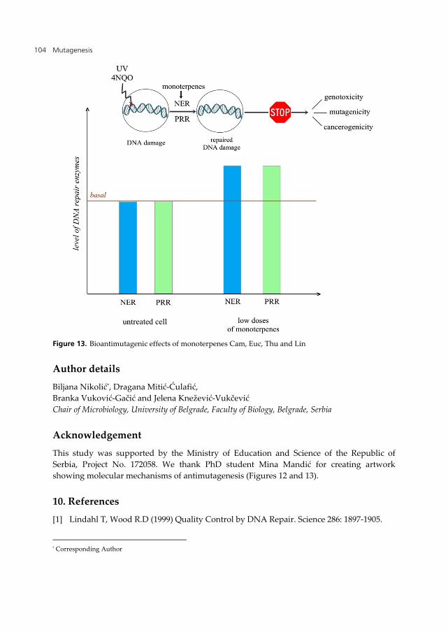
The identification of natural substances with antigenotoxic/antimutagenic potential and estimation of molecular mechanisms involved are very important to establish their value for chemoprevention strategies [19]. Our comparative study of sage and basil extracts and pure monoterpenes showed that multiple mechanisms are involved in their antimutagenicity/antigenotoxicity. Desmutagenic mechanisms of antioxidants from sage and basil include radical scavenging activity of Lin, Myr and Euc, and the inhibition of metabolic activation of promutagen by high molecular weight terpenes (Figure 12). Bioantimutagenic mechanism involves increased efficiency of error-free DNA repair, mainly NER, by Cam, Euc, Thu and Lin (Figure 13).
1 Radical scavenging activity of monoterpenes Lin, Myr and Euc 2 Inhibition of metabolic activation of promutagen EtBr
Figure 12. Desmutagenic effects of sage and basil
Dietary use of plant antimutagens has been seen by many authors as the most practical way of primary chemoprevention of cancer and many chronic degenerative diseases. Due to low cost and commercial availability of studied monoterpenes, they might be interesting candidates for further chemoprevention studies, but their genotoxicity must be taken in consideration and carefully analyzed.
Mutagenesis 104
Figure 13. Bioantimutagenic effects of monoterpenes Cam, Euc, Thu and Lin
Author details
Biljana Nikolić*, Dragana Mitić-Ćulafić, Branka Vuković-Gačić and Jelena Knežević-Vukčević Chair of Microbiology, University of Belgrade, Faculty of Biology, Belgrade, Serbia
Acknowledgement
This study was supported by the Ministry of Education and Science of the Republic of Serbia, Project No. 172058. We thank PhD student Mina Mandić for creating artwork showing molecular mechanisms of antimutagenesis (Figures 12 and 13).
10. References
[1] Lindahl T, Wood R.D (1999) Quality Control by DNA Repair. Science 286: 1897-1905.
* Corresponding Author
Molecular Mechanisms of Action of Antimutagens from Sage (Salvia officinalis) and Basil (Ocimum basilicum) 105
[2] Friedberg E.C, Walker G.C, Siede W, Wood R.D, Schultz R.A, Elleberger T (2006) DNA Repair and Mutagenesis, 2nd Edition, Washington D.C: ASM Press. 9 p.
[3] Eisen J.A, Hanavalt P.C (1999) A phylogenomic study of DNA repair genes, proteins and processes. Mutat. Res. 435: 171-213.
[4] Hoeijmakers J.H.J (2001) Genome maintenance mechanisms for preventing cancer. Nature 411: 366-374.
[5] De Flora S, Izzotti A, Randerath K, Randerath E, Bartsch H, Nair J, Balansky R, van Schooten F, Degan P, Fronza G, Valsh D, Lewtas J (1996) DNA adducts and chronic degenerative diseases, Pathogenic relevanse and imlications in preventive medicine. Mutat. Res. 366: 197-238.
[6] Finkel T, Holbrook N.J (2000) Oxidants, oxidative stress and the biology of ageing. Nature 408: 239-247.
[7] Olinski R, Gackowski D, Foksinski M, Rozalski R, Roszkowski K, Jaruga P (2002) Oxidative DNA damage: assessment of the role in carcinogenesis, atherosclerosis, and acquired immunodeficiency syndrome. Free Radic. Biol. Med. 33: 192-200.
[8] Davydov V, Hansen L.A, Shackelford D.A (2003) Is DNA repair compromised in Alzheimer’s disease? Neurobiol. Aging 5809: 1-16.
[9] Coppede F, Migliore L (2009) DNA damage and repair in Alzheimer’s disease. Curr. Alzheimer Res. 6: 36–47.
[10] Loeb L.A (1991) Mutator phenotype may be required for multistage carcinogenesis. Cancer Res. 51: 3075–3079.
[11] Marnett L.J (2000) Oxyradicals and DNA damage. Carcinogenesis 21: 361-370. [12] Loeb L.A, Loeb K.R, Anderson J.P (2003) Multiple mutations and cancer. Proc Natl
Acad Sci USA 100: 776–781. [13] Mills K.D, Ferguson D.O, Alt F.W (2003) The role of DNA breaks in genomic instability
and tumorigenesis. Immunol Rev. 194: 77-95. [14] Bielas J.H, Loeb K.R, Rubin B.P, True L.D, Loeb L.A (2006) Human cancers express a
mutator phenotype. Proc Natl Acad Sci USA 103: 18238–18242. [15] Hoffmann J-S, Cazaux C (2010) Aberrant expression of alternative DNA polymerases: A
source of mutator phenotype as well as replicative stress in cancer. Semin.cancer biol. 20: 312-319.
[16] Kada T, Kaneko K, Matsuzaki T, Hara Y (1985) Detection and chemical identification of natural bioantimutagens: A case of green tea factor. Mutat. Res. 150: 127-132.
[17] Kuroda Y (1990) Antimutagenesis studies in Japan. In: Kuroda Y, Shankel D.M, Waters M.D, editors. Antimutagenesis and Anticancerogenesis Mechanisms II, New York and London: Plenum Press pp. 1-22.
[18] De Flora F, Izzotti A, D’Agostini F, Balansky R.M, Noonan D, Albini A (2001) Multiple points of intervention in the prevention of cancer and other mutation-related diseases. Mutat. Res. 480-481: 9-22.
[19] De Flora F, Ferguson R.L (2005) Overview of mechanisms of cancer chemopreventive agents. Mutat. Res. 591: 8-15.
[20] Ferguson L.R, Philpott M, Karunasinghe N (2004) Dietary cancer and prevention using antimutagens. Toxicology 198: 147–159.
Mutagenesis 106
[21] Ferguson L.R (2011) Antimutagenesis studies: Where have they been and where are they heading?. Genes Environ. 33: 71-78.
[22] Simić D, Vuković-Gačić B, Knežević-Vukčević J, Trninić S, Jankov R.M (1997) Antimutagenic effect of terpenoids from sage (Salvia officinalis L.). J. Environ. Pathol. Toxicol. Oncol. 16: 293-301.
[23] Kada T, Inoue T, Ohta T, Shirasu Y (1986) Antimutagens and their modes of action, In: Shankel D.M, Hartman P.H, Kada T, Hollaender A, editors. Antimutagenesis and Anticarcinogenesis Mechanisms. New York: Plenum Press. pp. 181-196.
[24] De Flora S (1998) Mechanisms of inhibitors of mutagenesis and carcinogenesis. Mutat. Res. 402: 151-158.
[25] Crowell P.L (1999) Prevention and therapy of cancer by dietary monoterpenes. J. Nutr. 129: 7755-7785.
[26] Paduch R, Kandefer-Szerszen M, Trytek M, Fiedurek J (2007) Terpenes: substances useful in human healthcare. Arch. Immunol. Ther. Exp. 55: 315-327.
[27] Bakkali F, Averbeck S, Averbeck D, Waomar M (2008) Biological effects of essential oils – a review. Food Chem. Toxicol. 46: 446-475.
[28] Vuković-Gačić B, Simić D (1993) Identification of natural antimutagens with modulating effects on DNA repair. In: G. Bronzzeti G, Hayatsu H, De Flora S, Waters M.D, Shankel D.M, editors. Antimutagenesis and Anticarcinogenesis Mechanisms III, New York: Plenum Press. pp. 269-277.
[29] Cuvelier M.E, Berset C, Richard H (1994) Antioxidant constituents in sage (Salvia officinalis). J. Agric. Food Chem. 42: 665-669.
[30] Chattopadhyay R.R (1999) A comparative evaluation of some blood sugar lowering agents of plant origin. J. Ethnopharmacol. 67: 367-372.
[31] Koga T, Hirota N, Takumi K (1999) Bactericidal activities of essential oils of Basil and Sage against a range of bacteria and the effect of these essential oils on Vibrio parahaemolyticus. Microbiol. Res. 154: 267-273.
[32] Offiah V.N, Chikwendu, U.A (1999) Antidiarrheal effects of Ocimum gratissimum leaf extract in experimental animals. J. Ethnopharmacol. 68: 327-330.
[33] Barićević D, Bartol T (2000) The biological/pharmacological activity of the Salvia genus V., Pharmacology, In: Kintzios S.E, editor. Sage, The Genus Salvia. Amsterdam: Haewood Academic Publishers. pp. 143-184.
[34] Klem M.A, Nair M.G, Sraassburg G.M, Dewitt D.L (2000) Antioxidant and cyclooxygenase inhibitory phenolic compounds from Ocimum sanctum Linn. Phytomedicine 7: 7-13.
[35] Zupko I, Hohmann J, Redei D, Falkay G, Janicsak G, Mathe I (2001) Antioxidant activity of leaves of Salvia species in enzyme-dependent and enzyme-independent system of lipid peroxidation and their phenolic constituents. Planta Med. 67: 366-368.
[36] Capasso R, Izzo A.I, Capasso F, Romussi G. Bisio A, Mascolo N (2004) A diterpenoid from Salvia cinnabarina inhibits mouse intestinal motility in vivo. Planta Med. 70: 375-377.
[37] Ren Y, Houghton P.J, Hider R.C, Howes M.J.R (2004) Novel diterpenoid acetylcholinesterase inhibitors from Salvia miltiorhiz. Planta Med. 70: 201-204.
Molecular Mechanisms of Action of Antimutagens from Sage (Salvia officinalis) and Basil (Ocimum basilicum) 107
[38] Mitić-Ćulafić D, Vuković-Gačić B, Knežević-Vukčević J, Stanković S, Simić D (2005) Comparative study on the antibacterial activity of volatiles from sage (Salvia officinalis L.). Arch. Biol. Sci. 57: 173-178.
[39] Šmidling D, Mitić-Ćulafić D, Vuković-Gačić B, Simić D, Knežević-Vukčević J (2008) Evaluation of antiviral activity of fractionated extracts of sage Salvia officinalis L. (Lamiaceae). Arch. Biol. Sci. 60: 421-429.
[40] Tsai K.D, Lin B.R, Perng D.S, Wei J.C, Yu Y.W, Cherng, J-M (2011) Immunomodulatory effects of aqueous extract of Ocimum basilicum (Linn.) and some of its constituents on human immune cells. J. Med. Plant Res. 5: 1873-1883.
[41] Gali-Muhtasib H.U, Affara N.I (2000) Chemopreventive effects of sage oil on skin papilomas in mice. Phytomedicine. 7: 129-136.
[42] Prakash J, Gupta S.K (2000) Chemopreventive activity of Ocimum sanctum seed oil. J. Ethnopharmacol. 72: 29-34.
[43] Vujošević M, Blagojević J (2004) Antimutagenic effects of extracts from sage (Salvia officinalis) in mammalian system in vivo. Acta Vet. Hung., 52: 439-443.
[44] Sidiqque Y.H, Ara G, Beg T, Afzal M (2007) Anti-genotoxic effect of Ocimum sanctum L. extract against cyproterone acetate induced genotoxic damage in cultured mammalian cells. Acta Biol. Hun. 58: 397-409.
[45] Patenković A, Stamenković-Radak M, Banjanac T, Anđelković, M (2009) Antimutagenic effect of sage tea in the wing spot test of Drosophila melanogaster. Food Chem Toxicol. 47: 180-183.
[46] Simić D, Vuković-Gačić B, Knežević-Vukčević J (1998) Detection of natural bioantimutagens and their mechanisms of action with bacterial assay-system. Mutat. Res. 402: 51-57.
[47] Vuković-Gačić B, Simić D, Knežević-Vukčević J (2006) Escherichia coli assay system for detection of plant antimutagens and their mechanisms of action. In: Verschaeve L, editor. Topical Issues in Applied Microbiology and Biotechnology, Kerala, India: Research Signpost. pp. 61-86.
[48] Tood P.A, Monti-Bragadini C, Glickman B.W (1979) MMS mutagenesis in strains of E. coli carrying the R46 mutagenic enhancing plasmid: phenotypic analysis of Arg+ revertants. Mutat. Res. 62: 227-237.
[49] Young L.C, Hays J.B, Tron V.A, Andrew S.E (2003) DNA mismatch repair proteins: potential guardians against genomic instability and tumorigenesis induced by ultraviolet photoproducts. J Invest Dermatol. 121: 435-440.
[50] Walker G.C (1984) Mutagenesis and inducible responses to DNAdamage in Escherichia coli. Microbiol. Rev. 48: 60-93.
[51] Walker G.C (1985) Inducible DNA system. Annu.Rev. Bioch. 54: 425-457. [52] Jones C.J, Edwards S.M, Waters R (1989) The repair of identified large DNA adducts
induced by 4-nitroquinoline-1-oxide in normal or xeroderma pigmentosum group A human fibroblasts, and the role of DNA polymerase alpha or delta. Carcinogenesis. 10: 1197-1201.
Mutagenesis 108
[53] Hömme M, Jacobi H, Juhl-Strauss U, Witte I (2000) Synergistic DNA damaging effects of 4-nitroquinoline-1-oxide and non-effective concentrations of methyl methanesulfonate in human fibroblasts. Mutat. Res. 461: 211-219.
[54] Truglio J.J, Croteau D.L, van Houten B, Kisker C (2006) Prokaryotic nucleotide excision repair: The UvrABC System. Chem.Rev. 106: 233-252.
[55] Knežević J, Simić D (1982) Induction of phage lambda by bleomycin and nalidixic acid in the lexA mutant of E. coli K12. Genetika. 14: 77-91.
[56] Stanojević J, Berić T, Opačić B, Vuković-Gačić B, Simić D, Knežević-Vukčević J (2008) The effect of essential oil of basil (Ocimum basilicum L.) on UV-induced mutagenesis in Escherichia coli and Saccharomyces cerevisiae. Arch. Biol. Sci. 60: 93-102.
[57] Nikolić B, Stanojević J, Mitić D, Vuković-Gačić B, Knežević-Vukčević J, Simić D (2004) Comparative study of the antimutagenic potential of Vitamin E in different E. coli strains. Mutat. Res. 564: 31-38.
[58] Nikolić B, Mitić-Ćulafić D, Vuković-Gačić B, Knežević-Vukčević J (2011) Modulation of genotoxicity and DNA repair by plant monoterpenes Camphor, Eucalyptol and Thujone in Escherichia coli and mammalian cells. Food Chem. Tox. 49: 2035-2045.
[59] Dutreix M, Moreau P.L, Bailone A, Galibert A, Battista J.R, Walker G.C, Devoret R (1989) New recA mutations that dissociate the various RecA protein activities in E. coli provide evidence for an additional role for RecA protein in UV mutagenesis. J. Bacteriol. 171: 2415-2423.
[60] Maron D.M, Ames B.N (1983) Revised methods for the Salmonella mutagenicity test. Mutat. Res. 113: 173-215.
[61] Martinez A, Urios A, Blanco M (2000) Mutagenicity of 80 chemicals in Escherichia coli tester strains IC203, deficient in OxyR, and its oxyR+ parent WP2 uvrA/pKM101: detection of 31 oxidative mutagens. Mutat. Res. 467: 41-53.
[62] Zimmermann F.K, Kern R. Rasenberger H.(1975).A yeast strain for simultaneous detection of induced mitotic crossing over, mitotic gene conversion and reverse mutation. Mutat. Res. 28: 381-388.
[63] Miloshev G, Mihaylov I, Anachkova B (2002) Application of the single cell gel electrophoresis on yeast cells. Mutat. Res. 513: 69-74.
[64] Radman M (1999) Enzymes of evolutionary change. Nature, 401: 866-869. [65] Quillardet P, Hofnung M (1993) The SOS chromotest: a review. Mutat. Res. 297: 235-279. [66] Konrad E.B (1977) Method for the isolation of Escherichia coli mutants with enhanced
recombination between chromosomal duplications. J. Bacteriol. 130: 167-172. [67] Lawery P.E, Kowalczykowski S.C (1992) Biochemical basis of the constitutive repressor
cleavage activity of RecA730 protein. J. Biol. Chem. 267: 20648-20658. [68] Ennis D.G, Levine A.S, Koch W.H, Woodgate R (1995) Analysis of recA mutants with
altered SOS functions. Mutat. Res., 336: 39-48. [69] Kohlimeir L, Simonsen M, Mottus K (1995) Dietary modifiers of carcinogenesis.
Environ. Health Perspect. 103: 177-184. [70] Urios A, Blanco M (1996) Specifity of spontaneous and t-butyl hydroperoxide-induced
mutations in oxyR strains of Escherichia coli differing with respect to the SOS mutagenesis proficiency and to the MutY and MutM functions. Mutat. Res. 354: 95-101.
Molecular Mechanisms of Action of Antimutagens from Sage (Salvia officinalis) and Basil (Ocimum basilicum) 109
[71] Fowler R.G, White S.J, Koyama C, Moore S.C, Dunn R.L, Schaaer R.M (2003) Interactions among the Escherichia coli mutT, mutM, and mutY damage prevention pathways. DNA Repair 2: 159-173.
[72] Mure K. Rossman,T.G (2001) Reduction of spontaneous mutagenesis in mismatch repair-deficient and proficient cells by dietary antioxidants. Mutat. Res. 480-481: 85-95.
[73] Jackson A.L, Loeb L.A.(1998) The mutation rate and cancer. Genetics 148: 1483-1490. [74] Jackson A.L, Loeb L.A (2000) Microsatellite instability induced by hydrogen peroxide in
Escherichia coli. Mutat. Res. 447: 187-198. [75] Boyer J.C, Yamada N.A. Roques C.N. Hatch S.B. Riess K, Farber R.A (2002) Sequence
dependent instability of mononucleotide microsatellites in cultured mismatch repair proficient and deficient mammalian cells. Hum. Mol. Genet. 11: 707-713.
[76] OECD (2007). ISSN : 2074-5788 (online) DOI : 10.1787/20745788. [77] Blanco M, Urios A, Martinez A (1998) New Escherichia coli WP2 tester strains highly
sensitive to reversion by oxidative mutagens. Mutat. Res. 413: 95-101. [78] Collins A.R (2004) The Comet assay for DNA damage and repair. Mol. Biotech. 26: 249-
261. [79] Đarmati Z, Jankov R.M, Vujičić Z, Csanadi J, Đulinac B, Švirtlih E, Đorđević A, Švan K
(1994) 12-Deoxocarnisol isolated from the wild type of sage from Dalmatia. J. Serb. Chem. Soc. 59: 291-299.
[80] Đarmati Z, Jankov R.M, Vujičić Z, Csanadi J, Švirtlih E, Đorđević A, Švan K (1993) Natural terpenoids isolated from grown variety of sage. J. Serb. Chem. Soc. 58: 515-523.
[81] Mitić D, Vuković-Gačić B, Knežević-Vukčević J, Berić T, Nikolić B, Stanković S, Simić D (2001) Natural antioxidants and their mechanisms in inhibition of mutagenesis, In: Kreft I, Škrabanja V, editors. Molecular and genetic interactions involving phytochemicals. Ljubljana, Slovenia: Univerza v Ljubljani in Slovenska akademija znanosti in umetnosti. pp. 67-74.
[82] Simić D, Vuković-Gačić B, Knežević-Vukčević J, Đarmati Z, Jankov R.M (1994) New assay system for detecting bioantimutagens in plant extracts. Arch. Biol. Sci. 46: 81-85.
[83] Brkić D, Stepanović B, Nastovski T, Brkić S (1999) Distilation of sage. In: Brkić D, editor. Sage (S. officinalis L.). Belgrade, Serbia: Institute for Medicinal Plant Research “Dr Josif Pančić”. pp. 131-136.
[84] Marinković B, Marin P.D, Knežević-Vukčević J, Soković M.D, Brkić D (2002) Activity of essential oils of three Micromeria species (Lamiaceae) against micromycetes and bacteria. Phytoter. Res. 16: 336-339.
[85] Knežević-Vukčević J, Vuković-Gačić B, Stević T, Stanojević J, Nikolić B, Simić D (2005) Antimutagenic effect of essential oil of sage (Salvia officinalis L.) and its fractions against UV-induced mutations in bacterial and yeast cells. Arch. Biol. Sci. 57: 163-172.
[86] Berić T, Nikolić B, Stanojević J, Vuković-Gačić B, Knežević-Vukčević J (2008) Protective effect of basil (Ocimum basilicum L.) against oxidative DNA damage and mutagenesis. Food Chem. Toxicol. 46: 724-732.
[87] Mitić-Ćulafić D, Žegura B. Nikolić B. Vuković-Gačić B. Knežević-Vukčević J. Filipič M (2009) Protective effect of linalool, myrcene and eucalyptol against t-butyl
Mutagenesis 110
hydroperoxide induced genotoxicity in bacteria and cultured human cells. Food Chem. Tox. 47: 260-266.
[88] Nikolić B, Mitić-Ćulafić D, Vuković-Gačić B, Knežević-Vukčević J (2011) The antimutagenic effect of monoterpenes against UV-irradiation-, 4NQO- and t-BOOH-induced mutagenesis in E. coli. Arch. Biol. Sci. 63: 117-128.
[89] Lazarova M, Labaj J, Eckl P, Slamenova D (2006) Comparative evaluationof DNA damage by genotoxicants in primary rat cells applying the comet assay. Toxicol. Lett. 164: 54-62.
[90] Chadha A, Madyastha K.M (1984) Metabolism of geraniol and linalool in the rat and effects on liver and lung microsomal-enzymes. Xenobiotica 14: 365-374.
[91] Madyastha M.K, Srivatsan M (1987) Metabolism of β-myrcene in vivo and in vitro: its effects on rat-liver microsomal enzymes. Xenobiotica 17: 539-549.
[92] Knasmüller S, Parzefall W, Sanyal R, Ecker S, Schwab C, Uhl M, Mersch-Sundermann V, Williamson G, Hietsch G, Langer T, Darroudi F, Natarajan A.T (1998) Use of metabolically competent human hepatoma cells for the detection of mutagens and antimutagens, Mutat. Res. 402: 185-202.
[93] Miyazawa M, Shindo M, Shimada T (2001) Oxidation of 1,8-cineole, the monoterpene cyclic ether originated from Eucalyptus polybractea, by cytochrome P450 3A enzymes in rat and human liver microsomes. Drug Metab. Disposit. 29: 200-205.
[94] Kassie F, Mersch-Sundermann V, Edenharder R, Platt L.K, Darroudi F, Lhoste E, Humbolt C, Muckel E, Uhl M, Kundi M, Knasmuller S (2003) Development and application of test methods for the detection of dietary constituents which protect against heterocyclic aromatic amines. Mutat.Res. 523-524: 183-192.
[95] Ishida T (2005) Biotransformation of terpenoids by mammals, microorganisms and plant-cultured cells. Chem. Biodiver. 2: 569-590.
[96] Belsito D, Bickers D, Bruze M, Calow P, Greim H, Hanifin J.M, Rogers A.E, Saurat J.H, Sipes I.G, Tagami H (2008) A toxicologic and dermatologic assessment of cyclic and non-cyclic terpene alcohols when used as fragrance ingredients. Food Chem. Tox. 46: S1-S71.
[97] Vuković-Gačić B, Nikčević S, Berić-Bjedov T, Knežević-Vukčević J, Simić D (2006) Antimutagenic effect of essential oil of sage (Salvia officinalis L.) and its monoterpenes against UV-induced mutations in Escherichia coli and Saccharomyces cerevisiae. Food Chem. Toxicol. 44: 1730-1738.
[98] Crowley D.J, Hanawalt P.C, (1998) Induction of the SOS response increases the efficiency of global nucleotide excision repair of cyclobutane pyrimidine dimmers, but not 6-4 photoproducts, in UV-irradiated Escherichia coli. J. Bacteriol. 180: 3345-3352.
[99] Calabrese E.J, Baldwin L.A (2001) Hormesis: U-shaped dose responses and their centrality in toxicology. Trends Pharmacol. Sci. 22: 285-291.
[100] Calabrese E.J (2010) Hormesis is central to toxicology, pharmacology and risk assessment. Hum. Exp. Toxicol. 29: 249-261.
[101] Gomes-Carneiro M.R, Elzenszwalb I.F, Paumgartten F.J (1998) Mutagenicity testing (+/-) – camphor, 1,8-cineole, citral, citronellal, (-)-menthol and terpineol with Salmonella/ microsome assay. Mutat. Res. 416: 129-136.
Molecular Mechanisms of Action of Antimutagens from Sage (Salvia officinalis) and Basil (Ocimum basilicum) 111
[102] Stajković O, Berić-Bjedov T, Mitić-Ćulafić D, Stanković S, Vuković-Gačić B, Simić D, Knežević-Vukčević J (2007) Antimutagenic properties of basil (Ocimum basilicum L.) in Salmonella typhimurium TA100. Food Tech. Biotech. 45: 213-217.
[103] Ribeiro D.A, Matsumoto M.A, Marques M.E.A, Salvadori D.M.F (2007) Biocompatibility of gutta-percha solvents using in vitro mammalian test-system. Oral Surg. Oral Med. Oral Pathol. Oral Radiol. Endod. 103: e106-e109.
[104] Horvathova E, Turcaniova V, Slamenova D (2007) Comparative study of DNA-damaging and DNA-protective effects of selected components of essential plant oils in human leukamic cells K562. Neoplasma 54: 478-483.
[105] Alakilli S.Y.M (2009) Evaluation of camphor mutagenicity in somatic cells of pregnant rats. Asian J. Biotech. 1: 111-117.
[106] Pavlidou V, Karpouhtsis I, Franzios G, Zambetaki A, Scouras Z, Mavragani-Tsipidou P (2004) Insecticidal and genotoxic effects of essential oils of Greek sage, Salvia fruticosa, and mint, Mentha pulegium, on Drosophila melanogaster and Bactrocera oleae (Diptera: Tepritidae). J. Agr. Urban Entomol. 21: 39-49.
[107] Koitabashi R, Suzuki T, Kawazu T, Sakai A, Kuroiwa H, Kuroiwa T (1997) 1,8- Cineole inhibits root growth and DNA synthesis in the root apical meristem of Brassica campestris L. J. Plant Res. 110: 1-6.
[108] Moteki H, Hibasami H, Yamada Y, Katsuzaki H, Imai K, Komiya T (2002) Specific induction of apoptosis by 1,8-cineole in two human leukemia cell lines, but not in human stomach cancer cell line. Oncol. Rep. 9: 757-760.
[109] Kim J.O, Kim Y.S, Lee J.H, Kim M.N, Rhee S.H, Moon S.H, Park K.Y (1992) Antimutagenic effect of the major volatile compounds identified from mugworth (Artemisiia asictica nakai) leaves. J. Korean Soc. Food Nutr. 21: 308-313.
[110] Goel H.C, Singh S, Singh S.P (1989) Radiomodifying influence of camphor on sister-chromatid exchange induction in mouse bone marrow. Mutat. Res. 224: 157-160.
[111] Lutz D, Eder E, Neudecker T (1982) Structure-mutagenicity relationship in α,β-unsaturated carbonylic compounds and their corresponding allylic alcohols. Mutat. Res. 93: 305-315.
[112] Ishidate M Jr, Sofuni T, Yoshikawa K, Hayashi M, Nohmi T, Sawada M, Matsuoka A (1984) Primary mutagenicity screening of food additives currently used in Japan. Food Chem. Tox. 22: 623-636.
[113] Yoo Y.S (1986) Mutagenic and antimutagenic activities of flavoring agents used in foodstuffs. Osaka-shi Igakkai Zasshi [J Osaka City Medical Center] 34: 267-288.
[114] Heck J.D, Vollmuth T.A, Cifone M.A, Jagannath D.R, Myhr V, Curren R.D (1989) An evaluation of food flavoring ingredients in a genetic toxicity screening battery. Toxicologist 9: 257-264.
[115] Sasaki Y.F, Imanishi H, Ohta T, Shirasu Y (1989) Modifying effects of components of plant essence on the induction of sister chromatid exchanges in cultured Chinese hamster ovary cells. Mutat. Res. 226: 103-110.
[116] Di Sotto A, Mazzanti G, Carbone F, Hrelia P, Maffei F (2011) Genotoxicity of lavander oil, linalyl acetate, and linalool on human lymphocytes in vitro. Environ. Mol. Mutagen. 52: 69-71.
Mutagenesis 112
[117] Minnunni M, Wolleb U, Muellera O, Pfeifera A, Aeschbachera H.U (1992) Natural antioxidants as inhibitors of oxygen species induced mutagenicity. Mutat. Res. 269: 193-200.