Molecular epidemiology of methicillin-resistant … epidemiology of methicillin-resistant...
12
Molecular epidemiology of methicillin-resistant Staphylococcus aureus R.H. Deurenberg ∗1 , S. Kalenic 2 , A.W. Friedrich 3 , F.H. van Tiel 1 and E.E. Stobberingh 1 1 Department of Medical Microbiology, Maastricht Infection Center (MINC), University Hospital Maastricht, P. Debyelaan 25, 6229 HX, Maastricht, The Netherlands 2 Department of Clinical and Molecular Microbiology, Clinical Hospital Centre Zagreb, Kišpati eva 12, HR-10000, Zagreb, Croatia 3 Department for Infection Control, Institute of Hygiene, University Hospital of Münster, Robert Koch Straße 41, D-48149 Münster, Germany Staphylococcus aureus can cause a wide spectrum of infections. Its strong adaptive power to antibiotics has resulted in the emergence of methicillin-resistant S. aureus (MRSA). Resistance to methicillin and other ß-lactam antibiotics is caused by the mecA gene, which is situated on a mobile genetic element, the Staphylococcal Cassette Chromosome mec (SCCmec). Six SCCmec types (I to VI) have been distinguished. To study the molecular epidemiology of MRSA, pulsed-field gel electrophoresis (PFGE), multilocus sequence typing (MLST), spa typing and SCCmec typing can be used. Several MRSA clones have emerged and disseminated worldwide. Although, in the past, MRSA strains were mainly hospital- acquired (HA-MRSA), from the late 1990s, community-acquired MRSA (CA-MRSA) has emerged. CA- MRSA harbours SCCmec type IV or V and is often associated with Panton-Valentine leukocidin (PVL). This chapter describes the recent developments concerning the structure of SCCmec, the molecular evolution of MRSA and the methods used to investigate the molecular epidemiology of MRSA. Keywords MLST, MRSA; PFGE, PVL; SCCmec, spa typing 1. Introduction Since its discovery in the 1880s, Staphylococcus aureus has been known as a potential pathogenic Gram- positive bacterium. It can cause a broad variety of diseases, ranging from minor infections of the skin to post-operative wound infections. Until the introduction of penicillin in the early 1940s and the treatment of S. aureus infections with this antibiotic, the mortality rate of patients with a S. aureus infection was about 80%. However, shortly after the introduction of penicillin for medical use, the first penicillin- resistant strains were isolated in 1942, first in hospitals, and later on in the community. Since 1960, around 80% of all S. aureus strains are penicillin resistant. In 1962, two years after the introduction of methicillin, S. aureus developed resistance to methicillin through the acquisition of the mecA gene [1, 2]. 2. Resistance determinant SCCmec The resistance of S. aureus to methicillin and other ß-lactam antibiotics is caused by the mecA gene, which encodes the 78-kDa penicillin-binding protein (PBP) 2a (or PBP2’) [3]. The 2.1-kb mecA gene is located on a mobile genetic element, the Staphylococcal Cassette Chromosome mec (SCCmec) [4]. Currently, six main types of SCCmec (type I to VI) are distinguished, ranging in size from 20.9 to 66.9 kb (Fig. 1). SCCmec types I (34.3 kb), IV (20.9 to 24.3 kb), V (28 kb) and VI (20.9 Kb) encode for resistance to β- lactam antibiotics only. SCCmec types II (53.0 kb) and III (66.9 kb) determine multi resistance, as these ∗ Corresponding author. E-mail: [email protected], Phone: +31 43 3874644, Fax: +31 43 3876643 766 Communicating Current Research and Educational Topics and Trends in Applied Microbiology A. Méndez-Vilas (Ed.) _____________________________________________________________________ ©FORMATEX 2007
Transcript of Molecular epidemiology of methicillin-resistant … epidemiology of methicillin-resistant...

Molecular epidemiology of methicillin-resistant Staphylococcus aureus
R.H. Deurenberg∗1, S. Kalenic2, A.W. Friedrich3, F.H. van Tiel1 and E.E. Stobberingh1
1 Department of Medical Microbiology, Maastricht Infection Center (MINC), University Hospital Maastricht, P. Debyelaan 25, 6229 HX, Maastricht, The Netherlands 2 Department of Clinical and Molecular Microbiology, Clinical Hospital Centre Zagreb, Kišpati eva 12, HR-10000, Zagreb, Croatia 3 Department for Infection Control, Institute of Hygiene, University Hospital of Münster, Robert Koch Straße 41, D-48149 Münster, Germany
Staphylococcus aureus can cause a wide spectrum of infections. Its strong adaptive power to antibiotics has resulted in the emergence of methicillin-resistant S. aureus (MRSA). Resistance to methicillin and other ß-lactam antibiotics is caused by the mecA gene, which is situated on a mobile genetic element, the Staphylococcal Cassette Chromosome mec (SCCmec). Six SCCmec types (I to VI) have been distinguished. To study the molecular epidemiology of MRSA, pulsed-field gel electrophoresis (PFGE), multilocus sequence typing (MLST), spa typing and SCCmec typing can be used. Several MRSA clones have emerged and disseminated worldwide. Although, in the past, MRSA strains were mainly hospital-acquired (HA-MRSA), from the late 1990s, community-acquired MRSA (CA-MRSA) has emerged. CA-MRSA harbours SCCmec type IV or V and is often associated with Panton-Valentine leukocidin (PVL). This chapter describes the recent developments concerning the structure of SCCmec, the molecular evolution of MRSA and the methods used to investigate the molecular epidemiology of MRSA.
Keywords MLST, MRSA; PFGE, PVL; SCCmec, spa typing
1. Introduction
Since its discovery in the 1880s, Staphylococcus aureus has been known as a potential pathogenic Gram-positive bacterium. It can cause a broad variety of diseases, ranging from minor infections of the skin to post-operative wound infections. Until the introduction of penicillin in the early 1940s and the treatment of S. aureus infections with this antibiotic, the mortality rate of patients with a S. aureus infection was about 80%. However, shortly after the introduction of penicillin for medical use, the first penicillin-resistant strains were isolated in 1942, first in hospitals, and later on in the community. Since 1960, around 80% of all S. aureus strains are penicillin resistant. In 1962, two years after the introduction of methicillin, S. aureus developed resistance to methicillin through the acquisition of the mecA gene [1, 2].
2. Resistance determinant SCCmec
The resistance of S. aureus to methicillin and other ß-lactam antibiotics is caused by the mecA gene, which encodes the 78-kDa penicillin-binding protein (PBP) 2a (or PBP2’) [3]. The 2.1-kb mecA gene is located on a mobile genetic element, the Staphylococcal Cassette Chromosome mec (SCCmec) [4]. Currently, six main types of SCCmec (type I to VI) are distinguished, ranging in size from 20.9 to 66.9 kb (Fig. 1). SCCmec types I (34.3 kb), IV (20.9 to 24.3 kb), V (28 kb) and VI (20.9 Kb) encode for resistance to β-lactam antibiotics only. SCCmec types II (53.0 kb) and III (66.9 kb) determine multi resistance, as these
∗ Corresponding author. E-mail: [email protected], Phone: +31 43 3874644, Fax: +31 43 3876643
766
Communicating Current Research and Educational Topics and Trends in Applied Microbiology A. Méndez-Vilas (Ed.)_____________________________________________________________________
©FORMATEX 2007

elements harbour additional drug resistance genes, which are carried on integrated plasmids, i.e. pUB110, pI258 and pT181, and a transposon (Tn554). Plasmid pUB110 carries the ant(4')
Fig. 1 Schematic drawing of SCCmec types I to VI [5-9]. The major elements of the six SCCmec types (ccr genes, IS431, IS1272, mecA, mecI/RI, orfX, pI258, pT181, pUB101 and Tn554) are presented, as are the six loci (A to F) used for SCCmec typing according to the method of Oliviera et al. [8, 10, 11] (Adapted from [11]).
gene, encoding for resistance to kanamycin, tobramycin and bleomycin, and pI258 codes for resistance to penicillins and heavy metals, such as mercury. Plasmid pT181 codes for tetracycline resistance, while transposon Tn554 carries the ermA gene, which is responsible for inducible macrolide, lincosamide and streptogramin (MLS) resistance (Fig. 1) [4, 9, 12]. S. aureus can also carry resistance genes on plasmids and on other sites of the chromosome. Recently, it has been shown that SCCmec type III is a composite element, consisting of SCCmec type III and SCCmercury, harbouring ccrC, pI258 and Tn554 [13]. Furthermore, SCCmec harbours insertion sequences, such as IS431, as well as genes responsible for the regulation of mecA transcription, i.e. ∆mecRI (on SCCmec types I, IV, V and VI), or mecRI and mecI (on SCCmec types II and III) [4-7, 9]. Both mecI and mecRI can be truncated by IS431 or IS1272, and this results in a de-repression of mecA [14]. These genes are situated on the so-called mec complexes, of which five major classes have been distinguished (Table 1) [4, 5, 14].
Genes encoding cassette chromosome recombinases (ccr) are located within the SCCmec elements. Their function is the integration of SCCmec into and excision of SCCmec from the chromosome at a specific site (attBscc; at the 3’ end of an open reading frame (ORF) of unknown function, named orfX [15]), The ccr genes are designated ccrA1 and ccrB1 (in SCCmec type I), ccrA2 and ccrB2 (in SCCmec types II and IV), ccrA3 and ccrB3 (in SCCmec type III), ccrA4 and ccrB4 (in SCCmec type VI) and ccrC (in
767
Communicating Current Research and Educational Topics and Trends in Applied Microbiology A. Méndez-Vilas (Ed.)_____________________________________________________________________
©FORMATEX 2007

SCCmec type V). The regions outside the mec and ccr complexes are designated the J (junkyard) regions (Fig. 1) [6, 7, 9, 14, 16, 17]. SCCmec elements are divided into three regions. The J1 region is located between the chromosome right junction to the ccr genes, while the region from the ccr genes to the mec complex is designated the J2 region. The J3 region ranges from the mec complex to the left extremity (orfX) of SCCmec [13, 18]. As shown in table 2, several variants of the major SCCmec elements have been described in S. aureus [4, 10, 17-23].
a mec complex A4, mecI-IS1182- mecI-mecRI-mecA b mec complex A3, IS1182- mecI-mecRI-mecA
SCCmec type I was harboured in the first MRSA strain (NCTC10442), isolated in 1961 in the United Kingdom (UK), and this so-called Archaic clone disseminated worldwide in the 1960s. SCCmec type II was found in a MRSA strain (N315) isolated in 1982 in Japan, and this New York/Japan clone spread around the world. In 1985, an MRSA strain (85/2082) harbouring SCCmec type III was discovered in
768
Communicating Current Research and Educational Topics and Trends in Applied Microbiology A. Méndez-Vilas (Ed.)_____________________________________________________________________
©FORMATEX 2007
New Zealand. In the 1990s, MRSA strains harbouring SCCmec type IV disseminated worldwide. At the beginning of the 21st century, the first MRSA strain (WIS) with SCCmec type V was described in Australia. SCCmec type VI, represented by MRSA strain HDE288, has until now only been found in MRSA strains from Portugal [6, 9, 24, 25].
3. Typing methods for MRSA
A thorough knowledge of the dissemination and the molecular epidemiology of MRSA strains are required to develop effective strategies to prevent the spread of MRSA. Various molecular typing techniques have been developed to investigate the spread and evolution of MRSA. The most commonly used techniques include pulsed-field gel electrophoresis (PFGE), multilocus sequence typing (MLST), spa typing, and SCCmec typing [26].
3.1 Pulsed-field gel electrophoresis
PFGE is still considered to be the golden standard for typing of MRSA isolates, and is one of the most discriminative typing methods. Therefore, PFGE is used to study MRSA outbreaks in hospitals and hospital-to-hospital transmission. PFGE is based on the digestion of chromosomal DNA with the restriction enzyme SmaI, followed by agarose gel electrophoresis. The PFGE patterns are analysed with a software package with Dice comparison and unweighted pair group matching analysis (UPGMA) settings according to the criteria of Tenover et al. [27]. Attempts to harmonize PFGE protocols and to establish a standardized nomenclature have proven only partially successful when judged by reproducibility, speed, and costs of analysis. Because of the need for strict adherence to standardized protocols, common databases were only realised on a national level, such as in the USA and The Netherlands. However, on an international level, attempts for a common PFGE nomenclature were not successful [28-30].
3.2 Multilocus sequence typing
MLST is an excellent tool to investigate the clonal evolution of MRSA. MLST is based on sequence analysis of circa 500-bp fragments from seven S. aureus housekeeping genes, i.e. arcC, aroE, glpF, gmk, pta, tpi and yqiL. The different sequences of each housekeeping gene are assigned as distinct alleles, and each MRSA strain is defined by the alleles of the seven genes. The result is an allelic profile or Sequence Type (ST). The so-called Iberian clone, for example, has MLST profile 3-3-1-12-4-4-16, which has been defined as ST247 (www.mlst.net). The MRSA nomenclature is currently based on MLST and the SCCmec type. For example, the Iberian clone is ST247-MRSA-I, harbouring SCCmec type I. The software package BURST (Based Upon Related Sequence Types) is used to define clonal complexes (CCs) and to investigate evolutionary events within the MRSA population (www.mlst.net). When 5 of the 7 housekeeping genes have identical sequences, MRSA isolates are grouped within a single CC. The ST with the largest number of single locus variants (SLV) is the ancestor of a CC. Furthermore, sub-group founders can be described as SLVs or double locus variants (DLV) of a founder of a CC that has become prevalent in a population, and may be diversified to produce its own set of SLVs and DLVs [31-34]. A disadvantage of MLST is that it is rather expensive, laborious and time consuming.
3.3 spa typing
The sequences of the polymorphic region X of the S. aureus protein A (spa) gene have been used by Frenay et al. to developed a single-locus sequence typing technique for MRSA [35]. The spa locus consists of a number of mainly 24-bp repeats, and its diversity is attributed to deletions and duplications of the repeats, and, more seldom, to point mutations [36, 37]. The discriminative power of spa typing lies between that of PFGE and MLST [38], and, in contrast to MLST, spa typing can be used to investigate
769
Communicating Current Research and Educational Topics and Trends in Applied Microbiology A. Méndez-Vilas (Ed.)_____________________________________________________________________
©FORMATEX 2007

both the molecular evolution and hospital outbreaks of MRSA [39]. The main advantage of spa typing over MLST is its simplicity, since it involves sequencing of only a single locus. Another advantage of spa typing is that several laboratories can use different sequencing platforms and analyze the resulting sequence chromatograms using special software. By this means, typing is made accessible not only to reference laboratories, but also to local laboratories. Comparability and a common nomenclature with excellent quality of data are available [40]. Two major nomenclature systems are commonly used, one published by Koreen et al. [39] and the other by Harmsen et al. [41]. This difference in nomenclature makes comparison of published spa typing data difficult. Ridom StaphType software (Ridom GmbH, Würzburg, Germany) is most widely used for the analysis of spa sequences in Europe. The laboratory typing data are synchronized with the central spa server (www.spaserver.ridom.de), which is curate by the SeqNet.org initiative (www.seqnet.org), ensuring a universal nomenclature and public access to the typing data [42]. Currently, the spa server database is one of the largest known sequence-based typing databases of S. aureus, and comprises over 2,600 spa types consisting of a combination of 170 spa repeats from nearly 35,000 isolates typed in 45 countries worldwide. For infection control purposes, it is possible to collect spa typing data continuously, and it can easily be adapted for electronic early warning algorithms for the automatic detection of MRSA outbreaks in regions or institutions where MRSA is endemic with a pool of heterogenic circulating spa types [43]. Because of its higher discriminatory power, several spa types correspond to a single ST as determined with MLST, but they remain within an assigned clonal cluster (Table 4) [33, 44]. The implementation of the clustering algorithm Based Upon Repeat Patterns (BURP) into StaphType makes cluster analysis based on spa typing data (spa clonal complexes) possible. A recent study has shown a very good concordance between PFGE, MLST and spa typing using BURP analyses [45].
3.4 SCCmec typing
Three methods are mainly used to characterize SCCmec. A multiplex PCR for SCCmec type I to IV, in which mecA and different loci on SCCmec are detected (Fig. 1 and Table 3), was developed by Oliveira et al. [10]. Ito et al. developed a method in which parts of the structure of the mec complex (Table 1) and the ccr genes are amplified by PCR (Table 3) [5, 46].
a Locus A (495 bp) is located downstream of the pls gene, locus B (284 bp) is internal to the kdp operon, locus C (209 bp) is internal to the mecI gene, locus D (342 bp) is internal to the dcs region, locus E (243 bp) is located in the region between integrated plasmid pI258 and transposon Tn554 and locus F (414 bp) is located in the region between Tn554 and the chromosomal right junction (orfX) [10]. Fig. 1 shows a graphic illustration of the different loci on SCCmec type I to VI.
However, these methods can give different results when the SCCmec type of the same MRSA strain was characterized [18]. Zhang et al. developed a multiplex PCR for the characterization of SCCmec type I to V. This method detects mecA and only a single locus on SCCmec [47]. Since these methods each determine different structural properties of SCCmec, a single, universal method for the classification of
770
Communicating Current Research and Educational Topics and Trends in Applied Microbiology A. Méndez-Vilas (Ed.)_____________________________________________________________________
©FORMATEX 2007

SCCmec needs to be developed. Recently, Chongtrakool et al. proposed a novel classification scheme for the nomenclature of SCCmec. This scheme is based on the ccr genes (indicated by a number) and the mec complex (indicated by an uppercase letter). Application of this nomenclature results in SCCmec type 1A (type I), type 2A (type II), type 3A (type III), type 2B (type IV), type 4B (type VI) and type 5C (type V). Furthermore, differences in the J1 region and the J2-J3 regions are designated with numbers, e.g. SCCmec type 2B.2.1 (type IVb) (Table 2). Finally, the ccr genes and the J regions are numbered in chronological order according to their time of discovery [13]. Kondo et al developed a PCR scheme using five multiplex PCR reactions for this nomenclature, but this method is not feasible for routine applications due to its relative large number of PCR reactions [48].
4. Molecular epidemiology of HA-MRSA
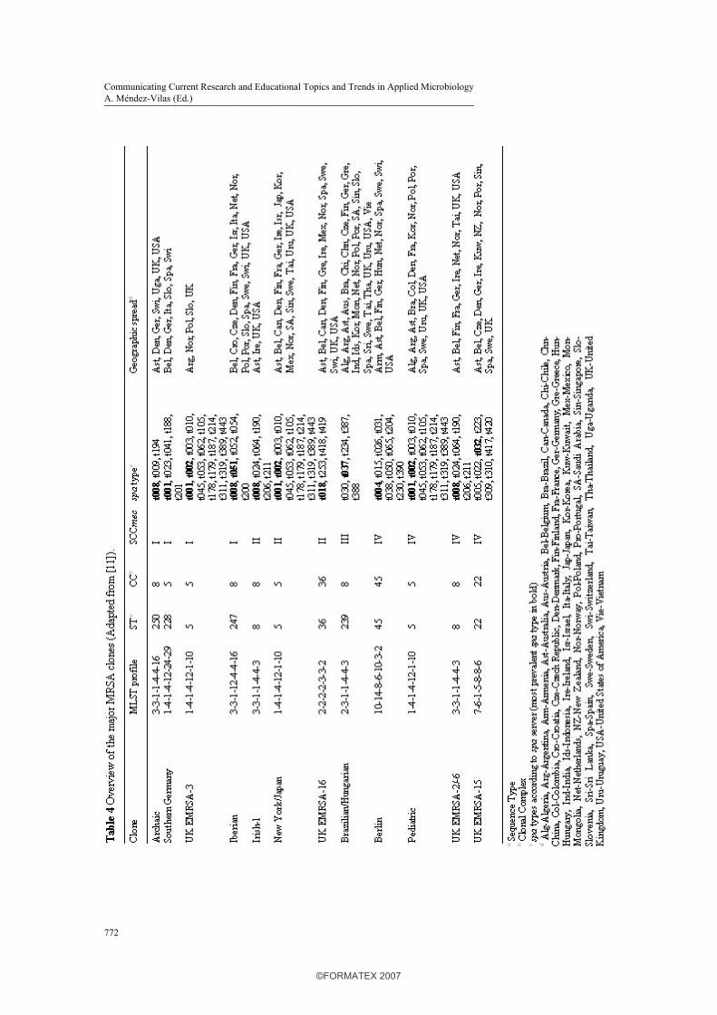
In 1961, two years after the introduction of methicillin, MRSA strains emerged. During the following years, MRSA strains disseminated to other European countries, whereas in the 1970s, MRSA spread worldwide, e.g. Australia, Japan and the USA. Currently, MRSA is a major cause of nosocomial infections worldwide, and the prevalence ranges from less than 1% in The Netherlands and Scandinavia to over 60% in Japan [49]. The worldwide spread of MRSA is driven by the dissemination of various clones with a specific genetic background (Table 4) [13, 26, 33, 50-84]. Two opposing theories have been suggested to describe the relationship between the first MRSA isolated and the recent MRSA clones. While the single-clone theory suggests that all MRSA clones have a common ancestor and that SCCmec was introduced only once in S. aureus [85], the multi-clone theory suggests that SCCmec was introduced several times into various S aureus lineages. The latter hypothesis is supported by a number of studies [33, 66, 86, 87]. Enright et al. investigated 359 MRSA and 553 MSSA isolates from 20 countries isolated between 1961 and 1999 with SCCmec typing and MLST. In the MRSA population, five CCs were found, and isolates with the same ST harboured different SCCmec elements (Table 4) [33]. The major MRSA clones, defined as groups of isolates from more than one country with the same ST and SCCmec element, belonged either to CC5, 8, 22, 30, or 45. Table 4 shows that different SCCmec elements have been acquired by S. aureus strains with a different genetic background, and this supports the multi-clone theory described above. Furthermore, it was shown that ST8-MSSA in CC8 is the ancestor of the first MRSA strain isolated, i.e. ST250-MRSA-I; ST250 differs from ST8 by a point mutation in the yqiL locus. ST8-MSSA is a common cause of epidemic MSSA disease and it has acquired SCCmec types I, II and IV (Fig. 2).
Fig. 2 Evolutionary origins of the major MRSA clones and the possible relation between CA-MRSA and HA-MRSA. The arrows indicating either 1) the acquisition of SCCmec, 2) a change of SCCmec, 3) a change of ST, or 4) the acquisition of PVL. The grey coloured circles represent the MRSA clones from CC30, while the white circles represent the MRSA clones from CC8. ST239-MRSA-III from CC8 has evolved by the transfer of a 557-kb fragment from the chromosome of ST30 into a ST8 background [11, 33, 88, 89] (Adapted from [11]).
771
Communicating Current Research and Educational Topics and Trends in Applied Microbiology A. Méndez-Vilas (Ed.)_____________________________________________________________________
©FORMATEX 2007
772
Communicating Current Research and Educational Topics and Trends in Applied Microbiology A. Méndez-Vilas (Ed.)_____________________________________________________________________
©FORMATEX 2007
Another ST250-related clone is ST247-MRSA-I (Iberian clone). These STs differ from each other by a single point mutation at the gmk locus. ST247-MRSA-I is one of the major MRSA clones currently isolated in European hospitals (Table 4). ST239-MRSA-III (Brazilian clone) is another major MRSA clone within CC8. It has evolved by the transfer through homologous recombination of a 557-kb fragment of the chromosome of ST30 into ST239-MRSA-III (Fig. 2) [44]. Furthermore, it has been shown that CC5, 22, 36 and 45 were all derived from epidemic MSSA lineages that acquired SCCmec, since they differed from each other and from ST8 at six or seven MLST loci (Table 4). MLST analyses also showed that some of the first vancomycin-intermediate S. aureus (VISA) isolates have emerged from ST5-MRSA-II, a pandemic MRSA clone known as the New York/Japan clone [33]. As shown by a study characterizing 147 geographically diverse MRSA strains, MRSA has emerged at least twenty times upon acquisition of SCCmec. That study also demonstrated that the acquisition of SCCmec by MSSA was four times more common than the replacement of one SCCmec element with another element. Furthermore, SCCmec type IV was found in twice as many MRSA clones than other SCCmec elements. This suggests that most clones arise by acquisition of SCCmec type IV in MSSA [90]. This is probably due to the smaller size of SCCmec type IV element compared to the other SCCmec elements, which may facilitate transfer of the cassette between staphylococcal species [89]. Furthermore, it has been shown that MRSA strains that belong to CC1, 5, 8, 22, 30, and 45, were easier to transform with mecA-expressing plasmids, compared to strains belonging to other CCs. This suggests that the genetic background of S. aureus is important for the stability of SCCmec [91]. Besides the major clones presented in table 4, certain MRSA strains are isolated in single hospitals (minor clones), or from single patients (sporadic isolates) [26]. Recently, several studies have described the clonal evolution within one hospital, such as in Mexico [56]. Although the majority of the MRSA strains are isolated in hospitals, CA-MRSA is an emerging problem outside the hospital.
5. Worldwide emergence of CA-MRSA
The emergence of CA-MRSA is a worldwide threat to both the community and the hospital environment since these strains are more virulent than HA-MRSA strains [92, 93]. Furthermore, CA-MRSA strains have started to replace HA-MRSA in health-care settings [94]. Since many definitions for CA-MRSA exist [95, 96], a general and international agreement has been reached on a universal definition of CA-MRSA. The Center for Disease Control and Prevention (CDC) defines CA-MRSA as strains isolated in an outpatient setting, or from patients within 48 hours of hospital admission. Furthermore, these patients must have no medical history of MRSA infection or colonisation, and no medical history in the past year of either hospitalisation (e.g. surgery), admission to a nursing home, or dialysis. Moreover, the patient should not have permanent indwelling catheters or medical devices that pass through the skin. In 1993, the first CA-MRSA strain was reported from patients in remote communities in Western Australia [97]. Interestingly, CA-MRSA strains were isolated from patients who have no known risk factors for MRSA colonisation. CA-MRSA is both phenotypically and genotypically different from HA-MRSA. In contrast to HA-MRSA, CA-MRSA strains are mainly susceptible to antibiotics other than β-lactam antibiotics. PFGE and MLST have shown that CA-MRSA strains belong to clonal types unrelated to clones isolated in hospitals [98, 99], and that CA-MRSA has greater clonal diversity than HA-MRSA [33, 46]. Although CA-MRSA mainly harbour SCCmec type IV or V [4, 6, 7], two reports described CA-MRSA isolates with SCCmec type I, II or III [51, 100]. The various reports are conflicting as to whether there is a relation between SCCmec type IV and Panton-Valentine leukocidin (PVL). PVL is a S. aureus-specific exotoxin, which is encoded by two co-transcribed genes, designated lukF-PV and lukS-PV, and is associated with skin and soft tissue infections, and severe necrotizing pneumonia [25, 101]. Vandenesch et al. showed that CA-MRSA was characterised by SCCmec type IV and that PVL was a stable genetic marker for CA-MRSA [25]. The relationship between CA-MRSA, SCCmec type IV and PVL was confirmed in a study by Shukla et al. in the USA [57]. However, another study by O’Brien et al. in Australia did not find a relationship between CA-MRSA, SCCmec type IV and PVL [58]. Further studies showed PVL–positive CA-MRSA strains
773
Communicating Current Research and Educational Topics and Trends in Applied Microbiology A. Méndez-Vilas (Ed.)_____________________________________________________________________
©FORMATEX 2007

harbouring SCCmec type I and III in The Netherlands [100] and PVL-positive HA-MRSA strains in Algeria [72]. In general, 40 to 90% of the MRSA strains that harbour SCCmec type IV carry PVL and less than 5% of the MRSA strains harbouring SCCmec type I to III carry PVL [102]. Further studies are needed to investigate the possible relation between SCCmec type IV (and V) and PVL in CA-MRSA strains. Recently, Müller-Premru et al described the first detection of PVL in MRSA with ST5 (spa t002), and CA-MRSA with ST152 (spa t454) in Slovenia associated with a clinical significant outbreak within members of a football team. The emergence of PVL in MRSA with ST5 is of particular concern, because of its epidemic potential with high capacity to spread [103]. Five main PVL-positive CA-MRSA clones are isolated worldwide. The ST1 clone [spa t127, t128, t174, t176, t386, t558] is found in Asia, Europe and the USA, the ST8 clone [spa t008, t024, t064, t190, t206, t211] in Europe and the USA, the ST30 clone [spa t012, t018, t019, t021, t138, t268, t276, t318, t338, t391] is found in Australia, Europe and South America, the ST59 clone [spa t199, t216, t437, t444] in Asia and the USA, and the ST80 clone [spa t044, t131] in Asia, Europe and the Middle-East [25, 104, 105]. Although these CA-MRSA clones harbour SCCmec type IV, the ST1 clone harbours SCCmec type IVa and the ST80 clone harbours SCCmec type IVc [106]. Furthermore, the ST80 clone harbours the farI gene, coding for resistance to fusidic acid. SCCmec type V is present in Australian CA-MRSA strains with ST5, 8, 45, 59, 152, 573 and 577, in Taiwanese isolates with ST59, in Finish strains with ST8 and 27, in strains from Uruguay with ST45, in Singaporean strains with ST1, 7, 8, 45, 59, 88, 188, 524 and 573, in Australian, Dutch, French, Greek and Swiss strains with ST377, and in strains from Kosovo with ST152 [21, 68-71, 80, 105, 107-109]. This shows that SCCmec type V harbouring CA-MRSA isolates have a diverse genetic background. Whether SCCmec of CA-MRSA has been acquired by MSSA strains in the community, or that CA-MRSA is derived from HA-MRSA remains unclear. Okuma et al showed that CA-MRSA are novel acquisitions of SCCmec type IV in the community [46]. Another study by Aires de Sousa et al. raised the possibility that some CA-MRSA strains may originate in hospitals, since several similarities between CA-MRSA and HA-MRSA strains were found [110]. A recent study showed that a HA-MRSA and a CA-MRSA clone have a common ancestor. In the 1950s, a penicillin-resistant S. aureus clone (phage type 80/81) emerged worldwide in hospitals and the community, but since the introduction of penicillinase-resistant ß-lactam antibiotics in the 1960s, this clone has disappeared. This PVL-harbouring ST30 clone re-emerged and acquired SCCmec type IV to become the ST30-MRSA-IV CA clone found in Australia. ST30-MSSA has also acquired SCCmec type II, possible through several intermediate steps, such as the acquisition of SCCmec type IV, to become ST36-MRSA-II, the pandemic EMRSA-16 clone (Fig. 2) [88]. This paragraph shows that there are still a number of questions to be answered about the molecular epidemiology of CA-MRSA.
References [1] FD Lowy, N Engl J Med 339, 520 (1998). [2] FD Lowy, J Clin Invest 111, 1265 (2003). [3] B Berger-Bachi, and S Rohrer, Arch Microbiol 178, 165 (2002). [4] T Ito, K Okuma, XX Ma, H Yuzawa, and K Hiramatsu, Drug Resist Updat 6, 41 (2003). [5] T Ito, Y Katayama, K Asada, N Mori, K Tsutsumimoto, C Tiensasitorn, and K Hiramatsu, Antimicrob Agents
Chemother 45, 1323 (2001). [6] T Ito, XX Ma, F Takeuchi, K Okuma, H Yuzawa, and K Hiramatsu, Antimicrob Agents Chemother 48, 2637
(2004). [7] RS Daum, T Ito, K Hiramatsu, F Hussain, K Mongkolrattanothai, M Jamklang, and S Boyle-Vavra, J Infect Dis
186, 1344 (2002). [8] RH Deurenberg, C Vink, GJ Oudhuis, JE Mooij, C Driessen, G Coppens, J Craeghs, E De Brauwer, S
Lemmen, H Wagenvoort, AW Friedrich, J Scheres, and EE Stobberingh, Antimicrob Agents Chemother 49, 4263 (2005).
[9] DC Oliveira, C Milheirico, and H de Lencastre, Antimicrob Agents Chemother 50, 3457 (2006). [10] DC Oliveira, and H de Lencastre, Antimicrob Agents Chemother 46, 2155 (2002).
774
Communicating Current Research and Educational Topics and Trends in Applied Microbiology A. Méndez-Vilas (Ed.)_____________________________________________________________________
©FORMATEX 2007
[11] RH Deurenberg, C Vink, S Kalenic, AW Friedrich, CA Bruggeman, and EE Stobberingh, Clinical Microbiology and Infection 13, 222 (2007).
[12] R Leclercq, Clin Infect Dis 34, 482 (2002). [13] P Chongtrakool, T Ito, XX Ma, Y Kondo, S Trakulsomboon, C Tiensasitorn, M Jamklang, T Chavalit, JH
Song, and K Hiramatsu, Antimicrob Agents Chemother 50, 1001 (2006). [14] Y Katayama, T Ito, and K Hiramatsu, Antimicrob Agents Chemother 45, 1955 (2001). [15] T Ito, Y Katayama, and K Hiramatsu, Antimicrob Agents Chemother 43, 1449 (1999). [16] K Hiramatsu, L Cui, M Kuroda, and T Ito, Trends Microbiol 9, 486 (2001). [17] DC Oliveira, A Tomasz, and H de Lencastre, Microb Drug Resist 7, 349 (2001). [18] A Shore, AS Rossney, CT Keane, MC Enright, and DC Coleman, Antimicrob Agents Chemother 49, 2070
(2005). [19] K Hisata, K Kuwahara-Arai, M Yamanoto, T Ito, Y Nakatomi, L Cui, T Baba, M Terasawa, C Sotozono, S
Kinoshita, Y Yamashiro, and K Hiramatsu, J Clin Microbiol 43, 3364 (2005). [20] XX Ma, T Ito, C Tiensasitorn, M Jamklang, P Chongtrakool, S Boyle-Vavra, RS Daum, and K Hiramatsu,
Antimicrob Agents Chemother 46, 1147 (2002). [21] S Boyle-Vavra, B Ereshefsky, CC Wang, and RS Daum, J Clin Microbiol 43, 4719 (2005). [22] NH Kwon, KT Park, JS Moon, WK Jung, SH Kim, JM Kim, SK Hong, HC Koo, YS Joo, and YH Park, J
Antimicrob Chemother (2005). [23] C Milheirico, DC Oliveira, and H de Lencastre, J Antimicrob Chemother (2007). [24] E van Duijkeren, MJ Wolfhagen, AT Box, ME Heck, WJ Wannet, and AC Fluit, Emerg Infect Dis 10, 2235
(2004). [25] F Vandenesch, T Naimi, MC Enright, G Lina, GR Nimmo, H Heffernan, N Liassine, M Bes, T Greenland, ME
Reverdy, and J Etienne, Emerg Infect Dis 9, 978 (2003). [26] MA de Sousa, and H de Lencastre, FEMS Immunol Med Microbiol 40, 101 (2004). [27] FC Tenover, RD Arbeit, RV Goering, PA Mickelsen, BE Murray, DH Persing, and B Swaminathan, J Clin
Microbiol 33, 2233 (1995). [28] S Murchan, ME Kaufmann, A Deplano, R de Ryck, M Struelens, CE Zinn, V Fussing, S Salmenlinna, J
Vuopio-Varkila, N El Solh, C Cuny, W Witte, PT Tassios, N Legakis, W van Leeuwen, A van Belkum, A Vindel, I Laconcha, J Garaizar, S Haeggman, B Olsson-Liljequist, U Ransjo, G Coombes, and B Cookson, J Clin Microbiol 41, 1574 (2003).
[29] FC Tenover, R Arbeit, G Archer, J Biddle, S Byrne, R Goering, G Hancock, GA Hebert, B Hill, R Hollis, and et al., J Clin Microbiol 32, 407 (1994).
[30] A van Belkum, W van Leeuwen, ME Kaufmann, B Cookson, F Forey, J Etienne, R Goering, F Tenover, C Steward, F O'Brien, W Grubb, P Tassios, N Legakis, A Morvan, N El Solh, R de Ryck, M Struelens, S Salmenlinna, J Vuopio-Varkila, M Kooistra, A Talens, W Witte, and H Verbrugh, J Clin Microbiol 36, 1653 (1998).
[31] MC Enright, and BG Spratt, Trends Microbiol 7, 482 (1999). [32] MC Enright, NP Day, CE Davies, SJ Peacock, and BG Spratt, J Clin Microbiol 38, 1008 (2000). [33] MC Enright, DA Robinson, G Randle, EJ Feil, H Grundmann, and BG Spratt, Proc Natl Acad Sci U S A 99,
7687 (2002). [34] BG Spratt, WP Hanage, B Li, DM Aanensen, and EJ Feil, FEMS Microbiol Lett 241, 129 (2004). [35] HM Frenay, AE Bunschoten, LM Schouls, WJ van Leeuwen, CM Vandenbroucke-Grauls, J Verhoef, and FR
Mooi, Eur J Clin Microbiol Infect Dis 15, 60 (1996). [36] B Shopsin, M Gomez, SO Montgomery, DH Smith, M Waddington, DE Dodge, DA Bost, M Riehman, S
Naidich, and BN Kreiswirth, J Clin Microbiol 37, 3556 (1999). [37] BC Kahl, A Mellmann, S Deiwick, G Peters, and D Harmsen, J Clin Microbiol 43, 502 (2005). [38] N Malachowa, A Sabat, M Gniadkowski, J Krzyszton-Russjan, J Empel, J Miedzobrodzki, K Kosowska-Shick,
PC Appelbaum, and W Hryniewicz, J Clin Microbiol 43, 3095 (2005). [39] L Koreen, SV Ramaswamy, EA Graviss, S Naidich, JM Musser, and BN Kreiswirth, J Clin Microbiol 42, 792
(2004). [40] M Aires-de-Sousa, K Boye, H de Lencastre, A Deplano, MC Enright, J Etienne, A Friedrich, D Harmsen, A
Holmes, XW Huijsdens, AM Kearns, A Mellmann, H Meugnier, JK Rasheed, E Spalburg, B Strommenger, MJ Struelens, FC Tenover, J Thomas, U Vogel, H Westh, J Xu, and W Witte, J Clin Microbiol 44, 619 (2006).
[41] D Harmsen, H Claus, W Witte, J Rothganger, H Claus, D Turnwald, and U Vogel, J Clin Microbiol 41, 5442 (2003).
[42] AW Friedrich, W Witte, D Harmsen, H de Lencastre, W Hryniewicz, J Scheres, and H Westh, Euro Surveill 11, E060112 4 (2006).
775
Communicating Current Research and Educational Topics and Trends in Applied Microbiology A. Méndez-Vilas (Ed.)_____________________________________________________________________
©FORMATEX 2007

[43] A Mellmann, AW Friedrich, N Rosenkotter, J Rothganger, H Karch, R Reintjes, and D Harmsen, PLoS Med 3, e33 (2006).
[44] DA Robinson, and MC Enright, J Bacteriol 186, 1060 (2004). [45] B Strommenger, C Kettlitz, T Weniger, D Harmsen, AW Friedrich, and W Witte, J Clin Microbiol 44, 2533
(2006). [46] K Okuma, K Iwakawa, JD Turnidge, WB Grubb, JM Bell, FG O'Brien, GW Coombs, JW Pearman, FC
Tenover, M Kapi, C Tiensasitorn, T Ito, and K Hiramatsu, J Clin Microbiol 40, 4289 (2002). [47] K Zhang, JA McClure, S Elsayed, T Louie, and JM Conly, J Clin Microbiol 43, 5026 (2005). [48] Y Kondo, T Ito, XX Ma, S Watanabe, BN Kreiswirth, J Etienne, and K Hiramatsu, Antimicrob Agents
Chemother (2006). [49] H Grundmann, M Aires-de-Sousa, J Boyce, and E Tiemersma, Lancet 368, 874 (2006). [50] DC Oliveira, A Tomasz, and H de Lencastre, Lancet Infect Dis 2, 180 (2002). [51] M Chung, G Dickinson, H De Lencastre, and A Tomasz, J Clin Microbiol 42, 542 (2004). [52] O Denis, A Deplano, C Nonhoff, R De Ryck, R de Mendonca, S Rottiers, R Vanhoof, and MJ Struelens,
Antimicrob Agents Chemother 48, 3625 (2004). [53] KS Ko, JY Lee, JY Suh, WS Oh, KR Peck, NY Lee, and JH Song, J Clin Microbiol 43, 421 (2005). [54] O Melter, M Aires de Sousa, P Urbaskova, V Jakubu, H Zemlickova, and H de Lencastre, J Clin Microbiol 41,
4998 (2003). [55] E Perez-Roth, F Lorenzo-Diaz, N Batista, A Moreno, and S Mendez-Alvarez, J Clin Microbiol 42, 4649
(2004). [56] ME Velazquez-Meza, M Aires De Sousa, G Echaniz-Aviles, F Solorzano-Santos, G Miranda-Novales, J Silva-
Sanchez, and H De Lencastre, J Clin Microbiol 42, 3877 (2004). [57] SK Shukla, ME Stemper, SV Ramaswamy, JM Conradt, R Reich, EA Graviss, and KD Reed, J Clin Microbiol
42, 3752 (2004). [58] FG O'Brien, TT Lim, FN Chong, GW Coombs, MC Enright, DA Robinson, A Monk, B Said-Salim, BN
Kreiswirth, and WB Grubb, J Clin Microbiol 42, 3185 (2004). [59] NA Faria, DC Oliveira, H Westh, DL Monnet, AR Larsen, R Skov, and H de Lencastre, J Clin Microbiol 43,
1836 (2005). [60] AM Hanssen, A Fossum, J Mikalsen, DS Halvorsen, G Bukholm, and JU Sollid, J Clin Microbiol 43, 2118
(2005). [61] HY Cha, DC Moon, CH Choi, JY Oh, YS Jeong, YC Lee, SY Seol, DT Cho, HH Chang, SW Kim, and JC Lee,
J Clin Microbiol 43, 3610 (2005). [62] LY Hsu, TH Koh, K Singh, ML Kang, A Kurup, and BH Tan, J Clin Microbiol 43, 2923 (2005). [63] MA de Sousa, T Conceicao, C Simas, and H de Lencastre, J Clin Microbiol 43, 5150 (2005). [64] C Berglund, P Molling, L Sjoberg, and B Soderquist, J Clin Microbiol 43, 4448 (2005). [65] F McAleese, E Murphy, T Babinchak, G Singh, B Said-Salim, B Kreiswirth, P Dunman, J O'Connell, SJ
Projan, and PA Bradford, Antimicrob Agents Chemother 49, 4521 (2005). [66] W Qi, M Ender, F O'Brien, A Imhof, C Ruef, N McCallum, and B Berger-Bachi, J Clin Microbiol 43, 5164
(2005). [67] H Wisplinghoff, B Ewertz, S Wisplinghoff, D Stefanik, G Plum, F Perdreau-Remington, and H Seifert, J Clin
Microbiol 43, 5445 (2005). [68] S Harbarth, P Francois, J Shrenzel, C Fankhauser-Rodriguez, S Hugonnet, T Koessler, A Huyghe, and D Pittet,
Emerg Infect Dis 11, 962 (2005). [69] XX Ma, A Galiana, W Pedreira, M Mowszowicz, I Christophersen, S Machiavello, L Lope, S Benaderet, F
Buela, W Vincentino, M Albini, O Bertaux, I Constenla, H Bagnulo, L Llosa, T Ito, and K Hiramatsu, Emerg Infect Dis 11, 973 (2005).
[70] GW Coombs, Emerg Infect Dis 12, 241 (2006). [71] LY Hsu, TH Koh, TY Tan, T Ito, XX Ma, RT Lin, and BH Tan, Singapore Med J 47, 20 (2006). [72] N Ramdani-Bouguessa, M Bes, H Meugnier, F Forey, ME Reverdy, G Lina, F Vandenesch, M Tazir, and J
Etienne, Antimicrob Agents Chemother 50, 1083 (2006). [73] C Sola, P Cortes, HA Saka, A Vindel, and JL Bocco, J Clin Microbiol 44, 192 (2006). [74] EE Udo, N Al-Sweih, and B Noronha, Clin Microbiol Infect 12, 262 (2006). [75] A Budimir, RH Deurenberg, V Plecko, C Vink, S Kalenic, and EE Stobberingh, J Antimicrob Chemother 57,
331 (2006). [76] A Vindel, P Trincado, E Gomez, R Cabrera, T Boquete, C Sola, S Valdezate, and JA Saez-Nieto, J Clin
Microbiol 44, 266 (2006).
776
Communicating Current Research and Educational Topics and Trends in Applied Microbiology A. Méndez-Vilas (Ed.)_____________________________________________________________________
©FORMATEX 2007
[77] D Orth, K Grif, L Erdenechimeg, C Battogtokh, T Hosbayar, B Strommenger, C Cuny, G Walder, C Lass-Florl, MP Dierich, and W Witte, Eur J Clin Microbiol Infect Dis 25, 104 (2006).
[78] AS Rossney, MJ Lawrence, PM Morgan, MM Fitzgibbon, A Shore, DC Coleman, CT Keane, and B O'Connell, Eur J Clin Microbiol Infect Dis 25, 79 (2006).
[79] G Durand, M Bes, H Meugnier, MC Enright, F Forey, N Liassine, A Wenger, K Kikuchi, G Lina, F Vandenesch, and J Etienne, J Clin Microbiol 44, 847 (2006).
[80] LY Hsu, YL Koh, NL Chlebicka, TY Tan, P Krishnan, RT Lin, N Tee, T Barkham, and TH Koh, J Clin Microbiol 44, 1090 (2006).
[81] AM Vivoni, BA Diep, AC de Gouveia Magalhaes, KR Santos, LW Riley, GF Sensabaugh, and BM Moreira, J Clin Microbiol 44, 1686 (2006).
[82] AE Fossum, and G Bukholm, Clin Microbiol Infect 12, 627 (2006). [83] YC Huang, LH Su, TL Wu, and TY Lin, J Clin Microbiol 44, 2268 (2006). [84] G Regev-Yochay, Y Carmeli, M Raz, E Pinco, J Etienne, A Leavitt, E Rubinstein, and S Navon-Venezia, Eur J
Clin Microbiol Infect Dis (2006). [85] B Kreiswirth, J Kornblum, RD Arbeit, W Eisner, JN Maslow, A McGeer, DE Low, and RP Novick, Science
259, 227 (1993). [86] JM Musser, and V Kapur, J Clin Microbiol 30, 2058 (1992). [87] JR Fitzgerald, DE Sturdevant, SM Mackie, SR Gill, and JM Musser, Proc Natl Acad Sci U S A 98, 8821
(2001). [88] DA Robinson, AM Kearns, A Holmes, D Morrison, H Grundmann, G Edwards, FG O'Brien, FC Tenover, LK
McDougal, AB Monk, and MC Enright, Lancet 365, 1256 (2005). [89] DA Robinson, and MC Enright, Clin Microbiol Infect 10, 92 (2004). [90] DA Robinson, and MC Enright, Antimicrob Agents Chemother 47, 3926 (2003). [91] Y Katayama, DA Robinson, MC Enright, and HF Chambers, J Clin Microbiol 43, 2380 (2005). [92] J Etienne, Clin Infect Dis 41, 591 (2005). [93] HF Chambers, Emerg Infect Dis 7, 178 (2001). [94] JA Otter, and GL French, The Lancet Infectious Diseases 6, 753 (2006). [95] TS Naimi, KH LeDell, K Como-Sabetti, SM Borchardt, DJ Boxrud, J Etienne, SK Johnson, F Vandenesch, S
Fridkin, C O'Boyle, RN Danila, and R Lynfield, Jama 290, 2976 (2003). [96] CD Salgado, BM Farr, and DP Calfee, Clin Infect Dis 36, 131 (2003). [97] EE Udo, JW Pearman, and WB Grubb, J Hosp Infect 25, 97 (1993). [98] AV Groom, DH Wolsey, TS Naimi, K Smith, S Johnson, D Boxrud, KA Moore, and JE Cheek, Jama 286,
1201 (2001). [99] TS Naimi, KH LeDell, DJ Boxrud, AV Groom, CD Steward, SK Johnson, JM Besser, C O'Boyle, RN Danila,
JE Cheek, MT Osterholm, KA Moore, and KE Smith, Clin Infect Dis 33, 990 (2001). [100] WJ Wannet, E Spalburg, ME Heck, GN Pluister, E Tiemersma, RJ Willems, XW Huijsdens, AJ de Neeling,
and J Etienne, J Clin Microbiol 43, 3341 (2005). [101] G Prevost, P Couppie, P Prevost, S Gayet, P Petiau, B Cribier, H Monteil, and Y Piemont, J Med Microbiol
42, 237 (1995). [102] S Deresinski, Clin Infect Dis 40, 562 (2005). [103] M Muller-Premru, B Strommenger, N Alikadic, W Witte, AW Friedrich, K Seme, NS Kucina, D Smrke, V
Spik, and M Gubina, Eur J Clin Microbiol Infect Dis 24, 848 (2005). [104] HF Chambers, N Engl J Med 352, 1485 (2005). [105] A Tristan, M Bes, H Meugnier, G Lina, B Bozdogan, P Courvalin, ME Reverdy, MC Enright, F Vandenesch,
and J Etienne, Emerg Infect Dis 13, 594 (2007). [106] W Witte, C Braulke, C Cuny, B Strommenger, G Werner, D Heuck, U Jappe, C Wendt, HJ Linde, and D
Harmsen, Eur J Clin Microbiol Infect Dis 24, 1 (2005). [107] GW Coombs, GR Nimmo, JM Bell, F Huygens, G O'Brien F, MJ Malkowski, JC Pearson, AJ Stephens, and
PM Giffard, J Clin Microbiol 42, 4735 (2004). [108] G O'Brien F, GW Coombs, JC Pearson, KJ Christiansen, and WB Grubb, Antimicrob Agents Chemother 49,
5129 (2005). [109] AM Kerttula, O Lyytikainen, J Vuopio-Varkila, S Ibrahem, N Agthe, M Broas, H Jagerroos, and A Virolainen,
J Clin Microbiol 43, 6161 (2005). [110] M Aires de Sousa, and H de Lencastre, J Clin Microbiol 41, 3806 (2003).
777
Communicating Current Research and Educational Topics and Trends in Applied Microbiology A. Méndez-Vilas (Ed.)_____________________________________________________________________
©FORMATEX 2007