Isolation of Methicillin-Resistant Coagulase-Negative ...
38
James Madison University JMU Scholarly Commons Senior Honors Projects, 2010-current Honors College Spring 2014 Isolation of methicillin-resistant coagulase-negative Staphylococcus (MRCoNS) from a fecal- contaminated stream in the Shenandoah Valley of Virginia Michael Timothy Partin James Madison University Follow this and additional works at: hps://commons.lib.jmu.edu/honors201019 is esis is brought to you for free and open access by the Honors College at JMU Scholarly Commons. It has been accepted for inclusion in Senior Honors Projects, 2010-current by an authorized administrator of JMU Scholarly Commons. For more information, please contact [email protected]. Recommended Citation Partin, Michael Timothy, "Isolation of Methicillin-Resistant Coagulase-Negative Staphylococcus (MRCoNS) from a fecal- contaminated stream in the Shenandoah Valley of Virginia" (2014). Senior Honors Projects, 2010-current. 459. hps://commons.lib.jmu.edu/honors201019/459
Transcript of Isolation of Methicillin-Resistant Coagulase-Negative ...
James Madison UniversityJMU Scholarly Commons
Senior Honors Projects, 2010-current Honors College
Spring 2014
Isolation of methicillin-resistant coagulase-negativeStaphylococcus (MRCoNS) from a fecal-contaminated stream in the Shenandoah Valley ofVirginiaMichael Timothy PartinJames Madison University
Follow this and additional works at: https://commons.lib.jmu.edu/honors201019
This Thesis is brought to you for free and open access by the Honors College at JMU Scholarly Commons. It has been accepted for inclusion in SeniorHonors Projects, 2010-current by an authorized administrator of JMU Scholarly Commons. For more information, please [email protected].
Recommended CitationPartin, Michael Timothy, "Isolation of Methicillin-Resistant Coagulase-Negative Staphylococcus (MRCoNS) from a fecal-contaminated stream in the Shenandoah Valley of Virginia" (2014). Senior Honors Projects, 2010-current. 459.https://commons.lib.jmu.edu/honors201019/459
Isolation of Methicillin-Resistant Coagulase-Negative Staphylococcus (MRCoNS) from a Fecal-Contaminated Stream in the Shenandoah Valley of Virginia
_____________________________
A Project Presented to
The Faculty of the Undergraduate
College of Science and Mathematics
James Madison University
_____________________________
In Partial Fulfillment of the Requirements
for the Degree of Bachelor of Science
_____________________________
by Michael Timothy Partin
May 2014
Accepted by the faculty of the Department of Biology, James Madison University, in partial fulfillment of the requirements for the Degree of Bachelor of Science.
FACULTY COMMITTEE: HONORS PROGRAM APPROVAL:
____________________________________________ ___________________________________________ Faculty Project Advisor: James Herrick, Ph. D., Barry Falk, Ph. D., Associate Professor, Biology Director, Honors Program ____________________________________________ Reader: Steve Cresawn, Ph. D., Associate Professor, Biology ____________________________________________ Reader: Kyle Seifert, Ph. D., Associate Professor, Biology
2
Table of Contents
List of Figures and Tables 3
Acknowledgements 4 Abstract 5 Introduction 7 Materials and Methods 11 Results 18 Discussion 27 Literature Cited 35

3
List of Figures and Tables
Figures
Figure 1. Example of colony morphology used to isolate putative Staphylococcus sp..
Figure 2. BOX-PCR of isolates used to differentiate isolated strains.
Figure 3. Zones of inhibition of Staphylococcus isolates to oxacillin.
Tables
Table 1. Primers used in this study.
Table 2. Locations and collection dates of eleven Staphylococcus isolates collected from Muddy
Creek in Hinton, Virginia.
Table 3. Summary of BLAST search results for the 16S rRNA nucleotide sequences from
isolates PS6-8, 10, and 11.
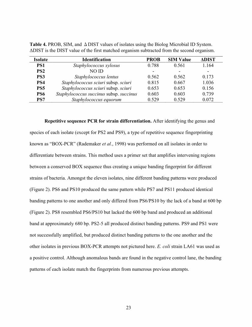
Table 4. PROB, SIM, and ΔDIST values of isolates using the Biolog Microbial ID System.

4
Acknowledgements
I would like to thank Dr. Herrick for his friendship, mentorship, advice, and wisdom
during this project over the past two years. I am extremely thankful for his influence on me both
personally and as a scientist during my time at James Madison University. I also would like to
thank Dr. Seifert for teaching me microbiology in two different courses over the past two years. I
want to thank Dr. Seifert and Dr. Cresawn for reading and editing my thesis as well as offering
suggestions of ways to improve my research. I would like to thank Dr. Vasudevan for assisting in
the 16S rRNA sequencing process. Additionally, I am thankful for Dr. Temple who collaborated
with our lab and provided helpful reagents in performing several of the experiments. I would like
to thank Brooke Sauder for providing reagents for the mecA PCR and assisting with the
experiment. I would like to thank Steven Mcbride for his assistance and guidance during the
Biolog experiment. Furthermore, I would like to thank my close friends Kevin Libuit, Curtis
Kapsack, and Jennifer Kurasz for providing countless hours to help me with this project. Finally,
I would like to thank James Madison University and the Department of Biology for providing the
resources and facilities for this project.
5
Abstract
Staphylococcus is comprised of 41 known species, of which 18 can colonize humans.
Despite the prevalence of infectious Staphylococcus within hospital settings and agriculture,
there are few reports of Staphylococcus in natural bodies of water. A recent study by the US
Food and Drug Administration found substantial contamination of poultry and other meats with
Staphylococcus. We hypothesized that intensive farming of poultry adjacent to streams would
result in contaminated runoff, resulting in at least transient occurrence of Staphylococcus spp. in
stream waters and sediments. In this study, we sought to determine whether Staphylococcus
occurs and persists within Muddy Creek, a stream located in Hinton, Virginia that originates at
the Appalachian Mountains of Virginia and runs through various agricultural fields and adjacent
to a poultry processing plant in the central Shenandoah Valley. Five different Staphylococcus spp.
were detected in water and sediment from Muddy Creek. Mannitol Salt Agar (MSA) was used to
isolate eleven Staphylococcus from both water and sediment. These isolates were Gram-positive,
catalase-positive, and oxidase-negative cocci that were capable of fermenting mannitol. In
addition, a method for screening putative staphylococci species from stream water and sediment
was developed. Ten out of the eleven tested isolates were oxacillin resistant (now used to
identify phenotypic methicillin-resistance) using a Kirby Bauer disc diffusion test. Furthermore,
the isolates were susceptible to trimethoprim/sulfamethoxazole, tetracycline, and gentamicin
while two of the isolates were resistant to erythromycin. Additionally, the BOX-PCR repetitive
sequence fingerprinting method verified the presence of nine different strains among the isolates.
Sequencing of the 16S rRNA gene identified five of the isolates as Staphylococcus equorum. The
Biolog identification protocol further identified the remaining isolates as Staphylococcus xylosus,
Staphylococcus lentus, Staphylococcus succinus, and Staphylococcus sciuri. Finally, polymerase

6
chain reaction amplification (PCR) confirmed that ten of the eleven isolates harbored the mecA
gene known to confer methicillin-resistance. Overall, the occurrence of coagulase-negative
staphylococci (MRCoNS) in stream water and sediment represents a potential environmental and
human health concern.
7
Introduction
Staphylococcus is a bacterial genus comprised of 41 known species, of which 18 possess
the ability to colonize humans (Schaechter, 2009). All staphylococci are Gram-positive cocci
with an approximate diameter of 0.7-1.2 µm. Growth patterns occur as single or paired cells in
liquid media and form grape-like clusters in solid media. Furthermore, staphylococci express the
catalase enzyme responsible for the decomposition of harmful hydrogen peroxide within the cell.
In general, staphylococci most commonly inhabit animals on the skin and the mucous
membranes. However, Staphylococcus spp. have occasionally been isolated from other sources
including soil, water, and food (Faria et al., 2009). Microbiologists generally divide the genus
based on the presence or absence of the coagulase enzyme, which is responsible for converting
fibrinogen to fibrin (Leboffe, 2006). For example, a laboratory coagulase test helps to distinguish
between S. aureus (coagulase-positive) and S. epidermidis (coagulase-negative), both of which
are common species known to inhabit humans.
Staphylococcus species – and particularly S. aureus – are responsible for potentially fatal
infections of the epidermal tissue in humans. Additionally, members of this genus are known to
cause endocarditis and septic shock. Staphylococcus release superantigens – a class of antigens
known to activate T-cells and cause cytokine release – leading to fever, capillary leak, and
multiorgan failure (Lowy, 1998). Staphylococcus species commonly contain regulatory genes,
such as agr, which modulate the expression of extracellular proteins known to contribute to the
organism’s virulence (Lowy, 1998). As the stages of infection progress, these genes are regulated
in order to attach to host cells for colonization purposes or to spread to adjacent tissues (Lowy,
1998).
In particular, S. aureus may acquire the mecA gene – responsible for methicillin-
resistance – through conjugation. Methicillin-resistant S. aureus (MRSA) was first isolated in

8
1961, one year after The Beecham Group developed methicillin from a penicillin derivative
(Soge, 2009). Initially, the majority of MRSA cases were hospital-acquired, but soon after in
1980, clinicians recorded the first notable cases of community-acquired MRSA (CA-
MRSA)(Huang, 2006). MRSA, similar to other staphylococcal infections, cause infection via
transmission from person to person through human-to-human interactions. However, MRSA
infections are exceptionally difficult to treat due to the resistance to methicillin and the lack of
other effective treatment options. The problem continues to grow as recent findings show that
MRSA strains are resistant to multiple antibiotics such as clindamycin and mupirocin (Wang et
al., 2012). With growing numbers of MRSA cases both in health care facilities and in the
community at large, research on this deadly bacterium has increased.
Although taxonomically and biochemically distinct from S. aureus, S. epidermidis is also
an opportunistic pathogen and represents an important species within the coagulase-negative
staphylococci (CoNS). With the discovery of a growing CoNS population able to infect humans,
the presence or absence of coagulase no longer distinguishes between pathogenic and non-
pathogenic Staphylococcus (Schaechter, 2009). Infections from CoNS commonly include skin
infections, urinary tract infections, and arthritis (Bhargava, 2012). Further concern has arisen as
methicillin-resistant coagulase-negative staphylococci (MRCoNS) have been isolated from
various types of livestock (Bhargava, 2012). In addition to their own increased potential for
resisting antibiotics used to treat CoNS infection, MRCoNS possess the capability to serve as
reservoirs of antimicrobial resistant genes that can horizontally transfer to other pathogenic
bacteria, including S. aureus (Bhargava, 2012).
Despite the prevalence of infectious Staphylococcus within hospital and even some
agricultural settings, few reports exist in the literature regarding the isolation and

9
characterization of Staphylococcus from natural bodies of water (Soge, 2009). In the study
performed by Soge, S. aureus was isolated from US West Coast marine beaches, which suggests
that humans are able to shed S. aureus in water environments and that S. aureus possesses the
necessary adaptive properties to survive, at least for a time, in salt water. Although
Staphylococcus has been found in and on livestock (Weese 2010), there are to date no known
reports of Staphylococcus occurring in agriculturally impacted bodies of fresh water. The
presence of contaminated runoff from nearby farms may introduce a novel population of
transient yet pathogenic bacteria. Due to growing reports of Staphylococcus isolation from
marine and livestock bacterial populations, agriculturally impacted streams represent a potential
new site for contamination with staphylococci. Implications of these interactions include
contamination of other waterways as well as of human food sources. The occurrence and
especially persistence of Staphylococcus in stream water and sediment may also lead to an
increased potential for antibiotic resistance gene transfer to and from native bacteria in these
systems.
The aims of this study were to develop a rapid isolation method to determine whether
Staphylococcus was present in Muddy Creek, to identify the genus and species of each isolate,
and to determine the susceptibility of Staphylococcus isolates to a range of relevant antibiotics,
including oxacillin. This study represents the first reported isolation of MRCoNS from
agriculturally impacted bodies of freshwater. Specifically, ten methicillin-resistant coagulase-
negative staphylococci were isolated from Muddy Creek. Although it is not yet known how
representative this result is even of streams in the Shenandoah Valley, the presence of
methicillin-resistant Staphylococccus in Muddy Creek has troubling implications for the possible

10
spread of resistance genes to and from native and introduced populations of Gram-positive
bacteria.

11
Materials and Methods
Sample collection. Sediment samples were collected from Muddy Creek located in
northwestern Rockingham County, Virginia. Muddy Creek flows from the foothill of the
Appalachian Mountains of Virginia into the Shenandoah Valley. The creek was chosen for this
study as it is heavily impacted by fecal runoff as a result of grazing cattle, poultry plant runoff
(site of “Muddy Creek” samples), and farm use. The creek empties into the Dry River and North
River, a tributary to the Shenandoah River. The creek ranges from a depth of 0.10 to 0.25 m and
an average width of 2.5 to 5.0 m. Samples of fine sediment were collected in sterile 50 mL
Falcon™ conical tubes (Fisher Scientific, Pittsburgh, PA). The top sediment layer was brushed
away by gloved hand and the tube was inserted into the sediment. Water samples were collected
in areas of standing water by inserting a sterile 50 mL Falcon™ conical tube approximately 5 cm
under the surface. The collected samples were transported to the lab on ice and refrigerated until
time of use. Samples were processed no later than 24 hrs after collection.
Staphylococcus isolation from sediment and water. A mixture of 95 mL of 0.1%
sodium pyrophosphate solution was applied to 5 g (wet weight) of sediment sample and mixed
thoroughly for 30 s to release the cells adhering to sediment (Holben et al., 1988). Afterwards,
volumes of 100 µL and 1 mL of the pyrophosphate-bacteria solution were pipetted directly onto
previously prepared Mannitol Salt Agar (MSA) plates. In addition, approximately 100 mL of the
water sample was filtered through a 0.5-micrometer vacuum filtration apparatus and then the
filter was applied directly to an MSA plate. Plates were placed in the 37ºC incubator for 24-48
hrs. Per liter, MSA contains 7.5% sodium chloride to select for halotolerant bacteria. It can also
differentiate between mannitol fermenters (phenol red in the agar turns yellow) and non-mannitol
fermenters (no color change).
12
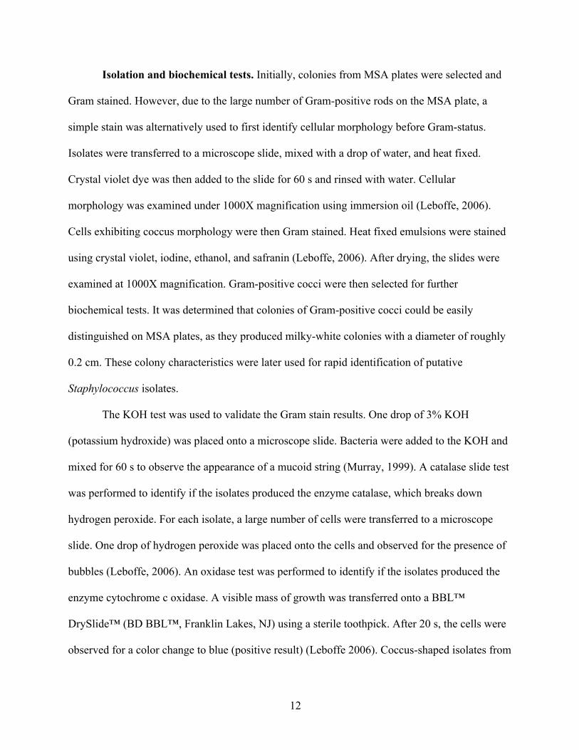
Isolation and biochemical tests. Initially, colonies from MSA plates were selected and
Gram stained. However, due to the large number of Gram-positive rods on the MSA plate, a
simple stain was alternatively used to first identify cellular morphology before Gram-status.
Isolates were transferred to a microscope slide, mixed with a drop of water, and heat fixed.
Crystal violet dye was then added to the slide for 60 s and rinsed with water. Cellular
morphology was examined under 1000X magnification using immersion oil (Leboffe, 2006).
Cells exhibiting coccus morphology were then Gram stained. Heat fixed emulsions were stained
using crystal violet, iodine, ethanol, and safranin (Leboffe, 2006). After drying, the slides were
examined at 1000X magnification. Gram-positive cocci were then selected for further
biochemical tests. It was determined that colonies of Gram-positive cocci could be easily
distinguished on MSA plates, as they produced milky-white colonies with a diameter of roughly
0.2 cm. These colony characteristics were later used for rapid identification of putative
Staphylococcus isolates.
The KOH test was used to validate the Gram stain results. One drop of 3% KOH
(potassium hydroxide) was placed onto a microscope slide. Bacteria were added to the KOH and
mixed for 60 s to observe the appearance of a mucoid string (Murray, 1999). A catalase slide test
was performed to identify if the isolates produced the enzyme catalase, which breaks down
hydrogen peroxide. For each isolate, a large number of cells were transferred to a microscope
slide. One drop of hydrogen peroxide was placed onto the cells and observed for the presence of
bubbles (Leboffe, 2006). An oxidase test was performed to identify if the isolates produced the
enzyme cytochrome c oxidase. A visible mass of growth was transferred onto a BBL™
DrySlide™ (BD BBL™, Franklin Lakes, NJ) using a sterile toothpick. After 20 s, the cells were
observed for a color change to blue (positive result) (Leboffe 2006). Coccus-shaped isolates from

13
the mixed MSA plate that were Gram-positive, catalase-positive, KOH-negative, and oxidase-
negative were purified on Tryptic Soy Agar (TSA) plates.
A coagulase test was also performed on each isolate in order to classify each putative
Staphylococcus as either coagulase-positive or -negative. A coagulase test assesses an
organism’s ability to coagulate rabbit plasma through the use of the coagulase enzyme. Rabbit
plasma was mixed with a heavy inoculum of bacteria and the tubes were incubated for 24 hrs at
37ºC. Following incubation, the tubes were examined for coagulation of the plasma (Leboffe,
2006).
Amplification and sequencing of the 16S rRNA gene. In order to identify the genus
and species of the isolates, a 16S rRNA PCR was performed. The forward and reverse primers
used were Bac8f and Univ1492r (Table 1), both at a final concentration of 0.125 µM. A small
number of cells were taken from isolated colonies on TSA plates and added to 10 µL of double
distilled water (ddH2O) in 0.2 mL PCR tubes. The cell suspension was placed into a Bio-Rad
C1000 Touch™ Thermal Cycler (Bio-Rad Laboratories, Hercules, CA) for 15 min at 95ºC to
lyse the cells. Ten microliters of Epicentre premix I (Epicenter Technologies, Madison, WI;
containing 100 mM of Tris-HCl (pH 8.3), 100 mM KCl, 400 µM of each dNTP, 7 mM of MgCl2,
and 8X FailSafe PCR Enhancer), 20 µL of primer mix, 5 µL of the Failsafe enzyme mix
(Epicentre), and 5 µL of cell lysate were mixed together. All PCR tubes were placed into the
Bio-Rad C1000 Touch™ Thermal Cycler and underwent the following automated amplification
cycles: 95ºC for 5 min and then 35 cycles of 95ºC for 60 s, 50ºC for 30 s, and 72ºC for 90 s
(Temple, 2008). After amplification, 10 µL of amplified DNA were visualized using an agarose
gel as described below. Visible bands at approximately 1500 bp represented the amplified 16S
rRNA PCR product. Successfully amplified isolates were sent to MWG Operon (Huntsville,

14
Alabama) for Sanger sequencing of the 16S rRNA gene using the Bac8f primer.
Table 1. Primers used in this study. “N/A” denotes that the BOX primers amplify variable intervening regions between conserved BOX sequences, which may result in varied product sizes and regions amplified.
Primer Name
Gene or region
Amplified
Nucleotide Sequence 5’-3’
Expected Product Size (bp)
Bac8f 16S rRNA AGAGTTTGATCCTGGCTCAG 1500
Univ1492r 16S rRNA GGTTACCTTGTTACGACTT 1500
BOX N/A CTACGGCAAGGCGACGCTGACG N/A
mecA_fwd mecA TGAAGTAGAAATGACTGAACGTCCG 1632
mecA_rev mecA TCTGCAGTACCGGATTTGCC 1632
mecA2_fwd mecA2 GGAGACCAGACGTAATAGTACCTGG 1559
mecA2_rev mecA2 AGCATTATAGCTGGCCATCCC 1559
mecA400_fwd mecA AACGTTGTAACCACCCCAAGA 407
mecA400_rev mecA GTTCTGCAGTACCGGATTTGCC 407
mecA2_500_fwd mecA2 GCCGTGTTTATCCATTGAACGAAGC 496
mecA2_500_rev mecA2 TGGGTTGAACCTGGTGATGTAGTG 496
Identification using the Biolog Microbial ID System. Further identification was
performed using the The Biolog Microbial ID System (Biolog, Hayward, CA) according to the
manufacturer’s protocol (Biolog, 2008). A colony from a pure culture growing on TSA was
selected and transferred to a new TSA plate and incubated at 33ºC for 24 hrs. The Biolog
protocol recommends using Biolog Universal Growth media (BUG), but states that TSA is a
valid substitute (Biolog, 2008). Cotton-tipped inoculator swabs were used to select an isolated

15
colony approximately 3 mm in diameter. A Biolog turbidimeter was calibrated and blanked using
fresh inoculating fluid (IF), which contains 0.03% Tween 40 and 0.25% Gellan Gum (Biolog,
2010). The swab containing bacteria was inserted into the tube containing IF and the swab tip
was rubbed against the bottom of the tube to release the bacteria into the liquid. A Vortex shaker
was used to create a homogenous cell suspension. The inoculated tube was adjusted to a percent
transmittance of 90-98% T. The adjusted cell suspension was poured into a multichannel pipet
reservoir and 100 µL of cell suspension was added to each well of a prepared 96-well plate using
an 8-channel repeating pipettor. A Biolog GEN III MicroPlate was used to analyze each isolate
through the use of 94 phenotypic tests, which creates a “phenotypic fingerprint” for identifying
the genus and species. The inoculated MicroPlate was covered with a lid and then incubated for
18 hrs at 33ºC. Afterwards, the plate was placed into the OmniLog reader and Biolog’s Microbial
Identification Systems software (OmniLog® Data Collection) was used to identify each isolate.
Throughout the experiment, a positive control (Staphylococcus epidermidis) was used to control
for each step of the protocol.
Repetitive sequence PCR for strain differentiation. Isolates were differentiated using
the BOX-PCR repetitive sequence fingerprinting method. This procedure works by amplifying
variable regions between a BOX sequence that is conserved amongst most bacterial species in
order to differentiate between possible strains. The procedure was carried out essentially
according to the protocol described by Rademaker et al. (1998). A small number of cells were
taken from isolated colonies on TSA plates and added to 10 µL of double distilled water (ddH2O)
in 0.2 mL PCR tubes. The cell suspension was placed into a Bio-Rad C1000 Touch™ Thermal
Cycler (Bio-Rad Laboratories, Hercules, CA) for 15 min at 95ºC to lyse the cells. In a separate
0.2 mL PCR tube, 13 pmol of BOX primer (Table 1), 0.1 µl Failsafe enzyme mix (Epicentre),
16
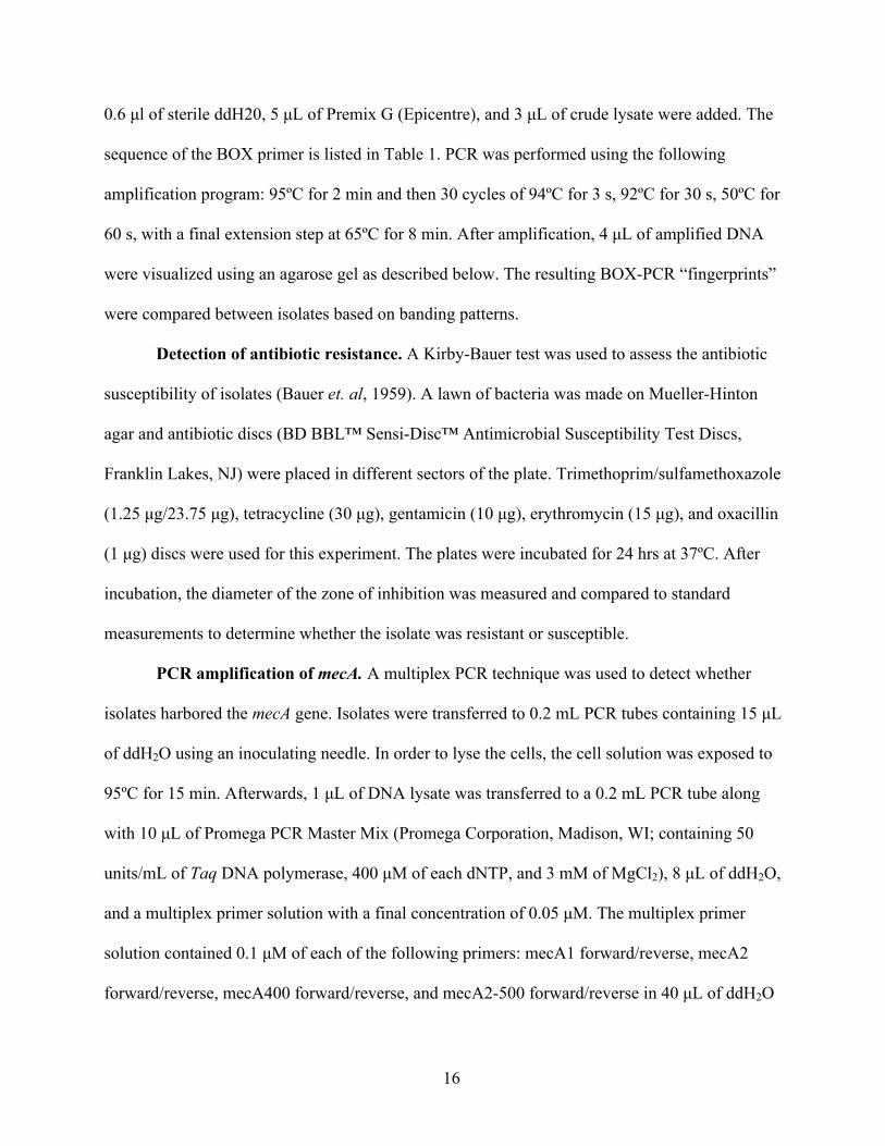
0.6 µl of sterile ddH20, 5 µL of Premix G (Epicentre), and 3 µL of crude lysate were added. The
sequence of the BOX primer is listed in Table 1. PCR was performed using the following
amplification program: 95ºC for 2 min and then 30 cycles of 94ºC for 3 s, 92ºC for 30 s, 50ºC for
60 s, with a final extension step at 65ºC for 8 min. After amplification, 4 µL of amplified DNA
were visualized using an agarose gel as described below. The resulting BOX-PCR “fingerprints”
were compared between isolates based on banding patterns.
Detection of antibiotic resistance. A Kirby-Bauer test was used to assess the antibiotic
susceptibility of isolates (Bauer et. al, 1959). A lawn of bacteria was made on Mueller-Hinton
agar and antibiotic discs (BD BBL™ Sensi-Disc™ Antimicrobial Susceptibility Test Discs,
Franklin Lakes, NJ) were placed in different sectors of the plate. Trimethoprim/sulfamethoxazole
(1.25 µg/23.75 µg), tetracycline (30 µg), gentamicin (10 µg), erythromycin (15 µg), and oxacillin
(1 µg) discs were used for this experiment. The plates were incubated for 24 hrs at 37ºC. After
incubation, the diameter of the zone of inhibition was measured and compared to standard
measurements to determine whether the isolate was resistant or susceptible.
PCR amplification of mecA. A multiplex PCR technique was used to detect whether
isolates harbored the mecA gene. Isolates were transferred to 0.2 mL PCR tubes containing 15 µL
of ddH2O using an inoculating needle. In order to lyse the cells, the cell solution was exposed to
95ºC for 15 min. Afterwards, 1 µL of DNA lysate was transferred to a 0.2 mL PCR tube along
with 10 µL of Promega PCR Master Mix (Promega Corporation, Madison, WI; containing 50
units/mL of Taq DNA polymerase, 400 µM of each dNTP, and 3 mM of MgCl2), 8 µL of ddH2O,
and a multiplex primer solution with a final concentration of 0.05 µM. The multiplex primer
solution contained 0.1 µM of each of the following primers: mecA1 forward/reverse, mecA2
forward/reverse, mecA400 forward/reverse, and mecA2-500 forward/reverse in 40 µL of ddH2O
17
(Kondo et al., 2007). The 5’-3’ nucleotide primer sequence for each primer is listed in Table 1.
The primers mecA1 and mecA2 amplify large regions of the mecA and mecA2 gene respectively.
The primers mecA400 and mecA2-500 amplify small regions of the mecA and mecA2 gene
respectively. All PCR tubes were placed into the Bio-Rad C1000 Touch™ Thermal Cycler (Bio-
Rad Laboratories, Hercules, CA) and underwent the following automated amplification cycles:
94ºC for 2 min and then 30 cycles of 94ºC for 2 min, 57ºC for 60 s, and 72ºC for 2 min, with a
final extension step of 2 min at 72ºC. After amplification, 10 µL of amplified DNA were
visualized using an agarose gel as described below. DNA was isolated from the N315 strain of
methicillin-resistant Staphylococcus aureus (MRSA) and was used as a positive control.
Gel electrophoresis of PCR products. Following PCR, amplified DNA was loaded into
a 1% agarose gel with 2 µL of 6X Blue/Orange Loading Dye (Promega Corporation, Madison,
WI) and electrophoresed at 80 V for 2 hours. A 1 kb Fisher BioReagents™ exACTGene™ DNA
Ladder (Fisher Scientific, Pittsburgh, PA) was used. The gel was stained using a 0.5% ethidium
bromide solution for 20 min. The gel was then destained with ddH2O for 10 min. Photographs of
the gel were taken with a Bio-Rad ChemiDoc™ XRS+ System (Bio-Rad Laboratories, Hercules,
CA) and analyzed using the Image Lab™ Software.
18
Results
Sample collection and processing. In January 2013, two sediment samples and two
water samples were collected from Muddy Creek in Hinton, Virginia. Sediment samples were
named “sediment up” and “sediment down” according to the collection site. “Sediment up”
samples were collected closer to the drainage pipe of the poultry processing plant whereas
“sediment down” samples were collected downstream from the waste pipe. A similar naming
system was applied to the water samples. Additionally, a second sampling was performed in
March 2013 with two sediment and two water samples collected.
After performing sediment cell release and water filtration, cells from the released cell
solution and from the water filter were plated onto MSA plates in order to selectively isolate
halotolerant bacteria such as staphylococci. A greater diversity of colony number, shape, size,
and morphology was found on the sediment plates (both the winter and spring samples)
compared to water.
Isolation and identification of Staphylococcus. Colonies were originally chosen at
random to determine cell morphology (cocci) using a Gram stain technique. However, the
majority of the colonies that grew on the MSA plate were Gram-positive rods. Therefore, a
simple stain was performed to first screen for cellular morphology and select all cocci for further
tests. After numerous rounds of simple staining, a general correlation between coccus cellular
morphology and colony morphology was determined. All of the elevated milky-white colonies
roughly 0.2 cm in diameter were determined to be cocci after performing a simple stain (Figure
1). Selecting colonies on MSA based on colony morphology created a rapid and simple initial
screening method for putative staphylococci.
19
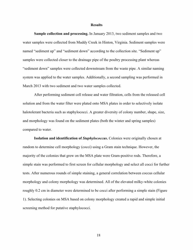
Figure 1. Example of colony morphology used to isolate putative Staphylococcus sp.. The black arrow points to a colony selected based on its milky-white appearance and ca. 0.2 cm diameter.
All of the elevated milky-white colonies roughly 0.2 cm in diameter (11) were selected
and were found to be coccus-shaped. The cells clustered in “grape-like” patterns, which is
common to the genus Staphylococcus. Ten of the eleven putative Staphylococcus (PS) were
isolated from the stream sediment (Table 2). A Gram stain, KOH, catalase, oxidase, and
coagulase test was performed on each of the eleven isolates for further identification. All eleven
isolates were Gram-positive, KOH-negative, catalase-positive, oxidase-negative, and coagulase-
negative cocci.
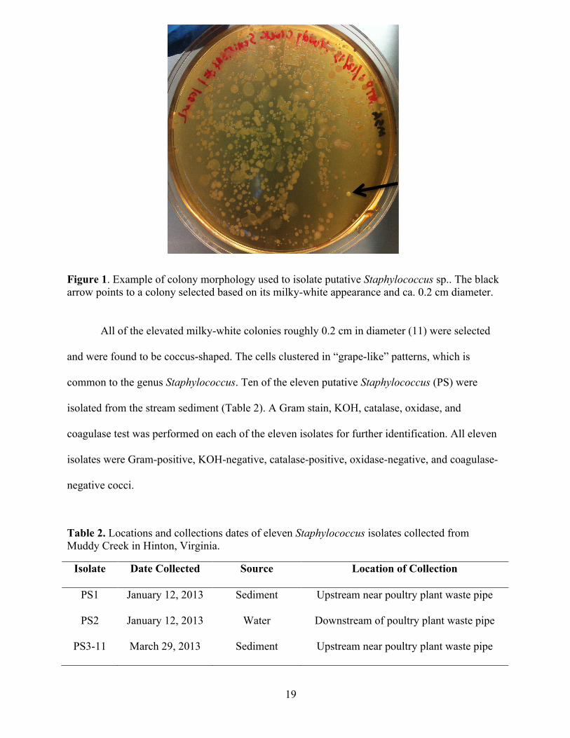
Table 2. Locations and collections dates of eleven Staphylococcus isolates collected from Muddy Creek in Hinton, Virginia.
Isolate Date Collected Source Location of Collection
PS1 January 12, 2013 Sediment Upstream near poultry plant waste pipe
PS2 January 12, 2013 Water Downstream of poultry plant waste pipe
PS3-11 March 29, 2013 Sediment Upstream near poultry plant waste pipe
20
The 16S rRNA genes of the isolates were amplified for sequencing using PCR; however,
only PS6-8, 10, and 11 produced an amplified 16S rRNA band at approximately 1500 bp.
Therefore, these isolates were submitted for partial sequencing of the 16S rRNA gene.
Sequences were compared to 16S rRNA genes in the nr (Non-redundant GenBank CDS
translations) database for identification using the National Center for Biotechnology
Information’s BLAST® program (Basic Local Alignment Search Tool). By inserting the
sequence of nucleotides for the 16S rRNA gene, the BLAST system is able to compare the
sequence from an isolate to a database of known sequences. The database generates a bit score
and an expect value (E-value) to determine if the match is statistically significant (Altschul et al.,
1997). Bit scores are based on identical alignments as well as gaps in the sequence whereas E-
values describe the number of expected matches generated by chance to a random database of
the same size. Thus, a high bit score and low E-value points to statistically significant matches.
Table 3 summarizes the results of the BLAST search by showing the top two matches for each
isolate tested as well as the bit score, E-value, percent match, and GenBank accession number.

21
Table 3. Summary of BLAST search results for the 16S rRNA nucleotide sequences from isolates PS6-8, 10, and 11. The top two matches based on bit score are listed for each isolate.
The remaining isolates (PS1-5), as well as PS6 and PS7 were identified using the Biolog
Microbial ID System (Biolog, 2010). By exposing each isolate to 94 different biochemical tests
all within one 96 well plate, a unique fingerprint is generated for each species of bacteria through
colorimetric changes. Biolog’s Microbial Identification Systems software (OmniLog® Data
Collection) then matches the optical density (OD) of the color in each well to a known database
of intensities to identify the unknown organism. After reading a plate, a number of possible
organisms appear along with a PROB, SIM, and DIST value. The PROB value allows for a
comparison of Biolog’s identification to other systems using similar identification calculations.
Isolate Description of 16S rRNA Gene
Max Bit Score
E-value
Percent Match
Accession Number
PS6
Staphylococcus sp. SB10-23
1622 0.0 96% GU595329.1
Staphylococcus equorum strain SCSGAB0145
1616 0.0 96% JX315320.1
PS7
Staphylococcus sp. SB10-23
1646 0.0 95% GU595329.1
Staphylococcus equorum strain SCSGAB0145
1640 0.0 95% JX315320.1
PS 8
Staphylococcus equorum strain C4052
1598 0.0 96% KF439737.1
Staphylococcus strain C4044
1598 0.0 96% KF439736.1
PS 10
Staphylococcus sp. SB10-23
1668 0.0 96% GU595329.1
Staphylococcus equorum isolate EO2001
1663 0.0 96% AY741060.1
PS 11
Staphylococcus sp. SB10-23
1733 0.0 98% GU595329.1
Staphylococcus equorum subsp. equorum
1727 0.0 98% AB735696.1

22
A SIM value is used to determine the accuracy of the identification with a “1” being a perfect
match and “0” meaning no match (Biolog, 2010). DIST represents the approximate number of
mismatches between the results of the unknown organism and information from the database of
the known organism proposed to be a match. Biolog’s User Guide explains that answering the
following three questions “yes” ensures a confident match for the top listed organism. First, do
the top listed organisms belong to the same or similar genus/genera? Second, for the number one
identification, is the SIM value greater than 0.5? Third, is the DIST value for the number one
identification at least two distance points away from the second choice (Biolog, 2010)?
PS2 was unable to be identified due to the low SIM value and therefore resulted in a “No
ID” result. The remaining isolates were all identified with a SIM value greater than 0.5 (Table 4).
Furthermore, the ΔDIST is the DIST value of the first matched organism subtracted from the
second organism and all of the identified isolates were less than two distance points (Table 4). If
the top-listed identifications all belong to the same genus, then the confidence level of
identification is strengthened. The Biolog Microbial ID System produced top-listed matches
from the genus Staphylococcus for PS 1, 4, and 6. PS 3, 5, and 7 were identified as
Staphylococcus spp. for the top match but subsequent matches showed different genera
consisting of Gram-positive rods. Staphylococcus epidermidis was used as a positive control and
the Biolog software identified the strain as Staphylococcus epidermidis with a PROB, SIM, and
ΔDIST of 0.995, 0.783, and 3.78 respectively. All of the other top-listed matches belonged to the
Staphylococcus genus. PS9 was unable to be identified either via 16S rRNA sequencing or
Biolog and remains unidentified. However, the biochemical characteristics as well as the cellular
morphology resembled all of the other isolates and therefore PS9 is likely to be an organism
belonging to the genus Staphylococcus.
23
Table 4. PROB, SIM, and ΔDIST values of isolates using the Biolog Microbial ID System. ΔDIST is the DIST value of the first matched organism subtracted from the second organism.
Repetitive sequence PCR for strain differentiation. After identifying the genus and
species of each isolate (except for PS2 and PS9), a type of repetitive sequence fingerprinting
known as “BOX-PCR” (Rademaker et al., 1998) was performed on all isolates in order to
differentiate between strains. This method uses a primer set that amplifies intervening regions
between a conserved BOX sequence thus creating a unique banding fingerprint for different
strains of bacteria. Amongst the eleven isolates, nine different banding patterns were produced
(Figure 2). PS6 and PS10 produced the same pattern while PS7 and PS11 produced identical
banding patterns to one another and only differed from PS6/PS10 by the lack of a band at 600 bp
(Figure 2). PS8 resembled PS6/PS10 but lacked the 600 bp band and produced an additional
band at approximately 680 bp. PS2-5 all produced distinct banding patterns. PS9 and PS1 were
not successfully amplified, but produced distinct banding patterns to the one another and the
other isolates in previous BOX-PCR attempts not pictured here. E. coli strain LA61 was used as
a positive control. Although anomalous bands are found in the negative control lane, the banding
patterns of each isolate match the fingerprints from numerous previous attempts.
Isolate Identification PROB SIM Value ΔDIST PS1 Staphylococcus xylosus 0.788 0.561 1.164 PS2 NO ID - - - PS3 Staphylococcus lentus 0.562 0.562 0.173 PS4 Staphylococcus sciuri subsp. sciuri 0.815 0.667 1.036 PS5 Staphylococcus sciuri subsp. sciuri 0.653 0.653 0.156 PS6 Staphylococcus succinus subsp. succinus 0.603 0.603 0.739 PS7 Staphylococcus equorum 0.529 0.529 0.072

24
Figure 2. BOX-PCR of isolates used to differentiate isolated strains using a 1% agarose gel and ethidium bromide staining. A 1 kb Fisher BioReagents™ exACTGene™ DNA Ladder (“L”) (Fisher Scientific, Pittsburgh, PA) was used as a size marker. Each isolate was labeled without the “PS” designation and with the isolate number only. Amplified products of each isolate were electrophoresed in duplicates and signified by the “A” and “B” following the isolate number. E. coli strain LA61 was used as a positive control (+). A negative control consisting of water and the PCR reagents was also used (-).
Antibiotic susceptibility. The Kirby-Bauer test was used to test for susceptibility to
oxacillin (now used to identify phenotypic methicillin-resistance), as well as susceptibility to
other antibiotics. Trimethoprim/sulfamethoxazole (1.25 µg/23.75 µg), tetracycline (30 µg),
gentamicin (10 µg), erythromycin (15 µg), and oxacillin (1 µg) discs were each placed on a lawn
of bacteria on Mueller-Hinton agar. The zone of inhibition diameter was measured and compared
to the interpretative chart supplied by BD BBL™. All isolates (PS1-11) were susceptible to
trimethoprim/sulfamethoxazole, tetracycline, and gentamicin. All of the isolates except for PS2
and PS3 were susceptible to erythromycin whereas PS2 and PS3 were resistant. Additionally, all
of the isolates were resistance to oxacillin (≤17 mm) except for PS6, which exhibited a zone of
inhibition diameter of 19 mm (Figure 3).

25
Figure 3. Zones of inhibition of Staphylococcus isolates to oxacillin. Isolates below the dotted line (17 mm) are resistant to oxacillin and isolates above the dotted line are susceptible. This breakpoint is according to the interpretative chart supplied by BD BBL™ for coagulase-negative staphylococci to oxacillin.
Detection of the mecA gene encoding resistance to oxacillin. Multiplex PCR was
performed in order to detect the mecA or mecA2 gene, which both confer resistance to oxacillin
and methicillin. The multiplex PCR combines different primer sets to amplify different regions
of the mecA and mecA2 genes (Kondo et al., 2007). The mecA primers amplify a 1632 bp region
of the mecA gene whereas the mecA2 primers amplify a 1559 bp region of the mecA2 gene. The
mecA400 primers amplify a 407 bp region of the mecA gene whereas the mecA2_500 primers
amplify a 496 bp region of the mecA2 gene (Table 1). All eleven isolates (PS1-11) were assessed
for the presence of the mecA gene. All isolates except for PS2 produced a faint band at
approximately 400 bp suggesting the presence of the amplified mecA400 product of the mecA
gene. However, the image of the gel is not included because in performing the mecA PCR a

26
negative control was not used and subsequent attempts in replicating the results were
unsuccessful.

27
Discussion
Sample collection and processing. This study aimed to isolate and to identify
methicillin-resistant Staphylococcus from Muddy Creek, a manure-contaminated stream in
Hinton, Virginia. Several studies have demonstrated the prevalence of MRSA from recreational
marine and fresh water beaches, stream water, and sand samples from beaches (Levin-Edens et
al., 2012). However, little information exists on the occurrence of Staphylococcus in streams
impacted by runoff from agricultural operations despite recent reports on the prevalence and ease
of isolating methicillin-resistant Staphylococcus from farm animals (Fang et al., 2014).
During January of 2013, two staphylococci (PS1 and PS2) were isolated from both water
and sediment samples. Nine more staphylococci were isolated in March of 2013, all from the
sediment sample. It is possible that a link may exist between temperature and the likelihood of
isolating of staphylococci found in Muddy Creek. During the sampling in January, the air
temperature was approximately 4.5ºC while the air temperature during the March sampling was
13ºC. Although samples were obtained from the same location, eight staphylococci were isolated
from the sediment sample in March as opposed to one in January. Besides the temperature,
runoff from area farms as well as the poultry plant may increase during Spring, thus contributing
more waste to the stream.
Isolation and identification of isolates. Initial examination of bacteria from both stream
and sediment samples growing on MSA plates revealed a large number of Gram-positive rods.
Although we tested Vogel and Johnson Agar and MSA, we were not able to find a selective
medium that selected against these Gram-positive rods to target staphylococci. However, after
randomly testing numerous colonies that grew on MSA, it was determined that Staphylococcus
spp. were relatively easily distinguished by their distinctive small size (0.2 cm diameter) and

28
milky-white color (Figure 1). Therefore, by first visually screening for these colonies on MSA
plates, followed by a simple crystal violet stain for cell shape, a relatively large numbers of
colonies could be examined quickly without using a great amount of time or resources. Although
a number of methods have been published for screening for and isolating Staphylococcus aureus
from natural samples, to our knowledge none exist that will do so for the entire genus (Soge,
2009).
All eleven isolates were tested for defining characteristics of the Staphylococcus genus.
The Gram stain, catalase test, oxidase test, KOH test, and coagulase test are extremely useful in
identifying members of the genus Staphylococcus. Each of our isolates was Gram-positive,
catalase-positive, KOH-negative, oxidase-negative, and coagulase-negative. Although none of
the isolates were S. aureus – which is coagulase positive – the threat of CoNS as human
pathogens may still exist. Coagulase-negative Staphylococcus were originally thought of as
harmless skin bacteria, but are now known to be the causative agent of 11% of all nosocomial
infections (Huebner et al., 1999). CoNS are known to cause endocarditis, bacteremia, and
urinary tract infections. Treatment methods for CoNS are continually complicated due to the
rapid development of antibiotic resistance, especially in hospital settings (Huebner et al., 1999).
Partial 16S rRNA gene sequencing and the Biolog Microbial ID System were used to
verify the identification of the isolates as members of the Staphylococcus genus, as well as to
determine, where possible, the species of each. In addition, the BOX-PCR fingerprinting method
was used to distinguish the different strains isolated, particularly since many of them were
closely related to one another, as determined by the 16S rRNA and Biolog results. A portion of
the 16S rRNA gene of five isolates (PS6-8, 10, and 11) was sequenced and matched to the Non-
redundant GenBank database using a BLAST search. Results of the BLAST search showed that
29
Staphylococcus equorum was listed as one of the top matches for all of the tested isolates (Table
3). Similarly, the BOX PCR results for PS6-8, 10, and 11 all showed similar banding patterns
(Figure 2).
Isolates PS6 and PS7 were the only isolates subjected to both 16S rRNA sequencing and
to the Biolog tests. The Biolog results identified both the first match for PS7 and the third match
for PS6 as Staphylococcus equorum. The top match from the 16S rRNA sequencing
(Staphylococcus sp. SB10-23) was only itself identified to the genus level; however, the top
Biolog result for PS6 was Staphylococcus succinus subsp. succinus. All of the top-rated matches
for PS6 belonged to the genus Staphylococcus, and the SIM value for the first match was greater
than 0.5 (Table 4). However, the ΔDIST (the DIST value of the first matched organism
subtracted from the second match) between the first and second match was less than 2. Although
two out of three of the Biolog accuracy parameters were met, the DIST value remained low.
Nonetheless, Biolog notes that similar DIST values may result between top-rated matches
because of the similar biochemical characteristics of the listed species (Biolog, 2010). Thus, a
confident identification was made despite the low ΔDIST value. Despite the differences between
the 16S rRNA sequencing and Biolog results, clearly PS6 belonged to the genus Staphylococcus
with uncertainty as to whether the species was equorum or succinus. PS7 produced a SIM value
greater than 0.5, but a ΔDIST value less than 2 and none of the top-rated generas matched
Staphylococcus. Therefore, the Biolog results for PS7 remained inconclusive yet still matched
the results of the 16S rRNA sequence, which may validate the accuracy of the Biolog experiment.
Biolog identified PS1 as Staphylococcus xylosus. The SIM value was greater than 0.5 and
the other top matches were all Staphylococcus (Table 4). Also, the ΔDIST value was less than
two (1.164), but was the highest ΔDIST of the isolates tested. Although not perfect, the accuracy

30
parameters for PS1, according to Biolog, demonstrated a confident match. None of the other
isolates were identified as Staphylococcus xylosus. PS1’s unique identification, among the other
isolates, was supported by its unique BOX-PCR banding pattern (Figure 2).
PS3 was identified by Biolog as Staphylococcus lentus. The SIM value was greater than
0.5, but the ΔDIST value was less than two. Besides Staphylococcus lentus, the other top-rated
matches were Gram-positive rods belonging to different genera (Table 4). However, isolate PS3
was clearly a Gram-positive coccus thus eliminating the other top matches as possibilities. This
finding along with the catalase and oxidase tests supported the Biolog identification as
Staphylococcus lentus.
PS4 and 5 were both identified by the Biolog system as Staphylococcus sciuri subsp.
sciuri. The Biolog Microbial ID System calculated a SIM value greater than 0.5 and a ΔDIST
value less than two for both isolates (Table 4). Additionally, all of the top-rated matches for PS4
consisted of species from Staphylococcus whereas those of PS5 consisted of Gram-positive rod
species from other genera. Similar to PS3, PS5 was indeed a Gram-positive coccus as confirmed
by a previous Gram stain. Although Biolog identified PS4 and PS5 as the same organism, the
different banding patterns from the BOX-PCR suggest separate strains (Figure 2).
PS2 and PS9 were unable to be identified by Biolog or 16S rRNA gene sequencing.
Despite the lack of identification, the cellular morphology and microbiological characteristics
matched the other isolates that were identified as coagulase-negative staphylococci. Additionally,
PS2 and PS9 each exhibited a unique BOX banding pattern relative to one another and to the
other isolates (Figure 2). Therefore, if these isolates belonged to the genus Staphylococcus, then
they may either be different strains or different species entirely when compared to the other
isolates.

31
Description of identified species. Staphylococcus equorum (PS6-8, 10, and 11) was first
isolated and characterized in 1984 from the skin of healthy horses (Nováková et al., 2006). Later,
isolates were obtained from the milk of cows with mastitis and from healthy goats (Nováková et
al., 2006). Isolating this organism from an agriculturally impacted stream comes as no surprise
due to the fact that both goat and cow farms exist upstream from the site of collection.
Additionally, worn paths are visible directly upstream from the poultry plant where cows
apparently cross Muddy Creek. The Biolog Microbial ID System identified PS6 as
Staphylococcus succinus. This species has been isolated from many sources including ripening
cheese and sausage (Nováková et al., 2006). Additionally, this species was originally isolated
from soil inclusions on Dominican amber (Lambert et al., 1998).
S. xylosus (PS1) commonly inhabits the skin of animals such as chickens, mice, pigs,
horses, and cows (Nováková et al., 2006). In humans, S. xylosus is one of the many agents
known to cause urinary tract infections and more rarely endocarditis or pneumonia (Nováková et
al., 2006). Similar to S. equorum, finding Staphylococcus xylosus in the sediment of an
agriculturally impacted stream is not surprising. S. lentus (PS3) is commonly isolated from the
skin of food producing animals such as chickens and cows (Schwendener et al., 2012).
Employees who commonly work with poultry or dairy animals have been found to carry S. lentus
and although rare, this organism may cause infection in humans (Schwendener et al., 2012). As
the poultry plant adjacent to Muddy Creek washes down carrier trucks or dumps waste, this
organism may be introduced into the water. S. sciuri (PS4 and 5) is mainly isolated from the skin
and mucosal surfaces of pets and farm animals (Dakić et al., 2005). Additionally, common
environmental reservoirs include soil, sand, and water. S. sciuri is also known to cause several

32
different complications in humans including endocarditis, urinary tract infections, and wound
infections (Dakić et al., 2005).
Determination of methicillin-resistance. Methicillin-resistance is conferred in all
known bacteria by variants of the mecA gene, which encodes for the penicillin-binding protein
PBP2a (Kondo et al., 2007). This gene is associated with the mobile genetic element SCCmec
and commonly inserts into the chromosome of staphylococci (Kondo et al., 2007). SCCmec
elements are characterized by four distinct properties. First, the element carries the mec gene
complex known to confer methicillin resistance. Second, the element contains a ccr gene
complex – containing ccr genes and open reading frames – responsible for the mobility of the
entire element. Third, direct repeat nucleotide sequences exist at both ends of the element and
lastly the element inserts into the 3’ end of orfX, an open reading frame of unknown function.
Although these common characteristics exist, structural differences within the ccr and
mec components have been identified (Kondo et al., 2007). For instance, four classes of mec
gene complexes – consisting of mecA, regulatory genes, and insertion sequences – have been
discovered (A, B, C, and D) and differ based on the arrangement and type of insertion sequence
used to integrate the SCCmec element into the chromosome of susceptible strains. In addition,
ccr and mec gene complexes combine differently on the SCCmec element creating genetically
divergent structures. As a result, microbiologists commonly classify MRSA clones based on the
SCCmec element makeup (IWG-SCC, 2009).
Due to the great variability in mecA gene complexes, we utilized a multiplex PCR
technique that combined primers known to amplify large and small portions of the mecA gene.
After performing the mecA PCR, bands were visible in all isolates except for PS2 at the expected
product size of approximately 400 bp (small region of the mecA gene). Despite the lack of a

33
negative control, these results matched the results of the Kirby Bauer disc diffusion test in which
all of the isolates were oxacillin-resistant except for PS6. However, due to the missing negative
control, we are unable to make definitive conclusions at this point regarding mecA. PS2 was
oxacillin-resistance in the disc diffusion test, but lacked the mecA gene. Lee et al. encountered
this same observation when studying 3,125 staphylococci isolates from patients in hospitals.
Specifically, 15 of the isolates were resistant to oxacillin, but did not have the mecA gene and 2
isolates were susceptible to oxacillin and harbored the mecA gene (Lee et al., 2007). The latter
cases may be explained by mutations in the mecA promoter, which when altered may affect the
levels of PBP2a translated (Chen et al., 2014). For instance, a G25A mutation (guanine changed
to adenine 25 bases upstream of the mecA translation start site) was correlated to a high
minimum inhibitory concentration (MIC) for oxacillin at 256 µg/mL (Chen et al., 2014). Other
mutations have been shown to decrease or increase the MIC for oxacillin. These findings support
the results for PS6 as this organism may have a mutation in the promoter region of the mecA
complex, which lowers the MIC and causes susceptibility to the oxacillin. PS2 may harbor the
mecA gene, but due to possible gene sequence divergence the primers used in the multiplex PCR
may not bind and therefore not amplify the gene. In order to determine the reason for this
anomaly, the putative mecA gene within PS2 could be sequenced.
Overall, in this study we isolated and identified eleven coagulase-negative staphylococci
of differing species from the sediments and water of Muddy Creek. Nine of the eleven isolates
were resistant to oxacillin and contained the mecA gene known to confer methicillin resistance.
PS6 was susceptible to oxacillin in the disc diffusion test, but contained the mecA gene thus
making ten of the eleven isolates carriers of the mecA gene. It is hypothesized that grazing farm
animals and waste from the adjacent poultry plant are introducing these bacteria into a novel
34
environment. Implications of these findings might include horizontal transfer of the SCCmec
element from such staphylococci to other (native or introduced) bacteria present in the water or
sediment. Their occurrence in the stream sediment is of particular concern as it may imply that
they are more persistent – or at least less transient – than bacteria found in the moving water
column of the stream. Additionally, these bacteria may pose a health threat as Muddy Creek
flows down stream into North River, which runs along the homes of many residents who swim
and drink the water.
Future research could include determining whether or the not the SCCmec element,
commonly found on chromosomal DNA, is plasmid-borne in these isolates and, if so, whether it
is transmissible to other bacteria via conjugation. Furthermore, through isolating, amplifying,
and sequencing the SCCmec element from these organisms, one may be able to determine
information regarding the taxonomy and origin of the element itself. Finally, this study could be
replicated but include sampling throughout the entire year to account for temperature and season
as well as additional samples from bodies of water both affected and unaffected by agriculture.
Although a direct causation does not exist between the poultry plant/farms and the presence of
these staphylococci, a strong link is present and deserves further attention in order to attenuate
the presence of these methicillin-resistant staphylococci.

35
Literature Cited
Altschul SF, Madden TL, Schaffer AA, Zhang J, Zhang Z, Miller W, Lipman DJ. 1997.
Gapped BLAST and PSI-BLAST: a new generation of protein database search programs.
Nucleic Acids Res. 25:3389–3402.
Bauer AW, Perry DM, Kirby WMM. 1959. Single disc antibiotic sensitivity testing of
Staphylococci. J. Am. Med. Assoc.. 104:208–216.
BD BBL™ Sensi-Disc™ Antimicrobial Susceptibility Test Discs. BD BBL™ Pamphlet. 1-5.
Bhargava K, Zhang Y. 2012. Multidrug-resistant coagulase-negative staphylococci in food
animals. J. Appl. Microbiol. 113: 1027-1036.
Biolog. 2008. GEN III MicroPlateTM Instructions for Use.
Biolog. 2010. MicroStationTM System/MicroLogTM User’s Guide. Biolog, Hayward, CA.
Biolog. 2012. Biolog Systems Brochure. Biolog, Hayward, CA.
Chen FJ, Wang C, Chen C, Hsu Y, Wang KT. 2014. Role of the mecA gene in oxacillin
resistance in a Staphylococcus aureus clinical strain with a pvl-positive ST59 genetic
background. Antimicrob. Agents Chemother. 58:1047-1054.
Dakić I, Morrison D, Vuković D, Savić B, Shittu A, Jezek P, Hauschild T, Stepanović S.
2005. Isolation and molecular characterization of Staphylococcus sciuri in the hospital
environment. J. Clin. Microbiol. 43:2782-2785.
Fang HW, Chiang PH, Huang YC. 2014. Livestock-associated methicillin-resistant
Staphylococcus aureus ST9 in pigs and related personnel in Taiwan. PLoS One. 9:1-7.
Holben WE, Jansson JK, Chelm BK, Tiedje JM. 1988. DNA probe method for the detection
of specific microorganisms in the soil bacterial community. Appl. Environ. Microbiol. 54:703-
711.
36
Huang H, Flynn NM, King JH, Monchaud C, Morita M, Cohen SH. 2006. Comparisons of
community-associated methicillin-resistant Staphylococcus aureus (MRSA) and hospital-
associated MSRA infections in Sacramento, California. J. Clin. Microbiol. 44:2423–2427.
Huebner J, Goldmann DA. 1999. Coagulase-negative staphylococci: role as pathogens. Annu
Rev Med. 50:223-36.
International Working Group on the Classification of Staphylococcal Cassette
Chromosome Elements (IWG-SCC). 2009. Classification of staphylococcal cassette
chromosome mec (SCCmec): guidelines for reporting novel SCCmec elements. Antimicrob.
Agents Chemother. 53:4961-4967.
Kondo Y, Ito T, Ma XX, Watanabe S, Kreiswirth BN, Etienne J, Hiramatsu K. 2007.
Combination of multiplex PCRs for staphylococcal cassette chromosome mec type assignment:
rapid identification system for mec, ccr, and major differences in junkyard regions. Antimicrob.
Agents Chemother. 51:264-274.
Lambert LH, Cox T, Mitchell K, Rosselló-Mora RA, Del Cueto C, Dodge DE, Orkand P,
Cano RJ. 1998. Staphylococcus succinus sp. nov., isolated from Dominican amber. Int J Syst
Bacteriol. 48: 511-518.
Leboffe MJ, Pierce BE. 2006. Microbiology: Laboratory Theory and Applications, 3rd ed.
Morton Publishing Co., Englewood CO.
Lee Y, Kim CK, Kim M, Yong D, Lee K, Chong Y. 2007. Detection of mecA in strains with
oxacillin and cefoxitin disk tests for detection of methicillin-resistant Staphylococcus. Korean J.
Lab. Med. 27:276-280.
37
Levin-Edens E, Soge OO, No D, Stiffarm A, Meschke JS, Roberts MC. 2012. Methicillin-
resistant Staphylococcus aureus from Northwest marine and freshwater recreational beaches.
FEMS Microbiol Ecol. 9:412-420.
Murray PA. 1999. Manual of Clinical Microbiology, 7th ed. ASM Press, Washington, DC.
Nováková D, Sedlácek I, Pantůcek R, Stetina V, Svec P, Petrás P. 2006. Staphylococcus
equorum and Staphylococcus succinus isolated from human clinical specimens. J. Med.
Microbiol.. 55: 523-528.
Rademaker JL, Louws FJ, Bruijn FJD. 1998. Characterization of the diversity of ecologically
important microbes by rep-PCR fingerprinting. Mol. Microb. Ecol. Man. 1-26.
Schaechter M. 2009. Staphylococcus. Encyclopedia of Microbiology, 3rd ed. Academic Press,
Waltham, MA.
Schwendener S, Perreten V. 2012. New MLSB resistance gene erm(43) in Staphylococcus
lentus. Antimicrob Agents Chemother. 56: 4746-4752.
Soge OO, Meschke JS, No DB, Roberts MC. 2009. Characterization of methicillin-resistant
Staphylococcus aureus and methicillin-resistant coagulase-negative Staphylococcus spp. isolated
from US West Coast public marine beaches. J Antimicrob Chemother. 64: 1148–1155.
Temple. 2008. Amplification of 16s Gene DNA by Polymerase Chain Reaction. Biology 380
Handout. James Madison University, Harrisonburg, VA.
Weese JS. 2010. Methicillin-resistant Staphylococcus aureus in animals. Inst. Lab. Anim. Res. J..
51:233-244.