Molecular Cell, Vol. 14, 699–711, June 18, 2004...
13
Molecular Cell, Vol. 14, 699–711, June 18, 2004, Copyright 2004 by Cell Press Targeted Proteomic Study of the Cyclin-Cdk Module contrast, in Schizosaccharomyces pombe, one Cdk Vincent Archambault, Emmanuel J. Chang, Benjamin J. Drapkin, Frederick R. Cross, 1 (Cdc2) functions with one of two G1 cyclins (Cig1 and 2) Brian T. Chait, 1 and Michael P. Rout 1, * and one B type cyclin (Cdc13). In vertebrates (including The Rockefeller University humans), at least eight Cdks function with at least nine 1230 York Avenue cyclins where only some cyclin-Cdk combinations are New York, New York 10021 functional and where tissue specificity adds another level of complexity (reviewed in Morgan, 1997). The yeast S. cerevisiae has provided a simple model Summary for cell cycle studies for more than 30 years. Genes involved in cell cycle functions were initially identified The cell division cycle of the yeast S. cerevisiae is by screens for mutants that failed to proceed through driven by one Cdk (cyclin-dependent kinase), which critical cell cycle transitions, followed by complementa- becomes active when bound to one of nine cyclin sub- tion of the mutation. This strategy allowed the identifica- units. Elucidation of Cdk substrates and other Cdk- tion of dozens of genes essential for cell division cycle associated proteins is essential for a full understand- (CDC genes; Hartwell et al., 1970). Genetic approaches ing of the cell cycle. Here, we report the results of a to investigate how specific cyclins participate in the cell targeted proteomics study using affinity purification cycle have been challenging, most likely because of the coupled to mass spectrometry. Our study identified cyclins’ functional redundancy. Initially, genetic studies numerous proteins in association with particular cyclin- suggested that G1 cyclins CLN1–3 were involved in Cdk complexes. These included phosphorylation sub- committing the cell to a division cycle, initiating spindle strates, ubiquitination-degradation proteins, adaptors, assembly, budding, and activating the S phase cyclins and inhibitors. Some associations were previously CLB5 and 6. These were found to activate DNA replica- known, and for others, we confirmed their specificity tion initiation and to prevent rereplication. The cyclins and biological relevance. Using a hypothesis-driven CLB1–4 were found to drive mitosis through the promo- mass spectrometric approach, we also mapped in vivo tion of spindle and bud morphogenesis (reviewed in phosphorylation at Cdk consensus motif-containing Miller and Cross, 2001). However, genetic experiments peptides within several cyclin-associated candidate failed to elucidate many of the protein targets of the Cdk substrates. Our results demonstrate that this different cyclin-Cdk complexes. One problem faced by approach can be used to detect a host of transient these studies is that communication between proteins and dynamic protein associations within a biological tends to be redundant, as crosstalk and backup path- module. ways involving proteins of partially overlapping function can add robustness and adaptability to a living system. Introduction Ultimately, the most direct way to study such networks of communicating proteins involves the detection of All eukaryotic organisms face the same fundamental their interactions. problems in their somatic cell division cycle. DNA must Understanding the molecular interface between be faithfully replicated once. Identical sets of genetic cyclin-Cdk complexes and the cellular events that they material must be physically segregated. Finally, cytoki- control is a central focus of the field today (Nurse, 2000). nesis must occur to separate the two progeny cells. Phosphorylation targets of cyclin-Cdk complexes have These cellular transformations must occur in an orderly, slowly begun to be discovered in a case by case manner. coordinated manner. In all eukaryotic organisms, a At the same time, a few proteins acting as inhibitors (e.g., cyclin-Cdk module appears to play the most central role Sic1; Donovan et al., 1994; Mendenhall, 1993; Schwob et in directing this program. An oscillation of cyclin-Cdk al., 1994) and adaptors (e.g., Cks1; Morris et al., 2003) activities essentially conducts a symphony of pathways, have been found. More recently, a study identified giving rise to a harmonious cell division cycle. Coordina- around 200 potential Cdk1 phosphorylation substrates tion of a highly complex set of tasks requires multiple in yeast (Ubersax et al., 2003). Only a few studies have avenues of communication involving dynamic and often begun to address the cyclin specificity of these interac- redundant pathways that involve multiple protein-pro- tions with targets and the mechanisms contributing to tein interactions. this specificity (reviewed in Miller and Cross, 2001). Cyclin-dependent kinases are activated upon binding In this report, we survey proteins associating with of cyclin subunits. The periodic synthesis and proteoly- cyclins in S. cerevisiae using a single step, Protein A sis of these cyclins ensures an oscillation in Cdk activity tag affinity purification technique followed by mass necessary to the cell cycle. In the yeast Saccharomyces cerevisiae, Cdc28 (Cdk1) is the only Cdk that is clearly spectrometric protein identification. We identified and involved in cell cycle coordination. It functions with three investigated numerous associations, both known and G1 cyclins (Cln1–3) and six B-type cyclins (Clb1–6). By previously unreported. In addition, we have mapped phosphopeptides containing in vivo phosphorylation sites in a subset of cyclin-associated candidate Cdk *Correspondence: [email protected] 1 These authors contributed equally to this work. substrates.
-
Upload
hoangkhanh -
Category
Documents
-
view
215 -
download
0
Transcript of Molecular Cell, Vol. 14, 699–711, June 18, 2004...
Molecular Cell, Vol. 14, 699–711, June 18, 2004, Copyright 2004 by Cell Press
Targeted Proteomic Study of the Cyclin-Cdk Module
contrast, in Schizosaccharomyces pombe, one CdkVincent Archambault, Emmanuel J. Chang,Benjamin J. Drapkin, Frederick R. Cross,1 (Cdc2) functions with one of two G1 cyclins (Cig1 and 2)Brian T. Chait,1 and Michael P. Rout1,* and one B type cyclin (Cdc13). In vertebrates (includingThe Rockefeller University humans), at least eight Cdks function with at least nine1230 York Avenue cyclins where only some cyclin-Cdk combinations areNew York, New York 10021 functional and where tissue specificity adds another
level of complexity (reviewed in Morgan, 1997).The yeast S. cerevisiae has provided a simple model
Summary for cell cycle studies for more than 30 years. Genesinvolved in cell cycle functions were initially identified
The cell division cycle of the yeast S. cerevisiae is by screens for mutants that failed to proceed throughdriven by one Cdk (cyclin-dependent kinase), which critical cell cycle transitions, followed by complementa-becomes active when bound to one of nine cyclin sub- tion of the mutation. This strategy allowed the identifica-units. Elucidation of Cdk substrates and other Cdk- tion of dozens of genes essential for cell division cycleassociated proteins is essential for a full understand- (CDC genes; Hartwell et al., 1970). Genetic approachesing of the cell cycle. Here, we report the results of a to investigate how specific cyclins participate in the celltargeted proteomics study using affinity purification cycle have been challenging, most likely because of thecoupled to mass spectrometry. Our study identified cyclins’ functional redundancy. Initially, genetic studiesnumerous proteins in association with particular cyclin- suggested that G1 cyclins CLN1–3 were involved inCdk complexes. These included phosphorylation sub- committing the cell to a division cycle, initiating spindlestrates, ubiquitination-degradation proteins, adaptors, assembly, budding, and activating the S phase cyclinsand inhibitors. Some associations were previously CLB5 and 6. These were found to activate DNA replica-known, and for others, we confirmed their specificity tion initiation and to prevent rereplication. The cyclinsand biological relevance. Using a hypothesis-driven CLB1–4 were found to drive mitosis through the promo-mass spectrometric approach, we also mapped in vivo tion of spindle and bud morphogenesis (reviewed inphosphorylation at Cdk consensus motif-containing
Miller and Cross, 2001). However, genetic experimentspeptides within several cyclin-associated candidate
failed to elucidate many of the protein targets of theCdk substrates. Our results demonstrate that this
different cyclin-Cdk complexes. One problem faced byapproach can be used to detect a host of transient
these studies is that communication between proteinsand dynamic protein associations within a biologicaltends to be redundant, as crosstalk and backup path-module.ways involving proteins of partially overlapping functioncan add robustness and adaptability to a living system.IntroductionUltimately, the most direct way to study such networksof communicating proteins involves the detection ofAll eukaryotic organisms face the same fundamentaltheir interactions.problems in their somatic cell division cycle. DNA must
Understanding the molecular interface betweenbe faithfully replicated once. Identical sets of geneticcyclin-Cdk complexes and the cellular events that theymaterial must be physically segregated. Finally, cytoki-control is a central focus of the field today (Nurse, 2000).nesis must occur to separate the two progeny cells.Phosphorylation targets of cyclin-Cdk complexes haveThese cellular transformations must occur in an orderly,slowly begun to be discovered in a case by case manner.coordinated manner. In all eukaryotic organisms, aAt the same time, a few proteins acting as inhibitors (e.g.,cyclin-Cdk module appears to play the most central roleSic1; Donovan et al., 1994; Mendenhall, 1993; Schwob etin directing this program. An oscillation of cyclin-Cdkal., 1994) and adaptors (e.g., Cks1; Morris et al., 2003)activities essentially conducts a symphony of pathways,have been found. More recently, a study identifiedgiving rise to a harmonious cell division cycle. Coordina-around 200 potential Cdk1 phosphorylation substratestion of a highly complex set of tasks requires multiplein yeast (Ubersax et al., 2003). Only a few studies haveavenues of communication involving dynamic and oftenbegun to address the cyclin specificity of these interac-redundant pathways that involve multiple protein-pro-tions with targets and the mechanisms contributing totein interactions.this specificity (reviewed in Miller and Cross, 2001).Cyclin-dependent kinases are activated upon binding
In this report, we survey proteins associating withof cyclin subunits. The periodic synthesis and proteoly-cyclins in S. cerevisiae using a single step, Protein Asis of these cyclins ensures an oscillation in Cdk activitytag affinity purification technique followed by massnecessary to the cell cycle. In the yeast Saccharomyces
cerevisiae, Cdc28 (Cdk1) is the only Cdk that is clearly spectrometric protein identification. We identified andinvolved in cell cycle coordination. It functions with three investigated numerous associations, both known andG1 cyclins (Cln1–3) and six B-type cyclins (Clb1–6). By previously unreported. In addition, we have mapped
phosphopeptides containing in vivo phosphorylationsites in a subset of cyclin-associated candidate Cdk*Correspondence: [email protected]
1These authors contributed equally to this work. substrates.
Molecular Cell700
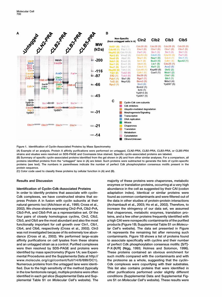
Figure 1. Identification of Cyclin-Associated Proteins by Mass Spectrometry
(A) Example of an analysis. Protein A affinity purifications were performed on untagged, CLN2-PRA, CLB2-PRA, CLB3-PRA, or CLB5-PRAstrains and eluates were resolved on SDS-PAGE and Coomassie blue stained. Specific cyclin-associated proteins are labeled.(B) Summary of specific cyclin-associated proteins identified from the gel shown in (A) and from other similar analyses. For a comparison, allproteins identified proteins from the “untagged” lane in (A) are listed. Such proteins were subtracted to generate the lists of cyclin-specificproteins (see text). The numbers in parentheses indicate the number of perfect Cdk phosphorylation consensus motifs present in theprotein sequence.(C) Color code used to classify these proteins by cellular function in (A) and (B).
Results and Discussion majority of these proteins were chaperones, metabolicenzymes or translation proteins, occurring at a very highabundance in the cell as suggested by their CAI (codonIdentification of Cyclin-Cdk-Associated Proteins
In order to identify proteins that associate with cyclin- adaptation index). Identical or similar proteins werefound as common contaminants and were filtered out ofCdk complexes, we have constructed strains that ex-
press Protein A in fusion with cyclin subunits at their the data in other studies of protein-protein interactions(Archambault et al., 2003; Ho et al., 2002). Therefore, tonatural genomic loci (Aitchison et al., 1995; Cross et al.,
2002). We chose strains expressing Cln2-PrA, Clb2-PrA, increase the stringency of our data set, we assumedthat chaperones, metabolic enzymes, translation pro-Clb3-PrA, and Clb5-PrA as a representative set. Of the
four pairs of closely homologous cyclins, Cln2, Clb2, teins, and a few other proteins frequently identified witha high CAI were nonspecific contaminants of the purifiedClb3, and Clb5 are the most abundant and also the most
functionally important for cell growth over Cln1, Clb1, products (Figure 1B; Supplemental Table S1 on Molecu-lar Cell’s website). The data set presented in FigureClb4, and Clb6, respectively (Cross et al., 2002). Cln3
was not investigated because of its extremely low abun- 1A represents the remaining list after removing suchcontaminants. Figure 1B shows a list of proteins founddance (Cross et al., 2002). We performed large-scale
affinity purifications on cell lysates from these strains to associate specifically with cyclins and their numberof perfect Cdk phosphorylation consensus motifs: [S/T]-and an untagged strain as a control. Purified complexes
were then resolved by SDS-PAGE and proteins were P-X-[K/R] (Nigg, 1993; Holmes and Solomon, 1996).These proteins contained an obvious enrichment foridentified by mass spectrometry (Figure 1A; see Experi-
mental Procedures and the Supplementa Data at http:// such motifs compared with the contaminants and withthe proteome as a whole, suggesting that the cyclin-www.molecule.org/cgi/content/full/14/6/699/DC1).
Numerous proteins from the untagged lane were identi- Cdk complexes were copurifying with their substrates.This list also contains proteins that were identified infied. Due to the high sensitivity of the method (typically
in the low femtomole range), multiple proteins were often other purifications performed under slightly differentconditions (Supplemental Data and Supplemental Fig-identified in each gel slice (Supplemental Data and Sup-
plemental Table S1 on Molecular Cell’s website). The ure S1 on Molecular Cell’s website). These results were
Proteomics of Cyclins701
highly reproducible when the same conditions wereused.
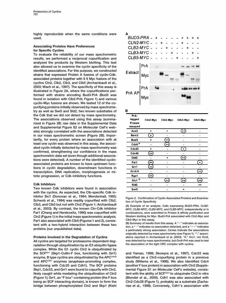
Associating Proteins Have Preferencesfor Specific CyclinsTo evaluate the reliability of our mass spectrometricresults, we performed a reciprocal copurification andanalyzed the products by Western blotting. This testalso allowed us to examine the cyclin specificity of theidentified associations. For this purpose, we constructedstrains that expressed Protein A fusions of cyclin-Cdk-associated proteins together with 9 X Myc fusions of thecyclins Cln2, Clb2, Clb3, and Clb5 (Archambault et al.,2003; Wach et al., 1997). The specificity of this assay isillustrated in Figure 2A, where the copurifications per-formed with strains encoding Bud3-PrA (Bud3 wasfound in isolation with Clb2-PrA; Figure 1) and variouscyclin-Myc fusions are shown. We tested 12 of the co-purifying proteins initially observed by mass spectrome-try as well as Swi5 and Sld2, two known substrates ofthe Cdk that we did not detect by mass spectrometry.The associations observed using this assay (summa-rized in Figure 2B; raw data in the Supplemental Dataand Supplemental Figure S2 on Molecular Cell’s web-site) strongly correlated with the associations detectedin our mass spectrometric screen (Figure 2B). Impor-tantly, for every protein where an association with atleast one cyclin was observed in this assay, the associ-ated cyclin initially detected by mass spectrometry wasconfirmed, strengthening our confidence in the massspectrometric data set (even though additional associa-tions were detected). A number of the identified cyclin-associated proteins are known to have upstream func-tions in cyclin degradation, downstream functions intranscription, DNA replication, morphogenesis or mi-totic progression, or Cdk inhibitory functions.
Cdk InhibitorsTwo known Cdk inhibitors were found in associationwith the cyclins. As expected, the Clb-specific Cdk in-hibitor Sic1 (Donovan et al., 1994; Mendenhall, 1993; Figure 2. Confirmation of Cyclin-Associated Proteins and Examina-Schwob et al., 1994) was readily copurified with Clb2, tion of Cyclin SpecificityClb3, and Clb5 but not with Cln2 (Figure 1; Archambault (A) Example of an analysis. Cells expressing BUD3-PRA, CLN2-et al., 2003). By contrast, the known Cln-Cdk inhibitor MYC, CLB2-MYC, CLB3-MYC, and CLB5-MYC, independently or in
combinations, were submitted to Protein A affinity purification andFar1 (Chang and Herskowitz, 1990) was copurified withWestern blotting for Myc. Bud3-PrA associated with Cln2-Myc andCln2 (Figure 1) in the initial mass spectrometric analysis.Clb2-Myc in this assay.Far1 also associated with Clb5 (Figures 1 and 2), consis-(B) Summary of results from this assay. A “�” indicates an associa-tent with a two-hybrid interaction between these twotion, a “�” indicates no association detected, and a “��” indicates
proteins (our unpublished data). a particularly strong association. Circles indicate the associationsoriginally detected by mass spectrometry (see Figure 1). “*,” associ-ations reported in Archambault et al. (2003). “#,” Orc1, not Orc6,Proteins Involved in the Degradation of Cyclinswas detected by mass spectrometry, but Orc6-PrA was used to testAll cyclins are targeted for proteasome-dependent deg-the association of the tight ORC complex with cyclins.radation through ubiquitination by an E3 ubiquitin ligase
complex. While the G1 cyclin Cln2 is ubiquitinated bythe SCFGrr1 (Skp1-Cullin-F box, functioning with Grr1) and Yamao, 1998; Skowyra et al., 1997). Cdc53 was
identified as a Cln2-copurifying protein in a previousenzyme, B type cyclins are ubiquitinated by the APCCdc20
and APCCdh1 enzymes (anaphase-promoting complex, study (Willems et al., 1996). We also identified Cdc4(another F box protein) in association with Cln2 (Supple-functioning with Cdc20 and Cdh1). The SCF proteins
Skp1, Cdc53, and Grr1 were found to copurify with Cln2, mental Figure S1 on Molecular Cell’s website), consis-tent with the ability of SCFCdc4 to ubiquinate Cln2 in vitrolikely caught while mediating the ubiquitination of Cln2
(Figure 1); Grr1, an “F box”-containing protein (the F box (Blondel et al., 2000). Cdh1 was also associated withCln2-Cdc28 (Figure 1), probably as a substrate (Zacha-being an SCF interacting domain), is known to form the
bridge between phosphorylated Cln2 and Skp1 (Kishi riae et al., 1998). Conversely, Cdh1’s association with

Molecular Cell702
Clb3 (Supplemental Figure S1 on Molecular Cell’s web- tagged proteins separately (Supplemental Figure S3 onMolecular Cell’s website). It is possible that there is ansite) was observed, most likely because of its role in
targeting this cyclin for degradation (Schwab et al., 2001; ATP requirement for this interaction or that the wholepool of Cln2-Ub conjugates is stably bound to Cdc48Visintin et al., 1997). The association of Cdc4 and Cdc53
with Clb3 (Supplemental Figure S1 on Molecular Cell’s before and after the lysis, preventing additional Cdc48from binding after the lysis.website), could have been bridged by Sic1, which is
known to be recognized and ubiquitinated by the SCFCdc4
(Feldman et al., 1997). No other components of APC Cln2-Ub Accumulates in cdc48-3 Mutant Cellsenzymes other than Cdh1 copurified with the B type Based on the above results and on the fact that Cln2 iscyclins, suggesting that these interactions are more a constitutively unstable protein (Schneider et al., 1998)transient. that is degraded by a ubiquitination-proteasome path-
way, we hypothesized that Cdc48 could function in facil-itating the degradation of polyubiquitinated Cln2 by theCdc48 Interacts Specifically with Cln2-Ubiquitin
Conjugates In Vivo proteasome. To test this idea, we used a temperature-sensitive allele of Cdc48 (cdc48-3) that allows cells toWe identified Cdc48 as a major Cln2-associated protein.
Originally, the essential gene CDC48 was isolated from grow at 23�C but causes them to arrest at 37�C as bud-ded cells containing an undivided nucleus (Moir et al.,mutants that arrested as budded cells with an undivided
nucleus at the restrictive temperatures (Moir et al., 1982). 1982). We grew cdc48-3 CLN2-MYC cells at 23�C andthen switched half of the culture to 37�C. Fractions ofThis gene was later found to encode a protein homolo-
gous to the mammalian protein VCP (Frohlich et al., the cultures were collected at different times followingthe temperature switch. Collected cells were then sub-1991). Cdc48 and VCP are members of a superfamily of
proteins termed AAA (ATPase associated with various mitted to a Myc immunoprecipitation (IP). Several fea-tures were noted. First, when probed for Myc, the IP ofcellular activities), so named for their multiple participa-
tions in cell cycle, vesicular transport, mitochondrial cdc48-3 CLN2-MYC revealed a large band that migratedmore slowly in the extract from cells incubated at 37�Cfunctions, peroxisome assembly, and proteolysis (Froh-
lich, 2001). Cdc48 is a polyubiquitin binding protein that than in the extract from cells incubated at 23�C (Figure3B). This indicated that covalently modified forms ofhas been proposed to participate in the degradation of
substrates of the proteasome (Dai and Li, 2001; Ghislain Cln2-Myc, possibly phosphoforms, accumulated in themutant at 37�C. Second, in the IP from cdc48-3 CLN2-et al., 1996). Although traces of Cdc48 (a very abundant
protein) were present in the control purification from MYC cells, a strong enrichment for a ubiquitin signal inthe �200 kDa range was observed relative to CDC48untagged cells, we hypothesized that the clear enrich-
ment observed in the Cln2-PrA purification (Figure 1A) CLN2-MYC cells. Thus, the ability to destroy Cln2-ubi-quitin conjugates has been impaired by the loss ofwas due to a specific interaction and so we did not
discard Cdc48 as a contaminant; perhaps Cdc48 targets Cdc48 function. Third, the ubiquitin signals were specificto the cdc48-3 CLN2-MYC cells but were observed atubiquitinated Cln2 for destruction, and it was various
ubiquitinated forms of Cln2-PrA that were associating both temperatures. This suggests that Cdc48 alreadypresents a loss of activity at 23�C in cdc48-3 mutants.with Cdc48 in the purification (Figure 1A).
Thus, we tested if Cdc48 associated specifically with Fourth, at the nonpermissive temperature the degree ofubiquitination increases (reflected in the shift to higherpoly-ubiquitin-Cln2 conjugates and whether Cdc48 as-
sociated with other cyclins. The Western blot from molecular weight forms of the ubiquitinated Cln2; Figure3B, bottom panel). Together, these results suggest thatCdc48-PrA purification products revealed the presence
of very slow migrating forms of Cln2-Myc, in the 150–200 Cln2 phosphoforms and polyubiquitinated forms accu-mulate in the context of a loss of function of Cdc48.kDa range (Figure 3A). No signal was observed around
95 kDa, where nonubiquitinated Cln2-Myc normally mi- This in turn implies that Cdc48 functions downstreamof the SCFGrr1 ubiquitin ligase in the pathway that allowsgrates. In contrast, a similar experiment performed using
Grr1-PrA as bait revealed an association with the 95 Cln2 degradation. In a cell cycle block-release experi-ment (using CDC48-PRA CLN2-MYC cdc20 GAL-CDC20kDa (nonubiquitinated) form of Cln2-Myc (Supplemental
Figure S2E on Molecular Cell’s website). No association cells), we observed that the amounts of Cln2-ubiquitinconjugates that copurify with Cdc48 followed closelyof either Cdc48-PrA or Grr1-PrA with Clb2-Myc, Clb3-
Myc, or Clb5-Myc was detected, suggesting that Cdc48 the profile of total Cln2 abundance (our unpublisheddata). Our findings are consistent with Cln2 being constitu-associates preferentially with SCF substrates rather
than with APC substrates. Preliminary experiments sug- tively turned over by a fast SCFGrr1-Cdc48-proteasomepathway.gested that Cdc48 does not significantly associate with
Sic1 (substrate of SCFCdc4) in this assay, suggesting We used another assay to test a role for Cdc48 inCln2 degradation. It involved the pheromone responsesome level of specificity for Cdc48 toward specific SCF
substrates. Nonetheless, the fact that Cdc48 associates pathway, which allows haploid cells to sense the pres-ence of a neighboring cell of the opposite mating type.with Far1 (Fu et al., 2003) (substrate of the SCFCdc4) in
addition to Cln2 (substrate of SCFGrr1) suggests that the The pheromone response involves the inactivation ofG1 cyclins (such as Cln2), which in turn leads to anassociation of Cdc48 with SCF targets is not restricted
by which F box protein subunit is used in the ubiquitin arrest in G1 in preparation for the fusion of the two cells(Valdivieso et al., 1993). By contrast, overexpression ofligation reaction. We demonstrated that the Cdc48-PrA/
Ub-Cln2-Myc association occurs in vivo and not postly- CLN2 represses the mating pathway (Oehlen and Cross,1994; Wassmann and Ammerer, 1997). Cdc48 has re-sis in our coaffinity purification assay by performing an
experiment on cell pools containing cells expressing the cently been shown to be required for normal degradation
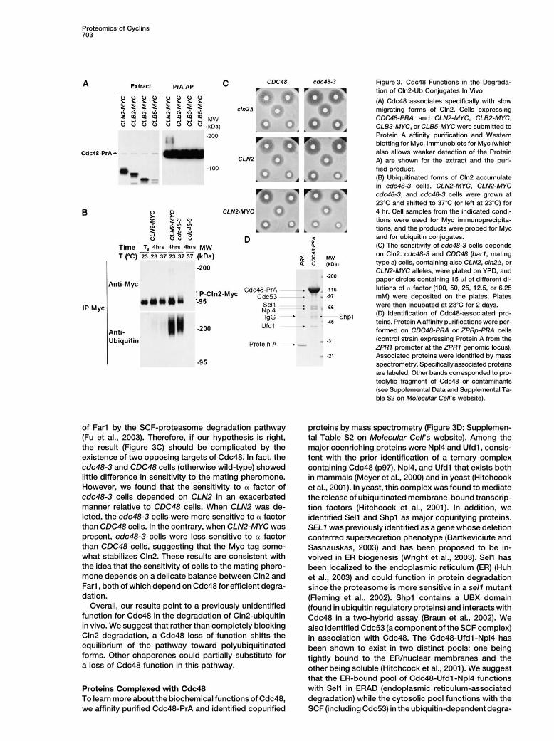
Proteomics of Cyclins703
Figure 3. Cdc48 Functions in the Degrada-tion of Cln2-Ub Conjugates In Vivo
(A) Cdc48 associates specifically with slowmigrating forms of Cln2. Cells expressingCDC48-PRA and CLN2-MYC, CLB2-MYC,CLB3-MYC, or CLB5-MYC were submitted toProtein A affinity purification and Westernblotting for Myc. Immunoblots for Myc (whichalso allows weaker detection of the ProteinA) are shown for the extract and the puri-fied product.(B) Ubiquitinated forms of Cln2 accumulatein cdc48-3 cells. CLN2-MYC, CLN2-MYCcdc48-3, and cdc48-3 cells were grown at23�C and shifted to 37�C (or left at 23�C) for4 hr. Cell samples from the indicated condi-tions were used for Myc immunoprecipita-tions, and the products were probed for Mycand for ubiquitin conjugates.(C) The sensitivity of cdc48-3 cells dependson Cln2. cdc48-3 and CDC48 (bar1, matingtype a) cells, containing also CLN2, cln2�, orCLN2-MYC alleles, were plated on YPD, andpaper circles containing 15 �l of different di-lutions of � factor (100, 50, 25, 12.5, or 6.25mM) were deposited on the plates. Plateswere then incubated at 23�C for 2 days.(D) Identification of Cdc48-associated pro-teins. Protein A affinity purifications were per-formed on CDC48-PRA or ZPRp-PRA cells(control strain expressing Protein A from theZPR1 promoter at the ZPR1 genomic locus).Associated proteins were identified by massspectrometry. Specifically associated proteinsare labeled. Other bands corresponded to pro-teolytic fragment of Cdc48 or contaminants(see Supplemental Data and Supplemental Ta-ble S2 on Molecular Cell’s website).
of Far1 by the SCF-proteasome degradation pathway proteins by mass spectrometry (Figure 3D; Supplemen-(Fu et al., 2003). Therefore, if our hypothesis is right, tal Table S2 on Molecular Cell’s website). Among thethe result (Figure 3C) should be complicated by the major coenriching proteins were Npl4 and Ufd1, consis-existence of two opposing targets of Cdc48. In fact, the tent with the prior identification of a ternary complexcdc48-3 and CDC48 cells (otherwise wild-type) showed containing Cdc48 (p97), Npl4, and Ufd1 that exists bothlittle difference in sensitivity to the mating pheromone. in mammals (Meyer et al., 2000) and in yeast (HitchcockHowever, we found that the sensitivity to � factor of et al., 2001). In yeast, this complex was found to mediatecdc48-3 cells depended on CLN2 in an exacerbated the release of ubiquitinated membrane-bound transcrip-manner relative to CDC48 cells. When CLN2 was de- tion factors (Hitchcock et al., 2001). In addition, weleted, the cdc48-3 cells were more sensitive to � factor identified Sel1 and Shp1 as major copurifying proteins.than CDC48 cells. In the contrary, when CLN2-MYC was SEL1 was previously identified as a gene whose deletionpresent, cdc48-3 cells were less sensitive to � factor conferred supersecretion phenotype (Bartkeviciute andthan CDC48 cells, suggesting that the Myc tag some- Sasnauskas, 2003) and has been proposed to be in-what stabilizes Cln2. These results are consistent with volved in ER biogenesis (Wright et al., 2003). Sel1 hasthe idea that the sensitivity of cells to the mating phero- been localized to the endoplasmic reticulum (ER) (Huhmone depends on a delicate balance between Cln2 and et al., 2003) and could function in protein degradationFar1, both of which depend on Cdc48 for efficient degra- since the proteasome is more sensitive in a sel1 mutantdation. (Fleming et al., 2002). Shp1 contains a UBX domain
Overall, our results point to a previously unidentified (found in ubiquitin regulatory proteins) and interacts withfunction for Cdc48 in the degradation of Cln2-ubiquitin Cdc48 in a two-hybrid assay (Braun et al., 2002). Wein vivo. We suggest that rather than completely blocking also identified Cdc53 (a component of the SCF complex)Cln2 degradation, a Cdc48 loss of function shifts the in association with Cdc48. The Cdc48-Ufd1-Npl4 hasequilibrium of the pathway toward polyubiquitinated been shown to exist in two distinct pools: one beingforms. Other chaperones could partially substitute for tightly bound to the ER/nuclear membranes and thea loss of Cdc48 function in this pathway. other being soluble (Hitchcock et al., 2001). We suggest
that the ER-bound pool of Cdc48-Ufd1-Npl4 functionswith Sel1 in ERAD (endoplasmic reticulum-associatedProteins Complexed with Cdc48degradation) while the cytosolic pool functions with theTo learn more about the biochemical functions of Cdc48,
we affinity purified Cdc48-PrA and identified copurified SCF (including Cdc53) in the ubiquitin-dependent degra-

Molecular Cell704
dation of proteins, such as Cln2. Future studies should enrichment of nuclear YFP-Ace2 fluorescence. In thistest, 27% of the clb3 clb4 cells showed a nuclear signal,investigate the function of these associations.compared with 20% for the wt cells, representing a 35%increase in nuclear localization (Figure 4A). Our resultPotential Phosphorylation Substrates of Cdc28is consistent with a role of Clb3,4-Cdc28 in negativelyThe proteins that specifically copurified with cyclinsregulating the nuclear localization of Ace2. Becauseshow a strong enrichment for optimal Cdk phosphoryla-some cytoplasmic Ace2 is still observed in the clb3 clb4tion consensus motifs, corresponding to the sequencecells, it seems likely that regulators other than Clb3,4-[S/T]-P-X-[K/R] (Figure 1B). This sequence is mostly ab-Cdc28 exist to exclude Ace2 from the nucleus. Somesent or else present only once in the proteins that weof these regulators may be alternative cyclin-Cdc28labeled as contaminants. This observation reinforcescomplexes. Consistent with this idea, we found thatthe idea that coenriching proteins are substrates. TheAce2-PrA also copurified with Cln2-Myc and Clb2-Myc,average number of [S/T]-P-X-[K/R] Cdk motifs in ouralthough with lesser efficiency when compared withspecifically associated proteins is 3.0, compared withClb3-Myc (Figure 4B). Moreover, when the same Protein0.3 in the whole yeast proteome. There are 24 (69%)A affinity purification was performed in cells deleted forproteins with 2 or fewer motifs and 11 (31%) with 3 orCLB3 and CLB4, a higher amount of Clb2-Myc reproduc-greater motifs in our cyclin-associated proteins. Thereibly copurified, suggesting that Clb2 can substitute forare 6015 (96%) proteins in the proteome with 2 or fewerthe absence of Clb3 and Clb4 (Figure 4B).motifs and 254 (4.0%) with 3 or more. The difference is
To visualize the cell cycle dependence of the Clb3-significant at a level of p � 1 � 10�6. When we considerAce2 association, we initially used cells tagged on bothone or fewer versus two or more motifs, our enrichmentAce2 and Clb3 that could be synchronized in mitosisfor proteins containing such motifs is also very signifi-and released into the cell cycle by switching from YEPDcant. Moreover, 15 of the 24 proteins that contained atto YEPGal medium (strain ACE2-PRA CLB3-MYC cdc20least one optimal Cdk phosphorylation consensus motifGAL-CDC20). Protein A affinity purification and Myc im-(namely Far1, Cdh1, Rga1, Bem3, Srl3, Ypl267, Sic1,munoblotting with samples from the time courseBud3, Lte1, Kel1, Ace2, Dbf2, Mob1, Orc1, and Spc42)showed that the Clb3-Ace2 association occurred in mi-have been reported to be Cdc28 substrates in vitro intosis (when Clb3-Myc is present) and that this associa-a recent study, where a selected fraction of the pro-tion coincided with a decrease in electrophoretic mobil-teome was assayed (Ubersax et al., 2003). By contrast,ity for Ace2 (characteristic of an increase in Ace2none of the proteins that we rejected as contaminantsphosphorylation; data not shown).were detected as phosphorylation substrates (Ubersax
Interestingly, we found that the Clb3-Ace2 associationet al., 2003). The known and potential phosphorylationcould occur efficiently post-cell lysis. We used this abil-substrates that we identified as cyclin/Cdc28-associat-ity to reconstitute the association in cell lysates to testing proteins function in a broad range of cellular events.if the efficiency of the association was dependent onBelow we provide examples of these cyclin-associatedthe cell cycle. Thus, we synchronized and then releasedproteins that are candidate Cdk substrates (Figure 1)ACE2-PRA cdc20 GAL-CDC20 cells, adding asynchro-and are involved in transcription, DNA replication, mor-nous CLB3-MYC cells to samples from the time coursephogenesis/signaling, and mitotic progression. We alsoimmediately before lysis. In this way, the same amountconfirmed that some of these candidate Cdk substratesof Clb3-Myc was present in each sample, and we couldwere phosphorylated in vivo.visualize the changes in efficiency of association inde-pendent of protein amounts (Ace2 did not change muchControl of Transcription: Clb3-Cdc28 Contributesin abundance). We observed a marked reduction in as-to Regulating the Localization of the Transcriptionsociation efficiency in G1, at a time when the fast migrat-Factor Ace2ing (i.e., likely hypophosphorylated) forms of Ace2 areThe G1 transcription factor Ace2 is a strong candidatepresent (Figure 4C, left). This loss of association wassubstrate for Cdc28 phosphorylation because it carriescompletely dependent on Sic1, as the same experimenta large number of consensus phosphorylation motifsperformed in parallel with ACE2-PRA cdc20 GAL-and has strong homology to the known Cdc28 substrateCDC20 sic1 cells resulted in constitutive associationSwi5. Reports from other groups have suggested thebetween Ace2-PrA and Clb3-Myc (Figure 4C, right). Fur-existence of a regulatory connection between Cdc28thermore, the faster migrating forms of Ace2 were al-and Ace2 (O’Conallain et al., 1999). Much like Swi5 (Mollmost absent in these cells throughout the time course.et al., 1991), Ace2 was thought to be phosphorylatedSic1 is known to function in keeping Clb-Cdc28 activityby Cdc28 on or near its nuclear localization signal (NLS),at its lowest in G1 (Schwob et al., 1994). Therefore,thereby excluding it from the nucleus. In our study, weour results suggest that Sic1 allows Ace2 to becomeobserved a strong association between Clb3 and Ace2dephosphorylated in G1 by inhibiting its interaction with(Figure 1A) and so hypothesized that Clb3-Cdc28 mayand phosphorylation by the Clb3,4-Cdc28 kinases.function in promoting the cell cycle-regulated nuclear
exclusion of Ace2. We therefore visualized the subcellu-lar localization of YFP-Ace2 in wt or in clb3 clb4 cells. Ace2 Is Phosphorylated at Multiple Sites In Vivo
To search for Cdk phosphorylation sites in Ace2, weIn addition to CLB3, we also deleted CLB4 because ofits high similarity to CLB3, both in sequence and in employed mass spectrometry (Figure 5A). Ace2-PrA ex-
pressed in yeast under the endogenous ACE2 promoterfunction (Miller and Cross, 2001; Morgan, 1997). In a“double blind” experiment, we scored between 1000 was affinity purified and submitted to a “hypothesis-
driven” mass-spectrometric analysis (Kalkum et al.,and 1200 cells of each type (wt and clb3 clb4) for their

Proteomics of Cyclins705
Figure 4. Clb3-Cdc28 Contributes to Regulating the Localization of Ace2
(A) Normal subcellular localization of Ace2 depends on the presence of Clb3 and Clb4. Between 1000 and 1200 asynchronous YFP-ACE2cells and YFP-ACE2 clb3 clb4 cells were scored for the enrichment of nuclear fluorescence signal in double blind experiments. The numbersof 20% and 27% have standard deviations of 1.1% and 1.3%, respectively.(B) Relative intensities of associations between cyclins and Ace2 in a wt or clb3 clb4 background. A Protein A affinity purification experimentperformed on either wt or clb3 clb4 cells expressing ACE2-PRA and CLN2-MYC, CLB2-MYC, CLB3-MYC, or CLB5-MYC. The numbers at thebottom refer to the ratios of cyclin-Myc/Ace2-PrA signals in the above anti-Myc blot from the Protein A purification.(C) Cell cycle dependence of the competence for association between Ace2 and Clb3. ACE2-PRA cdc20 GAL-CDC20 cells, SIC1-wt, or sic1were grown in YEPGal, arrested at mitosis in YEPD for 3 hr, and released in YEPGal. Cells were collected every 10 min and were mixed withasynchronous CLB3-MYC cells before being submitted to Protein A affinity purifications. A lanes, arrested ACE2-PRA cdc20 GAL-CDC20(SIC1-wt or sic1) cells were mixed with wt cells. B lanes, wt cells were mixed with CLB3-MYC cells. Extracts were probed for Protein A, Myc,and Clb3. Purified products were probed for Protein A and Myc. The percentages of unbudded cells and binucleate cells are shown for thetime courses.
2003; Chang et al., 2004). Using this approach, we identi- ylation could be mapped to specific residues. Serine 80(in a SP motif), serine 428 (in a SP motif), threonine 501fied 15 phosphorylated Ace2 peptides, 14 of which con-
tained at least 1 [S/T]-P minimal Cdk phosphorylation (in a TPVK motif), and serine 557 (in a SP motif) wereidentified as four previously unknown in vivo phosphory-consensus motif (Figure 5A). These peptides (containing
at least 17 different sites of phosphorylation) covered lation sites. Three of the observed phosphopeptidescontained serine 714 (in a SPVK motif), one of three26% of the Ace2 sequence and included 16 of its 21
[S/T]-P motifs. For four of these peptides, the phosphor- proposed Cdk phosphorylation sites present in the con-
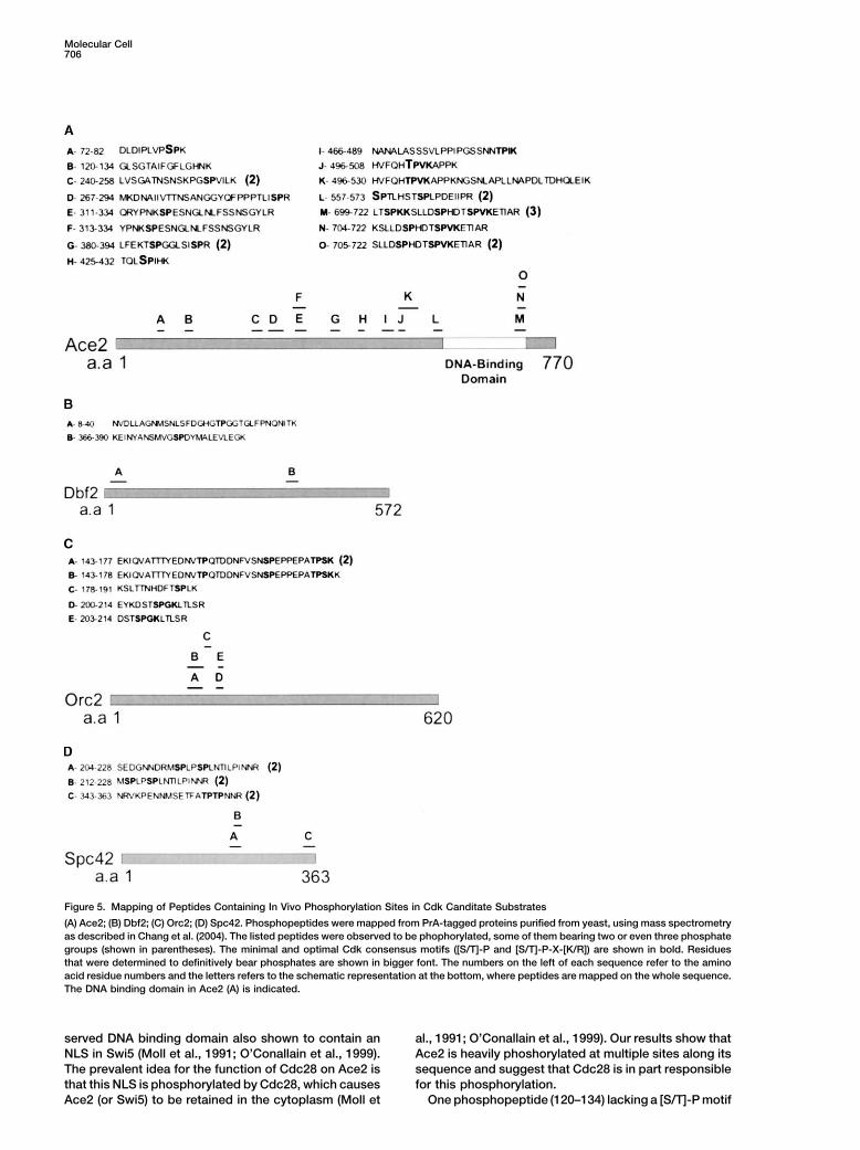
Molecular Cell706
Figure 5. Mapping of Peptides Containing In Vivo Phosphorylation Sites in Cdk Canditate Substrates
(A) Ace2; (B) Dbf2; (C) Orc2; (D) Spc42. Phosphopeptides were mapped from PrA-tagged proteins purified from yeast, using mass spectrometryas described in Chang et al. (2004). The listed peptides were observed to be phophorylated, some of them bearing two or even three phosphategroups (shown in parentheses). The minimal and optimal Cdk consensus motifs ([S/T]-P and [S/T]-P-X-[K/R]) are shown in bold. Residuesthat were determined to definitively bear phosphates are shown in bigger font. The numbers on the left of each sequence refer to the aminoacid residue numbers and the letters refers to the schematic representation at the bottom, where peptides are mapped on the whole sequence.The DNA binding domain in Ace2 (A) is indicated.
served DNA binding domain also shown to contain an al., 1991; O’Conallain et al., 1999). Our results show thatAce2 is heavily phoshorylated at multiple sites along itsNLS in Swi5 (Moll et al., 1991; O’Conallain et al., 1999).
The prevalent idea for the function of Cdc28 on Ace2 is sequence and suggest that Cdc28 is in part responsiblefor this phosphorylation.that this NLS is phosphorylated by Cdc28, which causes
Ace2 (or Swi5) to be retained in the cytoplasm (Moll et One phosphopeptide (120–134) lacking a [S/T]-P motif
Proteomics of Cyclins707
was also identified, indicating that Ace2 can be phos- ular Cell’s website), consistent with the observation thatefficient Sld2 phosphorylation is dependent on CLB5phorylated by at least one other kinase. This kinase
could be the Cbk1-Mob2 complex, which functions in and CLB6 (Masumoto et al., 2002).targeting Ace2 to daughter cells only at mitotic exit, amechanism that is required for normal asymmetric Control of Morphogenesisgrowth (Colman-Lerner et al., 2001; Racki et al., 2000; Cln2-PrA copurified with Ste20, confirming a previousWeiss et al., 2002). In fact, peptide 120–134 contains observation (Oda et al., 1999), but Ste20 was not ob-residue G128, which when mutated to a glutamic acid served in complex with other cyclins tested. Ste20 is aresidue bypasses the requirement for Cbk1 for nuclear kinase involved in the control of numerous cell growthlocalization (Racki et al., 2000; Colman-Lerner et al., and pheromone response pathways and is efficiently2001). Interestingly, we detected Cbk1 in the Clb3-PrA and specifically phosphorylated by Cln1,2-Cdc28 com-purification (Figure 1). Instead of being bound directly plexes in vivo (Oehlen and Cross, 1998; Wu et al., 1998).to Clb3, Cbk1 could have been bound to Ace2, which The association of Cln2-Cdc28 with Ste20 may be partwas present in large amounts in the Clb3-PrA copurifica- of a mechanism allowing Ste20 to switch its functiontion product (Figure 1A). from the mating response pathway to morphogenesis
(Oda et al., 1999; Oehlen and Cross, 1998; Wu et al.,1998). Two GAPs (GTPase activating proteins), Rga1Other Links between Cyclin-Cdk and Transcription
We observed that Swi5, a close homolog of Ace2, also and Bem3, were found to associate with Cln2-PrA (Fig-ure 2 and Supplemental Figure S2 on Molecular Cell’spreferentially associated with Clb3 (Supplemental Fig-
ure S2 on Molecular Cell’s website), suggesting a pre- website). Rga1 and Bem3 (along with Rga2 and Bem3)stimulate GTP hydrolysis by the Rho-like GTPase Cdc42,ferred function for Clb3 in regulating these closely re-
lated transcription factors with partially overlapping which functions in polarity establishment (review in Gulliand Peter, 2001). Since Rga1 acts to repress the phero-functions (Doolin et al., 2001). The Cln2-Sin3 association
provides another potential connection with transcrip- mone pathway (Stevenson et al., 1995), its phosphorylationby Cln2-Cdc28 could stimulate its function. Consistenttion. Sin3 is a member of a histone deacetylase complex
involved in transcriptional repression (Kadosh and with this idea, overexpression of CLN2 in a rga1-deletedstrain no longer represses the mating response pathwayStruhl, 1998) and is phosphorylated at an SP Cdc28
consensus site (Ficarro et al., 2003). These observations (Wassmann and Ammerer, 1997). Phosphorylation ofCdc42’s GAPs by Cln2-Cdc28 could provide anotherlead us to postulate a potential link between Cln2-Cdc28
activity and transcriptional repression by Sin3. level of control of the cyclin-Cdk machine over morpho-genesis, stimulating polarized growth.
Cyclin-Mediated Control of DNA ReplicationWe identified two components of the pre-replicative Control of Mitotic Progression
The B type cyclins are essential for mitotic progressioncomplex (pre-RC; required for DNA replication initiation),Cdc6 and Orc1, in association with Clb2 and Clb5, re- and must be inactivated in order to allow termination of
mitosis. We found that Nap1 associates specifically withspectively (Supplemental Figure S1 on Molecular Cell’swebsite; Archambault et al., 2003). Cdc6 is a licensing Clb2 (Figure 1), confirming published work (Kellogg et
al., 1995). Nap1 was found to be required for the normalfactor required for the loading of the hexameric MCMhelicase (Mcm2-7) to form the prereplicative complex, mitotic functions of Clb2, including a shift from polarized
to isotropic bud growth (Kellogg and Murray, 1995). Thewhile Orc1 is known to form a stable complex withOrc2–6 (reviewed in Bell and Dutta, 2002). Orc1, Orc2, axial bud site selection protein Bud3 was also clearly
recovered in specific association with Clb2 (Figure 1Band Orc6 all contain Cdk consensus phosphorylationmotifs and are Cdc28 substrates in vitro (Ubersax et al., and Supplemental Figure S1 on Molecular Cell’s web-
site). A recent report showed that Bud3 interacts with2003). Clb5 interacts specifically with Orc6, and a stableinteraction between Clb5-Cdc28 and Orc6 at origins Clb2 in a two-hybrid assay and that this interaction func-
tions to target Clb2 to the mother bud neck, which seemsprevents refiring of DNA replication (Wilmes et al., 2004).It is likely that Orc6 as well as other ORC subunits were to stimulate cytokinesis (Bailly et al., 2003). We also
identified the karyopherin � (Kap60/Srp1) in associationpresent in trace amounts in the same purification andthat we failed to detect them by mass spectrometry. In with Clb2, consistent with the existence of both a bud
neck-associated pool and a nuclear pool of Clb2—wheresuch a case, the Clb5-Orc1 association could be bridgedby other ORC subunits. Using the same strategy as for the nuclearly localized Clb2 is dependent on Kap60-
mediated import (Hood et al., 2001).Ace2, we identified several in vivo Cdk motif-containingphosphopeptides in Orc2 (Figure 5C) and Orc6 (docu- A series of elegant genetic experiments has sug-
gested that Cln1–3 promote SPB duplication, Clb1–6mented in Chang et al., 2004). We submitted Orc1 tothe same analysis and found no phosphopeptides. promote spindle pole body (SPB) maturation, and mi-
totic Clb1–4 inhibit SPB licensing and re-reduplicationPhosphopeptides observed in Orc2 and Orc6 containCdk consensus sites that were mutated and that were (Haase et al., 2001). Clb5-Cdc28 kinase activity is re-
quired for coordinated spindle assembly and orientationimplicated in preventing reinitiation of replication (Ngu-yen et al., 2001; Wilmes et al., 2004). (Segal et al., 2000). We found that Clb5 copurifies with
Spc42, a phosphoprotein required for SPB duplication.We also tested whether a specific cyclin could associ-ate with Sld2, the only known obligatory substrate of We also observed this interaction in a yeast two-hybrid
screen for Clb5 binding proteins (our unpublished data).Cdc28 in DNA replication (Masumoto et al., 2002). Wefound that Sld2 preferentially associated with Clb5 (Sup- We identified multiple in vivo phosphorylation at Cdk
motif-containing peptides in Spc42 (Figure 5D).plemental Data and Supplemental Figure S2H on Molec-
Molecular Cell708
study; therefore, we have not evaluated the mechanisticrole for the Cdk phosphorylation sites in the regulationof the targets that we have identified. We can speculatethat the cyclin-Cdk module could use different molecu-lar mechanisms to act on its targets. On one hand, acyclin-Cdk complex could act as a kinase on its target,thereby changing the activity of this target effector. Onthe other hand, it is theoretically possible for a cyclinbinding target to function primarily as a “landing pad”or adaptor for a cyclin-Cdk complex. Once bound, thecomplex could not only phosphorylate the binding targetitself but also other proteins in the vicinity; alternatively,the bound cyclin-Cdk module could change (and soregulate) the accessibility of its target for other interac-tions. By its nature, our study could have enriched forthese more stable cyclin-Cdk/target interactions relativeto the more transient kinase-substrate interactions.
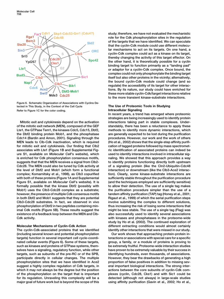
Figure 6. Schematic Organization of Associations with Cyclins De-The Use of Proteomic Tools in Studyingtected in This Study, in the Context of the Cell CycleIntracellular SignalingRefer to Figure 1C for the color coding.In recent years, a trend has emerged where proteomicstrategies are being increasingly used to identify proteininteractions taking part in stable complexes. Under-Mitotic exit and cytokinesis depend on the activation
of the mitotic exit network (MEN), composed of the GEF standably, there has been a reluctance to apply thesemethods to identify more dynamic interactions, whichLte1, the GTPase Tem1, the kinases Cdc5, Cdc15, Dbf2,
the Dbf2 binding protein Mob1, and the phosphatase are generally expected to be lost during the purificationprocedures. However, our work and the work of othersCdc14 (Bardin and Amon, 2001). Signaling through the
MEN leads to Clb-Cdk inactivation, which is required (Ho et al., 2002) shows that the single-step affinity purifi-cation of tagged proteins followed by mass spectromet-for mitotic exit and cytokinesis. Our finding that Clb2
associates with Lte1 (Figure 1B and Supplemental Fig- ric identification of associated proteins can indeed beused to identify interactions involved in intracellular sig-ure S1, available on Molecular Cell’s website), which
is enriched for Cdk phosphorylation consensus motifs, naling. We showed that this approach provides a wayto identify proteins functioning directly both upstreamsuggests that that the MEN receives a signal from Clb2-
Cdc28. The MEN could also be tuned by Cdk activity at of a signaling protein (like the Cln2-Ubiquitin-Cdc48interaction) or downstream (like the Clb3-Ace2 interac-the level of Dbf2 and Mob1 (which form a functional
complex; Komarnitsky et al., 1998), as Clb3 copurified tion). Clearly, some kinase-substrate interactions aresufficiently stable throughout the purification procedurewith both of these proteins (Figure 1A and Supplemental
Figure S1, available on Molecular Cell’s website). It is (and the techniques employed are sufficiently sensitive)to allow their detection. The use of a single tag makesformally possible that the kinase Dbf2 (possibly with
Mob1) uses the Clb3-Cdc28 complex as a substrate; the purification procedure simpler than the use of atandem affinity purification (TAP) tag (Honey et al., 2001;however, the presence of multiple Cdk consensus motifs
in both Dbf2 and Mob1 suggests that they are potential Rigaut et al., 1999) of which the serial purification stepsinvolve submitting the complex to different solutions,Clb3-Cdc28 substrates. In fact, we observed in vivo
phosphorylation of Dbf2 in two peptides containing min- thus increasing the risk of losing some interactions thatmight be less stable. The use of a single tag (Flag) wasimal Cdk motifs (Figure 5B). These results suggest the
existence of a feedback loop between the MEN and Clb- also successfully used to identify several associationswith kinases and phosphatases in the proteome-wideCdk activity.study by Ho et al. (2002). The continued exploration ofdifferent extracting conditions would almost certainlyMolecular Mechanisms of Cyclin-Cdk Action
The cyclin-Cdk-associated proteins that we identified identify other interactions that were missed in our study.Our work shows that approaching protein-protein in-(including several known and potential phosphorylation
targets) function in several important cell cycle-coordi- teractions or associations with special care for a specificgroup, a family, or a module of proteins is proving tonated cellular events (Figure 6). Some of these targets,
such as kinases and proteins of GTPase systems, them- be extremely fruitful. Proteome-wide interaction studieshave proven to be extremely valuable by their success inselves have a signaling, regulatory function, while other
targets, such as subunits of the pre-RC or of the SPB, identifying hundreds, even thousands, of associations.However, they bear the drawbacks of generating a highparticipate directly in cellular changes. The multiple
phosphorylation sites that we have identified in Ace2 proportion of false positives in addition to missing sev-eral important biological interactions. The strong inter-suggest a highly complex regulation of Cdk targets, in
which it may not always be the degree but the position actions between the core subunits of cyclin-Cdk com-plexes (cyclin, Cdc28, Cks1) and with Sic1 could beof the phosphorylation on the target that is important
for its regulation. Unraveling this complexity will be a detected (although not always) in large-scale studiesusing affinity purification (Gavin et al., 2002; Ho et al.,major goal of future work but is beyond the scope of this
Proteomics of Cyclins709
an integrated MALDI-QqTOF/MALDI-IT mass spectrometery system2002) or the two-hybrid system (Ito et al., 2001; Uetz et(Krutchinsky et al., 2000, 2001; Qin and Chait, 1997).al., 2000). However, of all the other specific associations
with cyclins that we detected in our study, none wereAcknowledgmentsdetected in those large-scale studies when either
cyclins or cyclin-associating proteins were used as We thank members of the Rout lab, the Chait lab, and the Crossbaits. Only Srl3 and Ypl014 were found as part of the lab for useful discussions. Special thanks go to Andrew Krutchinsky“Cdc28-associated complex” (Gavin et al., 2002). and Markus Kalkum for sage advice with the mass spectrometry.
Thanks to Randy Schekman for providing the cdc48-3 strain. Fund-Once an associated protein is identified, this proteining was provided by NIH grants RR00862 (to B.T.C.) and CA89810 (tocan in turn be tagged and affinity purified to identifyB.T.C., M.P.R., and F.R.C.). E.J.C. was supported by the Buroughsinteracting proteins, as we did for Cdc48. This strategyWellcome Fund.
in effect allows a walk though the proteome in action.The genomic tagging at the endogenous locus, unlike
Received: December 16, 2003overexpression, minimizes the chance of disturbing the Revised: April 29, 2004physiology of the cell and we assume that the interac- Accepted: May 18, 2004
Published: June 17, 2004tions recovered are more likely to be relevant in vivo. Aswe showed with Ace2 and many other cyclin-associated
ReferencesCdk candidate substrates, single-step affinity purifica-tion using the Protein A tag also allows the identification
Aitchison, J.D., Rout, M.P., Marelli, M., Blobel, G., and Wozniak,of in vivo phosphorylation sites on a protein of interest.R.W. (1995). Two novel related yeast nucleoporins Nup170p and
We are currently exploring ways to perform the phos- Nup157p: complementation with the vertebrate homologuephosite analysis in a quantitative manner. This will allow Nup155p and functional interactions with the yeast nuclear pore-us to examine the dynamics of the phosphorylation pro- membrane protein Pom152p. J. Cell Biol. 131, 1133–1148.file of a protein through the cell cycle. Archambault, V., Li, C.X., Tackett, A.J., Wasch, R., Chait, B.T., Rout,
We emphasize that this strategy has great potential for M.P., and Cross, F.R. (2003). Genetic and biochemical evaluationof the importance of cdc6 in regulating mitotic exit. Mol. Biol. Cellthe study of intracellular signaling. Important pathways14, 4592–4604.often show a high level of redundancy, providing ro-Bailly, E., Cabantous, S., Sondaz, D., Bernadac, A., and Simon, M.N.bustness and adapatability to living organisms. Unlike(2003). Differential cellular localization among mitotic cyclins frommost genetic approaches, the strategy employed hereSaccharomyces cerevisiae: a new role for the axial budding proteinworks independently of the existence of “backup path-Bud3 in targeting Clb2 to the mother-bud neck. J. Cell Sci. 116,
ways” that exist in a signaling network. Restricting a 4119–4130.protein-protein association analysis to a specific protein Bardin, A.J., and Amon, A. (2001). Men and sin: what’s the differ-module allows tailoring of the conditions used for the ence? Nat. Rev. Mol. Cell Biol. 11, 815–826.optimal gain of biologically meaningful information. This Bartkeviciute, D., and Sasnauskas, K. (2003). Studies of yeast Kluy-proteomic approach could be useful to study protein veromyces lactis mutations conferring super-secretion of recombi-interactions involving a broad range of signaling mole- nant proteins. Yeast 20, 1–11.cules, such as kinases, phosphatases, and GTPases. Bell, S.P., and Dutta, A. (2002). DNA replication in eukaryotic cells.
Annu. Rev. Biochem. 71, 333–374.
Experimental Procedures Blondel, M., Galan, J.M., Chi, Y., Lafourcade, C., Longaretti, C.,Deshaies, R.J., and Peter, M. (2000). Nuclear-specific degradation
Protein A Affinity Purification for Isolation of Associated Proteins of Far1 is controlled by the localization of the F-box protein Cdc4.and Protein Identification by Mass Spectrometry EMBO J. 19, 6085–6097.These procedures were performed as described elsewhere (Arch- Braun, S., Matuschewski, K., Rape, M., Thoms, S., and Jentsch, S.ambault et al., 2003); modifications are described in the Supplemen- (2002). Role of the ubiquitin-selective CDC48(UFD1/NPL4)chaper-tal Data available on Molecular Cell’s website. one (segregase) in ERAD of OLE1 and other substrates. EMBO J.
21, 615–621.Coaffinity Purifications, Immunoprecipitation, Chang, F., and Herskowitz, I. (1990). Identification of a gene neces-and Western Blotting sary for cell cycle arrest by a negative growth factor of yeast: FAR1Small-scale Protein A affinity purifications for visualization of copuri- is an inhibitor of a G1 cyclin, CLN2. Cell 63, 999–1011.fied Myc-tagged proteins were performed as described (Archam- Chang, E.J., Archambault, V., McLachlin, D.T., Krutchinsky, A.N.,bault et al., 2003). Anti-Myc immunoprecipitations were carried out and Chait, B.T. (2004). Analysis of protein phosphorylation by hy-in a similar way. Details are provided in the Supplemental Data pothesis-driven multiple stage mass spectrometry. Anal. Chem.,available on Molecular Cell’s website. in press.
Colman-Lerner, A., Chin, T.E., and Brent, R. (2001). Yeast Cbk1Microscopy and FACS and Mob2 activate daughter-specific genetic programs to induceFluorescence and DIC images were acquired on a Zeiss Axioplan asymmetric cell fates. Cell 107, 739–750.2 microscope (Carl Zeiss, Inc., Thornwood, NY, USA), with a 100�
Cross, F.R., Archambault, V., Miller, M., and Klovstad, M. (2002).1.4 NA Planapochromat objective (Vermont Optechs, Charlotte, VT)Testing a mathematical model of the yeast cell cycle. Mol. Biol. Cellfitted with a Hamamatsu digital CCD camera (Sciscope Instrument13, 52–70.Co., Iowa City, IA) controlled by Openlab software (Improvision Inc,Dai, R.M., and Li, C.C. (2001). Valosin-containing protein is a multi-Lexington, MA). DNA content analysis by FACS was performed asubiquitin chain-targeting factor required in ubiquitin-proteasomedescribed elsewhere (Epstein and Cross, 1992).degradation. Nat. Cell Biol. 3, 740–744.
Donovan, J.D., Toyn, J.H., Johnson, A.L., and Johnston, L.H. (1994).Phosphopeptide MappingP40SDB25, a putative CDK inhibitor, has a role in the M/G1 transitionPhosphopeptide mapping of Ace2-PrA, Dbf2-PrA, Orc2-PrA, andin Saccharomyces cerevisiae. Genes Dev. 8, 1640–1653.Spc42-PrA was accomplished by hypothesis-driven mass spec-
trometry as described (Kalkum et al., 2003; Chang et al., 2004) using Doolin, M.T., Johnson, A.L., Johnston, L.H., and Butler, G. (2001).

Molecular Cell710
Overlapping and distinct roles of the duplicated yeast transcription Rpd3 histone deacetylase complex generates a highly localizeddomain of repressed chromatin in vivo. Mol. Cell. Biol. 18, 5121–factors Ace2p and Swi5p. Mol. Microbiol. 40, 422–432.5127.Epstein, C.B., and Cross, F.R. (1992). CLB5: a novel B cyclin from
budding yeast with a role in S phase. Genes Dev. 6, 1695–1706. Kalkum, M., Lyon, G.J., and Chait, B.T. (2003). Detection of secretedpeptides by using hypothesis-driven multistage mass spectrometry.Feldman, R.M., Correll, C.C., Kaplan, K.B., and Deshaies, R.J. (1997).Proc. Natl. Acad. Sci. USA 100, 2795–2800.A complex of Cdc4p, Skp1p, and Cdc53p/cullin catalyzes ubiquiti-
nation of the phosphorylated CDK inhibitor Sic1p. Cell 91, 221–230. Kellogg, D.R., and Murray, A.W. (1995). NAP1 acts with Clb1 toperform mitotic functions and to suppress polar bud growth in bud-Ficarro, S., Chertihin, O., Westbrook, V.A., White, F., Jayes, F., Kalab,ding yeast. J. Cell Biol. 130, 675–685.P., Marto, J.A., Shabanowitz, J., Herr, J.C., Hunt, D.F., et al. (2003).
Phosphoproteome analysis of capacitated human sperm. Evidence Kellogg, D.R., Kikuchi, A., Fujii-Nakata, T., Turck, C.W., and Murray,of tyrosine phosphorylation of a kinase-anchoring protein 3 and A.W. (1995). Members of the NAP/SET family of proteins interactvalosin-containing protein/p97 during capacitation. J. Biol. Chem. specifically with B-type cyclins. J. Cell Biol. 130, 661–673.278, 11579–11589. Kishi, T., and Yamao, F. (1998). An essential function of Grr1 for theFleming, J.A., Lightcap, E.S., Sadis, S., Thoroddsen, V., Bulawa, degradation of Cln2 is to act as a binding core that links Cln2 toC.E., and Blackman, R.K. (2002). Complementary whole-genome Skp1. J. Cell Sci. 111, 3655–3661.technologies reveal the cellular response to proteasome inhibition Komarnitsky, S.I., Chiang, Y.C., Luca, F.C., Chen, J., Toyn, J.H.,by PS-341. Proc. Natl. Acad. Sci. USA 99, 1461–1466. Winey, M., Johnston, L.H., and Denis, C.L. (1998). DBF2 proteinFrohlich, K.U. (2001). An AAA family tree. J. Cell Sci. 114, 1601–1602. kinase binds to and acts through the cell cycle-regulated MOB1
protein. Mol. Cell. Biol. 18, 2100–2107.Frohlich, K.U., Fries, H.W., Rudiger, M., Erdmann, R., Botstein, D.,and Mecke, D. (1991). Yeast cell cycle protein CDC48p shows full- Krutchinsky, A.N., Zhang, W., and Chait, B.T. (2000). Rapidlylength homology to the mammalian protein VCP and is a member switchable matrix-assisted laser desorption/ionization and electro-of a protein family involved in secretion, peroxisome formation, and spray quadropole-time-of-flight mass spectrometry for protein iden-gene expression. J. Cell Biol. 114, 443–453. tification. J. Am. Soc. Mass Spectrom. 11, 493–504.Fu, X., Ng, C., Feng, D., and Liang, C. (2003). Cdc48p is required Krutchinsky, A.N., Kalkum, M., and Chait, B.T. (2001). Automaticfor the cell cycle commitment point at Start via degradation of the identification of proteins with a MALDI-quadrupole ion trap massG1-CDK inhibitor Far1p. J. Cell Biol. 163, 21–26. spectrometer. Anal. Chem. 73, 5066–5077.Gavin, A.C., Bosche, M., Krause, R., Grandi, P., Marzioch, M., Bauer, Masumoto, H., Muramatsu, S., Kamimura, Y., and Araki, H. (2002).A., Schultz, J., Rick, J.M., Michon, A.M., Cruciat, C.M., et al. (2002). S-Cdk-dependent phosphorylation of Sld2 essential for chromo-Functional organization of the yeast proteome by systematic analy- somal DNA replication in budding yeast. Nature 415, 651–655.sis of protein complexes. Nature 415, 141–147. Mendenhall, M.D. (1993). An inhibitor of p34CDC28 protein kinaseGhislain, M., Dohmen, R.J., Levy, F., and Varshavsky, A. (1996). activity from Saccharomyces cerevisiae. Science 259, 216–219.Cdc48p interacts with Ufd3p, a WD repeat protein required for ubi- Meyer, H.H., Shorter, J.G., Seemann, J., Pappin, D., and Warren, G.quitin-mediated proteolysis in Saccharomyces cerevisiae. EMBO J. (2000). A complex of mammalian ufd1 and npl4 links the AAA-15, 4884–4899. ATPase, p97, to ubiquitin and nuclear transport pathways. EMBOGulli, M.P., and Peter, M. (2001). Temporal and spatial regulation of J. 19, 2181–2192.Rho-type guanine-nucleotide exchange factors: the yeast perspec-
Miller, M.E., and Cross, F.R. (2001). Cyclin specificity: how manytive. Genes Dev. 15, 365–379.
wheels do you need on a unicycle? J. Cell Sci. 114, 1811–1820.Haase, S.B., Winey, M., and Reed, S.I. (2001). Multi-step control of
Moir, D., Stewart, S.E., Osmond, B.C., and Botstein, D. (1982). Cold-spindle pole body duplication by cyclin-dependent kinase. Nat. Cell
sensitive cell-division-cycle mutants of yeast: isolation, properties,Biol. 3, 38–42.
and pseudoreversion studies. Genetics 100, 547–563.Hartwell, L.H., Culotti, J., and Reid, B. (1970). Genetic control of the
Moll, T., Tebb, G., Surana, U., Robitsch, H., and Nasmyth, K. (1991).cell division cycle in yeast. I. Detection of mutants. Proc. Natl. Acad.
The role of phosphorylation and the CDC28 protein kinase in cellSci. USA 66, 352–359.
cycle-regulated nuclear import of the S. cerevisiae transcription fac-Hitchcock, A.L., Krebber, H., Frietze, S., Lin, A., Latterich, M., and tor SWI5. Cell 66, 743–758.Silver, P.A. (2001). The conserved npl4 protein complex mediates
Morgan, D.O. (1997). Cyclin-dependent kinases: engines, clocks,proteasome-dependent membrane-bound transcription factor acti-and microprocessors. Annu. Rev. Cell Dev. Biol. 13, 261–291.vation. Mol. Biol. Cell 12, 3226–3241.Morris, M.C., Kaiser, P., Rudyak, S., Baskerville, C., Watson, M.H.,Ho, Y., Gruhler, A., Heilbut, A., Bader, G.D., Moore, L., Adams, S.L.,and Reed, S.I. (2003). Cks1-dependent proteasome recruitment andMillar, A., Taylor, P., Bennett, K., Boutilier, K., et al. (2002). System-activation of CDC20 transcription in budding yeast. Nature 424,atic identification of protein complexes in Saccharomyces cerevis-1009–1013.iae by mass spectrometry. Nature 415, 180–183.Nguyen, V.Q., Co, C., and Li, J.J. (2001). Cyclin-dependent kinasesHolmes, J.K., and Solomon, M.J. (1996). A predictive scale for evalu-prevent DNA re-replication through multiple mechanisms. Natureating cyclin-dependent kinase substrates. A comparison of p34cdc2411, 1068–1073.and p33cdk2. J. Biol. Chem. 271, 25240–25246.Nigg, E.A. (1993). Cellular substrates of p34cdc and its companionHoney, S., Schneider, B.L., Schieltz, D.M., Yates, J.R., and Futcher,cyclin-dependent kinases. Trends Cell Biol. 3, 296–301.B. (2001). A novel multiple affinity purification tag and its use inNurse, P. (2000). A long twentieth century of the cell cycle andidentification of proteins associated with a cyclin-CDK complex.beyond. Cell 100, 71–78.Nucleic Acids Res. 29, E24.O’Conallain, C., Doolin, M.T., Taggart, C., Thornton, F., and Butler,Hood, J.K., Hwang, W.W., and Silver, P.A. (2001). The Saccharo-G. (1999). Regulated nuclear localisation of the yeast transcriptionmyces cerevisiae cyclin Clb2p is targeted to multiple subcellularfactor Ace2p controls expression of chitinase (CTS1) in Saccharo-locations by cis- and trans-acting determinants. J. Cell Sci. 114,myces cerevisiae. Mol. Gen. Genet. 262, 275–282.589–597.
Oda, Y., Huang, K., Cross, F.R., Cowburn, D., and Chait, B.T. (1999).Huh, W.K., Falvo, J.V., Gerke, L.C., Carroll, A.S., Howson, R.W.,Accurate quantitation of protein expression and site-specific phos-Weissman, J.S., and O’Shea, E.K. (2003). Global analysis of proteinphorylation. Proc. Natl. Acad. Sci. USA 96, 6591–6596.localization in budding yeast. Nature 425, 686–691.
Oehlen, L.J., and Cross, F.R. (1994). G1 cyclins CLN1 and CLN2Ito, T., Chiba, T., Ozawa, R., Yoshida, M., Hattori, M., and Sakaki,repress the mating factor response pathway at Start in the yeastY. (2001). A comprehensive two-hybrid analysis to explore the yeastcell cycle. Genes Dev. 8, 1058–1070.protein interactome. Proc. Natl. Acad. Sci. USA 98, 4569–4574.
Kadosh, D., and Struhl, K. (1998). Targeted recruitment of the Sin3- Oehlen, L.J., and Cross, F.R. (1998). Potential regulation of Ste20

Proteomics of Cyclins711
function by the Cln1-Cdc28 and Cln2-Cdc28 cyclin-dependent pro- vides an origin-localized replication control switch. Genes Dev.18, 981–991.tein kinases. J. Biol. Chem. 273, 25089–25097.
Wright, R., Parrish, M.L., Cadera, E., Larson, L., Matson, C.K., Gar-Qin, J., and Chait, B.T. (1997). Identification and characterization ofrett-Engele, P., Armour, C., Lum, P.Y., and Shoemaker, D.D. (2003).posttranslational modifications of proteins by MALDI ion trap massParallel analysis of tagged deletion mutants efficiently identifiesspectrometry. Anal. Chem. 69, 4002–4009.genes involved in endoplasmic reticulum biogenesis. Yeast 20,Racki, W.J., Becam, A.M., Nasr, F., and Herbert, C.J. (2000). Cbk1p,881–892.a protein similar to the human myotonic dystrophy kinase, is essen-Wu, C., Leeuw, T., Leberer, E., Thomas, D.Y., and Whiteway, M.tial for normal morphogenesis in Saccharomyces cerevisiae. EMBO(1998). Cell cycle- and Cln2p-Cdc28p-dependent phosphorylationJ. 19, 4524–4532.of the yeast Ste20p protein kinase. J. Biol. Chem. 273, 28107–28115.Rigaut, G., Shevchenko, A., Rutz, B., Wilm, M., Mann, M., and Ser-Zachariae, W., Schwab, M., Nasmyth, K., and Seufert, W. (1998).aphin, B. (1999). A generic protein purification method for proteinControl of cyclin ubiquitination by CDK-regulated binding of Hct1complex characterization and proteome exploration. Nat. Biotech-to the anaphase promoting complex. Science 282, 1721–1724.nol. 17, 1030–1032.
Schneider, B.L., Patton, E.E., Lanker, S., Mendenhall, M.D., Witten-berg, C., Futcher, B., and Tyers, M. (1998). Yeast G1 cyclins areunstable in G1 phase. Nature 395, 86–89.
Schwab, M., Neutzner, M., Mocker, D., and Seufert, W. (2001). YeastHct1 recognizes the mitotic cyclin Clb2 and other substrates of theubiquitin ligase APC. EMBO J. 20, 5165–5175.
Schwob, E., Bohm, T., Mendenhall, M.D., and Nasmyth, K. (1994).The B-type cyclin kinase inhibitor p40SIC1 controls the G1 to Stransition in S. cerevisiae. Cell 79, 233–244.
Segal, M., Clarke, D.J., Maddox, P., Salmon, E.D., Bloom, K., andReed, S.I. (2000). Coordinated spindle assembly and orientationrequires Clb5p-dependent kinase in budding yeast. J. Cell Biol.148, 441–452.
Skowyra, D., Craig, K.L., Tyers, M., Elledge, S.J., and Harper, J.W.(1997). F-box proteins are receptors that recruit phosphorylatedsubstrates to the SCF ubiquitin-ligase complex. Cell 91, 209–219.
Stevenson, B.J., Ferguson, B., De Virgilio, C., Bi, E., Pringle, J.R.,Ammerer, G., and Sprague, G.F., Jr. (1995). Mutation of RGA1, whichencodes a putative GTPase-activating protein for the polarity-estab-lishment protein Cdc42p, activates the pheromone-response path-way in the yeast Saccharomyces cerevisiae. Genes Dev. 9, 2949–2963.
Ubersax, J.A., Woodbury, E.L., Quang, P.N., Paraz, M., Blethrow,J.D., Shah, K., Shokat, K.M., and Morgan, D.O. (2003). Targets ofthe cyclin-dependent kinase Cdk1. Nature 425, 859–864.
Uetz, P., Giot, L., Cagney, G., Mansfield, T.A., Judson, R.S., Knight,J.R., Lockshon, D., Narayan, V., Srinivasan, M., Pochart, P., et al.(2000). A comprehensive analysis of protein-protein interactions inSaccharomyces cerevisiae. Nature 403, 623–627.
Valdivieso, M.H., Sugimoto, K., Jahng, K.Y., Fernandes, P.M., andWittenberg, C. (1993). FAR1 is required for posttranscriptional regu-lation of CLN2 gene expression in response to mating pheromone.Mol. Cell. Biol. 13, 1013–1022.
Visintin, R., Prinz, S., and Amon, A. (1997). CDC20 and CDH1: afamily of substrate-specific activators of APC-dependent proteoly-sis. Science 278, 460–463.
Wach, A., Brachat, A., Alberti-Segui, C., Rebischung, C., and Phil-ippsen, P. (1997). Heterologous HIS3 marker and GFP reporter mod-ules for PCR-targeting in Saccharomyces cerevisiae. Yeast 13,1065–1075.
Wassmann, K., and Ammerer, G. (1997). Overexpression of the G1-cyclin gene CLN2 represses the mating pathway in Saccharomycescerevisiae at the level of the MEKK Ste11. J. Biol. Chem. 272, 13180–13188.
Weiss, E.L., Kurischko, C., Zhang, C., Shokat, K., Drubin, D.G., andLuca, F.C. (2002). The Saccharomyces cerevisiae Mob2p-Cbk1pkinase complex promotes polarized growth and acts with the mitoticexit network to facilitate daughter cell-specific localization of Ace2ptranscription factor. J. Cell Biol. 158, 885–900.
Willems, A.R., Lanker, S., Patton, E.E., Craig, K.L., Nason, T.F., Ma-thias, N., Kobayashi, R., Wittenberg, C., and Tyers, M. (1996). Cdc53targets phosphorylated G1 cyclins for degradation by the ubiquitinproteolytic pathway. Cell 86, 453–463.
Wilmes, G.M., Archambault, V., Austin, R.J., Jacobson, M.D., Bell,S.P., and Cross, F.R. (2004). Interaction of the S-phase cyclin Clb5with an “RXL” docking sequence in the initiator protein Orc6 pro-