Molecular Cell Article - Rockefeller...
13
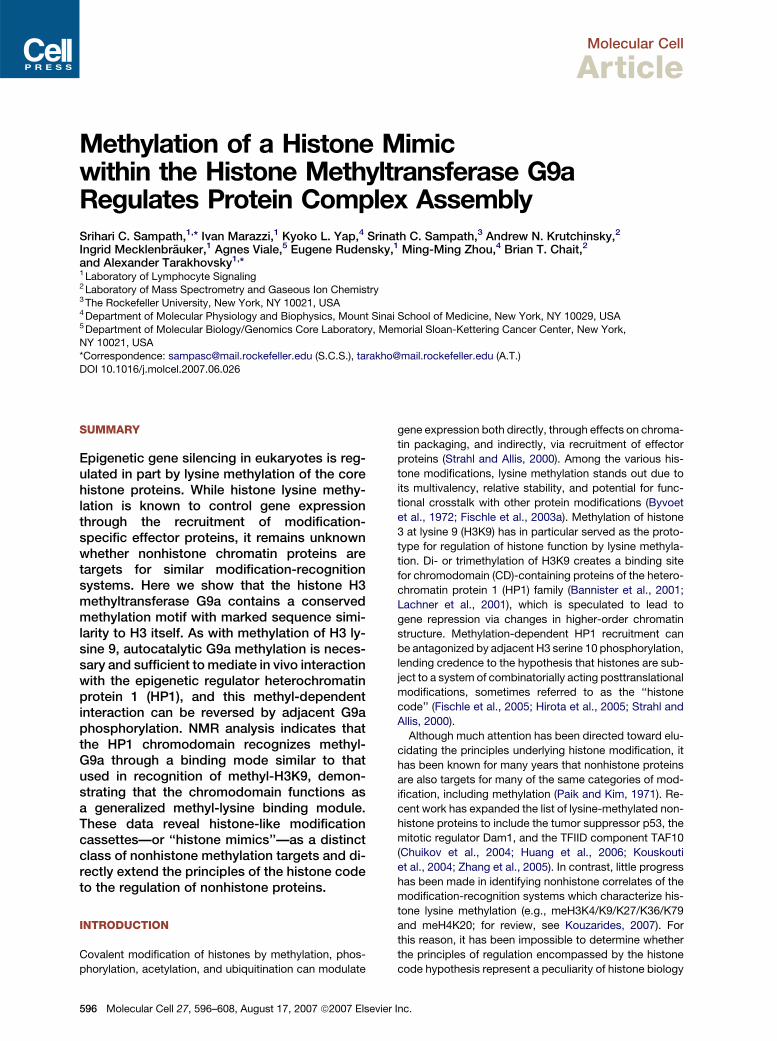
Molecular Cell Article Methylation of a Histone Mimic within the Histone Methyltransferase G9a Regulates Protein Complex Assembly Srihari C. Sampath, 1, * Ivan Marazzi, 1 Kyoko L. Yap, 4 Srinath C. Sampath, 3 Andrew N. Krutchinsky, 2 Ingrid Mecklenbra ¨ uker, 1 Agnes Viale, 5 Eugene Rudensky, 1 Ming-Ming Zhou, 4 Brian T. Chait, 2 and Alexander Tarakhovsky 1, * 1 Laboratory of Lymphocyte Signaling 2 Laboratory of Mass Spectrometry and Gaseous Ion Chemistry 3 The Rockefeller University, New York, NY 10021, USA 4 Department of Molecular Physiology and Biophysics, Mount Sinai School of Medicine, New York, NY 10029, USA 5 Department of Molecular Biology/Genomics Core Laboratory, Memorial Sloan-Kettering Cancer Center, New York, NY 10021, USA *Correspondence: [email protected] (S.C.S.), [email protected] (A.T.) DOI 10.1016/j.molcel.2007.06.026 SUMMARY Epigenetic gene silencing in eukaryotes is reg- ulated in part by lysine methylation of the core histone proteins. While histone lysine methy- lation is known to control gene expression through the recruitment of modification- specific effector proteins, it remains unknown whether nonhistone chromatin proteins are targets for similar modification-recognition systems. Here we show that the histone H3 methyltransferase G9a contains a conserved methylation motif with marked sequence simi- larity to H3 itself. As with methylation of H3 ly- sine 9, autocatalytic G9a methylation is neces- sary and sufficient to mediate in vivo interaction with the epigenetic regulator heterochromatin protein 1 (HP1), and this methyl-dependent interaction can be reversed by adjacent G9a phosphorylation. NMR analysis indicates that the HP1 chromodomain recognizes methyl- G9a through a binding mode similar to that used in recognition of methyl-H3K9, demon- strating that the chromodomain functions as a generalized methyl-lysine binding module. These data reveal histone-like modification cassettes—or ‘‘histone mimics’’—as a distinct class of nonhistone methylation targets and di- rectly extend the principles of the histone code to the regulation of nonhistone proteins. INTRODUCTION Covalent modification of histones by methylation, phos- phorylation, acetylation, and ubiquitination can modulate gene expression both directly, through effects on chroma- tin packaging, and indirectly, via recruitment of effector proteins (Strahl and Allis, 2000). Among the various his- tone modifications, lysine methylation stands out due to its multivalency, relative stability, and potential for func- tional crosstalk with other protein modifications (Byvoet et al., 1972; Fischle et al., 2003a). Methylation of histone 3 at lysine 9 (H3K9) has in particular served as the proto- type for regulation of histone function by lysine methyla- tion. Di- or trimethylation of H3K9 creates a binding site for chromodomain (CD)-containing proteins of the hetero- chromatin protein 1 (HP1) family (Bannister et al., 2001; Lachner et al., 2001), which is speculated to lead to gene repression via changes in higher-order chromatin structure. Methylation-dependent HP1 recruitment can be antagonized by adjacent H3 serine 10 phosphorylation, lending credence to the hypothesis that histones are sub- ject to a system of combinatorially acting posttranslational modifications, sometimes referred to as the ‘‘histone code’’ (Fischle et al., 2005; Hirota et al., 2005; Strahl and Allis, 2000). Although much attention has been directed toward elu- cidating the principles underlying histone modification, it has been known for many years that nonhistone proteins are also targets for many of the same categories of mod- ification, including methylation (Paik and Kim, 1971). Re- cent work has expanded the list of lysine-methylated non- histone proteins to include the tumor suppressor p53, the mitotic regulator Dam1, and the TFIID component TAF10 (Chuikov et al., 2004; Huang et al., 2006; Kouskouti et al., 2004; Zhang et al., 2005). In contrast, little progress has been made in identifying nonhistone correlates of the modification-recognition systems which characterize his- tone lysine methylation (e.g., meH3K4/K9/K27/K36/K79 and meH4K20; for review, see Kouzarides, 2007). For this reason, it has been impossible to determine whether the principles of regulation encompassed by the histone code hypothesis represent a peculiarity of histone biology 596 Molecular Cell 27, 596–608, August 17, 2007 ª2007 Elsevier Inc.
Transcript of Molecular Cell Article - Rockefeller...
Molecular Cell
Article
Methylation of a Histone Mimicwithin the Histone Methyltransferase G9aRegulates Protein Complex AssemblySrihari C. Sampath,1,* Ivan Marazzi,1 Kyoko L. Yap,4 Srinath C. Sampath,3 Andrew N. Krutchinsky,2
Ingrid Mecklenbrauker,1 Agnes Viale,5 Eugene Rudensky,1 Ming-Ming Zhou,4 Brian T. Chait,2
and Alexander Tarakhovsky1,*1 Laboratory of Lymphocyte Signaling2 Laboratory of Mass Spectrometry and Gaseous Ion Chemistry3 The Rockefeller University, New York, NY 10021, USA4 Department of Molecular Physiology and Biophysics, Mount Sinai School of Medicine, New York, NY 10029, USA5 Department of Molecular Biology/Genomics Core Laboratory, Memorial Sloan-Kettering Cancer Center, New York,NY 10021, USA
*Correspondence: [email protected] (S.C.S.), [email protected] (A.T.)
DOI 10.1016/j.molcel.2007.06.026
SUMMARY
Epigenetic gene silencing in eukaryotes is reg-ulated in part by lysine methylation of the corehistone proteins. While histone lysine methy-lation is known to control gene expressionthrough the recruitment of modification-specific effector proteins, it remains unknownwhether nonhistone chromatin proteins aretargets for similar modification-recognitionsystems. Here we show that the histone H3methyltransferase G9a contains a conservedmethylation motif with marked sequence simi-larity to H3 itself. As with methylation of H3 ly-sine 9, autocatalytic G9a methylation is neces-sary and sufficient to mediate in vivo interactionwith the epigenetic regulator heterochromatinprotein 1 (HP1), and this methyl-dependentinteraction can be reversed by adjacent G9aphosphorylation. NMR analysis indicates thatthe HP1 chromodomain recognizes methyl-G9a through a binding mode similar to thatused in recognition of methyl-H3K9, demon-strating that the chromodomain functions asa generalized methyl-lysine binding module.These data reveal histone-like modificationcassettes—or ‘‘histone mimics’’—as a distinctclass of nonhistone methylation targets and di-rectly extend the principles of the histone codeto the regulation of nonhistone proteins.
INTRODUCTION
Covalent modification of histones by methylation, phos-
phorylation, acetylation, and ubiquitination can modulate
596 Molecular Cell 27, 596–608, August 17, 2007 ª2007 Elsevie
gene expression both directly, through effects on chroma-
tin packaging, and indirectly, via recruitment of effector
proteins (Strahl and Allis, 2000). Among the various his-
tone modifications, lysine methylation stands out due to
its multivalency, relative stability, and potential for func-
tional crosstalk with other protein modifications (Byvoet
et al., 1972; Fischle et al., 2003a). Methylation of histone
3 at lysine 9 (H3K9) has in particular served as the proto-
type for regulation of histone function by lysine methyla-
tion. Di- or trimethylation of H3K9 creates a binding site
for chromodomain (CD)-containing proteins of the hetero-
chromatin protein 1 (HP1) family (Bannister et al., 2001;
Lachner et al., 2001), which is speculated to lead to
gene repression via changes in higher-order chromatin
structure. Methylation-dependent HP1 recruitment can
be antagonized by adjacent H3 serine 10 phosphorylation,
lending credence to the hypothesis that histones are sub-
ject to a system of combinatorially acting posttranslational
modifications, sometimes referred to as the ‘‘histone
code’’ (Fischle et al., 2005; Hirota et al., 2005; Strahl and
Allis, 2000).
Although much attention has been directed toward elu-
cidating the principles underlying histone modification, it
has been known for many years that nonhistone proteins
are also targets for many of the same categories of mod-
ification, including methylation (Paik and Kim, 1971). Re-
cent work has expanded the list of lysine-methylated non-
histone proteins to include the tumor suppressor p53, the
mitotic regulator Dam1, and the TFIID component TAF10
(Chuikov et al., 2004; Huang et al., 2006; Kouskouti
et al., 2004; Zhang et al., 2005). In contrast, little progress
has been made in identifying nonhistone correlates of the
modification-recognition systems which characterize his-
tone lysine methylation (e.g., meH3K4/K9/K27/K36/K79
and meH4K20; for review, see Kouzarides, 2007). For
this reason, it has been impossible to determine whether
the principles of regulation encompassed by the histone
code hypothesis represent a peculiarity of histone biology
r Inc.
Molecular Cell
Regulation of G9a by Methylation of a Histone Mimic
or whether they in fact represent a particular instance of
a potentially more universal ‘‘protein code’’ (Margueron
et al., 2005).
Here we demonstrate that the essential H3K9 methyl-
transferase G9a is lysine methylated on multiple H3K9-
like sites in vivo. We further show that methylation of
one of these sites recapitulates many aspects of H3K9
regulation, including methylation to multiple valencies,
structural recognition and binding by modification-
specific effector proteins, and reversibility of effector bind-
ing by concomitant phosphorylation. These findings re-
veal that both the modification and recognition systems
governing the methyl-H3K9 epigenetic mark are con-
served in a nonhistone protein, constituting one instance
of a phenomenon that we refer to as ‘‘histone mimicry.’’
On the basis of these data, we suggest that the core tenets
of the histone code hypothesis are of much broader rele-
vance than previously appreciated and are in fact applica-
ble to nonhistone proteins as well.
RESULTS
The Histone Methyltransferase G9a Is Lysine
Methylated In Vivo
G9a is a member of the Suvar(3–9) family of SET domain
HMTases, all members of which methylate lysine 9 of H3
(Dillon et al., 2005; Rea et al., 2000). In addition to G9a,
this family also includes a G9a-like protein known as
GLP or EuHMTase-1 (Ogawa et al., 2002). Alignment of
the amino termini of murine G9a and GLP identified an
identical stretch of eight amino acids that strongly resem-
bled the H3K9 methylation site targeted by both G9a and
GLP (Figure 1A). This sequence was conserved in the G9a
homolog from Xenopus, and the core motif was also found
in Drosophila G9a (Figure 1A).
We hypothesized that the similar sequence context of
G9a K165 and H3K9 could allow G9a K165 lysine methyl-
ation in vivo. Indeed, we found that a ‘‘multimethyl’’ lysine-
specific antibody originally raised against a branched-
peptide H3K9 me2 antigen (a4xH3K9me2; Perez-Burgos
et al., 2004) recognized both a chemically methylated
G9a K165 peptide (Figure 1B) as well as endogenous
G9a immunoprecipitated using an affinity-purified N-
terminal G9a antibody (aG9a-N; Figure 1C and see Fig-
ure S1 in the Supplemental Data available with this article
online). Furthermore, an antibody raised specifically
against dimethylated G9a K165 (Figure 1D) recognized
both full-length endogenous G9a and an alternatively tran-
scribed G9a isoform (Figure 1E, asterisk; Tachibana et al.,
2002), demonstrating that G9a is dimethylated on K165 in
vivo. In addition to G9a, the aK165 me2 antibody also rec-
ognized an �200 kD protein (Figure 1E, arrowhead). Anal-
ysis of this protein by mass spectrometry (MS) identified it
as the human homolog of mAM, a critical component of
the ESET H3K9 methyltransferase complex (Wang et al.,
2003; Figure S2A, see the Discussion).
Molecul
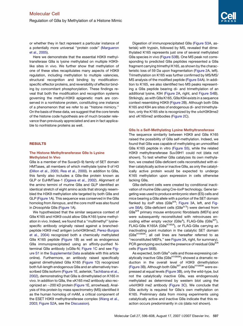
Digestion of immunoprecipitated G9a (Figure S3A, as-
terisk) with trypsin, followed by MS, revealed that dime-
thylated K165 represents just one of several methylated
G9a species in vivo (Figure S3B). One MS peak not corre-
sponding to predicted G9a peptides represented a G9a
fragment carrying trimethyl K165, as shown by the charac-
teristic loss of 59 Da upon fragmentation (Figure 2A, left).
Trimethylation on K165 was further confirmed by MS/MS/
MS analysis of the modified peptide (Figure S4A). In addi-
tion to K165, we also identified two MS peaks represent-
ing a G9a peptide bearing di- and trimethylation of an
additional lysine, K94 (Figure 2A, right, and Figure S4B).
Strikingly, as with G9a K165, G9a K94 exists in a sequence
context resembling H3K9 (Figure 2B). Although both G9a
K165 and K94 are sites of endogenous di- and trimethyla-
tion, only the K165 site is recognized by the a4xH3K9me2
and aK165me2 antibodies (Figure 2C).
G9a Is a Self-Methylating Lysine Methyltransferase
The sequence similarity between H3K9 and G9a K165
raised the possibility of G9a self-methylation. Indeed, we
found that G9a was capable of methylating an unmodified
G9a K165 peptide in vitro (Figure S5), while the related
H3K9 methyltransferase Suv39H1 could not (data not
shown). To test whether G9a catalyzes its own methyla-
tion, we created G9a-deficient cells reconstituted with ei-
ther catalytically active or inactive G9a, as only the catalyt-
ically active protein would be expected to undergo
K165 methylation upon expression in cells otherwise
lacking G9a.
G9a-deficient cells were created by conditional inacti-
vation of murine G9a using Cre-loxP technology. Gene tar-
geting was used to produce embryonic stem (ES) cells and
mice bearing a G9a allele with a portion of the SET domain
flanked by loxP sites (G9afl/fl; Figure 3A, left, and Fig-
ure S6A). G9a-deficient cells (G9aD/D) were derived from
G9afl/fl primary mouse embryonic fibroblasts (MEFs) and
were subsequently reconstituted with retroviruses en-
coding either empty vector (G9aD), FLAG-G9a (G9aWT),
FLAG-G9a K165A (G9aK165A), or FLAG-G9a carrying an
inactivating point mutation in the catalytic SET domain
(G9aH1093K; all cell lines are hereafter referred to as
‘‘reconstituted MEFs,’’ see Figure 3A, right, for summary).
PCR genotyping excluded the presence of residual G9afl/fl
cells (Figure S6B).
As expected, both G9aD cells and those expressing cat-
alytically inactive G9a (G9aH1093K) showed a dramatic re-
duction in the overall level of H3K9 dimethylation
(Figure 3B). Although both G9aWT and G9aH1093K were ex-
pressed at equal levels (Figure 3B), only the wild-type, but
not the catalytically inactive G9a, was endogenously
methylated as determined by western blot using the
a4xH3K9 me2 antibody (Figure 3C). We conclude that
G9a activity is required for G9a’s own methylation on
K165. Preliminary data from mixing experiments using
catalytically active and inactive G9a indicate that this re-
action occurs predominantly in cis (data not shown).
ar Cell 27, 596–608, August 17, 2007 ª2007 Elsevier Inc. 597

Molecular Cell
Regulation of G9a by Methylation of a Histone Mimic
Figure 1. The Histone Methyltransferase G9a Is Lysine Methylated In Vivo
(A) An H3K9-like sequence is located in the noncatalytic N terminus of the G9a protein. (Top) Schematic representation of human G9a. (Bottom) Partial
protein sequence alignment of G9a and GLP homologs from Homo sapiens (h), Mus musculus (m), Xenopus laevis (x), and Drosophila melanogaster
(d), together with a portion of the human H3 amino-terminal tail. Residues identical in at least four of six sequences are highlighted in red, and the
region used in G9a K165 peptides is indicated by a solid line. Residue numbers are indicated where known.
(B) Western blot analysis of G9a K165 and H3(1–20) peptides using a multimethyl a4xH3K9me2 antibody. Loading was monitored by Ponceau S stain-
ing, which preferentially stains the G9a peptide due to sequence effects (data not shown; Sarma et al. [2004]). Un, unmodified peptide; Me, peptide
dimethylated at K165 (G9a) or K9 (H3); KA, G9a K165A mutant peptide.
(C) Endogenous G9a was immunoprecipitated from 293T cells using either control nonimmune antibody (aIgG) or an antibody raised against the G9a
N terminus (aG9a-N; see Figure S1), followed by western blot using the antibodies indicated. a-tubulin blot was used to control for protein loading.
(D) The specificity of antibodies raised against dimethyl G9a K165 peptide (aK165me2) was assessed by dot blot analysis of 3-fold dilutions of the
indicated peptides. Ponceau S staining control is shown above the corresponding dot blots.
(E) Western blot analysis of 293T cell cytosolic (Cy) and nuclear (Nu) extracts using either aK165me2 preimmune (left) or immune (right) serum. The
molecular weight of endogenous hG9a is indicated with an asterisk, and the band corresponding to endogenous human mAM (hAM) is indicated with
an arrowhead (see Figure S2 and the Discussion).
598 Molecular Cell 27, 596–608, August 17, 2007 ª2007 Elsevier Inc.
Molecular Cell
Regulation of G9a by Methylation of a Histone Mimic
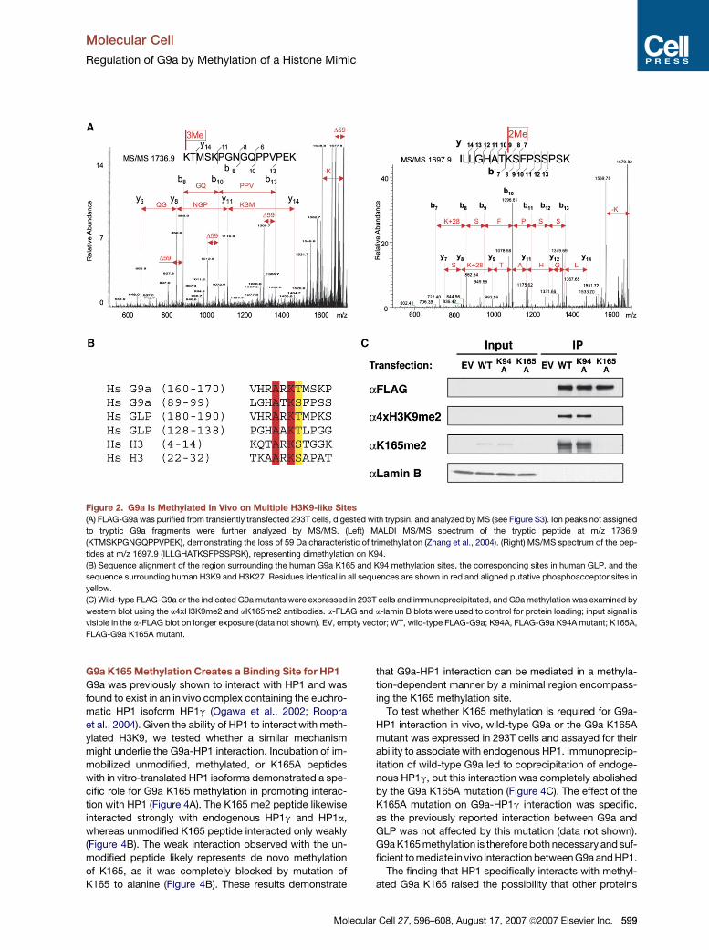
Figure 2. G9a Is Methylated In Vivo on Multiple H3K9-like Sites
(A) FLAG-G9a was purified from transiently transfected 293T cells, digested with trypsin, and analyzed by MS (see Figure S3). Ion peaks not assigned
to tryptic G9a fragments were further analyzed by MS/MS. (Left) MALDI MS/MS spectrum of the tryptic peptide at m/z 1736.9
(KTMSKPGNGQPPVPEK), demonstrating the loss of 59 Da characteristic of trimethylation (Zhang et al., 2004). (Right) MS/MS spectrum of the pep-
tides at m/z 1697.9 (ILLGHATKSFPSSPSK), representing dimethylation on K94.
(B) Sequence alignment of the region surrounding the human G9a K165 and K94 methylation sites, the corresponding sites in human GLP, and the
sequence surrounding human H3K9 and H3K27. Residues identical in all sequences are shown in red and aligned putative phosphoacceptor sites in
yellow.
(C) Wild-type FLAG-G9a or the indicated G9a mutants were expressed in 293T cells and immunoprecipitated, and G9a methylation was examined by
western blot using the a4xH3K9me2 and aK165me2 antibodies. a-FLAG and a-lamin B blots were used to control for protein loading; input signal is
visible in the a-FLAG blot on longer exposure (data not shown). EV, empty vector; WT, wild-type FLAG-G9a; K94A, FLAG-G9a K94A mutant; K165A,
FLAG-G9a K165A mutant.
G9a K165 Methylation Creates a Binding Site for HP1
G9a was previously shown to interact with HP1 and was
found to exist in an in vivo complex containing the euchro-
matic HP1 isoform HP1g (Ogawa et al., 2002; Roopra
et al., 2004). Given the ability of HP1 to interact with meth-
ylated H3K9, we tested whether a similar mechanism
might underlie the G9a-HP1 interaction. Incubation of im-
mobilized unmodified, methylated, or K165A peptides
with in vitro-translated HP1 isoforms demonstrated a spe-
cific role for G9a K165 methylation in promoting interac-
tion with HP1 (Figure 4A). The K165 me2 peptide likewise
interacted strongly with endogenous HP1g and HP1a,
whereas unmodified K165 peptide interacted only weakly
(Figure 4B). The weak interaction observed with the un-
modified peptide likely represents de novo methylation
of K165, as it was completely blocked by mutation of
K165 to alanine (Figure 4B). These results demonstrate
Molecu
that G9a-HP1 interaction can be mediated in a methyla-
tion-dependent manner by a minimal region encompass-
ing the K165 methylation site.
To test whether K165 methylation is required for G9a-
HP1 interaction in vivo, wild-type G9a or the G9a K165A
mutant was expressed in 293T cells and assayed for their
ability to associate with endogenous HP1. Immunoprecip-
itation of wild-type G9a led to coprecipitation of endoge-
nous HP1g, but this interaction was completely abolished
by the G9a K165A mutation (Figure 4C). The effect of the
K165A mutation on G9a-HP1g interaction was specific,
as the previously reported interaction between G9a and
GLP was not affected by this mutation (data not shown).
G9a K165 methylation is therefore both necessary and suf-
ficient to mediate in vivo interaction between G9a and HP1.
The finding that HP1 specifically interacts with methyl-
ated G9a K165 raised the possibility that other proteins
lar Cell 27, 596–608, August 17, 2007 ª2007 Elsevier Inc. 599

Molecular Cell
Regulation of G9a by Methylation of a Histone Mimic
Figure 3. Automethylation of G9a on K165 In Vivo
(A) (Left) G9a conditional mutagenesis strategy. Exons 23 and 24 of the murine G9a locus were flanked by loxP sites (floxed), causing out-of-frame
splicing and nonsense-mediated decay of the mutant transcript after Cre-mediated recombination (data not shown). Numbered box, exon; triangle,
loxP site; hexagon, FRT site; neo, neomycin (G418) resistance cassette; H, HindIII restriction site. (Right) Strategy for production and reconstitution of
G9a-deficient MEFs. G9a-deleted cells were reconstituted with either empty vector (D), wild-type FLAG-G9a (WT), FLAG-G9a K165A (KA), or FLAG-
G9a H1093K catalytic mutant (HK). RV-TAg, retroviral SV40 large-T antigen; Imm, immortalized; Ad-Cre, adenoviral Cre.
(B) Nuclear lysates from G9afl/fl (fl/fl) or reconstituted MEFs were analyzed by western blot using the indicated antibodies. a-lamin B blot was used to
control for protein loading.
(C) Analysis of G9a automethylation. FLAG-G9a immunoprecipitated from the reconstituted MEFs described was analyzed by western blot using the
a4xH3K9me2 antibody. Protein expression and loading were monitored by a-FLAG and a-lamin B blots, respectively.
might also bind to K165. Indeed, we found that immuno-
precipitation of G9a led to copurification of CDYL (chro-
modomain Y-like; Figure S3A, arrowhead), a known
transcriptional repressor containing an N-terminal chro-
modomain and a C-terminal coenzyme A-binding domain
of unknown function (Caron et al., 2003). Consistent with
its putative function as a transcriptional repressor, CDYL
was previously copurified in an in vivo complex containing
G9a, GLP, the transcriptional corepressor CtBP, and the
histone lysine demethylase LSD1 (Shi et al., 2003).
By analogy to the G9a-HP1 interaction, we tested
whether G9a K165 was necessary for G9a-CDYL interac-
tion. Whereas wild-type G9a could coimmunoprecipitate
endogenous CDYL from nuclear extracts, this interaction
was completely lost in the G9a K165A mutant (Figure 4D),
demonstrating that K165 serves as a binding site for both
HP1 and CDYL. We therefore examined whether, as with
HP1, K165 methylation mediates interaction with CDYL.
Unlike HP1, the G9a-CDYL interaction could not be reca-
pitulated in vitro using either unmodified, K165me, or
K165meT166ph peptides (data not shown). Considering
that CDYL exhibits HAT activity in vitro (Lahn et al.,
2002) and has been suggested to acetylate nonhistone
600 Molecular Cell 27, 596–608, August 17, 2007 ª2007 Elsevie
proteins (Caron et al., 2003), it is possible that CDYL bind-
ing is induced by alternative K165 modifications (e.g.,
phosphoacetylation or combinatorial lysine methylation),
as was shown for the chromodomain-containing DNA
methyltransferase CMT3 (Lindroth et al., 2004).
Taken together, these data demonstrate that G9a K165
is an in vivo binding site for multiple chromodomain-con-
taining effector proteins. As recognition of methyl-G9a
by HP1 to our knowledge represents the first nonhistone
protein target for chromodomain binding, these results
furthermore identify the chromodomain as a generalized
methyl-lysine binding module.
Structural Recognition of Methylated G9a
To address the structural basis for HP1 recognition of both
methylated H3K9 and methylated G9a K165, we used nu-
clear magnetic resonance (NMR) spectroscopy to study
both sets of interactions. In the absence of lysine-
methylated G9a K165 peptide, the backbone amide reso-
nances of 15N-labeled HP1g chromodomain (CD) were
well-dispersed in the 2D [15N,1H]-HSQC (heteronuclear
single quantum coherence) spectrum, indicating a folded
protein (Figure 5A). Upon addition of trimethylated G9a
r Inc.
Molecular Cell
Regulation of G9a by Methylation of a Histone Mimic
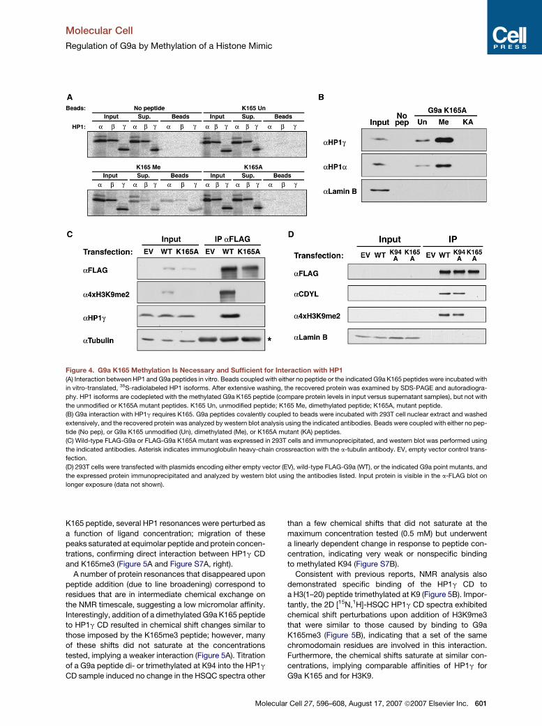
Figure 4. G9a K165 Methylation Is Necessary and Sufficient for Interaction with HP1
(A) Interaction between HP1 and G9a peptides in vitro. Beads coupled with either no peptide or the indicated G9a K165 peptides were incubated with
in vitro-translated, 35S-radiolabeled HP1 isoforms. After extensive washing, the recovered protein was examined by SDS-PAGE and autoradiogra-
phy. HP1 isoforms are codepleted with the methylated G9a K165 peptide (compare protein levels in input versus supernatant samples), but not with
the unmodified or K165A mutant peptides. K165 Un, unmodified peptide; K165 Me, dimethylated peptide; K165A, mutant peptide.
(B) G9a interaction with HP1g requires K165. G9a peptides covalently coupled to beads were incubated with 293T cell nuclear extract and washed
extensively, and the recovered protein was analyzed by western blot analysis using the indicated antibodies. Beads were coupled with either no pep-
tide (No pep), or G9a K165 unmodified (Un), dimethylated (Me), or K165A mutant (KA) peptides.
(C) Wild-type FLAG-G9a or FLAG-G9a K165A mutant was expressed in 293T cells and immunoprecipitated, and western blot was performed using
the indicated antibodies. Asterisk indicates immunoglobulin heavy-chain crossreaction with the a-tubulin antibody. EV, empty vector control trans-
fection.
(D) 293T cells were transfected with plasmids encoding either empty vector (EV), wild-type FLAG-G9a (WT), or the indicated G9a point mutants, and
the expressed protein immunoprecipitated and analyzed by western blot using the antibodies listed. Input protein is visible in the a-FLAG blot on
longer exposure (data not shown).
K165 peptide, several HP1 resonances were perturbed as
a function of ligand concentration; migration of these
peaks saturated at equimolar peptide and protein concen-
trations, confirming direct interaction between HP1g CD
and K165me3 (Figure 5A and Figure S7A, right).
A number of protein resonances that disappeared upon
peptide addition (due to line broadening) correspond to
residues that are in intermediate chemical exchange on
the NMR timescale, suggesting a low micromolar affinity.
Interestingly, addition of a dimethylated G9a K165 peptide
to HP1g CD resulted in chemical shift changes similar to
those imposed by the K165me3 peptide; however, many
of these shifts did not saturate at the concentrations
tested, implying a weaker interaction (Figure 5A). Titration
of a G9a peptide di- or trimethylated at K94 into the HP1g
CD sample induced no change in the HSQC spectra other
Molecu
than a few chemical shifts that did not saturate at the
maximum concentration tested (0.5 mM) but underwent
a linearly dependent change in response to peptide con-
centration, indicating very weak or nonspecific binding
to methylated K94 (Figure S7B).
Consistent with previous reports, NMR analysis also
demonstrated specific binding of the HP1g CD to
a H3(1–20) peptide trimethylated at K9 (Figure 5B). Impor-
tantly, the 2D [15N,1H]-HSQC HP1g CD spectra exhibited
chemical shift perturbations upon addition of H3K9me3
that were similar to those caused by binding to G9a
K165me3 (Figure 5B), indicating that a set of the same
chromodomain residues are involved in this interaction.
Furthermore, the chemical shifts saturate at similar con-
centrations, implying comparable affinities of HP1g for
G9a K165 and for H3K9.
lar Cell 27, 596–608, August 17, 2007 ª2007 Elsevier Inc. 601
Molecular Cell
Regulation of G9a by Methylation of a Histone Mimic
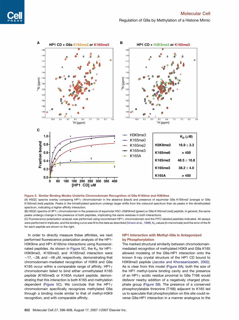
Figure 5. Similar Binding Modes Underlie Chromodomain Recognition of G9a K165me and H3K9me
(A) HSQC spectra overlay comparing HP1g chromodomain in the absence (black) and presence of equimolar G9a K165me2 (orange) or G9a
K165me3 (red) peptide. Peaks in the trimethylated spectrum undergo larger shifts from the unbound spectrum than do peaks in the dimethylated
spectrum, indicating a higher-affinity interaction.
(B) HSQC spectra of HP1g chromodomain in the presence of equimolar H3(1–20)K9me3 (green) or G9a K165me3 (red) peptide. In general, the same
peaks undergo change in the presence of both peptides, implicating the same residues in both interactions.
(C) Fluorescence polarization analysis was performed using recombinant HP1g chromodomain and the FITC-labeled peptides indicated. All assays
were performed in triplicate, and the binding curve was fit to the data as described (Vinson et al., 1998). KD values (in micromolar) and the error of the fit
for each peptide are shown to the right.
In order to directly measure these affinities, we next
performed fluorescence polarization analysis of the HP1-
H3K9me and HP1-K165me interactions using fluorescei-
nated peptides. As shown in Figure 5C, the KD for HP1-
H3K9me3, -K165me3, and -K165me2 interaction were
�17, �38, and �48 mM, respectively, demonstrating that
chromodomain-mediated recognition of H3K9 and G9a
K165 occur within a comparable range of affinity. HP1g
chromodomain failed to bind either unmethylated K165
peptide (K165me0) or K165A mutant peptide, demon-
strating that this interaction is both K165 and methylation
dependent (Figure 5C). We conclude that the HP1g
chromodomain specifically recognizes methylated G9a
through a binding mode similar to that of methyl-H3K9
recognition, and with comparable affinity.
602 Molecular Cell 27, 596–608, August 17, 2007 ª2007 Elsevie
HP1 Interaction with Methyl-G9a Is Antagonized
by Phosphorylation
The marked structural similarity between chromodomain-
mediated recognition of methylated H3K9 and G9a K165
allowed modeling of the G9a-HP1 interaction onto the
known X-ray crystal structure of the HP1 CD bound to
H3K9me3 peptide (Jacobs and Khorasanizadeh, 2002).
As is clear from this model (Figure 6A), both the size of
the HP1 methyl-lysine binding cavity and the presence
of an HP1g acidic residue proximal to G9a T166 would
disfavor nearby addition of a negatively charged phos-
phate group (Figure S8). The presence of a conserved
phosphorylatable threonine (T166) adjacent to K165 led
us to speculate that phosphorylation on this site could re-
verse G9a-HP1 interaction in a manner analogous to the
r Inc.
Molecular Cell
Regulation of G9a by Methylation of a Histone Mimic
recently described H3K9-H3 serine 10 ‘‘methyl-phos
switch’’ (Fischle et al., 2005; Hirota et al., 2005).
In the case of H3K9, mitotic H3S10 phosphorylation by
the kinase Aurora B serves to release chromatin-bound
HP1 (Fischle et al., 2005; Hirota et al., 2005). We therefore
tested whether Aurora B could phosphorylate G9a T166.
Indeed, both an H3(1–20) peptide and an unmodified
G9a K165 peptide containing T166 were robustly phos-
phorylated in vitro by Aurora B complex (Figure 6B). This
phosphorylation was not significantly affected by methyl-
ation on K165, consistent with recent reports demonstrat-
ing a minimal effect of H3K9 methylation on H3S10 phos-
phorylation by Aurora B in vitro (Fischle et al., 2005). We
further found that a peptide synthesized to carry both
K165 methylation and T166 phosphorylation was com-
pletely resistant to subsequent phosphorylation, demon-
strating that the serine 10 kinase activity is specific for
T166, the single residue aligning with H3 serine 10
(Figure 2B).
To determine whether phosphorylation of T166 can
modulate G9a-HP1 interaction in vitro, immobilized un-
modified, methylated, or methyl-phosphorylated G9a
K165 peptides were incubated with in vitro-translated
HP1g (Figure 6C, left; quantified at right). As expected,
methylated K165 peptide bound HP1g strongly; however,
this methyl-dependent binding was completely eliminated
by concomitant phosphorylation on T166. The lack of
HP1g binding to the methyl-phosphorylated G9a peptide
was not due to reduced coupling efficiency of this peptide,
since binding could be quantitatively restored by pretreat-
ment of the phosphorylated peptide matrix with protein
phosphatase 1 (Figure 6C). Consistent with this finding,
a phospho-mimic threonine to glutamic acid substitution
at position 166 (T166E) blocked interaction of G9a with
HP1g in vivo (Figure 6D).
The antagonistic effect of T166 phosphorylation on
HP1g CD binding to methylated G9a K165 could also be
demonstrated by NMR, as titration of the HP1g CD with
methyl-phosphorylated G9a K165 peptide induced a con-
siderably smaller number of NMR perturbations when
compared to titrations with methylated G9a K165 peptide
(Figures 5B and 6E). Furthermore, the chemical shift
changes observed with the methyl-phosphorylated pep-
tide did not saturate at the maximal concentration tested,
indicating that phosphorylation significantly weakens the
interaction of G9a with the HP1 chromodomain.
G9a Activity, but Not K165 Methylation, Is Required
for Control of Gene Expression
Considering the known function of G9a as a transcriptional
regulator (Shi et al., 2003; Tachibana et al., 2002), we
tested the possibility that G9a methylation contributes to
target gene regulation in vivo. Indirect immunofluores-
cence analysis of G9aD cells demonstrated that complete
G9a deficiency dramatically reduced euchromatic H3K9
dimethylation, with concomitant redistribution of HP1g
from euchromatin to pericentric heterochromatin
(Figure S9; Tachibana et al., 2002, 2005). These changes
Molecul
were associated with dysregulated expression of �300–
400 genes, as determined by comparing microarray
gene expression analyses of G9aD and G9aWT cells
(Figure S10).
Similar changes were also seen in cells reconstituted
with the G9a H1093K catalytic mutant, demonstrating
that G9a methyltransferase activity is necessary for
H3K9 dimethylation, HP1g localization, and proper in
vivo control of gene expression (Figure 3B, Figures S9
and S10). In contrast, expression of the G9a K165A mu-
tant caused only a mild increase in the amount of HP1g
localized to pericentric heterochromatin (Figure S9), con-
sistent with the unaltered levels and distribution of
H3K9me2 in these cells (Figure 3B). Lack of K165 methyl-
ation likewise caused statistically significant dysregulation
of only one gene, Hey1 (Figure S10).
DISCUSSION
Starting from a sequence comparison, we have demon-
strated that the histone methyltransferase G9a contains
a lysine methylation cassette that mimics multiple features
of histone lysine methylation, including sequence context,
effector protein recognition, and secondary regulation by
phosphorylation. Although striking on its own, there are in-
dications that such histone-like modification cassettes—
or histone mimics—may exist in many nonhistone pro-
teins, particularly those that are chromatin associated,
and may involve additional layers of complexity.
Nonhistone Targets of Lysine Methylation
To date, attempts to identify targets of nonhistone lysine
methylation have largely relied on candidate approaches,
utilizing methyltransferases and/or putative targets of em-
piric interest (Chuikov et al., 2004; Kouskouti et al., 2004;
Zhang et al., 2005). While an attempt was also recently
made to identify methylated proteins in a more unbiased
fashion using antibody enrichment and MS, all of the
new targets identified were in fact arginine methylated
(Ong et al., 2004). In contrast, our findings suggest that bi-
oinformatic prediction of methylation targets may in fact
be possible, based on similarity to known lysine methyla-
tion sites in histones. Our own initial analysis has yielded
a number of proteins bearing evolutionarily conserved
motifs likely to be targets of lysine methylation; strikingly,
many of these are found within chromatin-associated pro-
teins (S.C.S. and A.T., unpublished data).
Among the proteins found in this study to contain a po-
tential methylation target site was mAM, a factor that
binds to and regulates the catalytic activity of the essential
H3K9 methyltransferase ESET (Wang et al., 2003). The hu-
man homolog of mAM (hAM) is recognized by our aK165
me2 antibody (Figure 1E), and inspection of the hAM/
mAM coding sequence identified a single conserved motif
identical to G9a K165 and highly similar to H3K9
(Figure S2A and Figure 6F). Similarly, it was recently re-
ported that HP1 is itself a target of phosphorylation on ser-
ine 83, a residue that we believe exists within an H3K9-like
ar Cell 27, 596–608, August 17, 2007 ª2007 Elsevier Inc. 603
Molecular Cell
Regulation of G9a by Methylation of a Histone Mimic
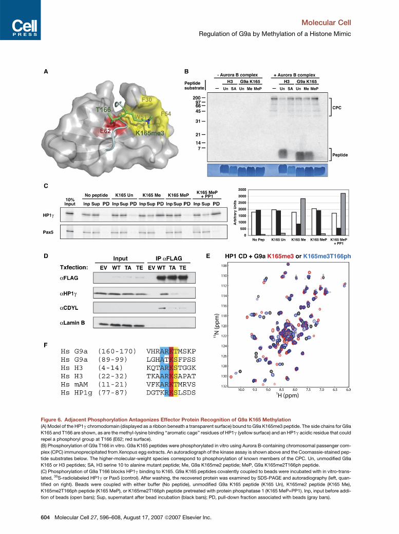
Figure 6. Adjacent Phosphorylation Antagonizes Effector Protein Recognition of G9a K165 Methylation
(A) Model of the HP1g chromodomain (displayed as a ribbon beneath a transparent surface) bound to G9a K165me3 peptide. The side chains for G9a
K165 and T166 are shown, as are the methyl-lysine binding ‘‘aromatic cage’’ residues of HP1g (yellow surface) and an HP1g acidic residue that could
repel a phosphoryl group at T166 (E62; red surface).
(B) Phosphorylation of G9a T166 in vitro. G9a K165 peptides were phosphorylated in vitro using Aurora B-containing chromosomal passenger com-
plex (CPC) immunoprecipitated from Xenopus egg extracts. An autoradiograph of the kinase assay is shown above and the Coomassie-stained pep-
tide substrates below. The higher-molecular-weight species correspond to phosphorylation of known members of the CPC. Un, unmodified G9a
K165 or H3 peptides; SA, H3 serine 10 to alanine mutant peptide; Me, G9a K165me2 peptide; MeP, G9a K165me2T166ph peptide.
(C) Phosphorylation of G9a T166 blocks HP1g binding to K165. G9a K165 peptides covalently coupled to beads were incubated with in vitro-trans-
lated, 35S-radiolabeled HP1g or Pax5 (control). After washing, the recovered protein was examined by SDS-PAGE and autoradiography (left, quan-
tified on right). Beads were coupled with either buffer (No peptide), unmodified G9a K165 peptide (K165 Un), K165me2 peptide (K165 Me),
K165me2T166ph peptide (K165 MeP), or K165me2T166ph peptide pretreated with protein phosphatase 1 (K165 MeP+PP1). Inp, input before addi-
tion of beads (open bars); Sup, supernatant after bead incubation (black bars); PD, pull-down fraction associated with beads (gray bars).
604 Molecular Cell 27, 596–608, August 17, 2007 ª2007 Elsevier Inc.
Molecular Cell
Regulation of G9a by Methylation of a Histone Mimic
histone mimic (Lomberk et al., 2006, and Figure 6F). If so,
this putative methylation site may in fact represent an in-
tramolecular binding partner for the HP1 chromodomain,
similar to the intramolecular SH2-phosphotyrosine inter-
action found in Src-family kinases (Roskoski, 2004).
The unexpected recurrence of H3K9 mimics in proteins
associated with H3K9 methyltransferases (or, in the case
of G9a and GLP, the methyltransferases themselves) sug-
gests a complex interplay between histone and nonhis-
tone methylation systems. While the functional details of
this interplay are yet to be determined (see below), our
recent observation that the H3K27 histone methyltransfer-
ase Ezh2 also undergoes automethylation (I. Su, M. Dobe-
necker, and A.T., unpublished data) suggests that self-
methylation of HMTases and their associated proteins is
likely to be a ubiquitous mode of HMTase regulation, anal-
ogous to kinase autophosphorylation. Furthermore, the
emerging literature on effector modules for other histone
methylation sites, such as H3K4 (Pray-Grant et al., 2005;
Sims et al., 2005; Wysocka et al., 2005; reviewed in Kou-
zarides, 2007), suggests that methylation-dependent
crosstalk between HMTases, their histone target sites,
and nonhistone ‘‘mimics’’ of these sites may well be the
rule rather than the exception.
The G9a Modification System
The organization of the G9a ‘‘tail’’—multiple methylation
sites with similar sequences located in close proximity—
raises a clear analogy to the H3 amino-terminal tail, in
which recognition of methylated H3K9 and H3K27 are
mediated by related but distinct classes of chromodo-
main-containing effectors (HP1 and Polycomb, respec-
tively; Fischle et al. [2003b]). Since G9a K94 methylation
is dispensable for both HP1 and CDYL binding
(Figure 2C and data not shown), it is likely that distinct ef-
fector proteins are involved in recognition of methylated
G9a K94. As with H3K9/K27, characterization of these
K94-specific binding proteins may clarify functional simi-
larities and differences between K165 and K94.
With regard to K165, it is now clear that this residue
constitutes a binding site for at least two chromodomain
proteins, HP1 and CDYL (Figures 4C and 4D). While
K165-HP1 binding clearly requires K165 methylation
(Figure 4A), it remains unclear which modification(s) medi-
ates K165-CDYL interaction, as neither K165-unmodified,
-methylated, or -methyl-phosphorylated peptides are ca-
pable of mediating G9a-CDYL interaction in vitro (data not
shown). Adding further complexity to this situation, we
Molecul
note the potential existence of even more K165-binding
proteins. For instance, in addition to G9a, GLP, and
CDYL, previous purification of the CtBP repressor com-
plex recovered Pc2, a chromodomain-containing SUMO
E3 ligase (Shi et al., 2003). Although in vivo histone targets
for the Pc2 CD are not known, it has been shown to bind
in vitro to H3K9me3 peptides (Bernstein et al., 2006),
and we in fact also observe in vitro binding of the Pc2
CD to K165me3 peptide (Figure S12). It should be noted,
however, that G9a and Pc2 are localized within largely
nonoverlapping nuclear domains (Saurin et al., 1998;
Tachibana et al., 2001), and it is therefore uncertain
whether G9a-Pc2 interaction occurs endogenously.
In addition to the further identification of K94/K165 bind-
ing partners, a final important mechanistic issue concerns
regulation of K165-directed G9a catalytic activity. G9a is
known to contribute significantly to H3K9 mono- and di-
methylation, but not to trimethylation (Peters et al., 2003;
Rice et al., 2003). In contrast, we observe di- and trimethy-
lation of K165 by MS, but not monomethylation (Figure 2A
and Figure S4). While these results may reflect technical
difficulties in identifying monomethylated K165 (e.g., low
abundance or concomitant modification on another site),
we favor the view that the methyltransferase activity of
G9a is altered in vivo to favor di- and trimethylation.
Such a scenario is quite likely, given that the related
HMTase ESET is regulated in this way by binding of the ac-
cessory protein mAM (Wang et al., 2003), and the fact that
mAM is itself recognized by our aK165 me2 antibody
(Figure 1E and Figure S2). Alternatively, it may be that
a dedicated G9a K165 trimethylase exists in vivo, as is
known to be the case for trimethylation of H3K9 (Peters
et al., 2003; Rice et al., 2003).
Functions of G9a Methylation
Determining the precise functions of G9a methylation, and
of histone-like modification cassettes in general, clearly
represents a central challenge for the future. Despite the
obvious similarities between self-methylation of G9a and
self-phosphorylation of Src-family kinases, a process
known to regulate catalytic activity (Roskoski, 2004), we
found no evidence of a role for G9a K165 methylation in
regulation of G9a enzymatic function (Figure S11). Immu-
nofluorescence analysis of G9a-deleted cells expressing
nonmethylatable G9aK165A similarly demonstrated that
G9a-HP1g interaction is largely dispensable for euchro-
matic localization of both proteins (Figure S9). However,
interpretation of these experiments is complicated by
(D) 293T cells were transfected with plasmids encoding either empty vector (EV), wild-type FLAG-G9a (WT), FLAG-G9a T166A (TA), or ‘‘phosphomi-
mic’’ FLAG-G9a T166E (TE). The expressed proteins were immunoprecipitated and examined by western blot as indicated. Note that the T166A
mutation reduces HP1g binding but that this is even further suppressed by the T166E mutation; the increased countereffect of the T166E mutation
is specific for HP1g and is not seen with CDYL.
(E) HSQC spectra of HP1g chromodomain in the presence of equimolar G9a K165me3 peptide with (blue) or without (red) adjacent phosphorylation at
T166. The smaller shift changes in the presence of phosphorylation demonstrate that this modification strongly counteracts methyl-G9a binding to
HP1g.
(F) Protein sequence alignment of the human G9a K165 and K94 methylation sites with H3K9, H3K27, the H3K9-like site of human mAM (see
Figure S2), and the predicted methylation site in HP1g (see the Discussion). Complete identity is highlighted in red, five of six identity in blue, and
shared phosphoacceptor sites in yellow.
ar Cell 27, 596–608, August 17, 2007 ª2007 Elsevier Inc. 605
Molecular Cell
Regulation of G9a by Methylation of a Histone Mimic
the multiple layers of redundancy operating on the G9a
methylation system, including the existence of multiple
methylation sites, multiple effector proteins, and duplica-
tion of the entire pattern of G9a histone mimics on GLP,
a protein known to heterodimerize with G9a in vivo (Tachi-
bana et al., 2005). It is therefore likely that definitive an-
swers regarding the functions of G9a methylation will re-
quire both single and combinatorial genetic ablation of
G9a and GLP methylation sites; such studies are currently
underway.
Despite the functional redundancy within this methyla-
tion system, several models can be envisaged for the
function of G9a methylation in vivo. Histone mimics such
as G9a K165 may allow effector-mediated targeting of as-
sociated proteins to particular nucleosomal sites (as has
been suggested to occur for the H3K9 methyltransferase
Suv39H; Maison and Almouzni, 2004) or conversely may
increase the efficiency of effector protein loading onto
histones. Since G9a exists in protein complexes contain-
ing HMTase, HDAC, and lysine demethylase activities
(Ogawa et al., 2002; Shi et al., 2003), these enzymes
may also regulate G9a function by directly modifying his-
tone mimic sites and thereby defining the array of G9a-
interacting effector proteins. Such a scenario would
represent an exact parallel of the combinatorial readout
proposed to occur on histones (Strahl and Allis, 2000). Im-
portantly, while mammalian histones are not amenable to
genetic analysis due to the multicopy nature of the histone
genes (Maxson et al., 1983), such analysis is possible in
the case of histone mimics. Therefore, in addition to their
intrinsic interest and unique functions, histone mimics
such as G9a K165 may unexpectedly provide the best
system in a higher eukaryote in which to test fundamental
predictions of the histone code hypothesis.
EXPERIMENTAL PROCEDURES
Please see the Supplemental Experimental Procedures for additional
protocols and experimental details.
Generation of G9afl/fl Mice
To create the G9a targeting vector, a BAC fragment (from RPCI24-
165L4; BL/6; CHORI, USA) containing the murine G9a locus was
subcloned into pBlueScriptIIKS+, and an oligonucleotide containing
the 50 loxP site was inserted into the intron separating exons 22 and
23. A fragment from pZeroloxP-FRT-neo-FRT(�) containing the
30 loxP site and an FRT-flanked neo gene was inserted in the intron
separating exons 24 and 25, and the resulting plasmid was subcloned
into pDTA-TK to produce the final targeting construct. E14.1 (129/Ola)
ES cells were transfected, selected, and used to produce chimeric
mice as described (Torres and Kuhn, 1997). Chimeras were crossed
to C57/BL6, and germline transmission was assessed by coat color.
Deletion of the FRT-flanked neo gene was carried out by crossing of
mice carrying the G9aTarg allele in the germline to FLPe-transgenic
mice (Rodriguez et al., 2000). Induced G9a deletion produces out-
of-frame splicing of exon 22 to exon 25, causing nonsense-mediated
decay of the mutant transcript (data not shown). G9a-targeted mice
were maintained on a mixed C57/BL6-129 genetic background and
were housed in the Rockefeller University Laboratory Animal Resource
Center under specific pathogen-free (SPF) conditions. All procedures
606 Molecular Cell 27, 596–608, August 17, 2007 ª2007 Elsevie
were approved by the Institutional Animal Care and Use Committee
(IACUC).
Peptide Pull-Down Assays
G9a peptides were coupled to SulfoLink beads (Pierce) as per manu-
facturer’s protocol. Twenty-five microliters (bed volume) of beads were
added to nuclear extracts (prepared as above), rotated overnight at
4�C, washed five times with M2 lysis buffer (50 mM Tris-HCl
[pH 7.4], 150 mM NaCl, 1 mM EDTA, 1% Triton X-100), and boiled in
1 3 Laemmli sample buffer. Binding to in vitro-translated proteins
was performed essentially as described (Lachner et al., 2001). Where
necessary, peptides were first dephosphorylated with 25 units PP1
(NEB) as per manufacturer’s protocol. Samples were separated by
7.5%–15% linear gradient SDS-PAGE, stained with Coomassie blue,
dried, and exposed to a PhosphorImager.
For Pc2 pull-down, peptides coupled as above were incubated with
5 mg recombinant Pc2 CD-GST fusion protein (kind gift of E. Bernstein
and C.D. Allis) and a 20-fold excess of BSA in assay buffer (150 mM
NaCl, 50 mM Tris-Cl [pH 8.0], 1% NP-40). Beads were washed six
times in assay buffer followed by PAGE and staining with colloidal
Coomassie blue.
NMR Spectroscopy
Uniformly 15N-labeled HP1g protein (pET16b, residues 20–70) was ex-
pressed by growing E. coli BL21(DE3) bacteria in minimal medium sup-
plemented with 15NH4Cl as the sole nitrogen source and inducing with
1 mM IPTG for 16 hr at 18�C. The protein was purified using nickel-NTA
affinity and size-exclusion chromatography. NMR samples contained
0.5 mM protein in PBS at pH 7.4, 5 mM DTT-d10, and 10% D2O. Pep-
tides were dissolved in NMR buffer prior to addition to samples. All
NMR spectra were acquired on Bruker 500 or 600 MHz spectrometers
at 25�C. The model of HP1g CD bound to G9a K165me3 peptide was
calculated using MODELER based on previous structures of the HP1
chromodomain bound to methylated H3 peptides (PDB codes 1KNE
and 1GUW).
Mass Spectrometry Analysis
Purified FLAG-G9a protein expressed by transient transfection in 293T
cells was digested with trypsin, and mass spectra were obtained using
an in-house-modified MALDI-QqTOF mass spectrometer equipped
with a compact disc (CD) sample stage (Krutchinsky et al., 2000,
2001). Masses of tryptic peptides were determined with 10 ppm accu-
racy. After obtaining tryptic maps of proteins in the QqTOF mass spec-
trometer, the CD MALDI target was transferred to an in-house-
constructed MALDI ion trap mass spectrometer for detailed MS/MS
analysis of the tryptic peptide ions (Krutchinsky et al., 2001).
Fluorescence Polarization
Anisotropy binding assays (Vinson et al., 1998) were performed essen-
tially as described (Fischle et al., 2003b). HP1g chromodomain was ex-
pressed and purified as for NMR (see the Supplemental Experimental
Procedures), and binding was analyzed in assay buffer (50 mM
Na2HPO4 [pH 7.0], 25 mM NaCl, 1 mM MgCl2, 2 mM DTT).
Peptides
All G9a peptides were produced by the Rockefeller University Proteo-
mics Resource Center and were at least 85% pure. All G9a peptides
included an exogenous C-terminal cysteine; G9a K165 peptides con-
tained hG9a residues 159–171 (final sequence, KVHRARKTMSKPGC),
and G9a K94 peptides contained hG9a residues 88–100 (final
sequence, LLGHATKSFPSSPC). H3 peptides were kindly provided
by C.D. Allis (Rockefeller University).
Antibodies
Sources and working dilutions of antibodies for western blotting were
as follows: aG9a-N (RU1061), rabbit, 1 mg/ml; a4xH3K9me2 (kind
gift of T. Jenuwein, IMP, Vienna), rabbit, 6 mg/ml; a-tubulin (DM-1A;
r Inc.
Molecular Cell
Regulation of G9a by Methylation of a Histone Mimic
Sigma), mouse, 1:5000; a-FLAG M2 (Sigma), mouse, 10 mg/ml;
aMeK165 (RU1218), rabbit, 2 mg/ml; a-lamin B (Santa Cruz Biotech),
goat, 1:500; aHP1g (Upstate), rabbit, 1:500; aHP1a (Upstate), rabbit,
1:500; and adiMeH3K9 (Upstate, #07-212), rabbit, 1:500.
Supplemental Data
Supplemental Data include Supplemental Experimental Procedures,
12 figures, and Supplemental References and can be found with
this article online at http://www.molecule.org/cgi/content/full/27/4/
596/DC1/.
ACKNOWLEDGMENTS
We thank T. Jenuwein (IMP, Vienna) for the gifts of a4xH3K9 me2 an-
tibody and HP1 expression constructs; E. Bernstein and C.D. Allis
(Rockefeller University, NY) for recombinant Pc2 CD-GST protein
and H3 peptides; D. O’Carroll for help with conditional mutagenesis;
A. Kelly for help with NMR analysis; C. Karan and E. Bernstein for as-
sistance with fluorescence polarization experiments; H. Zebroski for
peptide synthesis; C. Schmedt and A. Kelly for critical reading of the
manuscript; and C.D. Allis, C. Rice, C. Nathan, and P. Nurse for
stimulating discussions. S.C.S. was supported by the Rudin Founda-
tion and by National Institutes of Health Medical Scientist Training Pro-
gram grant GM07739 to the Cornell/Rockefeller/Sloan-Kettering Tri-
Institutional MD-PhD program. K.L.Y. was supported by a fellowship
from the Terry Fox Foundation through the National Cancer Institute
of Canada. Work in the lab of A.T. is supported by the Irene Diamond
Foundation. B.T.C. acknowledges National Institutes of Health NCRR
Grant R00862 for support.
Received: February 12, 2007
Revised: May 19, 2007
Accepted: June 12, 2007
Published: August 16, 2007
REFERENCES
Bannister, A.J., Zegerman, P., Partridge, J.F., Miska, E.A., Thomas,
J.O., Allshire, R.C., and Kouzarides, T. (2001). Selective recognition
of methylated lysine 9 on histone H3 by the HP1 chromo domain.
Nature 410, 120–124.
Bernstein, E., Duncan, E.M., Masui, O., Gil, J., Heard, E., and Allis, C.D.
(2006). Mouse polycomb proteins bind differentially to methylated
histone H3 and RNA and are enriched in facultative heterochromatin.
Mol. Cell. Biol. 26, 2560–2569.
Byvoet, P., Shepherd, G.R., Hardin, J.M., and Noland, B.J. (1972). The
distribution and turnover of labeled methyl groups in histone fractions
of cultured mammalian cells. Arch. Biochem. Biophys. 148, 558–567.
Caron, C., Pivot-Pajot, C., van Grunsven, L.A., Col, E., Lestrat, C.,
Rousseaux, S., and Khochbin, S. (2003). Cdyl: a new transcriptional
co-repressor. EMBO Rep. 4, 877–882.
Chuikov, S., Kurash, J.K., Wilson, J.R., Xiao, B., Justin, N., Ivanov,
G.S., McKinney, K., Tempst, P., Prives, C., Gamblin, S.J., et al.
(2004). Regulation of p53 activity through lysine methylation. Nature
432, 353–360.
Dillon, S.C., Zhang, X., Trievel, R.C., and Cheng, X. (2005). The SET-
domain protein superfamily: protein lysine methyltransferases. Ge-
nome Biol. 6, 227. Published online August 2, 2005. 10.1186/gb-
2005-6-8-227.
Fischle, W., Wang, Y., and Allis, C.D. (2003a). Binary switches and
modification cassettes in histone biology and beyond. Nature 425,
475–479.
Fischle, W., Wang, Y., Jacobs, S.A., Kim, Y., Allis, C.D., and Khorasa-
nizadeh, S. (2003b). Molecular basis for the discrimination of repres-
Molecul
sive methyl-lysine marks in histone H3 by Polycomb and HP1 chromo-
domains. Genes Dev. 17, 1870–1881.
Fischle, W., Tseng, B.S., Dormann, H.L., Ueberheide, B.M., Garcia,
B.A., Shabanowitz, J., Hunt, D.F., Funabiki, H., and Allis, C.D. (2005).
Regulation of HP1-chromatin binding by histone H3 methylation and
phosphorylation. Nature 438, 1116–1122.
Hirota, T., Lipp, J.J., Toh, B.H., and Peters, J.M. (2005). Histone H3
serine 10 phosphorylation by Aurora B causes HP1 dissociation from
heterochromatin. Nature 438, 1176–1180.
Huang, J., Perez-Burgos, L., Placek, B.J., Sengupta, R., Richter, M.,
Dorsey, J.A., Kubicek, S., Opravil, S., Jenuwein, T., and Berger, S.L.
(2006). Repression of p53 activity by Smyd2-mediated methylation.
Nature 444, 629–632.
Jacobs, S.A., and Khorasanizadeh, S. (2002). Structure of HP1 chro-
modomain bound to a lysine 9-methylated histone H3 tail. Science
295, 2080–2083.
Kouskouti, A., Scheer, E., Staub, A., Tora, L., and Talianidis, I. (2004).
Gene-specific modulation of TAF10 function by SET9-mediated meth-
ylation. Mol. Cell 14, 175–182.
Kouzarides, T. (2007). Chromatin modifications and their function. Cell
128, 693–705.
Krutchinsky, A.N., Zhang, W., and Chait, B.T. (2000). Rapidly switch-
able matrix-assisted laser desorption/ionization and electrospray
quadrupole-time-of-flight mass spectrometry for protein identification.
J. Am. Soc. Mass Spectrom. 11, 493–504.
Krutchinsky, A.N., Kalkum, M., and Chait, B.T. (2001). Automatic iden-
tification of proteins with a MALDI-quadrupole ion trap mass spec-
trometer. Anal. Chem. 73, 5066–5077.
Lachner, M., O’Carroll, D., Rea, S., Mechtler, K., and Jenuwein, T.
(2001). Methylation of histone H3 lysine 9 creates a binding site for
HP1 proteins. Nature 410, 116–120.
Lahn, B.T., Tang, Z.L., Zhou, J., Barndt, R.J., Parvinen, M., Allis, C.D.,
and Page, D.C. (2002). Previously uncharacterized histone acetyl-
transferases implicated in mammalian spermatogenesis. Proc. Natl.
Acad. Sci. USA 99, 8707–8712.
Lindroth, A.M., Shultis, D., Jasencakova, Z., Fuchs, J., Johnson, L.,
Schubert, D., Patnaik, D., Pradhan, S., Goodrich, J., Schubert, I.,
et al. (2004). Dual histone H3 methylation marks at lysines 9 and 27
required for interaction with CHROMOMETHYLASE3. EMBO J. 23,
4286–4296.
Lomberk, G., Bensi, D., Fernandez-Zapico, M.E., and Urrutia, R.
(2006). Evidence for the existence of an HP1-mediated subcode within
the histone code. Nat. Cell Biol. 8, 407–415.
Maison, C., and Almouzni, G. (2004). HP1 and the dynamics of hetero-
chromatin maintenance. Nat. Rev. Mol. Cell Biol. 5, 296–304.
Margueron, R., Trojer, P., and Reinberg, D. (2005). The key to develop-
ment: interpreting the histone code? Curr. Opin. Genet. Dev. 15, 163–
176.
Maxson, R., Cohn, R., Kedes, L., and Mohun, T. (1983). Expression
and organization of histone genes. Annu. Rev. Genet. 17, 239–277.
Ogawa, H., Ishiguro, K., Gaubatz, S., Livingston, D.M., and Nakatani,
Y. (2002). A complex with chromatin modifiers that occupies E2F-
and Myc-responsive genes in G0 cells. Science 296, 1132–1136.
Ong, S.E., Mittler, G., and Mann, M. (2004). Identifying and quantifying
in vivo methylation sites by heavy methyl SILAC. Nat. Methods 1, 119–
126.
Paik, W.K., and Kim, S. (1971). Protein methylation. Science 174, 114–
119.
Perez-Burgos, L., Peters, A.H., Opravil, S., Kauer, M., Mechtler, K.,
and Jenuwein, T. (2004). Generation and characterization of methyl-
lysine histone antibodies. Methods Enzymol. 376, 234–254.
ar Cell 27, 596–608, August 17, 2007 ª2007 Elsevier Inc. 607
Molecular Cell
Regulation of G9a by Methylation of a Histone Mimic
Peters, A.H., Kubicek, S., Mechtler, K., O’Sullivan, R.J., Derijck, A.A.,
Perez-Burgos, L., Kohlmaier, A., Opravil, S., Tachibana, M., Shinkai,
Y., et al. (2003). Partitioning and plasticity of repressive histone meth-
ylation states in mammalian chromatin. Mol. Cell 12, 1577–1589.
Pray-Grant, M.G., Daniel, J.A., Schieltz, D., Yates, J.R., III, and Grant,
P.A. (2005). Chd1 chromodomain links histone H3 methylation with
SAGA- and SLIK-dependent acetylation. Nature 433, 434–438.
Rea, S., Eisenhaber, F., O’Carroll, D., Strahl, B.D., Sun, Z.W., Schmid,
M., Opravil, S., Mechtler, K., Ponting, C.P., Allis, C.D., and Jenuwein,
T. (2000). Regulation of chromatin structure by site-specific histone
H3 methyltransferases. Nature 406, 593–599.
Rice, J.C., Briggs, S.D., Ueberheide, B., Barber, C.M., Shabanowitz,
J., Hunt, D.F., Shinkai, Y., and Allis, C.D. (2003). Histone methyltrans-
ferases direct different degrees of methylation to define distinct chro-
matin domains. Mol. Cell 12, 1591–1598.
Rodriguez, C.I., Buchholz, F., Galloway, J., Sequerra, R., Kasper, J.,
Ayala, R., Stewart, A.F., and Dymecki, S.M. (2000). High-efficiency
deleter mice show that FLPe is an alternative to Cre-loxP. Nat. Genet.
25, 139–140.
Roopra, A., Qazi, R., Schoenike, B., Daley, T.J., and Morrison, J.F.
(2004). Localized domains of G9a-mediated histone methylation are
required for silencing of neuronal genes. Mol. Cell 14, 727–738.
Roskoski, R., Jr. (2004). Src protein-tyrosine kinase structure and reg-
ulation. Biochem. Biophys. Res. Commun. 324, 1155–1164.
Sarma, K., Nishioka, K., and Reinberg, D. (2004). Tips in analyzing an-
tibodies directed against specific histone tail modifications. Methods
Enzymol. 376, 255–269.
Saurin, A.J., Shiels, C., Williamson, J., Satijn, D.P., Otte, A.P., Sheer,
D., and Freemont, P.S. (1998). The human polycomb group complex
associates with pericentromeric heterochromatin to form a novel
nuclear domain. J. Cell Biol. 142, 887–898.
Shi, Y., Sawada, J., Sui, G., Affar el, B., Whetstine, J.R., Lan, F.,
Ogawa, H., Luke, M.P., and Nakatani, Y. (2003). Coordinated histone
modifications mediated by a CtBP co-repressor complex. Nature
422, 735–738.
Sims, R.J., III, Chen, C.F., Santos-Rosa, H., Kouzarides, T., Patel, S.S.,
and Reinberg, D. (2005). Human but not yeast CHD1 binds directly and
selectively to histone H3 methylated at lysine 4 via its tandem chromo-
domains. J. Biol. Chem. 280, 41789–41792.
608 Molecular Cell 27, 596–608, August 17, 2007 ª2007 Elsevie
Strahl, B.D., and Allis, C.D. (2000). The language of covalent histone
modifications. Nature 403, 41–45.
Tachibana, M., Sugimoto, K., Fukushima, T., and Shinkai, Y. (2001).
Set domain-containing protein, G9a, is a novel lysine-preferring mam-
malian histone methyltransferase with hyperactivity and specific selec-
tivity to lysines 9 and 27 of histone H3. J. Biol. Chem. 276, 25309–
25317.
Tachibana, M., Sugimoto, K., Nozaki, M., Ueda, J., Ohta, T., Ohki, M.,
Fukuda, M., Takeda, N., Niida, H., Kato, H., and Shinkai, Y. (2002). G9a
histone methyltransferase plays a dominant role in euchromatic his-
tone H3 lysine 9 methylation and is essential for early embryogenesis.
Genes Dev. 16, 1779–1791.
Tachibana, M., Ueda, J., Fukuda, M., Takeda, N., Ohta, T., Iwanari, H.,
Sakihama, T., Kodama, T., Hamakubo, T., and Shinkai, Y. (2005). His-
tone methyltransferases G9a and GLP form heteromeric complexes
and are both crucial for methylation of euchromatin at H3-K9. Genes
Dev. 19, 815–826.
Torres, R.M., and Kuhn, R. (1997). Laboratory Protocols for Condi-
tional Gene Targeting (Oxford: Oxford University Press).
Vinson, V.K., De La Cruz, E.M., Higgs, H.N., and Pollard, T.D. (1998).
Interactions of Acanthamoeba profilin with actin and nucleotides
bound to actin. Biochemistry 37, 10871–10880.
Wang, H., An, W., Cao, R., Xia, L., Erdjument-Bromage, H., Chatton,
B., Tempst, P., Roeder, R.G., and Zhang, Y. (2003). mAM facilitates
conversion by ESET of dimethyl to trimethyl lysine 9 of histone H3 to
cause transcriptional repression. Mol. Cell 12, 475–487.
Wysocka, J., Swigut, T., Milne, T.A., Dou, Y., Zhang, X., Burlingame,
A.L., Roeder, R.G., Brivanlou, A.H., and Allis, C.D. (2005). WDR5 asso-
ciates with histone H3 methylated at K4 and is essential for H3 K4
methylation and vertebrate development. Cell 121, 859–872.
Zhang, K., Yau, P.M., Chandrasekhar, B., New, R., Kondrat, R., Imai,
B.S., and Bradbury, M.E. (2004). Differentiation between peptides con-
taining acetylated or tri-methylated lysines by mass spectrometry:
an application for determining lysine 9 acetylation and methylation of
histone H3. Proteomics 4, 1–10.
Zhang, K., Lin, W., Latham, J.A., Riefler, G.M., Schumacher, J.M.,
Chan, C., Tatchell, K., Hawke, D.H., Kobayashi, R., and Dent, S.Y.
(2005). The Set1 methyltransferase opposes Ipl1 aurora kinase func-
tions in chromosome segregation. Cell 122, 723–734.
r Inc.