Molecular approach to characterize ectomycorrhizae … · Molecular approach to characterize...
8
Molecular approach to characterize ectomycorrhizae fungi from Mediterranean pine stands in Portugal Carla Ragonezi 1 , A. Teresa Caldeira 2,3 , M. Rosário Martins 1,2 , Cátia Salvador 3 , Celeste Santos-Silva 1,4 , Elsa Ganhão 4 , Krystyna Klimaszewska 5 , Amely Zavattieri 1,4 1 Instituto de Ciências Agrárias e Ambientais Mediterrânicas, Universidade de Évora, Núcleo da Mitra, Évora, Portugal. 2 Departamento de Química, Universidade de Évora, Évora, Portugal. 3 Centro de Química de Évora, Universidade de Évora, Évora, Portugal. 4 Departamento de Biologia, Universidade de Évora, Évora, Portugal. 5 Natural Resources Canada, Canadian Forest Service, Laurentian Forestry Centre, Stn. Sainte-Foy, Québec, Canada. Submitted: March 07, 2012; Approved: July 23, 2012. Abstract Stone pine (Pinus pinea L.), like other conifers, forms ectomycorrhizas (ECM), which have benefi- cial impact on plant growth in natural environments and forest ecosystems. An in vitro co-culture of stone pine microshoots with pure mycelia of isolated ECM sporocarps was used to overcome the root growth cessation not only in vitro but also to improve root development during acclimation phase. Pisolithus arhizus (Scop.) Rauschert and Lactarius deliciosus (L. ex Fr.) S.F. Gray fungi, were col- lected, pure cultured and used in in vitro co-culture with stone pine microshoots. Samples of P. arhizus and L. deliciosus for the in vitro co-cultures were collected from the pine stands southwest Portugal. The in situ characterization was based on their morphotypes. To confirm the identity of the collected material, ITS amplification was applied using the pure cultures derived from the sporo- carps. Additionally, a molecular profile using PCR based genomic fingerprinting comparison was executed with other genera of Basidiomycetes and Ascomycetes. Our results showed the effective- ness of the techniques used to amplify DNA polymorphic sequences, which enhances the characte- rization of the genetic profile of ECM fungi and also provides an option to verify the fungus identity at any stage of plant mycorrhization. Key words: Pisolithus arhizus, Lactarius deliciosus, Pinus pinea, M13-PCR, ITS. Introduction Ectomycorrhizal fungi (ECM) are major components of the soil fungal communities in most forests around the world and, are ecologically and economically important (Mello et al. 2006). Plants in Betulaceae, Pinaceae and Fagaceae families form obligate association with ECM (Smith and Read, 1997). ECM fungi include species from multiple families in the Basidiomycetes, Ascomycetes and some from the Zygomycetes (Bruns et al., 2002). Globally, as many as 10,000 fungus species and 8,000 plant species maybe involved in ECM associations (Taylor and Alexan- der, 2005). Development and growth of pine (Pinus spp.) roots are regulated in nature by ECM (Smith and Read, 1997). In- oculation with specific fungi can enhance pine root forma- tion and/or subsequent root branching of cuttings (Normand et al., 1996; Karabaghli et al., 1998; Niemi et al., 2000). Some research results demonstrated the potential use of ECM fungi in the vegetative propagation of conifers (Gay, 1990; Niemi et al., 2005) and during in vitro rooting of pine shoots (Zavattieri et al., 2009; Ragonezi et al., 20010a). Inoculations enhanced plant performance and Brazilian Journal of Microbiology 44, 2, 663-670 (2013) Copyright © 2013, Sociedade Brasileira de Microbiologia ISSN 1678-4405 www.sbmicrobiologia.org.br Send correspondence to M.R. Martins. Instituto de Ciências Agrárias e Ambientais Mediterrânicas, Universidade de Évora, Núcleo da Mitra, Ap. 94 7002-554 Évora, Portugal. E-mail: [email protected]. Research Paper
Transcript of Molecular approach to characterize ectomycorrhizae … · Molecular approach to characterize...

Molecular approach to characterize ectomycorrhizae fungi from Mediterranean pinestands in Portugal
Carla Ragonezi1, A. Teresa Caldeira2,3, M. Rosário Martins1,2, Cátia Salvador3,Celeste Santos-Silva1,4, Elsa Ganhão4, Krystyna Klimaszewska5, Amely Zavattieri1,4
1Instituto de Ciências Agrárias e Ambientais Mediterrânicas,Universidade de Évora, Núcleo da Mitra, Évora, Portugal.
2Departamento de Química, Universidade de Évora, Évora, Portugal.3Centro de Química de Évora, Universidade de Évora, Évora, Portugal.
4Departamento de Biologia, Universidade de Évora, Évora, Portugal.5Natural Resources Canada, Canadian Forest Service, Laurentian Forestry Centre,
Stn. Sainte-Foy, Québec, Canada.
Submitted: March 07, 2012; Approved: July 23, 2012.
Abstract
Stone pine (Pinus pinea L.), like other conifers, forms ectomycorrhizas (ECM), which have benefi-cial impact on plant growth in natural environments and forest ecosystems. An in vitro co-culture ofstone pine microshoots with pure mycelia of isolated ECM sporocarps was used to overcome the rootgrowth cessation not only in vitro but also to improve root development during acclimation phase.Pisolithus arhizus (Scop.) Rauschert and Lactarius deliciosus (L. ex Fr.) S.F. Gray fungi, were col-lected, pure cultured and used in in vitro co-culture with stone pine microshoots. Samples of P.
arhizus and L. deliciosus for the in vitro co-cultures were collected from the pine stands southwestPortugal. The in situ characterization was based on their morphotypes. To confirm the identity of thecollected material, ITS amplification was applied using the pure cultures derived from the sporo-carps. Additionally, a molecular profile using PCR based genomic fingerprinting comparison wasexecuted with other genera of Basidiomycetes and Ascomycetes. Our results showed the effective-ness of the techniques used to amplify DNA polymorphic sequences, which enhances the characte-rization of the genetic profile of ECM fungi and also provides an option to verify the fungus identityat any stage of plant mycorrhization.
Key words: Pisolithus arhizus, Lactarius deliciosus, Pinus pinea, M13-PCR, ITS.
Introduction
Ectomycorrhizal fungi (ECM) are major componentsof the soil fungal communities in most forests around theworld and, are ecologically and economically important(Mello et al. 2006). Plants in Betulaceae, Pinaceae andFagaceae families form obligate association with ECM(Smith and Read, 1997). ECM fungi include species frommultiple families in the Basidiomycetes, Ascomycetes andsome from the Zygomycetes (Bruns et al., 2002). Globally,as many as 10,000 fungus species and 8,000 plant species
maybe involved in ECM associations (Taylor and Alexan-der, 2005).
Development and growth of pine (Pinus spp.) rootsare regulated in nature by ECM (Smith and Read, 1997). In-oculation with specific fungi can enhance pine root forma-tion and/or subsequent root branching of cuttings(Normand et al., 1996; Karabaghli et al., 1998; Niemi et al.,2000). Some research results demonstrated the potentialuse of ECM fungi in the vegetative propagation of conifers(Gay, 1990; Niemi et al., 2005) and during in vitro rootingof pine shoots (Zavattieri et al., 2009; Ragonezi et al.,20010a). Inoculations enhanced plant performance and
Brazilian Journal of Microbiology 44, 2, 663-670 (2013) Copyright © 2013, Sociedade Brasileira de MicrobiologiaISSN 1678-4405 www.sbmicrobiologia.org.br
Send correspondence to M.R. Martins. Instituto de Ciências Agrárias e Ambientais Mediterrânicas, Universidade de Évora, Núcleo da Mitra, Ap. 947002-554 Évora, Portugal. E-mail: [email protected].
Research Paper

contributed to alleviation of stress related with acclimationin a nursery and the subsequent growth in the field.
Stone pine (Pinus pinea L.) is one of the most impor-tant pines economically (due to the valued edible nut pro-duction) in the Mediterranean basin and it forms ecto-mycorrhizas. Rincón et al. (1999) reported that at leasteight genera of ECM were associated with P. pinea seed-lings in the nursery (Amanita, Hebeloma, Laccaria,Lactarius, Pisolithus, Rhizopogon, Scleroderma, andSuillus). Two species of fungi are commonly used for inoc-ulation in controlled mycorrhization programs associatedwith P. pinea: Pisolithus arhizus (Scop.) Rauschert, (Marxet al., 1982; Burgess et al., 1995) a cosmopolitan funguswhich grows in warm temperate regions of the world and iseasy to propagate in vitro (Marx et al., 1982; Cline et al.,1987) and Lactarius deliciosus (L. ex Fr.) S.F. Gray, typi-cally a Basidiomycetes which produces high commerciallyvaluable edible fruiting bodies (Singer, 1986; Hutchison,1999; FAO, 2004; Hortal et al., 2006).
In nature, and also in controlled inoculations, geneti-cally distinct mycelia of the same ECM species were foundon the root system of a single tree (Guidot et al., 1999). Thiswas also demonstrated by other studies with Pinus
banksiana (De La Bastide et al., 1995) and Pinus pinaster
(Gryta et al., 1997). Even in cases where the in vitro inocu-lation was controlled, genetic diversity has been found in ex
vitro phases caused by the lack of effective sterilization ofthe mixed substrates, contamination from the environmentin the growth chamber and in some cases from the irrigationsource. On the other hand, ECM fungi are relatively selec-tive of host plant species (Allen et al., 1995) and host re-sponses could be partially attributable to variation betweendifferent fungus taxa and strains. For all these reasons, ac-curate characterization and identification of the ECM fungiare fundamental requirements for in vivo or in vitro mycor-rhization programs.
The traditional method of fungal identification bycolour, shape and other macroscopic features and micro-scopic characteristics (Agerer, 1987-2002) could be ap-plied only to a limited number of fungal species (Iotti andZambonelli, 2006). Neverthless, today a wide range of mo-lecular techniques can be used to distinguish DNA se-quence for the identification of ECM fungi (Gardes et al.,1991a, 1991b; Henrion et al., 1992; Hortal et al., 2006) andalso to verify the genetic variation within a specific group(Alves et al., 2007; Caldeira et al., 2009).
Amplification of the internal transcribed spacer (ITS)regions in the ribosomal genes (rDNA) usually revealsinterspecific variations (Bruns et al., 1991; Gomes et al.,2000; Horton 2002). This region has four primary advan-tages over other regions: 1 - it is multicopy, so the amountof sample material needed for successful amplification islow; 2 - it has well-conserved fungal specific priming sitesdirectly adjacent to multiple highly variable regions; 3 -there are many sequences already available for comparison,
which facilitates the identification of unknown samples;and 4 - it correlates well with morphologically defined spe-cies in many groups (Smith et al., 2007).
Genetic profiles and polymorphic sequences on theother hand, are important tools for rapid and effective cha-racterization of ECM species (Caldeira et al., 2009). Thepolymerase chain reaction (PCR) based genomic finger-printing is a good alternative to methods that rely on specif-ically targeted primers. This technique, which analyzes thewhole genome, has been shown to be relatively robust anddiscriminatory (Alves et al., 2007). PCR fingerprinting isalso used in the study of genetic variability in yeast and fila-mentous fungi (Godoy et al., 2004; Alves et al., 2007;Lopes et al., 2007).
The goals of the present study were, first to identifyECM fungi associated with stone pine stands through PCRamplification of the ITS region of the ribosomal genes andto use them in in vitro mycorrhization experiments. Secondgoal was to test the applicability of the M13-PCR finger-printing methodology for monitoring different species ofBasidiomycetes and Ascomycetes which can be found inassociation between P. pinea and ECM fungi.
Materials and Methods
Collection of mushrooms from stone pine (Pinuspinea L.) stand
Fruiting bodies of Pisolithus arhizus (Scop.)Rauschert and Lactarius deliciosus (L. ex Fr.) S.F. Graywere collected from a pure stand of stone pine (N 38°25’;W 7°56’) in January of 2010. Morphological identificationwas done in situ at the collection time. Specimens werestored at 4 °C prior to sterilization and isolation procedures.Voucher specimens of Pisolithus arhizus and Lactarius
deliciosus were deposited at Évora University Herbariumwith the numbers UEVH-FUNGI 2001610 and UEVH-FUNGI 2001712, respectively.
Mycelia isolation and fungal cultures
For the asepsis, the fruiting bodies were cut into largepieces, placed in running water for 10 min followed by 70%ethanol for 2 min. Then, pieces were rinsed with sterile dis-tilled water in a laminar flow unit, placed in 20% (v/v) com-mercial bleach (� 5% active chlorine) for 10 min and rinsedfour times with sterile water. The larger pieces were thencut in smaller pieces (50 mm3) for growth and subsequentlyDNA extraction or were stored at -20 °C. Isolates were cul-tured in Hagen medium (Modess, 1941). The formulationof modified Hagen per liter was: KH2PO4 0.5 g, NH4CL0.5 g, MgSO4 7H2O 0.5 g, FeCL3 (1%) 0.5 mL, glucose 5 g,malt extract 5 g, thiamine HCL 50 �g and agar 15 g andthe pH was adjusted to 4.5-5.0. With the purpose to avoidthe contamination by bacteria, 100 mg/mL of Rifampicin(Sigma-Aldrich®) was added to the media after cooling.Pieces of sporocarps were kept in Petri dishes filled with
664 Ragonezi et al.
Hagen medium, grown at 25 °C in the dark and subculturedat weekly intervals. Isolates have been growing in Hagenslants for 14 days at 25 °C and stored at 4 °C. Fungal iso-lates of Pisolithus arhizus and Lactarius deliciosus weredeposited in the Culture Collection of the BiotechnologyLaboratory of University of Évora and preserved at -80 °Cin cryovials containing 10% glycerol.
DNA extraction
The extraction of the genomic DNA from the smallerfragments of sporocarps and from the mycelia (after14 days of culture) was performed using the modifiedmicrosphere method (Martins, 2004; Guimarães et al.,2011). The quality and quantity of the obtained DNA wasevaluated by agarose gel.
ITS region amplification and sequencing
The region containing partial portions of the smallsubunit (18S), both internal transcribed spacers (ITS) andthe 5.8S of the rDNA repeat unit was amplified using theoligonucleotides primers ITS5(5’-GGAAGTAAAAGTCGTAACAAGG-3’) and ITS4(5’-TCCTCCGCTTATTGATATGC-3’) (Gardes andBruns, 1993). PCR reactions were carried out on aMyCycler Thermal Cycler (BIO-RAD) and consisted ofinitial denaturing at 95 °C for 3 min followed by 30 cyclesat 92 °C each 30 s, 55 °C for 30 s, and 72 °C for 1 min. Thereaction was completed by a 10-min extension at 72 °C.PCR products were analyzed by agarose gel (1%) electro-phoresis, purified with the NucleoSpin Extract II Kit(Macherey-Nagel) and sequenced by capillary electropho-resis using the ABI PRISM 3730 xl sequencer (AppliedBiosystems) with the Kit BDT v1.1 (Applied Biosystems).
M13-PCR amplification
The M13 primer (5’- GAGGGTGGCGGTTCT-3’)was used for the PCR. The PCR conditions consisted of aninitial denaturing step of 5 min at 94 °C followed by 40 cy-cles of 1 min at 94 °C, 1 min at 50 °C and 2 min at 72 °C.The reaction was completed with a final extension at 72 °Cfor 5 min and then cooled at 4 °C. A sample of each PCR re-action product was electrophoresed in a 1.5% agarose geland visualized, by staining with ethidium bromide, in a UVtransilluminator (BIO-RAD). To evaluate the repro-ducibility of the assay, each sample was analyzed in at leastthree independent PCR reactions. A negative control (with-out DNA template) was included in every run. Subse-quently, DNA sequence analysis was employed forconfirmation of the fingerprint technique characterization.
Data analysis
The nucleotide sequences of the ITS region werealigned with those of related fungal species retrieved fromthe GenBank (National Center for Biotechnology Informa-tion - NCBI) databases for the homology analysis using the
BLASTN 2.2.25+ program. The phylogenetic relationshipsbetween different species were inferred after multiplealignments using CLUSTAL W (Thompson et al., 1994).The distances of the DNA arrays were calculated with theoption of Jukes-Cantor and from these matrixes, using theNeighbor-Joining method, the phylogenetic tree was con-structed, using the program Mega 5 (Tamura, 2011).
For the M13-PCR analysis, the phylogenetic tree wasgenerated by the Unweighted Pair Group Method witharithmetic Average (UPGMA), through the use of the Dicecoefficient of similarity using Quantity One 1-D Analysissoftware (BIO-RAD).
Results and Discussion
Collection of fruiting bodies from stone pine stand
Representative voucher specimens of Pisolithus
arhizus and Lactarius deliciosus fruiting bodies are shownin Figure 1a and 2a. Based on preliminary tests, we have se-lected Hagen medium as the most suitable for isolation andgrowth of the mycelia from sporocarps. The cultured myce-lia were characterized by yellowish-ochraceous with palermargin in the case of P. arhizus (Figure 1b) and pinkishwith paler margin for L. deliciosus (Figure 2b). The micro-scopic features showed the secondary mycelia at the septaof a Basidiomycota hypha (Figures 1c and 2c).
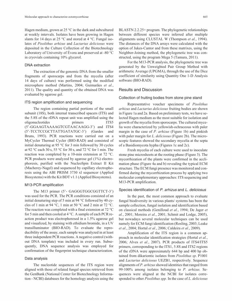
Fresh mycelia of each culture were used to inoculatestone pine microshoots at the rooting phase (Figure 3). Themycorrhization of the plants were confirmed in the accli-mation phase (Figure 4a and b) revealing the typical ECMstructure. The ECM fungi presence was monitored and con-firmed during the mycorrhization process by applying twomolecular complementary approaches: ITS sequencing andM13-PCR amplification.
Species identification of P. arhizus and L. deliciosus
In the past, the most common approach to evaluatefungal biodiversity in various plants/ systems has been thesample collection, fungal isolation and identification basedon classical methods (Genilloud et al., 1994; De Jager et
al., 2001; Moreira et al., 2001; Schmit and Lodge, 2005),but nowadays several molecular techniques can be usednamely for ECM fungi identification (Rosling, 2003; Leakeet al., 2004; Hortal et al., 2006; Caldeira et al., 2009).
Amplification of the ITS region is a common ap-proach in molecular identification strategies (Hortal et al.,2006; Alves et al., 2007). PCR products of ITS4/ITS5primers, corresponding to the ITS1, 5.8S and ITS2 regionsof the rDNA were approximately 644 bp and 400 bp ob-tained from dikariontic isolates from Pisolithus sp. P1001and Lactarius deliciosus UEZB1, respectively. Sequencealignments of P. arhizus showed identities that ranged from99-100% among isolates belonging to P. arhizus. Se-quences were aligned at the NCBI for isolates corre-sponded to other Pisolithus spp. In the case of L. deliciosus
Molecular approach to characterize ectomycorrhizae 665

the homology was over 99%. The most similar sequencesof P. arhizus and L. deliciosus are shown in Table 1. Thephylogenetic tree (Figure 5) was obtained from the align-ment of these sequences. We identified two different clus-ters, Pisolithus sp. P1001 and L. deliciosus isolate UEZB1(Figure 5). Multiple alignment of Pisolithus sp. cluster cor-responded to a partial sequence of 18S RNA gene and ITS1,5.8S ribosomal RNA gene and ITS2, and partial sequenceof 28S RNA ribosomal region. L. deliciosus UEZB1 corre-
sponded to partial sequence of ITS1, 5.8S ribosomal RNAgene, ITS2 and partial sequence of 28S ribosomal RNA.Both sequences were published in GenBank with accessionnumber HQ896485 and JQ066791, respectively.
Intraspecies identification by M13-PCR
The amplification using the M13 primer has gener-ated a profile with 7-14 DNA fragments ranging from 100to 2700 bp in the Basidiomycetes sporocarps species (P.
666 Ragonezi et al.
Figure 1 - Pisolithus arhizus sporocarp collected in a stone pine stand selected for Pinus pinea-ECM associations study (a). The mycelia cultured inHagen medium (b). Secondary mycelia. Each interval 2.5 �m (c).
Figure 2 - Lactarius deliciosus sporocarps collected in a pine stand selected for Pinus pinea-ECM associations study (a). The mycelia cultured in Hagenmedium (b). Secondary mycelia. Each interval 2.5 �m (c).
arhizus, L. deliciosus and R. roseolus), Pisolithus sp. iso-lated P1001 and select Ascomycetes (P. brevicompactum,A. niger, Cladosporium sp.1, and F. oxysporum). TheseAscomycetes species could live in association with ECMfungi and were commonly found in the isolation process.
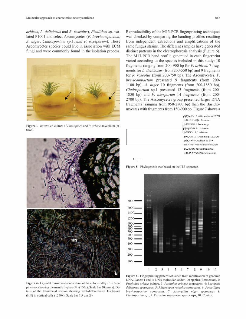
Reproducibility of the M13-PCR fingerprinting techniqueswas checked by comparing the banding profiles resultingfrom independent extractions and amplifications of thesame fungus strains. The different samples have generateddistinct patterns in the electrophoresis analysis (Figure 6).The M13-PCR band profile generated in each fingerprintvaried according to the species included in this study: 10fragments ranging from 200-900 bp for P. arhizus, 7 frag-ments for L. deliciosus (from 200-550 bp) and 9 fragmentsfor R. roseolus (from 200-750 bp). The Ascomycetes, P.
brevicompactum presented 9 fragments (from 200-1100 bp), A. niger 10 fragments (from 200-1850 bp),Cladosporium sp.1 presented 13 fragments (from 200-1850 bp) and F. oxysporum 14 fragments (from 200-2700 bp). The Ascomycetes group presented larger DNAfragments (ranging from 950-2700 bp) than the Basidio-mycetes with fragments from 150-900 bp. Figure 7 shows a
Molecular approach to characterize ectomycorrhizae 667
Figure 3 - In vitro co-culture of Pinus pinea and P. arhizus mycelium (ar-rows).
Figure 4 - Cryostat transversal root section of the colonized by P. arhizus
pine root showing the mantle hyphae (M) (100x); Scale bar 20 �m (a). De-tails of the transversal section showing well-differentiated Hartig-net(HN) in cortical cells (1250x); Scale bar 7.5 �m (b).
Figure 5 - Phylogenetic tree based on the ITS sequence.
Figure 6 - Fingerprinting patterns obtained from mplification of genomicDNA. Lanes: 1 and 11 DNA molecular ladder 100 bp plus (Fermentas), 2:Pisolithus arhizus culture, 3: Pisolithus arhizus sporocarps, 4: Lactarius
deliciosus sporocarps, 5: Rhizopogon roseolus sporocarps, 6: Penicillium
brevicompactum sporocarps, 7: Aspergillus niger sporocarps 8:Cladosporium sp., 9: Fusarium oxysporum sporocarps, 10: Control.
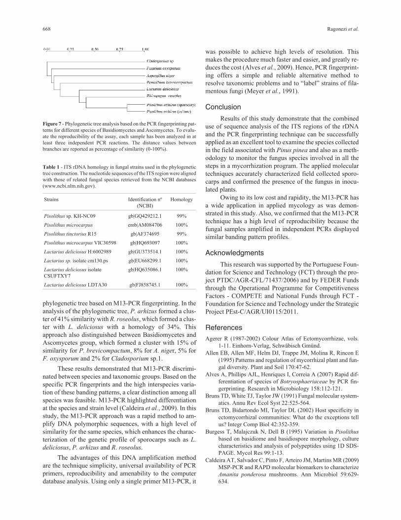
phylogenetic tree based on M13-PCR fingerprinting. In theanalysis of the phylogenetic tree, P. arhizus formed a clus-ter of 41% similarity with R. roseolus, which formed a clus-ter with L. deliciosus with a homology of 34%. Thisapproach also distinguished between Basidiomycetes andAscomycetes group, which formed a cluster with 15% ofsimilarity for P. brevicompactum, 8% for A. niger, 5% forF. oxysporum and 2% for Cladosporium sp.1.
These results demonstrated that M13-PCR discrimi-nated between species and taxonomic groups. Based on thespecific PCR fingerprints and the high interspecies varia-tion of these banding patterns, a clear distinction among allspecies was feasible. M13-PCR highlighted differentiationat the species and strain level (Caldeira et al., 2009). In thisstudy, the M13-PCR approach was a rapid method to am-plify DNA polymorphic sequences, with a high level ofsimilarity for the same species, which enhances the charac-terization of the genetic profile of sporocarps such as L.
deliciosus, P. arhizus and R. roseolus.
The advantages of this DNA amplification methodare the technique simplicity, universal availability of PCRprimers, reproducibility and amenability to the computerdatabase analysis. Using only a single primer M13-PCR, it
was possible to achieve high levels of resolution. Thismakes the procedure much faster and easier, and greatly re-duces the cost (Alves et al., 2009). Hence, PCR fingerprint-ing offers a simple and reliable alternative method toresolve taxonomic problems and to “label” strains of fila-mentous fungi (Meyer et al., 1991).
Conclusion
Results of this study demonstrate that the combineduse of sequence analysis of the ITS regions of the rDNAand the PCR fingerprinting technique can be successfullyapplied as an excellent tool to examine the species collectedin the field associated with Pinus pinea and also as a meth-odology to monitor the fungus species involved in all thesteps in a mycorrhization program. The applied moleculartechniques accurately characterized field collected sporo-carps and confirmed the presence of the fungus in inocu-lated plants.
Owing to its low cost and rapidity, the M13-PCR hasa wide application in applied mycology as was demon-strated in this study. Also, we confirmed that the M13-PCRtechnique has a high level of reproducibility because thefungal samples amplified in independent PCRs displayedsimilar banding pattern profiles.
Acknowledgments
This research was supported by the Portuguese Foun-dation for Science and Technology (FCT) through the pro-ject PTDC/AGR-CFL/71437/2006) and by FEDER Fundsthrough the Operational Programme for CompetitivenessFactors - COMPETE and National Funds through FCT -Foundation for Science and Technology under the StrategicProject PEst-C/AGR/UI0115/2011.
References
Agerer R (1987-2002) Colour Atlas of Ectomycorrhizae, vols.1-11. Einhorn-Verlag, Schwäbisch Gmünd.
Allen EB, Allen MF, Helm DJ, Trappe JM, Molina R, Rincon E(1995) Patterns and regulation of mycorrhizal plant and fun-gal diversity. Plant and Soil 170:47-62.
Alves A, Phillips AJL, Henriques I, Correia A (2007) Rapid dif-ferentiation of species of Botryosphaeriaceae by PCR fin-gerprinting. Research in Microbiology 158:112-121.
Bruns TD, White TJ, Taylor JW (1991) Fungal molecular system-atics. Annu Rev Ecol Syst 22:525-564.
Bruns TD, Bidartondo MI, Taylor DL (2002) Host specificity inectomycorrhizal communities: What do the exceptions tellus? Integr Comp Biol 42:352-359.
Burgess T, Malajczuk N, Dell B (1995) Variation in Pisolithus
based on basidiome and basidiospore morphology, culturecharacteristics and analysis of polypeptides using 1D SDS-PAGE. Mycol Res 99:1-13.
Caldeira AT, Salvador C, Pinto F, Arteiro JM, Martins MR (2009)MSP-PCR and RAPD molecular biomarkers to characterizeAmanita ponderosa mushrooms. Ann Microbiol 59:629-634.
668 Ragonezi et al.
Table 1 - ITS rDNA homology in fungal strains used in the phylogenetictree construction. The nucleotide sequences of the ITS region were alignedwith those of related fungal species retrieved from the NCBI databases(www.ncbi.nlm.nih.gov).
Strains Identification nº(NCBI)
Homology
Pisolithus sp. KH-NC09 gb|GQ429212.1 99%
Pisolithus microcarpus emb|AM084706 100%
Pisolithus tinctorius R15 gb|AF374695 99%
Pisolithus microcarpus VIC30598 gb|HQ693097 100%
Lactarius deliciosus H:6002989 gb|GU373514.1 100%
Lactarius sp. isolate cm130.ps gb|EU668299.1 100%
Lactarius deliciosus isolateCSUFTXY7
gb|HQ635086.1 100%
Lactarius deliciosus LDTA30 gb|FJ858745.1 100%
Figure 7 - Phylogenetic tree analysis based on the PCR fingerprinting pat-terns for different species of Basidiomycetes and Ascomycetes. To evalu-ate the reproducibility of the assay, each sample has been analyzed in atleast three independent PCR reactions. The distance values betweenbranches are reported as percentage of similarity (0-100%).

Cline ML, France RC, Reid CPP (1987) Intraspecific and inter-specific growth variation of ectomycorrhizal fungi at differ-ent temperatures. Can J Bot 65:869-875.
De Jager ES, Wehner FC, Korsten L (2001) Microbial ecology ofthe mango phylloplane. Microbial Ecology 42:201-207.
De La Bastide PY, Kropp BR, Piche Y (1995) Population-struc-ture and mycelial phenotypic variability of the ectomy-corrhizal basidiomycete Laccaria bicolor (Maire) OrtonMycorrhiza 5:371-379.
FAO (2004) Wild edible fungi. A global overview of their use andimportance to people. In: Boa E. (ed) Non-wood ForestProducts, No. 17. FAO, Rome.
Gardes M, Mueller GM, Fortin JA, Kropp BR (1991a) Mitochon-drial-DNA polymorphisms in Laccaria-Bicolor, L-Laccata,
L-Proxima and L-Amethystina. Mycological Research95:206-216.
Gardes M, White TJ, Fortin JA, Bruns TD, Taylor JW (1991b)Identification of indigenous and introduced symbiotic fungiin ectomycorrhizae by amplification of nuclear and mito-chondrial ribosomal DNA. Can J Bot 69:180-190.
Gardes M, Bruns TD (1993) ITS primers with enhanced specific-ity for basidiomycetes - Application to the identification ofmycorrhizae and rusts. Molecular Ecology 2:113-118.
Gay G (1990) Effect of the ectomycorrhizal fungus Hebeloma
hiemale on adventitious root-formation in derooted Pinus
halepensis shoot hypocotyls. Can J Bot 68:1265-1270.
Genilloud O, Pelaez F, Gonzalez I, Dyez MT (1994) Diversity ofactinomycetes and fungi on seaweeds from the Iberiancoasts. Microbiologia SEM 10:413-422.
Godoy P, Cano J, Gene J, Guarro J, Hofling-Lima AL, ColomboAL (2004) Genotyping of 44 isolates of Fusarium solani, themain agent of fungal keratitis in Brazil. J Clin Microbiol42:4494-4497.
Gomes EA, de Abreu LM, Borges AC, de Araujo EF (2000) ITSsequences and mitochondrial DNA polymorphism inPisolithus isolates. Mycological Research 104:911-918.
Gryta H, Debaud JC, Effosse A, Gay G, Marmeisse R (1997)Fine-scale structure of populations of the ectomycorrhizalfungus Hebeloma cylindrosporum in coastal sand dune for-est ecosystems. Mol Ecol 6:353-364.
Guidot A, Lumini E, Debaud JC, Marmeisse R (1999) The nuclearribosomal DNA intergenic spacer as a target sequence tostudy intraspecific diversity of the ectomycorrhizal basi-diomycete Hebeloma cylindrosporum directly on Pinus rootsystems. Appl Environ Microb 65:903-909.
Guimaraes JB, Pereira P, Chambel L, Tenreiro R (2011) Assess-ment of filamentous fungal diversity using classic and mo-lecular approaches: Case study - Mediterranean ecosystem.Fungal Ecology 4:309-321.
Henrion B, Letacon F, Martin F (1992) Rapid Identification ofGenetic-Variation of Ectomycorrhizal Fungi by Amplifica-tion of Ribosomal-RNA Genes. New Phytol 122:289-298.
Hortal S, Pera J, Galipienso L, Parlade J (2006) Molecular identi-fication of the edible ectomycorrhizal fungus Lactarius deli-
ciosus in the symbiotic and extraradical mycelium stages. JBiotechnol 126:123-134.
Horton TR (2002) Molecular approaches to ectomycorrhizal di-versity studies: Variation in ITS at a local scale. Plant Soil244:29-39.
Hutchison LJ (1999) Lactarius. In: Cairney JWG, Chambers SM(eds) Ectomycorrhizal Fungi: Key Genera in Profile.Springer-Verlag, Berlin, pp 269-285.
Iotti M, Zambonelli A (2006) A quick and precise technique foridentifying ectomycorrhizas by PCR. Mycol Res 110:60-65.doi:S0953-7562(05)00029-8[pii]0.1016/j.mycres.2005.09.010.
Karabaghli C, Frey-Klett P, Sotta B, Bonnet M, Le Tacon F(1998) In vitro effects of Laccaria bicolor S238 N and Pseu-
domonas fluorescens strain BBc6 on rooting of de-rootedshoot hypocotyls of Norway spruce. Tree Physiol 18:103-111.
Leake JR, McKendrick SL, Bidartondo M, Read DJ (2004) Sym-biotic germination and development of the myco-hetero-troph Monotropa hypopitys in nature and its requirement forlocally distributed Tricholoma spp. New Phytologist163:405-423.
Lopes M, Silva D, Freitas G, Tenreiro R (2007) Simultaneousidentification and typing of Candida species by MSP-PCRand AFLP: Study of clinical isolates from a Portuguese pedi-atric hospital. J Mycol Médicale 17:157-67.
Martins M (2004) Degradação Biológica de Fungicidas emAmostras de Solo. Tese de Doutoramento, Departamento deQuímica, Universidade de Évora, Évora, pp 140-142.
Marx DH, Ruehle JL, Kenney DS, Cordell CE, Riffle JW, MolinaRJ, Pawuk WH, Navratil S, Tinus RW, Goodwin OC (1982)Commercial vegetative inoculum of Pisolithus tinctorius
and inoculation techniques for development of ectomycor-rhizae on container-grown tree seedlings. For Sci 28:373-400.
Mello A, Ghignone S, Vizzini A, Sechi C, Ruiu P, Bonfante P(2006) ITS primers for the identification of marketable bo-letes. J Biotechnol 121:318-329.
Meyer W, Koch A, Niemann C, Beyermann B, Epplen J T, BornerT (1991) Differentiation of species and strains among fila-mentous fungi by DNA fingerprinting. Curr Genet 19:239-242.
Modess O (1941) Zur kenntnis der mykorrhizabildner von kieferund fichte. Symb Bot Upsala 5:1-147.
Moreira SR, Schwan RF, Carvalho EP, Wheals AE (2001) Isola-tion and identification of yeasts and filamentous fungi fromyogurts in Brazil. Braz J Microbiol 32:117-122.
Niemi K, Salonen M, Ernstsen A, Heinonen-Tanski H, HäggmanH (2000) Application of ectomycorrhizal fungi in rooting ofScots pine fascicular shoots. Can J For Res 30:1221-1230.
Niemi K, Julkunen-Tiitto R, Tegelberg R, Haggman H (2005)Light sources with different spectra affect root and mycor-rhiza formation in Scots pine in vitro. Tree Physiol 25:123-128.
Normand L, Bärtschi H, Debaud JC, Gay G (1996) Rooting andacclimatization of micropropagated cuttings of Pinus
pinaster and Pinus sylvestris are enhanced by the ectomy-corrhizal fungus Hebeloma cylindrosporum. Physiol Plant98:759-766.
Ragonezi C, Castro MR, Klimaszewska K, Lima M, ZavattieriMA (2010a) Influence of light quality and intensity on ad-ventitious root formation in microshoots of Pinus pinea L.Acta Hort (ISHS) 865:287-291.
Rincón A, Alvarez IF, Pera J (1999) Ectomycorrhizal fungi ofPinus pinea L. in northern Spain. Mycorrhiza 8:271-276.
Molecular approach to characterize ectomycorrhizae 669

Rosling A (2003) Responses of Ectomycorrhizal Fungi to MineralSubstrates. Ph.D. Thesis, Swedish University of Agricul-tural Sciences, Sweden.
Schmit JP, Lodge DJ (2005) Chapter 10. Classical methods andmodern analysis for studying fungal diversity. In: Dighton J,White Jr JF, Oudemans P (eds) The Fungal Community, itsOrganization and Role in the Ecosystem. 3rd edition. CRCPress, Taylor & Francis, Boca Raton, pp 193-214.
Singer R (1986) The Agaricales in ModernTaxonomy. 4th edi-tion. Koeltz Scientific Books, Koenigstein, 981 pp.
Smith SE, Read DJ (1997) Mycorrhizal Symbiosis. 2nd edition.Academic Press, London.
Smith ME, Douhan GW, Rizzo DM (2007) Intra-specific andintra-sporocarp ITS variation of ectomycorrhizal fungi asassessed by rDNA sequencing of sporocarps and pooledectomycorrhizal roots from a Quercus woodland. Mycorrhi-za 18:15-22.
Tamura K, Peterson D, Peterson N, Stecher G, Nei M, Kumar S(2011) MEGA5: Molecular evolutionary genetics analysisusing maximum likelihood, evolutionary distance, and max-imum parsimony methods. Mol Biol Evol 28:2731-2739.
Taylor AFS, Alexander I (2005) The ectomycorrhizal symbiosis:Life in the real world. Mycologist 19:102-112.
Thompson J, Higgins D, Gibson T (1994) ClustalW: Improvingthe sensitivity of progressive multiple sequence alignmentthrough sequence weighting, position-speci®c gap penaltiesand weight matrix choice. Nucl Acids Res 22:4673-4690.
Zavattieri A, Lima M, Sobral V, Oliveira P, Costa A (2009) Ef-fects of carbon source, carbon concentration and cultureconditions on in vitro rooting of Pinus pinea L. microshoots.Acta Hort (ISHS) 812:173-180.
All the content of the journal, except where otherwise noted, is licensed under aCreative Commons License CC BY-NC.
670 Ragonezi et al.