Module on - Educypediaeducypedia.karadimov.info/library/anatomy_hiv.pdf · HIV belongs to the...
12
Anatomy and pathophysiology of HIV 4-1 Clinical Tract Module on The anatomy and pathophysiology of HIV
Transcript of Module on - Educypediaeducypedia.karadimov.info/library/anatomy_hiv.pdf · HIV belongs to the...

Anatomy and pathophysiology of HIV 4-1
Clinical Tract
Module on
The anatomy and pathophysiology
of HIV

Anatomy and pathophysiology of HIV 4-2
LEARNING OUTCOMES FOR SOCIAL WORKERS, LABORATORY TECHNICIANS, PHARMACISTS, NURSING STAFF AND DOCTORS After completion of this module the learner should be able to: • Give a basic description of the anatomy of the virus. • Describe the important steps in the life cycle of the virus and the different
enzymes involved. • Describe the organs making up the human immune system and understand how
the immune system is damaged. • Understand the basic disease processes. This module is technically the most difficult module in the manual. It is not meant to be studied by counsellors and data typists. Even other members of the team should only have a w orking know ledge of HIV and the immune system and are nor required to give a detailed description.

Anatomy and pathophysiology of HIV 4-3
1. THE ANATOMY OF THE VIRUS HIV belongs to the family of human retroviruses (Retroviridae) and the subfamily Lentiviruses. It is an RNA virus and its hallmark is the reverse transcription of viral RNA to DNA by reverse transcriptase enzyme, a unique characteristic of retroviruses. All the other non-retroviral viruses lead to the transcription of new RNA from a DNA strand. There are tw o types of HIV, HIV-1 and HIV-2 w ith the former being the predominant type in most parts of the w orld. HIV virion morphology
Figure 1. Schematic structure of the HIV virion The virus detaches from the surface of the host cell membrane by a process called budding. Part of the host cell membrane therefore forms the envelope of the new virion w ith some of the host proteins becoming incorporated into the envelope in the process. Of most signif icance is the major histocompatibility complex (MHC) class I and II host proteins. The envelope is also studded by specif ic glycoproteins that facilitate attachment and subsequent entry of the virus into the host cell. These envelope glycoproteins are, the outer membrane glycoprotein (gp120) and the transmembrane glycoprotein (gp41). On the inside, the envelope is lined by a structural protein called the matrix protein (p17) w hile another structural protein, capsid (p24) forms a cone-shaped casing around the core of the virion. The core consists of tw o single-stranded RNA molecules w hich form the RNA genome of the virus, and enzymatic proteins, reverse transcriptase, integrase and proteases.

Anatomy and pathophysiology of HIV 4-4
Glycoproteins The surface glycoprotein, gp120 is attached to the viral membrane via the transmembrane glycoprotein gp41. It recognizes specif ic receptors called the CD4 receptors on the surface of the target cells. gp41 the transmembrane glycoprotein traverses the virion membrane and mediates fusion of the host and viral membrane thereby allow ing HIV to enter the host cell. HIV genome Common to most retroviruses are genes that encode the structural proteins of the virus, Gag-Pol-Env genes. Gag encodes the proteins that form the core of the virion, Env encodes the envelope glycoproteins and Pol encodes the viral enzymes, reverse transcriptase, proteases and integrase. HIV-1 is however more complex than other retroviruses and contains other genes, HIV-regulatory genes w hich code for proteins involved in the regulation of gene expression (tat, rev, nef, vif, vpr, and vpu). Some of these regulatory genes play a role in pathogenesis of HIV disease. Target cells of HIV Any cell that expresses the CD4 receptor together w ith the co-receptor molecules (chemokine receptors) can be a target. The principal targets are the CD4 T-lymphocyte cells (T helper cells), monocytes and macrophage lineage cells. Others with few CD4 receptors are follicular dendritic cells (FDC) and the epidermal Langerhans cells present in the skin.
Figure 2. Target cells of HIV (From; Robin’s Pathologic Basis of Disease, 6th Edition; pg 241) Normally, tw o surface receptors are required for the virus to gain entry into the host cells. Chemokine receptors (co-receptors for HIV) belong to the large group of seven-transmembrane domain G protein-coupled cellular receptors. The tw o most

Anatomy and pathophysiology of HIV 4-5
important co-receptors for HIV-1 are CCR5 (R5) and CXCR4 (X4). These co-receptors also help in determining the cellular tropism of the HIV-1. The T-tropic HIV-1 strains bind to X4 co-receptor cells, mainly the T-cells w hile the M-tropic strains bind to the R5 co-receptor cells like the macrophages. 2. REPLICATION CYCLE OF HIV
1. The replication cycle begins w ith binding of the viral gp120 to the target host cell CD4 receptor.
2. gp41-mediated fusion of the host and viral membrane then follows leading to internalization of the HIV genomic RNA and its associated proteins into the target cell. The co-receptor is vital for this stage.
3. Reverse transcriptase enzyme then catalyses the reverse transcription of the viral RNA to produce a double-stranded viral DNA.
4. Viral DNA translocates to the nucleus w here it is integrated randomly into the host cell genome w ith the help of yet another viral enzyme, integrase.
5. Integrated proviral DNA may remain inactive (latent) in the nucleus or it may lead to gene expression and transcription forming genomic viral RNA or mRNA if the infected cell is activated.
6. HIV mRNA is translated into viral proteins 7. Viral particle is formed by the assembly of the HIV proteins, enzymes and
genomic RNA at the cell plasma membrane. 8. Budding of the immature virus occurs at the host cell membrane and the core
acquires its external envelope. 9. During or just after budding, the third viral enzyme protease, cleaves the Gag-
Pol precursor proteins yielding a mature virion that is infectious.
Figure 3. The replication cycle of HIV (From; Principles of Internal Medicine, Harrison’s 15th Edition; pg 1855) Looking at the different stages of the life cycle, there is a potential for therapeutic intervention at each one of them.
Anatomy and pathophysiology of HIV 4-6
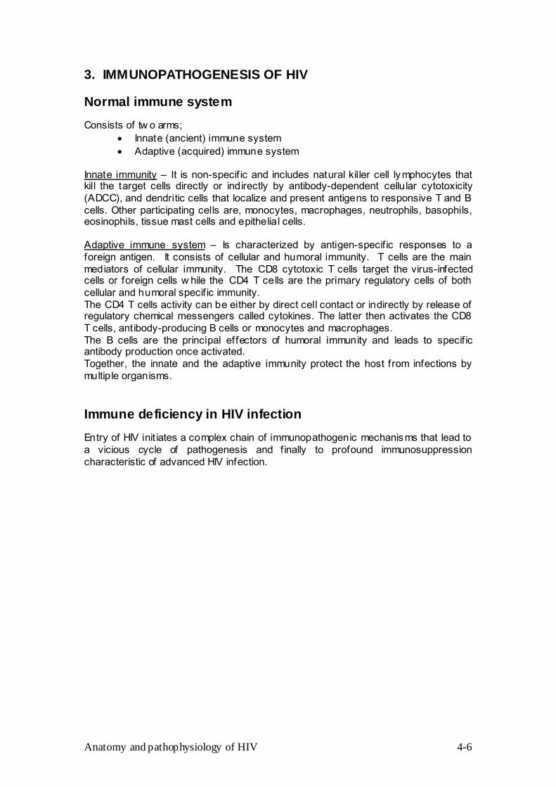
3. IMMUNOPATHOGENESIS OF HIV Normal immune system Consists of tw o arms;
• Innate (ancient) immune system • Adaptive (acquired) immune system
Innate immunity – It is non-specif ic and includes natural killer cell lymphocytes that kill the target cells directly or indirectly by antibody-dependent cellular cytotoxicity (ADCC), and dendritic cells that localize and present antigens to responsive T and B cells. Other participating cells are, monocytes, macrophages, neutrophils, basophils, eosinophils, tissue mast cells and epithelial cells. Adaptive immune system – Is characterized by antigen-specif ic responses to a foreign antigen. It consists of cellular and humoral immunity. T cells are the main mediators of cellular immunity. The CD8 cytotoxic T cells target the virus-infected cells or foreign cells w hile the CD4 T cells are the primary regulatory cells of both cellular and humoral specif ic immunity. The CD4 T cells activity can be either by direct cell contact or indirectly by release of regulatory chemical messengers called cytokines. The latter then activates the CD8 T cells, antibody-producing B cells or monocytes and macrophages. The B cells are the principal effectors of humoral immunity and leads to specif ic antibody production once activated. Together, the innate and the adaptive immunity protect the host from infections by multiple organisms. Immune deficiency in HIV infection Entry of HIV init iates a complex chain of immunopathogenic mechanisms that lead to a vicious cycle of pathogenesis and f inally to profound immunosuppression characteristic of advanced HIV infection.

Anatomy and pathophysiology of HIV 4-7
Figure 4 – The vicious cycle of HIV pathogenesis (From; Pantaleo G, Fauci A S.1995. Annu Rev Immunnol;13:487-512) Primary infection is accompanied by w idespread dissemination of the virus. Rapid replication of the virus in this early stage of infection triggers an appropriate immune response that is HIV-specif ic. This leads to a marked reduction in the initial viremia. How ever, this immune response is inadequate and only partially eliminates the virus from the body. A stage of clinical ‘latency’ that is seemingly dormant and asymptomatic develops. During this ‘latent’ stage, viral replication continues especially in the lymph nodes. A chronic and persistent state of viral replication ensues and gradual deterioration of the immune system occurs. This f inally leads to clinically apparent disease. Factors affecting the pathogenesis of HIV
• Host factors - Cell-mediated immunity - Humoral immunity - Genetic factors
• Viral factors

Anatomy and pathophysiology of HIV 4-8
• Other co-factors Cell-mediated immune response
1) CD4 T lymphocyte The depletion and dysfunction of the CD4 T cells w ith persistent HIV infection is central to the pathogenesis of HIV disease. This occurs via direct and indirect mechanisms.
a) Direct HIV-cytopathic effects This causes single-cell killing and cell fusion w ith syncytia formation. Many uninfected CD4 cells fuse w ith a single HIV-infected cell via CD4-gp120 interaction forming giant multinucleated cells (syncytium) that may be short lived. Several mechanisms are thought to contribute to the single-cell cytotoxicity of HIV infection;
• Accumulation of unintegrated viral DNA • Inhibit ion of cellular protein synthesis by impairing cellular RNA
processing • Excessive production and accumulation of gp120 also has a
direct cell-killing (cytopathic) effect apart from accelerating the virus-cell and cell-cell fusion
• Viral budding from the cell plasma membrane causes loss of membrane integr ity
b) Inhibited lymphopoiesis
Lack of the ability to completely regenerate or repopulate the peripheral blood CD4 T lymphocytes that are persistently diminished in HIV infection. This is due to destruction and depletion of lymphoid precursor cells in the lymphoid organs and in the bone marrow . The thymic progenitor cells in the thymus gland are also affected. This leads to depressed formation of new blood cells, and other hematological abnormalit ies.
2) CD8 T lymphocytes
a) Cytotoxic CD8 T lymphocytes (CTL)
HIV-specif ic immune response has both humoral and cellular immune responses. The HIV-specif ic cytotoxic CD8 T lymphocytes are predominantly involved in the cell-mediated immune response. When activated, they are cytotoxic to HIV-infected CD4 cells and other target cells expressing identical MHC class I molecules associated w ith HIV antigen. The CTL are also cytotoxic to uninfected CD4 cells (innocent bystanders) w ith surface-bound envelope proteins like gp120. The HIV-specif ic CD8 CTLs appear early in primary HIV infection and play a major role in reduction of the viremia in the initial partial immune response. There is a signif icant inverse correlation betw een the CTLs and the viral load. As the disease progresses, their specif ic

Anatomy and pathophysiology of HIV 4-9
functional capability decreases and in late stages of the disease, the numbers of CTLs may be signif icantly reduced. Their functional integrity is partly dependent on adequate inductive signals from CD4 T cells, the main regulatory cells of adaptive immunity. Therefore, the defect in advanced disease might be compounded by the marked CD4 cell reduction.
b) Suppressor CD8 T lymphocytes – Another group of CD8 T cells that
cause suppression of HIV replication w ithout killing the invaded host cell. Suppression is mediated via chemical messengers in form of antiviral cytokines and chemokines.
3) B cells – Many functional abnormalities of B cells occur in patients w ith HIV
infection like, polyclonal activation, hypergammaglobulinaemia, autoantibody production and also impaired primary and secondary (memory) antibody responses to microorganisms.
4) Natural killer lymphocyte cells (NK cells) – Involved in the killing of HIV-
infected cells either directly or indirectly via the A DCC w here the NK cells w ith specif ic antibodies against HIV antigens are directed towards HIV-expressing cells.
5) Monocytes and macrophages
HIV infection usually leads to death of the invaded target host cell. It how ever has a low cell-killing effect on monocyte-lineage cells like monocytes and macrophages that are also targets of HIV. These cells therefore act as reservoirs of HIV infection, a function that may be an obstacle in eradication of HIV infection w ith antiretroviral treatment.
6) Dendrit ic and Langerhans cells
They are the f irst cells to appear at inf lamed mucous membrane sites and are vital in init iat ing T cell response. On entering an inflamed site, HIV is bound by dendritic cells and carried to the lymphoid organs rich in CD4 T cells. They are also thought to init iate the partial HIV-specif ic cell mediated immune response by interacting w ith CTLs in the lymphoid organs.
Humoral immune response HIV-specif ic humoral immune response is important in the init ial dow nregulation of HIV replication. HIV-binding antibodies produced by the B cells w ithin six to tw elve weeks are directed to multiple HIV antigens. The antibodies, w hich can be detected by ELISA or w estern blot do not have a clear definite function. Some have a protective function while others enhance the pathogenesis of HIV infection. Protective function • By participating in ADCC after binding to the NK cells. Leads to the killing of the
HIV-infected cells. • Neutralizing antibodies neutralize the HIV directly therefore preventing the spread
of infection. The envelope glycoproteins that induce production of these neutralizing antibodies can be considered as a component for a potential HIV vaccine.

Anatomy and pathophysiology of HIV 4-10
Pathogenic role HIV-specif ic antibodies may also contribute to disease progression by: • Antibody enhancement mechanism – production of antibodies that facilitate
infection of other cells like macrophages and NK cells. • Bystander killing – Anti-gp120 antibodies involved in ADCC might also kill
uninfected CD4 T cells expressing free gp120. Other mechanisms in pathogenesis of HIV Autoimmune mechanisms Several auto-antibodies directed against individual’s ow n normal antigens in the body, have been detected in HIV-infected patients. The possible explanations of the autoimmune processes are:
• B cell polyclonal proliferation and antibody production due to luck of T cell regulation in HIV infection after depletion of the CD4 T cells.
• Molecular mimicry – some degree of structural resemblance is shared betw een the HIV proteins and the major histocompatibility complex (MHC) class II molecules. There could therefore be a cross-reaction betw een the antibodies to the HIV protein and the human leucocyte antigens (HLA) class II molecules. Several auto-antibodies have been detected in HIV infection.
Apoptosis (Programmed cell death) This is a normal mechanism of cell death w here the body eliminates autoreactive clones of T cells. HIV infection of CD4 T cells may lead to activation-induced cell death sometimes w ithout direct infection of the CD4 T cells by HIV. It is thought that cross-linking of the CD4-gp120 or gp120-anti gp120 immune complexes prepares the cells for apoptosis. Lymphoid organs in HIV infection When HIV enters the body, it is w idely disseminated especially to the lymphoid tissues. The latter plays a major role as a reservoir of HIV w here the virus particles are trapped w ithin the follicular dendritic cells (FDC) in the germinal center and persistent viral replication occurs even during the clinical latency period. The FDCs facilitate infection of CD4 T helper cells and memory T cells w ithin the germinal centers of the lymph nodes. Persistent activation of the immune cells in the lymphoid organs further facilitates their infection by trapped virus and its replication once inside the cells. The expanding netw ork of FDCs and the chronic, persistent viral replication leads to follicular hyperplasia clinically presenting as enlarged lymph nodes. In advanced HIV disease, degeneration of the follicular dendritic cell netw ork and involution of the lymph node germinal center occurs releasing the trapped HIV virions into circulation w ith resultant massive increase in plasma viremia in late disease. Genetic factors in HIV pathogenesis Some host genetic factors have been described that may influence the pathogenesis and course of HIV disease; • MHC class I and II alleles – specif ic alleles of the HLA loci are associated w ith
different rates of progression and varying susceptibility to HIV infection.

Anatomy and pathophysiology of HIV 4-11
• Chemokine receptor (co-receptor) mutations and mutations of the chemokines, chemical messengers specif ic for these receptors w ill interfere with viral binding to the host cell membrane and subsequent progression of the infection.
• Certain immunodominant HIV T helper or cytotoxic T lymphocyte epitopes lead to a relatively protective immune response against the HIV.
These varying genetic factors are associated w ith different clinical conditions in HIV-infected persons and also w ith different clinical courses of the disease. • Long-term nonprogressors or slow progressors
CCR5 mutations (homozygous defect) are associated w ith decreased susceptibility to infection w hile the heterozygous defect causes delayed progression of the disease. Approximately 1% of Caucasians of w estern European origin have the homozygous mutation w hile up to 20% are heterozygotes of the CCR5 mutation. These mutations are extremely rare in Africans. CCR2 heterozygous mutations are also associated w ith slow progression.
• Rapid progression – HIV-infected patients w ith homozygous mutation of
CX3CR1 co-receptor showed rapid progression to AIDS. HLA B35 is also associated w ith rapid progression.
• Exposed yet uninfected individuals – they remain negative despite repetit ive
sexual exposure to HIV in high risk environment. In vitro tests done on these individuals specimens revealed that they had homozygous defects for the CCR5 mutation, the co-receptor for R5 strains of HIV-1 but w ere rapidly infected w ith X4 strains.
Viral (HIV) factors that affect pathogenesis of disease • Viral escape from immune response by mutations. • Viral attenuation – attenuated HIV can slow the rate of disease progression, like a
deletion in the nef gene (a regulator gene) slows progression. • Viral tropism – progression from M-tropic to T-tropic viruses is associated w ith
increased pathogenicity and progression of disease. • Viral subtypes – different subtypes (M,N,O) have different virulence and
transmissibility. Co-factors contributing to HIV pathogenesis Co-factors can be endogenous or exogenous factors. They alter the pathogenesis by several mechanisms like induction of HIV expression. • Cytokines – Pro-inflammatory cytokines like TNFα and β, IL-1 and IL-6
produced in response to most infections or inf lammations are also increased in HIV-infected persons. They enhance HIV replication w hile interferon α and β and IL-16 suppress HIV replication.
• Co-infection – Co-infection or simultaneous cotransfection of cells w ith HIV
and other viruses like cytomegalovirus, human herpes virus type 6, herpes simplex virus type 1, Epstein-Barr virus, human T lymphotrophic virus type 1 and others can up-regulate the expression of HIV. Other microbes like Mycobacterium tuberculosis and Mycoplasma also have a similar effect.

Anatomy and pathophysiology of HIV 4-12
4. CONCLUSION While appreciating all the research efforts in the last tw o decades that have provided much insight into the pathogenesis of HIV infection, these complex immunopathogenic mechanisms are still not completely understood. A better understanding of these mechanisms w ould help a great deal in the development of new therapeutic agents and hopefully the development of an effective vaccine. 5. FURTHER READING
• Pantaleo G, Graziosi C, Fauci AS. The immunopathogenesis of human immunodeficiency virus infection, N Engl J Med, 1993;328:327-335
• Fauci AS, Pantaleo G, Stanley S. Immunopathogenic mechanisms of HIV infection, Ann Intern Med, 1996;124:654-653
• Braunw ald E, Fauci AS, Kasper DL, et al. HIV:AIDS and related diseases, Principles of Internal Medicine, Harrisson’s 15th Edition;2001:chapter 309
• Hogan CM, Hammer SM. Host determinants in HIV infection and diseases. Ann Intern Med, 2001;134:761-776 and 978-996