Modifying Antigen-Encapsulating Liposomes with KALA ... · 1 Modifying antigen-encapsulating...
64
Instructions for use Title Modifying Antigen-Encapsulating Liposomes with KALA Facilitates MHC Class I Antigen Presentation and Enhances Anti-tumor Effects Author(s) Miura, Naoya; Akita, Hidetaka; Tateshita, Naho; Nakamura, Takashi; Harashima, Hideyoshi Citation Molecular therapy, 25(4), 1003-1013 https://doi.org/10.1016/j.ymthe.2017.01.020 Issue Date 2017-04-05 Doc URL http://hdl.handle.net/2115/68705 Type article (author version) File Information WoS_78901_harashima.pdf Hokkaido University Collection of Scholarly and Academic Papers : HUSCAP
Transcript of Modifying Antigen-Encapsulating Liposomes with KALA ... · 1 Modifying antigen-encapsulating...

Instructions for use
Title Modifying Antigen-Encapsulating Liposomes with KALA Facilitates MHC Class I Antigen Presentation and EnhancesAnti-tumor Effects
Author(s) Miura, Naoya; Akita, Hidetaka; Tateshita, Naho; Nakamura, Takashi; Harashima, Hideyoshi
Citation Molecular therapy, 25(4), 1003-1013https://doi.org/10.1016/j.ymthe.2017.01.020
Issue Date 2017-04-05
Doc URL http://hdl.handle.net/2115/68705
Type article (author version)
File Information WoS_78901_harashima.pdf
Hokkaido University Collection of Scholarly and Academic Papers : HUSCAP
1
Modifying antigen-encapsulating liposomes with KALA
facilitates MHC class-I antigen presentation and enhances
anti-tumor effects
Naoya Miura1*, Hidetaka Akita2*, Naho Tateshita2, Takashi Nakamura1 and Hideyoshi
Harashima1
1Department of Molecular Design of Pharmaceutics, Faculty of Pharmaceutical Sciences,
Hokkaido University, Kita 12, Nishi 6, Kita-ku, Sapporo City, Hokkaido, Japan
2Laboratory of Pharmacology and Toxicology, Graduate School of Pharmaceutical Sciences,
Chiba University, 1-8-1 Inohana, Chuou-ku, Chiba City, Chiba, Japan
Correspondence should be addressed to H.A. ([email protected]). Tel: +81-43-226-2893;
Fax: +81-43-226-2893; Address: 1-8-1 Inohana, Chuou-ku, Chiba City, Chiba, Japan
Correspondence may also be addressed to H.H. ([email protected]). Tel: +81 11
706 3919; Fax: +81 11 706 3879; Address: Kita 12, Nishi 6, Kita-ku, Sapporo, Hokkaido, Japan
*These authors contributed equally to the paper as first authors.
Short title: A KALA-modified liposome as anti-cancer vaccine

2
Key words: anti-cancer vaccines, protein vaccines, liposomes, KALA peptides, dendritic cells,
MHC class-I presentation

3
ABSTRACT
For a successful anti-cancer vaccine, antigen presentation on the major histocompatibility
complex (MHC) class-I is a requirement. To accomplish this, an antigen must be delivered to the
cytoplasm by overcoming the endosome/lysosome is required. We previously reported that a
lipid-nanoparticle modified with a KALA peptide
(WEAKLAKALAKALAKHLAKALAKALKA), an α-helical cationic peptide, permits the
encapsulated-pDNA to be efficiently delivered to the cytoplasm in bone marrow-derived
dendritic cells (BMDCs). Herein, we report on the use of KALA-modified liposomes as an
antigen carrier, in an attempt to induce potent antigen-specific cellular immunity. The
subcutaneous injection of KALA-modified ovalbumin (OVA)-encapsulating liposomes
(KALA-OVA-LPs) elicited a much more potent OVA-specific cytotoxic T-lymphocyte activity
and anti-tumor effect in comparison with particles that were modified with octaarginine (R8), a
cell-penetrating peptide (R8-OVA-LPs). In addition, the numbers of OVA-specific CD8+ T cells
were increased by immunization the KALA-OVA-LPs. The treatment of BMDCs with
KALA-OVA-LPs induced a substantial MHC class-I antigen presentation. Furthermore, the

4
acidic pH-dependent membrane destabilization activity of KALA-OVA-LPs strongly suggests
that they are able to escape from endosomes/lysosomes, and thereby deliver their cargos to the
cytoplasm. Collectively, the KALA modified liposome is a potential antigen delivery platform
for use as a protein vaccine.
5
INTRODUCTION
Antigen presentation on the major histocompatibility complex (MHC) class-I in
antigen-presenting cells (APCs) (e.g. dendritic cells (DCs), macrophage) is particularly
important for the induction of cellular immunity, an immunological action which plays a key role
in effective cancer immunotherapy via the activation of antigen-specific cytotoxic
T-lymphocytes (CTL).1-5 An exogenous antigen is usually degraded into peptide fragments in
endosome/lysosomes and presented in the context of MHC class-II, and thereby activate CD4+ T
cells for inducing humoral immunity. Therefore, a strategy for delivering the antigen to the
cytoplasm is prerequisite for the effective activation of cellular immunity, since the antigen must
be subjected to proteosomal degradation for antigen presentation onto the MHC class-I (“cross
presentation”).
To accomplish an efficient antigen delivery, considerable efforts have been devoted to develop
various drug delivery carriers.6-8 The liposome is one of the most useful carriers for antigens
since they contain an inner aqueous phase in which water-soluble molecules can be

6
encapsulated.9-11 Furthermore, when the surfaces of liposomes are modified with functional
molecules (i.e. peptides, carbohydrates, and antibodies), this can facilitate the efficient delivery
of an antigen.6,10,12-15 For efficient cellular uptake, the use of cationic liposomes (i.e.
N-[1-(2,3-Dioleoyloxy)propyl]-N, N, N-trimethylammonium methyl-sulfate: DOTAP) can be
beneficial, since it can be inherently absorbed on the cellular surface. Moreover, it is well known
that cationic nanoparticles are taken up by macrophages in vitro and in vivo (i.e. Kupffer
cells).16,17 We previously reported on the development of a liposome-based antigen carrier,
octaarginine (R8)-modified liposomes.18 Modification with R8, which mimics the TAT peptide
of the influenza virus, enhanced endosomal escape, as well as the uptake of the liposomes in
murine bone marrow-derived dendritic cells (BMDCs), compared with simple cationic liposomes.
Furthermore, the R8-modified liposomes that contained encapsulated ovalbumin (OVA) elicited
efficient antigen presentation in the context of MHC class-I and an OVA-specific anti-tumor
effect, while conventional DOTAP liposomes preferred MHC class II-mediated antigen
presentation. Thus, the rational design of functional molecules for delivering an antigen to the
cytoplasm allowed us to develop an effective anti-tumor vaccine system.

7
We recently reported on the development of a KALA-modified lipid nanoparticle containing
pDNA (a multi-functional envelope-type nano device: KALA-MEND) as a carrier of a DNA
vaccine.19,20 The key component (KALA) in this system adopts an α-helical structure at
physiological pH for membrane destabilization.21 To modify the liposomal structure with it, the
peptide was stearylated. As a result, the KALA-MEND exhibited a >2 orders of magnitude
higher gene transfection activity in comparison with R8-modified one (R8-MEND) in BMDC.
Moreover, it unexpectedly induced the production of a large variety of cytokines and chemokines
via the stimulation of cytosolic DNA sensors (i.e. TBK1/STING pathway, AIM-2
inflammasome). These results indicate that KALA-modified liposomes are more beneficial for
delivering encapsulating cargos to the cytoplasm in comparison with R8-modified particles. This
observation prompted us to evaluate the possibility of using the KALA peptide to deliver an
antigen protein to cells for a cancer vaccine. The focus of this study was on the induction of the
antigen presentation on MHC class-I molecules, and the resulting cellular immunity for cancer
therapeutics.

8
RESULTS
Measurement of OVA-specific CTL activity in mice immunized with KALA-OVA-LPs
To evaluate the efficiency of the KALA peptide for antigen protein delivery, we designed
KALA peptide-modified OVA-encapsulating liposomes (KALA-OVA-LPs). The liposomes
were composed of 3 types of lipids; DOPE (a membrane fusogenic lipid for overcoming the
biomembrane barrier), CHEMS (a pH-sensitive lipid for creating sensitivity to a low pH
environment: endosomes/lysosomes and EPC (a membrane stabilizing lipid for the formation of
liposomes), as reported previously.18 To increase the membrane fusogenic activity of the
liposomes, DOPE was employed in the lipid composition, while an EPC-based particle one was
used as a non-fusogenic control, in order to clarify the relationship between membrane-fusogenic
property and vaccine activity. The OVA protein was encapsulated in liposomes by repetitive
freeze/thaw cycles. After liposomal formation, the surfaces of the liposomes were modified with
the KALA peptide by adding a lipid derivative of the KALA peptide (STR-KALA). As a control
for the cationic peptide, octaarginine (R8) was used to modify the particles, instead of KALA
(R8-OVA-LPs). The physicochemical properties of each liposome preparation are shown in

9
Table 1. The modification with STR-KALA or STR-R8, resulted in a large increase in
ζ-potential (approximately to +50 mV), while the size and polydispersity index (PdI) were
comparable to the values for non-modified OVA-LPs (approximately 160-170 nm and 0.15,
respectively).
In initial experiments, we evaluated the impact of the modification of the KALA peptide on
the induction of antigen-specific cellular immunity in vivo. To investigate this, in vivo
OVA-specific CTL activity was evaluated after subcutaneous immunization of the
KALA-OVA-LPs. The liposomes containing the indicated amount of OVA, free-OVA or empty
KALA-modified liposomes (KALA-LPs) were subcutaneously injected to the back of the necks
of the mice 1 time. OVA-specific CTL activity was evaluated at 7 days after immunization (Fig.
1). First, immunization with the OVA protein per se did not induce CTL activity. In contrast, a
dose-dependent increase in CTL activity was achieved when the OVA was immunized using the
DOPE-incorporated KALA-OVA-LPs whereas the CTL activity for the R8-OVA-LPs was
marginal, even in the case of a maximum dose (50 μg). Of note, the corresponding activities for
non-modified OVA-LPs were at the background level. These data collectively indicate that
KALA is a key component for the effective induction of CTL activity. While the
EPC-incorporated KALA-OVA-LPs also induced significant CTL activity, the activity was
10
inferior to that for the DOPE-based KALA-OVA-LPs, suggesting that modification with the
fusogenic lipid envelope and KALA synergistically potentiated the immune stimulative
activity.20 Furthermore, the KALA-OVA-LPs induced significant CTL activity by single
immunization at a dose of 13 µg OVA, a smaller dose in comparison with previous reports.22,23
Collectively, these data suggest that the DOPE-based KALA-OVA-LPs have the potential for
use as an anti-cancer vaccine system.
Anti-tumor effect of KALA-OVA-LPs
To prove the potential of KALA-OVA-LPs as an anti-cancer vaccine, the prophylactic
anti-tumor effect against E.G7-OVA, a mouse lymphoma EL4 expressing the OVA gene, was
evaluated. Mice were immunized with KALA-OVA-LPs, R8-OVA-LPs, non-modified
OVA-LPs (equivalent to 25 μg of OVA) or empty KALA-LPs (the same amount of lipid as
KALA-OVA-LPs) subcutaneously 1 time. The immunized mice were inoculated with
E.G7-OVA in the right flank 1 week after immunization. Tumor volume was then monitored for
up to 27 days after tumor inoculation (Fig. 2a). The immunization by the non-modified
OVA-LPs failed to inhibit tumor growth, suggesting that the induction of OVA-specific

11
immunity was insufficient. Consistent with data from a previous study, immunization with the
R8-OVA-LPs inhibited tumor growth, although the effect was marginal at this dose. On the other
hand, immunization with the KALA-OVA-LPs exhibited a significant anti-tumor effect: tumor
growth was completely suppressed up to day 15, and furthermore, in 3 of 5 mice, tumor growth
was not detectable, even on day 27 (Fig. 2b). Consequently, antigen-encapsulating KALA
modified liposomes were more potent as an anti-cancer vaccine in comparison with the
R8-modified particles.
To demonstrate the anti-tumor effect of the KALA-OVA-LPs in a more representative clinical
setting, therapeutic anti-tumor experiments were carried out. Mice were inoculated with
E.G7-OVA cells, followed by immunization with the KALA-OVA-LPs. Furthermore, to
evaluate the synergistic effect of an immune checkpoint inhibitor on the therapeutic effect, mice
were treated with an anti-PD-1 antibody when they were immunized with KALA-OVA-LPs. As
a result, tumor volume tended to decrease on average as the result of immunization with the
KALA-OVA-LPs (Figure S1a). Since the tumor volume of vehicle control animals was largely
varied (tumor rejection occurred on day 20 in only 1 of 5 mice), no significant differences were
observed. However, the administration of the KALA-OVA-LPs suppressed the tumor growth in
four of five mice (under 500 mm3 on day 20), whereas only two out of five mice were found to

12
be under 500 mm3 on day 20 in the vehicle treatment group (Figure S1b). Furthermore, while
anti-PD-1 antibody-treated group reached an average of 1000 mm3 on day 20, the group that had
the combined KALA-OVA-LPs and anti-PD-1 treatment decreased on average by half (Figure
S1a). These results suggest that KALA-OVA-LPs are potent in anti-tumor vaccines in a clinical
setting and that they enhance the action of immune checkpoint inhibitors.
MHC class-I antigen presentation by KALA-OVA-LPs
To elucidate the mechanism responsible for the more prominent induction of in vivo
OVA-specific immunity, we evaluated the antigen presentation activity of the KALA-OVA-LPs
in vitro. In this analysis, BMDCs were used, since it is well known that DCs have a crucial role
in the induction of antigen-specific immunity.24-25 We first evaluated the innate immune
activation of BMDCs in terms of enhancing the activation of CD80/86, markers of immune
stimulation in BMDCs. However, no activation was observed even when KALA-OVA-LPs were
administered (Figure S2). This is consistent with the fact that, an inflammatory immune
response (IL-6 production in serum) was not observed after the injection of the KALA-OVA-LPs
(Figure S3). We then investigated a OVA-specific antigen presentation on MHC class-I. Antigen

13
presentation was quantified by co-cultivation with B3Z T-cell hybridoma, a reporter cell line that
produces the lacZ protein in response to the OVA257-264 epitope bound to H-2Kb.26 As a result,
modification with STR-KALA drastically enhanced antigen presentation from BMDCs,
depending on the amount of modification (Fig. 3a). The effect of KALA modification
approached saturation at 5mol% of total lipids, where almost no antigen presentation was
observed in R8-OVA-LPs treatment at the corresponding density. Comparing the dose-response
curves, the antigen presentation efficiency of the KALA-OVA-LPs at a dose of 16 µM
approached those of R8-OVA-LPs at a dose of 128 µM, suggesting that KALA modification
resulted in an enhancement in antigen presentation activity that was 8 times higher than that for
R8 modification (Fig. 3b). The BMDCs treated with KALA-OVA-LPs elicited anti-tumor
effects against E.G7-OVA when they were immunized via the footpad. This suggests that the
enhanced antigen presentation in DCs is one of the mechanisms responsible for the anti-tumor
activity of KALA-OVA-LPs (Figure S4). Furthermore, the enhancement of antigen presentation
by modification with KALA was reduced when the lipid was replaced from DOPE to the
non-fusogenic EPC (Fig. 3a). This result is consistent with the results of in vivo CTL assays. It
appears that the KALA peptide is a potent activator for a liposomal antigen delivery system for
the achievement of antigen presentation to MHC class-I.
14
In vitro/in vivo cellular uptake and membrane-fusogenic activity of the KALA-OVA-LPs
To gain further insights into the mechanism underlying the difference in immunological action
between KALA-OVA-LPs and R8-OVA-LPs, in vitro/in vivo cellular uptake and
membrane-fusogenic activity were evaluated. The kinetics of the ex vivo cellular uptake of
KALA-OVA-LPs and R8-OVA-LPs using BMDCs were first compared by means of flow
cytometry. Liposomes encapsulating a fluorescence-labeled OVA (Alexa Fluor 488-conjugated
OVA, 25% of whole OVA) were prepared to quantify the uptake of OVA. As shown in Figure
S5, the uptake of both liposomes was observed on 6 and 24 hours after the treatment.
Quantitative analysis revealed that the geomean values for the uptake of both liposomes were
comparable after 5 hr, while the initial uptake speed (at 2 hours) was significantly higher in the
case of the KALA-OVA-LPs (Figure S5a). The most significant difference was their
homogeneity: the coefficient variance (CV) value for the cellular uptake of KALA-OVA-LPs
was significantly lower compared with that of R8-OVA-LPs (Figure S5b). Furthermore, the
difference in CV values became larger with increasing time, dominantly due to the increase in
15
the corresponding value specifically in R8-OVA-LPs. This difference might be relevant with the
anti-tumor effect of the immunization of ex vivo antigen-treated cells (Figure S4).
To elucidate the behavior of the liposomes in vivo, flow-cytometric analyses of draining
lymph nodes were performed after an s.c. injection of the liposomes. As a result, both liposomes,
which were labeled with a fluorescently labeled lipid, were found to have accumulated in the
draining lymph nodes at comparable levels (Figure S6a). Furthermore, while the large fraction
of the liposomes were taken up by CD11c‒F4/80‒ cells, which were expected to be B cells, a
portion of the liposomes were taken up by CD11c+F4/80+ cells, which are likely dendritic cells
or macrophages (Figure S6b). Importantly, the level of in vivo cellular uptake to these cells was
comparable between the KALA-OVA-LPs and the R8-OVA-LPs. These results suggest that the
uptake process was not a major determinant for explaining the difference in the in vivo
antigen-specific immune response between the KALA-OVA-LPs and R8-OVA-LPs.
To investigate the membrane-fusogenic activity of the KALA-OVA-LPs and the
R8-OVA-LPs under conditions of physiological or endosomal pH, the membrane disruption
activity of RBCs was monitored after incubation with the liposomes by measuring the amount of
hemoglobin that was released into the supernatant. In this experiments RBCs were mixed with
various amounts of liposomes in PBS adjusted to pH 5.5, 6.5 and 7.4, which mimic late

16
endosomes, early/recycling endosomes, and the cytoplasm/extracellular space, respectively.27 As
a result, the KALA-OVA-LPs induced membrane disruption depending on the lipid
concentration at pH 5.5 and 6.5, whereas membrane disruption was marginal in the case of the
R8-OVA-LPs (Fig. 4). Furthermore, antigen presentation in the presence of chloroquine, an
endosome disruptive agent, was evaluated, to investigate whether the membrane disruptive
activity of the KALA peptide amplifies antigen presentation. As a result, the chloroquine
treatment enhanced antigen presentation in cells that were treated with the R8-OVA-LPs, while
the activity of the KALA-OVA-LPs was not enhanced, but rather decreased (Fig. 5). These
results suggest that modifying liposomes with the KALA peptide induced a higher fusion with
biomembranes in comparison with R8, especially under acidic pH conditions.
Immunological analysis of the mice immunized with the KALA-OVA-LPs
To clarify the precise mechanism responsible for the immune response induced by the
KALA-OVA-LPs, we performed further immunological analyses in vivo. We first evaluated the
contribution of CD8- and CD4-positive T cells to the final output of in vivo CTL activity by
depleting these cells using an antibody (Fig. 6a). Mice were immunized with the
KALA-OVA-LPs on day 0, and subsequently, 200 μg of an anti-CD4 or anti-CD8 antibody were
17
injected intraperitoneally on day ‒1, 1, 3, 6. OVA-specific CTL activity was evaluated one week
after immunization by an in vivo CTL assay. Almost all of the CD4 or CD8 T cells were depleted
on day 7. As a result, CD4 depletion failed to attenuate the CTL activity induced by the
KALA-OVA-LPs, but rather slightly enhanced the effect. In contrast, CD8 depletion drastically
impaired CTL activity. These collective results suggest that CD4 T cells were not involved in the
antigen-specific CTL activity induced by the KALA-OVA-LPs. Thereafter, we also evaluated
the number of OVA-specific T cells induced by immunization with the KALA-OVA-LPs and
R8-OVA-LPs by staining for intracellular cytokines. To assess the frequency of OVA-specific
CD8+ T cells, splenocytes from immunized mice were isolated and stimulated by treatment with
the OVA257-264 peptide (MHC-I peptide) in the presence of a protein transport inhibitor, followed
by staining for intracellular IFN-γ. As a result, immunization with the KALA-OVA-LPs
significantly increased the frequency of IFN-γ+CD8+ T cells (Fig. 6b). This is consistent with the
fact that the KALA-OVA-LPs induced significant in vivo CTL activity via CD8+ T cells (Fig.1,
6a). Furthermore, stimulation of the OVA323-339 peptide (MHC-II peptide) revealed that
OVA-specific Th2 immunity was induced by immunization with KALA-OVA-LPs whereas Th1
and Th17 was not (Fig. 6c). Collectively, these results suggest that the numbers of OVA-specific

18
CD8+ T cells and CD4+ (Th2 phonotype) were expanded as the result of immunization with the
KALA-OVA-LPs.

19
DISCUSSION
In the present study, we report on the development of a system that permits the efficient
delivery of an antigen for cancer immunotherapy using an α-helical peptide: KALA. In general,
positively charged nanoparticles are inherently taken up by a variety of cells, since they bind
easily to the cell surface, which is negatively charged and covered by anionic proteoglycans.28,29
Consistent with previous observations, both KALA-OVA-LPs and R8-OVA-LPs were efficiently
taken up by BMDCs (Figure S5a). However, KALA-OVA-LPs were taken up more
homogenously in comparison with R8-OVA-LPs, (Figure S5b). Since the surface charges and
ζ-potentials of both particles (prepared with DOPE as a helper lipid) were quite comparable,
these differences cannot be simply explained by the physicochemical characteristics. One
possible reason is the differences in secondary structure, i,e, an α-helix structure. As reported
previously, the KALA peptide forms an α-helical structure when the liposomal membrane exists,
whereas the oligo-arginine peptide (especially in R8 and R9) forms a random coil structure.21,30,31
Several previous studies have reported that the α-helical structure interacts with
glycosaminoglycans, whose binding is independent of electrostatic interactions.32,33 While these
electrostatic and structural interactions might contribute to the efficient and homogeneous uptake
20
of the KALA-OVA-LPs in BMDCs, the extent of accumulation in the lymph node between the
KALA-OVA-LPs and the R8-OVA-LPs was comparable in vivo (Figure S6). Thus, the
homogenous uptake of the KALA-OVA-LPs might only contribute to applications of ex vivo
protein vaccines (Figure S4).
The most drastic difference between the KALA-OVA-LPs and R8-OVA-LPs was their
membrane-fusogenic activity, especially at acidic conditions (Fig. 4). It is plausible that this
ability contributes to the remarkable induction of antigen presentation by KALA-OVA-LPs (Fig.
3) since this process is prerequisite for MHC class-I antigen presentation to release the antigen
into the cytoplasm by overcoming endosome/lysosomes membranes. The experiments using
chloroquine also strongly suggest that the KALA peptide amplifies endosomal escape followed
by the higher cytosolic delivery of antigen protein to a higher degree in comparison with R8 (Fig.
5). However, the reason why the fusogenic ability of KALA was elicited at acidic conditions
remains to be determined. Of note, the data reported herein are quite inconsistent with an original
design and function of KALA peptide: it formed an α-helix at physiological pH. In fact, we also
developed a KALA-MEND to allow the encapsulating pDNA to enter the nucleus via step-wise
membrane fusion with the plasma membrane and the nuclear membrane at cytoplasmic pH
(pH=7.4). Thus, the prominently higher gene transfection of the KALA-MEND was not derived

21
from the nuclear membrane fusion-mediated nuclear delivery of pDNA as we designed.19
Wyman et al. reported that the KALA-peptide can form an α-helix structure even under
conditions of acidic pH, when the peptide is present with liposomes whereas its structure
changed to a random coil without liposomes.21 Therefore, it is highly possible that the KALA
might form a α-helix when it is modified onto the lipid envelope. Therefore, the pH-dependent
membrane-fusogenic activity can be explained by assuming that the membrane-fusogenic
activity might be perturbed via electrostatic interactions between KALA and the negatively
charged helper lipid (CHEMS) that is incorporated in the liposomal component. However, when
the KALA-LPs were exposed in an acidic pH in a range close to its pKa (~5.8), the fraction of
ionized CHEMS is decreased by the protonation of the carboxyl group. In this situation, the
electrostatic interactions become less intense, and the fusogenic function of KALA would be
recovered. Such a pH-dependent loss of negative charge was conventionally used as a trigger for
endosomal membrane fusion via the formation of an inverted hexagonal HII phase in liposomes
composed of DOPE, a cone-shape lipid and CHEMS.34,35 Collectively, the KALA-OVA-LPs
might achieve an accelerated endosomal escape via a dual mechanism in response to pH via the
protonation of CHEMS: switching on the KALA function by cancelling the electrostatic
interactions with the peptide, and the loss of lamellar structure to form an inverse cone packaging

22
formation for membrane fusion. Another possible reason is the presence of a histidine (His)
residue in KALA. It is known that a His residue can be employed to facilitate viral membrane
fusion in response to a low pH in some α-helical viral proteins.36,37 Further investigations will be
needed in order to clarify the effect of His residues on the membrane fusogenic property of the
KALA peptide.
As shown in Figure 1 and 3a, the induction of CTL activity and antigen presentation was
weak in EPC-based liposomes, presumably because EPC-based liposome lacks membrane
fusiogenic activity. Phosphatidylcholine derivatives, including EPC are considered to be a
stabilizer of lipid bilayers, since they form a cylindrical structure in the lipid membrane. Thus, it
is difficult to overcome the endosomal membrane barrier, and subsequently for the antigen to be
released to the cytoplasm. To support this. we previously demonstrated that KALA-modified
DOPE liposomes that encapsulate pDNA could stimulate the BMDCs via the stimulation of
cytosolic DNA sensors, whereas the EPC-based showed no activity.20 Thus, EPC is inadequate in
terms of cytoplasmic delivery, at least when used in combination with KALA peptides.
As shown in Figure 3, KALA-OVA-LPs induced potent antigen presentation in BMDCs. In
addition, subsequent immunization with these DCs (ex vivo immunization) efficiently inhibited
tumor growth in comparison with those of R8-OVA-LPs treated BMDCs (Figure S4). However,

23
this ex vivo approach conferred less anti-tumor effect in comparison with a direct in vivo
immunization of KALA-OVA-LPs: the complete prevention of the tumor growth was observed
up to day 15 after tumor inoculation (Fig. 2a). These data suggest that DCs are not a unique cell
population that contributes to the anti-tumor effect of the KALA-OVA-LPs in vivo. One of the
considerable populations is macrophages, a type of APC as well as DCs. In fact, while the large
fraction of the liposomes was taken up by CD11c‒F4/80‒ cells, which have the characteristics of
B cells, a part of the liposomes was taken up by CD11c+F4/80+ cells, which is considered to be
dendritic cells/macrophages (Figure S6b). It is generally thought that the DCs are major, even
unique, APCs that can activate naïve T cells, and macrophages are considered to be less potent
APCs in terms of their activation. However, an accumulating body of evidence has revealed that
a specific subpopulation of macrophages in lymph nodes, CD169-positive macrophage,
contribute to the antigen-specific CTL response and subsequent anti-tumor effect.38-40 For
example, Muraoka et al. revealed that an antigen peptide encapsulated within a nanoparticulate
hydrogel (nanogel, composed of cholesteryl pullulan) was efficiently incorporated into
CD169-positive medullary macrophages and elicited a potent anti-tumor effect.40 Although a
cationic nanogel was reported to be less incorporated into macrophages in draining lymph nodes
in comparison with neutral ones, it has also been reported that a cationic agarose hydrogel could

24
deliver incorporated-nucleic acids into CD169-positive macrophages.41 These facts suggest that
macrophages, in particular CD169-positive macrophages, may contribute to the anti-tumor effect
of KALA-OVA-LPs in vivo. Additional strategies for targeting draining lymph nodes may
improve the efficiency of the KALA-OVA-LPs, such as PEGylation for shielding the surface
charge to avoid trapping by the extracellular matrix or ligand modification for the targeting of
the specific cell type.42,43
In conclusion, we report herein on the development of a carrier for delivering an antigen
protein that has the potential for use in cancer immunotherapy. Modifying antigen-encapsulating
liposomes with KALA facilitated antigen-specific CTL activity and anti-tumor effects when the
liposomes were administered subcutaneously. Furthermore, OVA-specific CD8+ T cells and
CD4+ cells (Th2 phenotype) were expanded by immunization with the KALA-OVA-LPs.
Considering that CD4+ T cells were not involved in OVA-specific CTL activity induced by the
KALA-OVA-LPs (Fig. 6a), the activation of Th2 immunity might not relevant with the activity
of the KALA-OVA-LPs as anti-cancer vaccine. However, this result suggests that a possibility
for an application of the KALA-OVA-LPs to infectious diseases via induction of humoral
immunity. Experiments using BMDCs suggested that the KALA-OVA-LPs induced a much
25
more potent OVA-specific MHC class-I restricted antigen presentation in comparison with
R8-OVA-LPs, that were reported to be a highly efficient carrier. This effect can be attributed to
the cytoplasmic delivery of the antigen via the acidic environment-specific membrane
destabilization activity in the KALA-OVA-LPs. Collectively, the KALA-modified
antigen-encapsulating liposomes are promising devices for use as an anti-cancer vaccine, which
can be combined with immune checkpoint inhibitors such as an anti-PD-1 antibody.
26
MATERIALS AND METHODS
Materials
1,2-dioleoyl sn-glycero-3-phosphoethanolamine (DOPE) and egg phosphatidylcholine (EPC)
were purchased from Avanti Polar Lipid (Alabaster, AL, USA). Cholesterol (Chol), cholesteryl
hemisuccinate (CHEMS), ovalbumin (OVA, grade VI) were purchased from Sigma (St. Louis,
MO, USA). Alexa Fluor® 488 conjugated OVA and 1,1'-dioctadecyl-3,3,3',3'-
tetramethylindodicarbocyanine,4-chlorobenzenesulfonate salt (DiD) were purchased from
Invitrogen (Carlsbad, CA, USA). Chlorophenol red β-D-galactopyranoside was purchased from
Roche Diagnostics (Minneapolis, MN, USA). Stearylated octaarginine (STR-R8) and stearylated
KALA (STR-KALA) were custom-synthesized by Kurabo (Osaka, Japan) as described
previously.21 Mouse recombinant granulocyte-macrophage stimulating factor (GM-CSF) and
Mouse IL-6 Quantikine ELISA were purchased from R&D systems (Minneapolis, MN, USA).
Fluorescent dye-conjugated monoclonal antibodies, including F4/80 (Clone: BM8, Cat#:
123109), CD11c (Clone: N418, Cat#: 117305), CD3 (Clone: 145-2C11, Cat#: 100311), CD4
(Clone: RM4-5, Cat#: 100511), CD8 (Clone: 53-6.7, Cat#: 100705), IFNγ (Clone: XMG1.2,

27
Cat#: 505807), IL-4 (Clone: 11B11, Cat#: 504103), IL-17 (Clone: TC11-18H10.1, Cat#:
506903), CD80 (Clone: 16-10A1, Cat#: 104707), CD86 (Clone: GL-1, Cat#: 105007), isotype
control antibody and Anti-mouse CD16/32 antibody (Clone: 93. Cat#: 101302) were purchased
from Biolegend (San Diego, CA, USA). OVA H-2Kb cytotoxic T-lymphocyte epitope peptide
(SIINFEKL, OVA257-264) was synthesized by TORAY research center, Inc. (Tokyo, Japan). OVA
H-2Kb helper T-lymphocyte epitope peptide (ISQAVHAAHAEINEAGR, OVA323-339) was
synthesized by Invitrogen. All other chemicals were commercially available and reagent grade
products.
Cell lines
E.G7-OVA cells, a murine lymphoma cell line EL4 expressing chicken OVA, were purchased
from the American Type Culture Collection (Manassas, VA, USA).B3Z, a CD8+ T-cell
hybridoma specific for the OVA257-264 epitope in the context of Kb,41 26, were a generous gift from
Dr. Shastri (University of California, Berkeley, CA, USA). These cells were cultured in Roswell
Park Memorial Institute (RPMI)-1640 medium containing 50 μM 2-mercaptethanol, 10 mM
28
HEPES, 1 mM sodium pyruvate, 100 U/mL penicillin, 100 μg/mL streptomycin and 10% fetal
calf serum.
Preparation of KALA-OVA-LPs and R8-OVA-LPs
DOPE-based OVA-LPs were composed of DOPE, CHEMS, EPC (7.5:1.25:23.75 molar ratio)
and EPC based OVA-LPs were composed of EPC, CHEMS, Chol (7:1:2). Each of the OVA-LPs
were prepared by the lipid film hydration method as reported previously with minor
modifications.18 Briefly, a chloroform solution of lipids was mixed in a test tube, and the solvent
was evaporated by a stream on nitrogen gas to produce a thin lipid film. The resulting lipid film
was hydrated with a 5 mg/mL solution of OVA in 10 mM HEPES (pH 7.4) for 10 minutes at
room temperature (300 μL, 10 mM of total lipid concentration). For FACS analysis, 25% of the
OVA was substituted by an Alexa Fluor® 488 conjugated one. The hydrated lipid film was then
gently sonicated to produce the liposomes. The liposome suspension was subjected to five
freeze/thaw cycles. After the treatment, the liposome suspension was extruded through
polycarbonate membrane filters (400 nm pore size; Nucleopore) with a Mini-extruder (Avanti
Polar Lipids) for sizing the liposomes. To remove unencapsulated OVA, the liposome
29
suspension was centrifuged at 80,000 g for 30 minutes at 4 degrees. After purification of the
liposomes, the surface of the liposomal membrane was modified with STR-KALA and STR-R8
(5 mol% of total lipids) by vortexing the liposome suspension. The diameter and ζ-potential of
the liposomes were determined using an electrophoretic light-scattering spectrophotometer
(Zetasizer; Malvern Instruments Ltd., Malvern, WR, UK). The concentration of lipid and protein
were determined using a phospholipid assay kit (Wako, Osaka, Japan) and a BCA protein assay
kit (PIERCE, Rockford, IL, USA) after precipitation.44
Tumor challenge
Female C57BL/6J (H-2b) mice (6-8 weeks old) were obtained from Japan SLC (Shizuoka,
Japan). The protocol for using the mice was approved by the Pharmaceutical Science Animal
Committee of Hokkaido University. In the prophylactic experiment, the mice were immunized
with KALA-OVA-LPs, R8-OVA-LPs, unmodified OVA-LPs containing 25 μg OVA and
KALA-LPs (the same amount of lipid as KALA-OVA-LPs) subcutaneously. At 7 days after
immunization, the left flank of the mice were subcutaneously inoculated with 8.0 × 10⁵
E.G7-OVA cells. In the therapeutic experiment, the mice were subcutaneously inoculated on the

30
left flank with 8.0 × 10⁵ E.G7-OVA cells. At 5, 9, 13 and 17 days after tumor inoculation, the
mice were subcutaneously immunized with the KALA-OVA-LPs containing 25 μg OVA.
Anti-PD-1 or isotype control antibody (Clone: RMP1-14, 2A3, Cat#: BE0146, BE0089,
respectively, BioXCell (West Lebanon, NH, USA)) also administered intraperitoneally at a dose
of 50 μg at 2 day intervals after the immunization. Tumor volume was calculated by the
following formula: major axis × minor axis2 × 0.52
In vivo CTL assay
In vivo CTL assays were performed as described previously.45 Briefly, C57BL/6J mice were
subcutaneously immunized with each sample. In the CD4/CD8 depletion experiments, 200 μg of
anti-CD4, CD8 or isotype control antibody (Clone: YTS 191, YTS 169.4 and LTF-2, Cat#:
BE0119, BE0117, and BE0090, respectively, BioXcell) was administered intraperitoneally at 1
day before and 1, 3, 6 days after immunization. After 7 days, splenocytes were prepared from
naive C57BL/6J mice and incubated for 1 hour at 37 degrees with the OVA257-264 peptide in
RPMI-1640 medium containing 50 μM 2-mercaptethanol, 10 mM HEPES, 1 mM sodium
pyruvate, 100 U/mL penicillin, 100 μg/mL streptomycin and 10% fetal calf serum. The

31
OVA257-264 peptide-presented splenocytes were then labeled by incubation for 10 minutes at 37
degrees with 5 μM carboxyfluorescein succinimidyl ester (CFSE) in PBS (CFSEHigh cells). The
naïve splenocytes were labeled by incubation for 10 minutes at 37 degrees with 0.5μM CFSE in
PBS (CFSELow cells). CFSE-labeled cells were washed with PBS. A mixture of 5 × 10⁶ cells
CFSEHigh cells and 5 × 10⁶ cells CFSELow cells was intravenously injected into the immunized
mice. After 20 hours, splenocytes from immunized mice were collected, and single-cell
suspensions were analyzed for the detection and quantification of CFSE-labeled cells by
FACSCaliber (BD, Franklin Lakes, NJ, USA). The numbers of CFSELow cells were essentially
the same in all samples including the non-treated group. The values for lysis were the number of
CFSEHigh cells corrected by the number of CFSELow cells. In this study, no non-specific lysis was
observed.
Measurement of IL-6 concentration in serum
The KALA-OVA-LPs and the R8-OVA-LPs were subcutaneously injected to the backs of
C57BL/6J mice. After, 1, 3, 6, 12 and 24 hours, blood was collected from the tail vein, followed

32
by centrifugation at 2,000 g, for 10 minutes at 4 degrees to obtain serum samples. IL-6
concentration of the serum was measured by a Mouse IL-6 Quantikine ELISA Kit.
Lymph nodes accumulation of the liposomes
To evaluate the lymph node accumulation of each of the OVA-LPs, 0.1 mol% DiD-labeled
each OVA-LPs were subcutaneously injected to the left and right flank of C57BL6/J mice. After
24 hours, inguinal lymph nodes were collected, mashed, and filtered through a nylon mesh. The
acquired cell suspension was washed with FACS buffer twice, followed by Fc-blocking
(anti-CD16/32 antibody) and staining with PE-conjugated anti-F4/80 (2.5 μg/mL) and
FITC-conjugated CD11c (5.0 μg/mL) antibody. After washing, the cells were analyzed by
FACSAriaII (Becton Dickinson). Dead cells were removed by means of a 7-AAD Viability
Staining Solution (Biolegend).
Preparation of mouse BMDCs

33
BMDCs of mice were prepared as described previously.18 Briefly, bone marrow cells were
cultured overnight in RPMI1640 medium containing 50 μM 2-mercaptethanol, 10 mM HEEPS, 1
mM sodium pyruvate, 100 U/mL penicillin, 100 μg/mL streptomycin and 10% fetal calf serum.
Non-adherent cells were harvested and cultured in the same medium supplemented with 10
ng/mL GM-CSF. On days 2 and 4, non-adherent cells were removed, and the remaining adherent
cells were cultured in fresh medium containing 10 ng/mL GM-CSF. On days 6, non-adherent
cells were used in experiment as immature BMDC.
Antigen presentation assay
BMDCs (1.0 × 10⁶ cells) were incubated with each OVA-LPs at an indicated lipid
concentration for 2 hours at 37 degrees in serum-free RPMI-1640 medium. RPMI-1640 medium
was then added to the cells, followed by a further 3 hours of incubation. GM-CSF was added in
the medium at 10 ng/mL throughout the incubation. In the chloroquine experiments, the BMDCs
were incubated with medium containing chloroquine for 30 minutes before being incubated with
the liposomes. The treated BMDCs were harvested by pipetting, followed by centrifugation at
500 g, for 5 minutes. The BMDCs (2.0 × 10⁵ cells) were mixed with B3Z (1.0 × 10⁵ cells) and

34
co-cultured in RPMI-1640 with 10% FCS in 96 well plate at 37 degrees for 15 hours. The
co-cultured cells were washed with PBS, followed by incubation with 100 μL chlorophenol red
β-D-garactopyranoside buffer (5 mM chlorophenol red β-D-garactopyranoside, 0.125% NP-40
and 9 mM MgCl2 in PBS) for 4 hours at 37 degrees. After the incubation, the absorbance at 595
nm of each well was measured by a microplate reader (Benchmark Plus; BioRad, Hercules, CA,
USA).
Flow cytometric analysis of BMDCs
To evaluate the kinetics of uptake of each OVA-LPs, BMDCs were incubated with the
OVA-LPs encapsulated Alexa Fluor® 488 conjugated OVA in serum-free RPMI-1640 for 2
hours, followed by a further 0, 4, 10, 22 hours (total incubation time was 2, 6, 12, 24 hours,
respectively). GM-CSF was added in the medium at 10 ng/mL throughout the incubation. After
the incubation, the cells were harvested by pipetting, followed by washing with RPMI-1640
medium 2 times, and PBS containing 0.5% bovine serum albumin and 0.1% NaN3 (FACS
buffer) 2 times. After washing, the BMDCs were analyzed by FACSCalibur (Becton
Dickinson, Franklin Lakes, NJ, USA).

35
Hemolysis assay
Hemolysis assays were performed as described previously with minor modifications.46 Briefly,
male ICR mice (4-6 weeks) were obtained from Japan SLC. Fresh red blood cells (RBCs) were
collected from ICR mice and suspended in PBS (pH 7.4) or 10 mM malic acid/PBS (pH 5.5 or
6.5). The RBC suspension was mixed with indicated concentration of OVA-LPs, and then
incubated at 37 degrees for 30 minutes. After the incubation, the absorbance of the supernatant at
545 nm was measured after centrifugation at 500 g, for 5 minutes. The RBCs lysed by incubation
with 0.02 %(w/v) Triton-X was used as a positive control. As a negative control, the RBCs
without OVA-LPs were also measured. The % lysis was represented as the % of the absorbance
of positive control.
Intracellular cytokine staining assay
C57BL/6J mice were immunized with the KALA-OVA-LPs or the R8-OVA-LPs at a dose of
25 μg OVA twice every 7 days. At 7 days after the second immunization, splenocytes were
harvested and incubated with 1 mM OVA257-264 (for MHC class-I stimulation) or OVA323-339 (for

36
MHC class-II stimulation for 1 hour at 37 degrees, followed by an additional 6 hours of
incubation with GoldiPlug (BD). Cells were treated with anti-mouse CD16/32 antibody to block
non-specific antibody binding, followed by staining the surface markers with APC-conjugated
anti-CD3 (2.5 μg/mL) and FITC-conjugated anti-CD4 or CD8 (2.5 μg/mL) antibody in FACS
buffer for 30 minutes at 4 degrees. Permeabilization/fixation was then performed using a
Transcription Factor Buffer Set (BD) according to the manufacturer’s protocol. Briefly, the
stained cells were incubated in the Fix/Perm Working Solution for 45 minutes at 4 degrees. The
cells were then washed twice with the Perm/Wash Working Solution, followed by staining the
intracellular cytokines by PE-conjugated anti-IFNγ, IL-4, or IL-17 (1.2 μg/mL) antibody in
Perm/Wash Working Solution for 45 minutes at 4 degrees, and analyzed by FACSAriaII (BD).
Statistical analysis
Comparisons between multiple treatment were performed by one-way analysis of variance,
followed by Bonferroni correction. Comparisons between two treatments were performed by
unpaired t-test. P value of <0.05 was considered to be a significant difference.
37
AUTHER CONTRIBUTION
N.M. and N.T. performed the experiments. N.M. and H.A. analyzed data and wrote the
manuscript with critical input from the coauthors. H.A., T.N. and H.H. supervised the project and
designed experiments. All authors contributed to discussing the results. All authors have given
approval to the final version of the manuscript.
ACKNOWLEDGEMENT
This work was supported by JSPS KAKENHI Grant Number 15H01806 and 15K14934a grant
from the Ministry of Education, Culture, Sports, Science and Technology of Japan (LR001 to
H.A.), The Mochida Memorial Foundation for Medical and Pharmaceutical Research (to H.A.),
and The Asahi Glass Foundation (to H.A.) and Takeda Science Foundation. We wish to thank Dr.
M.S. Feather for his helpful advice in writing the English manuscript.
Conflict of interest statement. None declared.

38
REFERENCES
1. Tacken, PJ, de Vries, IJ, Torensma, R, and Figdor, CG (2007). Dendritic-cell immunotherapy: from ex vivo loading to in vivo targeting. Nature reviews Immunology 7: 790-802.
2. Apostolopoulos, V, Pietersz, GA, Gordon, S, Martinez-Pomares, L, and McKenzie, IF (2000). Aldehyde-mannan antigen complexes target the MHC class I antigen-presentation pathway. European journal of immunology 30: 1714-1723.
3. Zhou, F, and Huang, L (1995). Delivery of protein antigen to the major histocompatibility complex class I-restricted antigen presentation pathway. Journal of drug targeting 3: 91-109.
4. Laus, R, Graddis, TJ, Hakim, I, and Vidovic, D (2000). Enhanced major histocompatibility complex class I-dependent presentation of antigens modified with cationic and fusogenic peptides. Nature biotechnology 18: 1269-1272.
5. Joffre, OP, Segura, E, Savina, A, and Amigorena, S (2012). Cross-presentation by dendritic cells. Nature reviews Immunology 12: 557-569.
6. Joshi, MD, Unger, WJ, Storm, G, van Kooyk, Y, and Mastrobattista, E (2012). Targeting tumor antigens to dendritic cells using particulate carriers. Journal of controlled release : official journal of the Controlled Release Society 161: 25-37.
7. Tahara, Y, and Akiyoshi, K (2015). Current advances in self-assembled nanogel delivery systems for immunotherapy. Advanced drug delivery reviews 95: 65-76.
8. Almeida, AJ, and Souto, E (2007). Solid lipid nanoparticles as a drug delivery system for peptides and proteins. Advanced drug delivery reviews 59: 478-490.
9. Schwendener, RA (2014). Liposomes as vaccine delivery systems: a review of the recent advances. Therapeutic advances in vaccines 2: 159-182.
10. Altin, JG, and Parish, CR (2006). Liposomal vaccines--targeting the delivery of antigen. Methods (San Diego, Calif) 40: 39-52.
11. Moon, JJ, Suh, H, Bershteyn, A, Stephan, MT, Liu, H, Huang, B, et al. (2011). Interbilayer-crosslinked multilamellar vesicles as synthetic vaccines for potent humoral and cellular immune responses. Nature materials 10: 243-251.
39
12. van Broekhoven, CL, Parish, CR, Demangel, C, Britton, WJ, and Altin, JG (2004). Targeting dendritic cells with antigen-containing liposomes: a highly effective procedure for induction of antitumor immunity and for tumor immunotherapy. Cancer research 64: 4357-4365.
13. Faham, A, and Altin, JG (2010). Antigen-containing liposomes engrafted with flagellin-related peptides are effective vaccines that can induce potent antitumor immunity and immunotherapeutic effect. Journal of immunology 185: 1744-1754.
14. Arigita, C, Bevaart, L, Everse, LA, Koning, GA, Hennink, WE, Crommelin, DJ, et al. (2003). Liposomal meningococcal B vaccination: role of dendritic cell targeting in the development of a protective immune response. Infection and immunity 71: 5210-5218.
15. Kawasaki, N, Rillahan, CD, Cheng, TY, Van Rhijn, I, Macauley, MS, Moody, DB, et al. (2014). Targeted delivery of mycobacterial antigens to human dendritic cells via Siglec-7 induces robust T cell activation. Journal of immunology 193: 1560-1566.
16. Xiao, K, Li, Y, Luo, J, Lee, JS, Xiao, W, Gonik, AM, et al. (2011). The effect of surface charge on in vivo biodistribution of PEG-oligocholic acid based micellar nanoparticles. Biomaterials 32: 3435-3446.
17. Oh, WK, Kim, S, Choi, M, Kim, C, Jeong, YS, Cho, BR, et al. (2010). Cellular uptake, cytotoxicity, and innate immune response of silica-titania hollow nanoparticles based on size and surface functionality. ACS nano 4: 5301-5313.
18. Nakamura, T, Moriguchi, R, Kogure, K, Shastri, N, and Harashima, H (2008). Efficient MHC class I presentation by controlled intracellular trafficking of antigens in octaarginine-modified liposomes. Molecular therapy : the journal of the American Society of Gene Therapy 16: 1507-1514.
19. Akita, H, Ishii, S, Miura, N, Shaheen, SM, Hayashi, Y, Nakamura, T, et al. (2013). A DNA microarray-based analysis of immune-stimulatory and transcriptional responses of dendritic cells to KALA-modified nanoparticles. Biomaterials 34: 8979-8990.
20. Miura, N, Shaheen, SM, Akita, H, Nakamura, T, and Harashima, H (2015). A KALA-modified lipid nanoparticle containing CpG-free plasmid DNA as a potential DNA vaccine carrier for antigen presentation and as an immune-stimulative adjuvant. Nucleic acids research 43: 1317-1331.
21. Wyman, TB, Nicol, F, Zelphati, O, Scaria, PV, Plank, C, and Szoka, FC, Jr. (1997). Design, synthesis, and characterization of a cationic peptide that binds to nucleic acids and permeabilizes bilayers. Biochemistry 36: 3008-3017.
40
22. Ahmed, KK, Geary, SM, and Salem, AK (2016). Development and Evaluation of Biodegradable Particles Coloaded With Antigen and the Toll-Like Receptor Agonist, Pentaerythritol Lipid A, as a Cancer Vaccine. Journal of pharmaceutical sciences 105: 1173-1179.
23. Kurosaki, T, Kitahara, T, Nakamura, T, Nishida, K, Fumoto, S, Kodama, Y, et al. (2012). Development of effective cancer vaccine using targeting system of antigen protein to APCs. Pharmaceutical research 29: 483-489.
24. Guermonprez, P, Valladeau, J, Zitvogel, L, Thery, C, and Amigorena, S (2002). Antigen presentation and T cell stimulation by dendritic cells. Annual review of immunology 20: 621-667.
25. Banchereau, J, and Steinman, RM (1998). Dendritic cells and the control of immunity. Nature 392: 245-252.
26. Shastri, N, and Gonzalez, F (1993). Endogenous generation and presentation of the ovalbumin peptide/Kb complex to T cells. Journal of immunology 150: 2724-2736.
27. Casey, JR, Grinstein, S, and Orlowski, J (2010). Sensors and regulators of intracellular pH. Nature reviews Molecular cell biology 11: 50-61.
28. Mislick, KA, and Baldeschwieler, JD (1996). Evidence for the role of proteoglycans in cation-mediated gene transfer. Proceedings of the National Academy of Sciences of the United States of America 93: 12349-12354.
29. Shin, EH, Li, Y, Kumar, U, Sureka, HV, Zhang, X, and Payne, CK (2013). Membrane potential mediates the cellular binding of nanoparticles. Nanoscale 5: 5879-5886.
30. Takechi, Y, Yoshii, H, Tanaka, M, Kawakami, T, Aimoto, S, and Saito, H (2011). Physicochemical mechanism for the enhanced ability of lipid membrane penetration of polyarginine. Langmuir : the ACS journal of surfaces and colloids 27: 7099-7107.
31. Eiriksdottir, E, Konate, K, Langel, U, Divita, G, and Deshayes, S (2010). Secondary structure of cell-penetrating peptides controls membrane interaction and insertion. Biochimica et biophysica acta 1798: 1119-1128.
32. Tchoumi Neree, A, Nguyen, PT, Chatenet, D, Fournier, A, and Bourgault, S (2014). Secondary conformational conversion is involved in glycosaminoglycans-mediated cellular uptake of the cationic cell-penetrating peptide PACAP. FEBS letters 588: 4590-4596.
33. Yang, J, Tsutsumi, H, Furuta, T, Sakurai, M, and Mihara, H (2014). Interaction of amphiphilic alpha-helical cell-penetrating peptides with heparan sulfate. Organic & biomolecular chemistry 12: 4673-4681.
41
34. Ellens, H, Bentz, J, and Szoka, FC (1984). pH-induced destabilization of phosphatidylethanolamine-containing liposomes: role of bilayer contact. Biochemistry 23: 1532-1538.
35. Lee, RJ, and Huang, L (1996). Folate-targeted, anionic liposome-entrapped polylysine-condensed DNA for tumor cell-specific gene transfer. The Journal of biological chemistry 271: 8481-8487.
36. Kampmann, T, Mueller, DS, Mark, AE, Young, PR, and Kobe, B (2006). The Role of histidine residues in low-pH-mediated viral membrane fusion. Structure (London, England : 1993) 14: 1481-1487.
37. Krishnan, A, Verma, SK, Mani, P, Gupta, R, Kundu, S, and Sarkar, DP (2009). A histidine switch in hemagglutinin-neuraminidase triggers paramyxovirus-cell membrane fusion. Journal of virology 83: 1727-1741.
38. Martinez-Pomares, L, and Gordon, S (2012). CD169+ macrophages at the crossroads of antigen presentation. Trends in immunology 33: 66-70.
39. Asano, K, Nabeyama, A, Miyake, Y, Qiu, CH, Kurita, A, Tomura, M, et al. (2011). CD169-positive macrophages dominate antitumor immunity by crosspresenting dead cell-associated antigens. Immunity 34: 85-95.
40. Muraoka, D, Harada, N, Hayashi, T, Tahara, Y, Momose, F, Sawada, S, et al. (2014). Nanogel-based immunologically stealth vaccine targets macrophages in the medulla of lymph node and induces potent antitumor immunity. ACS nano 8: 9209-9218.
41. Huang, Z, Zhang, Z, Zha, Y, Liu, J, Jiang, Y, Yang, Y, et al. (2012). The effect of targeted delivery of anti-TNF-alpha oligonucleotide into CD169+ macrophages on disease progression in lupus-prone MRL/lpr mice. Biomaterials 33: 7605-7612.
42. Zhuang, Y, Ma, Y, Wang, C, Hai, L, Yan, C, Zhang, Y, et al. (2012). PEGylated cationic liposomes robustly augment vaccine-induced immune responses: Role of lymphatic trafficking and biodistribution. Journal of controlled release : official journal of the Controlled Release Society 159: 135-142.
43. Wang, C, Liu, P, Zhuang, Y, Li, P, Jiang, B, Pan, H, et al. (2014). Lymphatic-targeted cationic liposomes: a robust vaccine adjuvant for promoting long-term immunological memory. Vaccine 32: 5475-5483.
44. Brown, RE, Jarvis, KL, and Hyland, KJ (1989). Protein measurement using bicinchoninic acid: elimination of interfering substances. Analytical biochemistry 180: 136-139.
42
45. Yang, Y, Huang, CT, Huang, X, and Pardoll, DM (2004). Persistent Toll-like receptor signals are required for reversal of regulatory T cell-mediated CD8 tolerance. Nature immunology 5: 508-515.
46. Sato, Y, Hatakeyama, H, Sakurai, Y, Hyodo, M, Akita, H, and Harashima, H (2012). A pH-sensitive cationic lipid facilitates the delivery of liposomal siRNA and gene silencing activity in vitro and in vivo. Journal of controlled release : official journal of the Controlled Release Society 163: 267-276.

43
FIGURE LEGENDS
Figure 1. In vivo CTL assay. C57BL/6 mice were immunized subcutaneously once with
KALA-OVA-LPs, R8-OVA-LPs, OVA-LPs (DOPE or EPC as a helper lipid) or free OVA
protein at a dose of 3.1, 13, 25 (KALA-OVA-LPs only) or 50 µg OVA. Fluorescent-labeled
target cells (OVA257-264 pulsed, CFSEHigh) and control cells (no peptide pulsed, CFSELow) were
injected intravenously at one week after immunization. The OVA-specific lysis was calculated
from target cell / control cell ratio measured by flow cytometer 20 hours after injection. Data are
mean + SEM (n = 3-5).
Figure 2. Anti-tumor effect of OVA-encapsulating liposomes. (a) C57BL/6 mice were
immunized subcutaneously once with KALA-OVA-LPs, R8-OVA-LPs, OVA-LPs or
KALA-LPs at a dose of 25 µg OVA. For the injection of KALA-LPs (empty liposomes), the
dose of the lipid was adjusted to that for the the KALA-OVA-LPs. One week after immunization,
mice were inoculated with 8.0 × 10⁵ cells of E.G7-OVA in the left flank. The tumor volume was
measured up to 27 days after inoculation. The plots represent the mean ± SEM (n = 5-6).

44
Statistical analyses were performed by the one-way ANOVA, followed by Bonferroni test. *P <
0.05 versus PBS group. (b) The tumor volumes in individual mice on day 27 were plotted.
Figure 3. Evaluation of MHC class-I antigen presentation. (a) BMDCs were treated with
several types of KALA-OVA-LPs (DOPE or EPC as a helper lipid), modified with the KALA
peptide (1, 3, 5, 7, 9 mol% of total lipid) or R8-OVA-LPs (DOPE as a helper lipid, modified
with R8 peptide (5 mol% of total lipid) at a dose of 10 µM of lipid. After 5 hours, the treated
cells were co-cultured with a B3Z T-cell hybridoma, specific for the OVA257-264 epitope in the
context of Kb, 41 for 15 hours at 37 degrees. The co-cultured cells were lysed and incubated with
chrolophenol red β-D-galactopyranoside buffer for 4 hours at 37 degrees. The absorbance at 595
nm was used as an index for antigen presentation activity. Data are mean + SD (n = 3). Statistical
analyses were performed by the one-way ANOVA, followed by Dunnett test. **P < 0.01 versus
5 mol% KALA-OVA-LPs composed of DOPE. N.S.: not significant. (b) BMDCs were treated
with the KALA-OVA-LPs or R8-OVA-LPs at several doses. Data are mean ± SD (n = 3)
Statistical analyses were performed by Student’s t-test. **P < 0.01.

45
Figure 4. pH-dependent membrane destabilization activity of the KALA-OVA-LPs and the
R8-OVA-LPs. RBCs were incubated with the KALA-OVA-LPs and the R8-OVA-LPs at
various pH values for 30 minutes at 37 degrees. The values are represented as relative values of
the positive control acquired by Triton X-100 treatment. Data are mean + SD (n=3).
Figure 5. Antigen presentation activity in the presence of chloroquine; an endosome
disruptive agent. BMDCs were pre-incubated with 0, 25, 50 µM chloroquine in medium,
followed by treatment of the KALA-OVA-LPs or the R8-OVA-LPs at a lipid dose of 32 µM.
After 5 hours, the treated cells were co-cultured with a B3Z T-cell hybridoma for 15 hours at 37
degrees. The co-cultured cells were lysed and incubated with chrolophenol red
β-D-galactopyranoside buffer for 4 hours at 37 degrees. The absorbance at 595 nm was used as
an index for antigen presentation activity. Data are the mean + SD (n = 6).
Figure 6. Immunological analysis of the mice immunized with the KALA-OVA-LPs. (a)
C57BL/6J mice were immunized subcutaneously once with KALA-OVA-LPs at a dose of 25 µg
OVA. At 1 days before and 1, 3, 6 days after the immunization, anti-CD4, anti-CD8 or isotype
46
control antibody was intraperitoneally administered at a dose of 200 µg. Fluorescent-labeled
target cells (OVA257-264 pulsed, CFSEHigh) and control cells (no peptide pulsed, CFSELow) were
injected intravenously at one week after immunization. The OVA-specific lysis was calculated
from target cell / control cell ratio measured by flow cytometer 20 hours after injection. Data are
mean + SEM (n = 3). Statistical analysis was performed by one-way ANOVA, followed by
Bonferroni test. **P < 0.01 versus Isotype control group. N.S.: not significant. (b, c) C57BL/6J
mice were immunized subcutaneously with KALA-OVA-LPs at a dose of 25 µg OVA twice
every 7 days. At 7 days after the second immunization, splenocytes were harvested and
re-stimulated by (b) OVA257-264 or (c) OVA323-339, followed by an additional incubation with the
protein transport inhibitor. Populations of cells in the spleen, which were positive in IFNγ (b, c),
IL-4, and IL-17 (c) were quantified by flow cytometry after the immune-staining for intracellular
cytokines. Each dot represents the percent of cytokine-positive cells in an individual mouse.
Statistical analysis was performed by one-way ANOVA, followed by Bonferroni test. *P < 0.05,
**P < 0.01 versus Non-treat group.
47
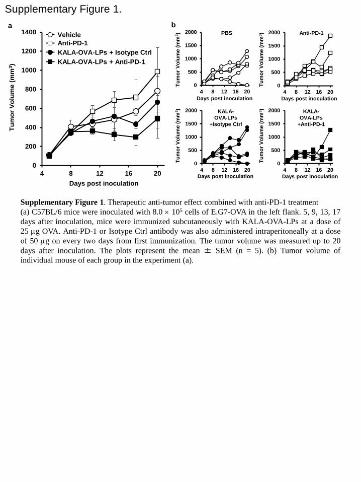
Supplemental Figure 1. Therapeutic anti-tumor effect combined with anti-PD-1 antibody
treatment. C57BL/6 mice were inoculated in the left flank with 8.0 × 10⁵ cells of E.G7-OVA.
At 5, 9, 13, 17 days after inoculation, the mice were immunized subcutaneously with
KALA-OVA-LPs at a dose of 25 µg OVA. An anti-PD-1 or a Isotype Ctrl antibody was also
administered intraperitoneally at a dose of 50 µg at two day intervals after the first immunization.
Tumor volume was measured up to 20 days after inoculation. The plots represent the mean ±
SEM (n = 5). Tumor volume of an individual mouse of each group in the experiment.
Supplemental Figure 2. CD80/86 expression in BMDCs that were treated with liposomes.
BMDCs (1.0 × 10⁶ cells) were treated with the KALA-OVA-LPs, the R8-OVA-LPs or the
non-modified OVA-LPs at a lipid dose of 32 µM. After 18 hours, the BMDCs were recovered
and stained by PE-labeled anti-mouse CD80 and CD86 (Biolegend).
Supplemental Figure 3. IL-6 concentration in serum after liposome injection. C57BL/6 mice
were subcutaneously administered KALA-OVA-LPs, R8-OVA-LPs at a dose of 25 µg OVA.
48
Blood from these mice was collected at 1, 3, 6, 12 and 24 hours after administration. Serum was
separated from blood and the IL-6 concentration was measured by ELISA.
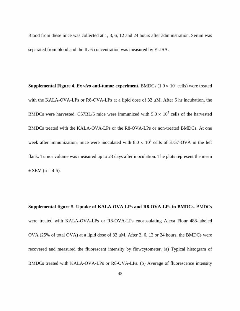
Supplemental Figure 4. Ex vivo anti-tumor experiment. BMDCs (1.0 × 10⁶ cells) were treated
with the KALA-OVA-LPs or R8-OVA-LPs at a lipid dose of 32 µM. After 6 hr incubation, the
BMDCs were harvested. C57BL/6 mice were immunized with 5.0 × 10⁵ cells of the harvested
BMDCs treated with the KALA-OVA-LPs or the R8-OVA-LPs or non-treated BMDCs. At one
week after immunization, mice were inoculated with 8.0 × 10⁵ cells of E.G7-OVA in the left
flank. Tumor volume was measured up to 23 days after inoculation. The plots represent the mean
± SEM (n = 4-5).
Supplemental figure 5. Uptake of KALA-OVA-LPs and R8-OVA-LPs in BMDCs. BMDCs
were treated with KALA-OVA-LPs or R8-OVA-LPs encapsulating Alexa Flour 488-labeled
OVA (25% of total OVA) at a lipid dose of 32 μM. After 2, 6, 12 or 24 hours, the BMDCs were
recovered and measured the fluorescent intensity by flowcytometer. (a) Typical histogram of
BMDCs treated with KALA-OVA-LPs or R8-OVA-LPs. (b) Average of fluorescence intensity

49
(Geo mean, left) and coefficient variance (CV) value (right). Data are mean ± SD (n=3).
Statistical analyses were performed by Student’s t-test. **P < 0.01.
Supplemental figure 6. Lymph node accumulation of the KALA-OVA-LPs and the
R8-OVA-LPs. C57BL/6 mice were subcutaneously administered KALA-OVA-LPs,
R8-OVA-LP modified with 0.1 mol% DiD at a dose of 25 µg OVA in the both flanks. After 24
hours, draining lymph nodes (inguinal lymph nodes) were isolated and homogenized. A nylon
mesh-filtered cell suspension was analyzed by flow cytometry for the uptake of the
fluorescently-labeled liposomes and the expression of F4/80 and CD11c. Data are the mean + SD
(n=3). Statistical analyses were performed by the Student’s t-test. N.S.: Not significant.

50
Liposome Size (nm) ζ-potential (mV)
OVA-Lp (DOPE) 158 ± 2 −19 ± 1
OVA-Lp (EPC) 162 ± 1 −37 ± 11
KALA-OVA-Lp (DOPE) 169 ± 4 50 ± 1
KALA-OVA-Lp (EPC) 185 ± 32 30 ± 1
R8-OVA-Lp (DOPE) 178 ± 4 52 ± 1
R8-OVA-Lp (EPC) 169 ± 2 52 ± 4
Table 1. Physicochemical properties of the various OVA-LPs.
Data were represented as the mean ± SEM value of at least three independent experiments
(n=3-15)

Figure 1.
0
20
40
60
80
100
120
3.1 13 25 50 50 50 50 50
CTL
act
ivity
(%
Lys
is)
50 50
DOPE-OVA-Lp EPC-OVA-Lp
OVA pro- tein
KALA R8 NM R8 NM KA LA
OVA (μg)

Figure 2.
0
1000
2000
3000
4000
5000
6000
5 15 25
Tum
or V
olum
e (m
m3 )
PBS OVA-Lp KALA-Lp R8-OVA-Lp KALA-OVA-Lp
Days post inoculation
*
0
2000
4000
6000
8000a b
Tum
or V
olum
e (m
m3 )

Figure 3.
0
4
8
12
16
1 4 16 64 256
Abso
rban
ce (A
bs59
5nm
)
Lipid concentration (μM)
**
**
**
b
a A
bsor
banc
e (A
bs59
5nm
)
0
1
2
3
4
5
6
1 3 5 7 9 5 5 mol%
KALA KA LA R8
DOPE EPC
peptide
lipid
**
**
** **
N.S. N.S.
: R8-OVA-Lp : KALA-OVA-Lp

Figure 4.
0
5
10
15
20
25
30
35KALA-OVA-Lp
pH 5.5
pH 6.5
pH 7.4
0
5
10
15
20
25
30
35R8-OVA-Lp
pH 5.5
pH 6.5
pH 7.4
Hem
olys
is a
ctiv
ity
Hem
olys
is a
ctiv
ity
Lipid concentration (μM) 2 4 8 16
Lipid concentration (μM) 2 4 8 16
a

Figure 5.
0
1
2
3
4
5Ab
sorb
ance
(Abs
595n
m)
Chloroquine concentration (μM)
: R8-OVA-Lp : KALA-OVA-Lp
0 25 50

Figure 6.
0
20
40
60
80
100
120
CTL
act
ivity
(%
Lys
is)
Isotype control
Anti- CD4
Anti- CD8
**
N.S.
KALA
0.0
0.5
1.0
1.5
2.0
2.5
IFNγ+
cel
ls/to
tal C
D8+
T c
ell (
%)
OVA257-264/IFNγ a b
0.0
0.4
0.8
1.2
1.6
IFNγ+
cel
ls/to
tal C
D4+
T c
ell (
%)
0.0
1.0
2.0
3.0
4.0
5.0
IL-4
+ cel
ls/to
tal C
D4+
T c
ell (
%)
0.00
0.02
0.04
0.06
0.08
0.10
IL-1
7+ c
ells
/tota
l CD
4+ T
cel
l (%
)
c OVA323-339/IFNγ OVA323-339/IL-4 OVA323-339/IL-17
R8 Non- treat
**
*
Table 1
Liposome Size (nm) ζ-potential (mV)
OVA-Lp (DOPE) 158 ± 2 −19 ± 1
OVA-Lp (EPC) 162 ± 1 −37 ± 11
KALA-OVA-Lp (DOPE) 169 ± 4 50 ± 1
KALA-OVA-Lp (EPC) 185 ± 32 30 ± 1
R8-OVA-Lp (DOPE) 178 ± 4 52 ± 1
R8-OVA-Lp (EPC) 169 ± 2 52 ± 4
0
200
400
600
800
1000
1200
1400
4 8 12 16 20
Supplementary Figure 1. Tu
mor
Vol
ume
(mm
3 )
Days post inoculation
Vehicle Anti-PD-1 KALA-OVA-LPs + Isotype Ctrl KALA-OVA-LPs + Anti-PD-1
Supplementary Figure 1. Therapeutic anti-tumor effect combined with anti-PD-1 treatment (a) C57BL/6 mice were inoculated with 8.0 × 10⁵ cells of E.G7-OVA in the left flank. 5, 9, 13, 17 days after inoculation, mice were immunized subcutaneously with KALA-OVA-LPs at a dose of 25 µg OVA. Anti-PD-1 or Isotype Ctrl antibody was also administered intraperitoneally at a dose of 50 µg on every two days from first immunization. The tumor volume was measured up to 20 days after inoculation. The plots represent the mean ± SEM (n = 5). (b) Tumor volume of individual mouse of each group in the experiment (a).
0
500
1000
1500
2000
4 8 12 16 20
Tum
or V
olum
e (m
m3 ) PBS
0
500
1000
1500
2000
4 8 12 16 20
0
500
1000
1500
2000
4 8 12 16 200
500
1000
1500
2000
4 8 12 16 20
Days post inoculation
Days post inoculation
Days post inoculation Days post inoculation
Tum
or V
olum
e (m
m3 )
Tum
or V
olum
e (m
m3 )
Tum
or V
olum
e (m
m3 )
Anti-PD-1
KALA- OVA-LPs
+Isotype Ctrl
KALA- OVA-LPs
+Anti-PD-1
a b

Supplementary Figure 2.
80
70
60 50
40
30
20
10
0 100 101 102 103 104
Cou
nt
Fluorescent intensity
CD80 80
70
60 50
40
30
20
10
0 100 101 102 103 104
Cou
nt
Fluorescent intensity
CD86
: OVA-LPs
: R8-OVA-LPs
: Non treatment
: Isotype control
: KALA-OVA-LPs
Supplementary Figure 2. CD80/86 expression in BMDCs that were treated with liposomes. BMDCs (1.0 × 10⁶ cells) were treated with the KALA-OVA-LPs, the R8-OVA-LPs or the non-modified OVA-LPs at a lipid dose of 32 µM. After 18 hours, the BMDCs were recovered and stained by PE-labeled anti-mouse CD80 and CD86 (Biolegend).

Supplementary Figure 3.
0
20
40
60
80
100
120
140
160
0 5 10 15 20 25
IL-6
con
cent
ratio
n (p
g / m
L)
Time after injection (hr)
Vehicle R8-OVA-LPs KALA-OVA-LPs
Supplementary Figure 3. IL-6 concentration in serum after liposome injection. C57BL/6 mice were administered subcutaneously with KALA-OVA-LPs, R8-OVA-LPs at a dose of 25 µg OVA. Blood of these mice was collected at 1, 3, 6, 12 and 24 hrs after administration. Serum was separated from blood and the IL-6 concentration was measured by ELISA.

Supplementary Figure 4.
0
500
1000
1500
2000
2500
3000
3500
5 10 15 20 25
Tum
or V
olum
e (m
m3 )
Days post inoculation
PBS Non-treated DC R8-OVA-Lp-treated DC KALA-OVA-Lp-treated DC
Supplementary Figure 4. Ex vivo anti-tumor experiment. BMDCs (1.0 × 10⁶ cells) were treated with the KALA-OVA-LPs or R8-OVA-LPs at a lipid dose of 32 µMd. After 6 hr incubation, the BMDCs were harvested. C57BL/6 mice were immunized with 5.0 × 10⁵ cells of the harvested BMDCs treated with the KALA-OVA-LPs or the R8-OVA-LPs or non-treated BMDCs. At one week after immunization, mice were inoculated with 8.0 × 10⁵ cells of E.G7-OVA in the left flank. Tumor volume was measured up to 23 days after inoculation. The plots represent the mean ± SEM (n = 4-5).

Supplementary figure 5.
0
50
100
150
0 10 20 30100
120
140
160
180
200
0 10 20 30
Cel
lula
r upt
ake
(Geo
Mea
n)
CV
valu
e
Time (hours) Time (hours)
: R8-OVA-Lp
: KALA-OVA-Lp
**
** **
**
**
80
70
60 50
40
30
20
10
0 100 101 102 103 104
Cou
nt
Fluorescent intensity
6 hours 80
70
60 50
40
30
20
10
0 100 101 102 103 104
Cou
nt
Fluorescent intensity
24 hours
: R8-OVA-Lp
: KALA-OVA-Lp
: Non treatment
a
b c
Supplementary figure 5. Uptake of KALA-OVA-LPs and R8-OVA-LPs in BMDCs. BMDCs were treated with KALA-OVA-LPs or R8-OVA-LPs encapsulating Alexa Flour 488-labeled OVA (25% of total OVA) at a lipid dose of 32 μM. After 2, 6, 12 or 24 hours, the BMDCs were recovered and measured the fluorescent intensity by flowcytometer. (a) Typical histogram of BMDCs treated with KALA-OVA-LPs or R8-OVA-LPs. (b) Average of fluorescence intensity (Geo mean, left) and coefficient variance (CV) value (right). Data are mean ± SD (n=3). Statistical analyses were performed by Student’s t-test. **P < 0.01.

Supplementary figure 6.
DiD+
F4/80+CD11c‒
F4/80‒CD11c‒
F4/80+CD11c+
F4/80‒CD11c+
DiD
+ cel
ls in
tota
l cel
ls (%
)
F4/8
0‒C
D11
c‒ c
ells
in
DiD
+ cel
ls(%
) F4
/80+
CD
11c‒
cel
ls
in D
iD+ c
ells
(%)
F4/8
0‒C
D11
c+ c
ells
in
DiD
+ cel
ls(%
) F4
/80+
CD
11c+
cel
ls
in D
iD+ c
ells
(%)
N.S.
N.S. N.S.
N.S. N.S.
Supplementary figure 6. Lymph node accumulation of KALA-OVA-LPs and R8-OBA-LPs C57BL/6 mice were administered subcutaneously with KALA-OVA-LPs, R8-OVA-LP modified with 0.1 mol% DiD at a dose of 25 µg OVA in the both side flanks. After 24 hours, the draining lymph nodes (inguinal lymph nodes) were isolated and mashed. Nylon mesh-filtered cell suspension were analyzed by flow cytometry for uptake of the fluorescently-labeled liposomes and expression of F4/80 and CD11c. Data are mean + SEM (n=3). Statistical analyses were performed by Student’s t-test. N.S.: Not significant.
a b
0.0
0.5
1.0
1.5
2.0
2.5
3.0
R8 KALA
0
20
40
60
80
100
R8 KALA0
2
4
6
8
10
12
R8 KALA
0
10
20
30
40
R8 KALA0.0
1.0
2.0
3.0
4.0
5.0
R8 KALA
![Three decades of messenger RNA vaccine development · information to antigen-presenting cells (APCs) [3, 4]. As pioneers, Martinon et al. showed that liposomes containing mRNA encoding](https://static.fdocuments.in/doc/165x107/60b8282dbb75f712773ecb5d/three-decades-of-messenger-rna-vaccine-development-information-to-antigen-presenting.jpg)