Modifications in cardiorenal sydrome Clinical approach to ...
26
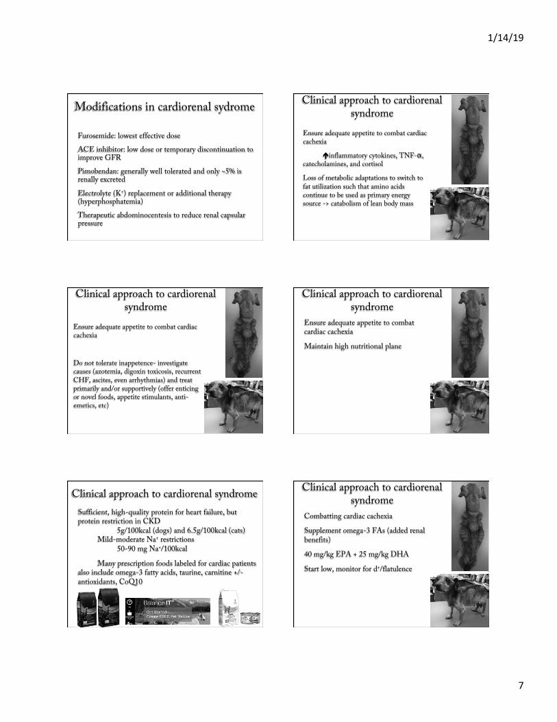
1/14/19 7 Modifications in cardiorenal sydrome Furosemide: lowest effective dose ACE inhibitor: low dose or temporary discontinuation to improve GFR Pimobendan: generally well tolerated and only ~5% is renally excreted Electrolyte (K + ) replacement or additional therapy (hyperphosphatemia) Therapeutic abdominocentesis to reduce renal capsular pressure Clinical approach to cardiorenal syndrome Ensure adequate appetite to combat cardiac cachexia éinflammatory cytokines, TNF-α, catecholamines, and cortisol Loss of metabolic adaptations to switch to fat utilization such that amino acids continue to be used as primary energy source -> catabolism of lean body mass Clinical approach to cardiorenal syndrome Ensure adequate appetite to combat cardiac cachexia Do not tolerate inappetence- investigate causes (azotemia, digoxin toxicosis, recurrent CHF, ascites, even arrhythmias) and treat primarily and/or supportively (offer enticing or novel foods, appetite stimulants, anti- emetics, etc) Clinical approach to cardiorenal syndrome Ensure adequate appetite to combat cardiac cachexia Maintain high nutritional plane Clinical approach to cardiorenal syndrome Sufficient, high-quality protein for heart failure, but protein restriction in CKD 5g/100kcal (dogs) and 6.5g/100kcal (cats) Mild-moderate Na + restrictions 50-90 mg Na + /100kcal Many prescription foods labeled for cardiac patients also include omega-3 fatty acids, taurine, carnitine +/- antioxidants, CoQ10 Clinical approach to cardiorenal syndrome Combatting cardiac cachexia Supplement omega-3 FAs (added renal benefits) 40 mg/kg EPA + 25 mg/kg DHA Start low, monitor for d + /flatulence
Transcript of Modifications in cardiorenal sydrome Clinical approach to ...
1/14/19
7
Modifications in cardiorenal sydrome
Furosemide: lowest effective dose
ACE inhibitor: low dose or temporary discontinuation to improve GFR
Pimobendan: generally well tolerated and only ~5% is renally excreted
Electrolyte (K+) replacement or additional therapy (hyperphosphatemia)
Therapeutic abdominocentesis to reduce renal capsular pressure
Clinical approach to cardiorenal syndrome
Ensure adequate appetite to combat cardiac cachexia
éinflammatory cytokines, TNF-α, catecholamines, and cortisol
Loss of metabolic adaptations to switch to fat utilization such that amino acids continue to be used as primary energy source -> catabolism of lean body mass
Clinical approach to cardiorenal syndrome
Ensure adequate appetite to combat cardiac cachexia
Do not tolerate inappetence- investigate causes (azotemia, digoxin toxicosis, recurrent CHF, ascites, even arrhythmias) and treat primarily and/or supportively (offer enticing or novel foods, appetite stimulants, anti-emetics, etc)
Clinical approach to cardiorenal syndrome
Ensure adequate appetite to combat cardiac cachexia
Maintain high nutritional plane
Clinical approach to cardiorenal syndrome Sufficient, high-quality protein for heart failure, but protein restriction in CKD
5g/100kcal (dogs) and 6.5g/100kcal (cats) Mild-moderate Na+ restrictions 50-90 mg Na+/100kcal
Many prescription foods labeled for cardiac patients also include omega-3 fatty acids, taurine, carnitine +/- antioxidants, CoQ10
Clinical approach to cardiorenal syndrome
Combatting cardiac cachexia
Supplement omega-3 FAs (added renal benefits)
40 mg/kg EPA + 25 mg/kg DHA
Start low, monitor for d+/flatulence
1/14/19
8
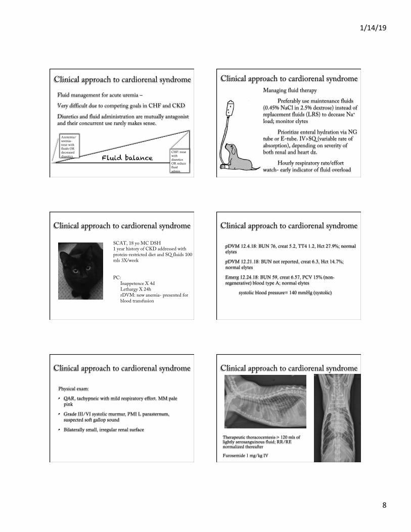
Fluid management for acute uremia –
Very difficult due to competing goals in CHF and CKD
Diuretics and fluid administration are mutually antagonist and their concurrent use rarely makes sense.
Clinical approach to cardiorenal syndrome
Fluid balance
Azotemia/uremia– treat with fluids OR decreased diuretics
CHF: treat with diuretics OR reduce fluid admin.
Managing fluid therapy
Preferably use maintenance fluids (0.45% NaCl in 2.5% dextrose) instead of replacement fluids (LRS) to decease Na+ load; monitor elytes
Prioritize enteral hydration via NG tube or E-tube. IV>SQ (variable rate of absorption), depending on severity of both renal and heart dz.
Hourly respiratory rate/effort watch- early indicator of fluid overload
Clinical approach to cardiorenal syndrome
SCAT, 18 yo MC DSH 1 year history of CKD addressed with protein-restricted diet and SQ fluids 100 mls 3X/week PC:
Inappetence X 4d Lethargy X 24h rDVM: new anemia- presented for blood transfusion
Clinical approach to cardiorenal syndrome Clinical approach to cardiorenal syndrome
pDVM 12.4.18: BUN 76, creat 5.2, TT4 1.2, Hct 27.9%; normal elytes
pDVM 12.21.18: BUN not reported, creat 6.3, Hct 14.7%; normal elytes
Emerg 12.24.18: BUN 59, creat 6.57, PCV 15% (non-regenerative) blood type A; normal elytes
systolic blood pressure= 140 mmHg (systolic)
Clinical approach to cardiorenal syndrome
Physical exam:
• QAR, tachypneic with mild respiratory effort. MM pale pink
• Grade III/VI systolic murmur, PMI L parasternum, suspected soft gallop sound
• Bilaterally small, irregular renal surface
Clinical approach to cardiorenal syndrome
Therapeutic thoracocentesis-> 120 mls of lightly serosanguinous fluid; RR/RE normalized thereafter
Furosemide 1 mg/kg IV
1/14/19
9
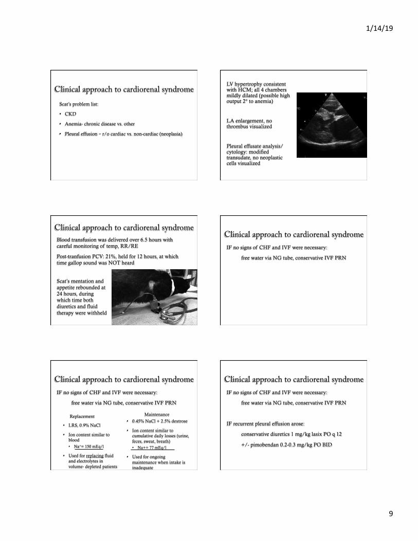
Scat’s problem list:
• CKD
• Anemia- chronic disease vs. other
• Pleural effusion – r/o cardiac vs. non-cardiac (neoplasia)
Clinical approach to cardiorenal syndrome LV hypertrophy consistent with HCM; all 4 chambers mildly dilated (possible high output 2° to anemia)
LA enlargement, no thrombus visualized
Pleural effusate analysis/cytology: modified transudate, no neoplastic cells visualized
Blood transfusion was delivered over 6.5 hours with careful monitoring of temp, RR/RE
Post-tranfusion PCV: 21%, held for 12 hours, at which time gallop sound was NOT heard
Clinical approach to cardiorenal syndrome
Scat’s mentation and appetite rebounded at 24 hours, during which time both diuretics and fluid therapy were withheld
IF no signs of CHF and IVF were necessary:
free water via NG tube, conservative IVF PRN
Clinical approach to cardiorenal syndrome
IF no signs of CHF and IVF were necessary:
free water via NG tube, conservative IVF PRN
Clinical approach to cardiorenal syndrome
Replacement Maintenance
• LRS, 0.9% NaCl
• Ion content similar to blood • Na+= 150 mEq/l
• Used for replacing fluid and electrolytes in volume- depleted patients
• 0.45% NaCl + 2.5% dextrose
• Ion content similar to cumulative daily losses (urine, feces, sweat, breath) • Na+= 77 mEq/l
• Used for ongoing maintenance when intake is inadequate
IF no signs of CHF and IVF were necessary:
free water via NG tube, conservative IVF PRN
IF recurrent pleural effusion arose:
conservative diuretics 1 mg/kg lasix PO q 12
+/- pimobendan 0.2-0.3 mg/kg PO BID
Clinical approach to cardiorenal syndrome
1/14/19
10
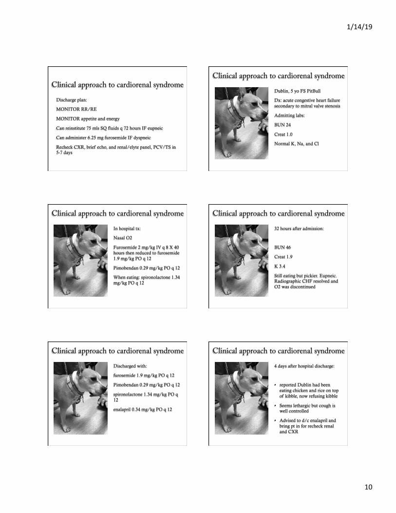
Discharge plan:
MONITOR RR/RE
MONITOR appetite and energy
Can reinstitute 75 mls SQ fluids q 72 hours IF eupneic
Can administer 6.25 mg furosemide IF dyspneic
Recheck CXR, brief echo, and renal/elyte panel, PCV/TS in 5-7 days
Clinical approach to cardiorenal syndrome Clinical approach to cardiorenal syndrome
Dublin, 5 yo FS PitBull
Dx: acute congestive heart failure secondary to mitral valve stenosis
Admitting labs:
BUN 24
Creat 1.0
Normal K, Na, and Cl
Clinical approach to cardiorenal syndrome
In hospital tx:
Nasal O2
Furosemide 2 mg/kg IV q 8 X 40 hours then reduced to furosemide 1.9 mg/kg PO q 12
Pimobendan 0.29 mg/kg PO q 12
When eating: spironolactone 1.34 mg/kg PO q 12
Clinical approach to cardiorenal syndrome
32 hours after admission:
BUN 46
Creat 1.9
K 3.4
Still eating but pickier. Eupneic. Radiographic CHF resolved and O2 was discontinued
Clinical approach to cardiorenal syndrome
Discharged with:
furosemide 1.9 mg/kg PO q 12
Pimobendan 0.29 mg/kg PO q 12
spironolactone 1.34 mg/kg PO q 12
enalapril 0.34 mg/kg PO q 12
Clinical approach to cardiorenal syndrome
4 days after hospital discharge:
• reported Dublin had been eating chicken and rice on top of kibble, now refusing kibble
• Seems lethargic but cough is well controlled
• Advised to d/c enalapril and bring pt in for recheck renal and CXR
1/14/19
11
Clinical approach to cardiorenal syndrome
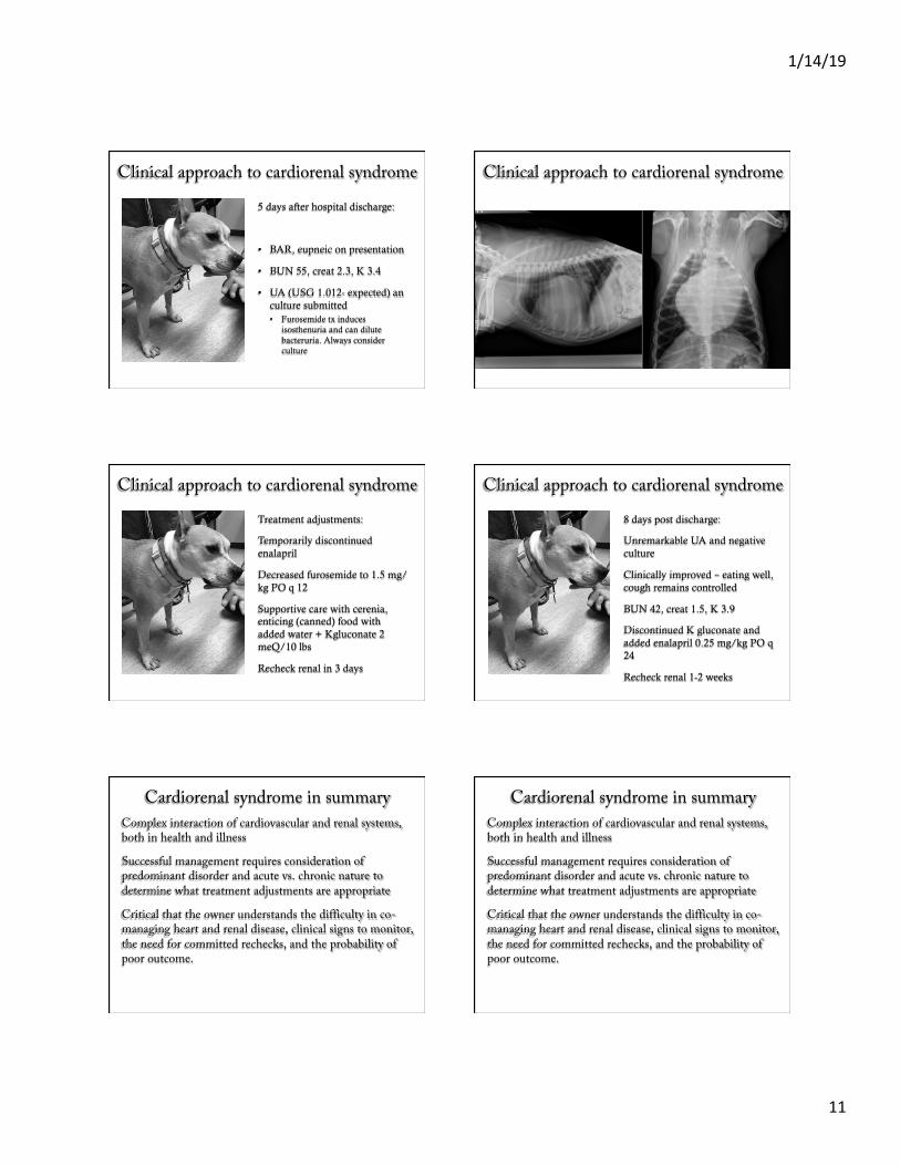
5 days after hospital discharge:
• BAR, eupneic on presentation
• BUN 55, creat 2.3, K 3.4
• UA (USG 1.012- expected) an culture submitted • Furosemide tx induces
isosthenuria and can dilute bacteruria. Always consider culture
Clinical approach to cardiorenal syndrome
Clinical approach to cardiorenal syndrome
Treatment adjustments:
Temporarily discontinued enalapril
Decreased furosemide to 1.5 mg/kg PO q 12
Supportive care with cerenia, enticing (canned) food with added water + Kgluconate 2 meQ/10 lbs
Recheck renal in 3 days
Clinical approach to cardiorenal syndrome
8 days post discharge:
Unremarkable UA and negative culture
Clinically improved – eating well, cough remains controlled
BUN 42, creat 1.5, K 3.9
Discontinued K gluconate and added enalapril 0.25 mg/kg PO q 24
Recheck renal 1-2 weeks
Complex interaction of cardiovascular and renal systems, both in health and illness
Successful management requires consideration of predominant disorder and acute vs. chronic nature to determine what treatment adjustments are appropriate
Critical that the owner understands the difficulty in co-managing heart and renal disease, clinical signs to monitor, the need for committed rechecks, and the probability of poor outcome.
Cardiorenal syndrome in summary Complex interaction of cardiovascular and renal systems, both in health and illness
Successful management requires consideration of predominant disorder and acute vs. chronic nature to determine what treatment adjustments are appropriate
Critical that the owner understands the difficulty in co-managing heart and renal disease, clinical signs to monitor, the need for committed rechecks, and the probability of poor outcome.
Cardiorenal syndrome in summary
1/14/19
12
Complex interaction of cardiovascular and renal systems, both in health and illness
Successful management requires consideration of predominant disorder and acute vs. chronic nature to determine what treatment adjustments are appropriate
Critical that the owner understands the difficulty in co-managing heart and renal disease, clinical signs to monitor, the need for committed rechecks, and the probability of poor outcome.
Cardiorenal syndrome in summary Complex interaction of cardiovascular and renal systems, both in health and illness
Successful management requires consideration of predominant disorder and acute vs. chronic nature to determine what treatment adjustments are appropriate
Critical that the owner understands the difficulty in co-managing heart and renal disease, clinical signs to monitor, the need for committed rechecks, and the probability of poor outcome.
Cardiorenal syndrome in summary
QUESTIONS?
Thank you for your time
1/12/19
1
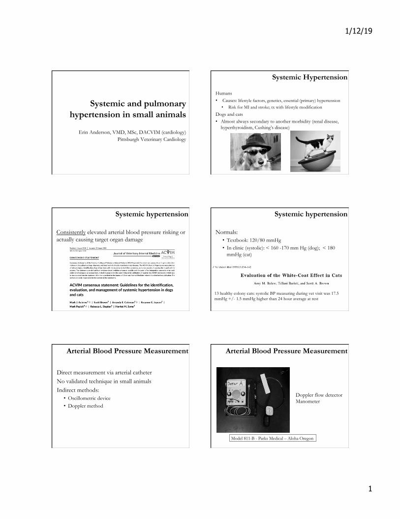
Systemic and pulmonary hypertension in small animals
Erin Anderson, VMD, MSc, DACVIM (cardiology) Pittsburgh Veterinary Cardiology
Systemic Hypertension
Humans • Causes: lifestyle factors, genetics, essential (primary) hypertension
• Risk for MI and stroke; tx with lifestyle modification
Dogs and cats • Almost always secondary to another morbidity (renal disease,
hyperthyroidism, Cushing’s disease)
Systemic hypertension
Consistently elevated arterial blood pressure risking or actually causing target organ damage
Systemic hypertension
Normals: • Textbook: 120/80 mmHg • In clinic (systolic): < 160 -170 mm Hg (dog); < 180
mmHg (cat)
13 healthy colony cats: systolic BP measuring during vet visit was 17.5 mmHg +/- 1.5 mmHg higher than 24 hour average at rest
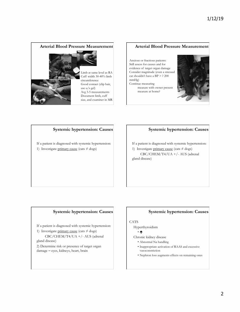
Direct measurement via arterial catheter No validated technique in small animals Indirect methods:
• Oscillometric device • Doppler method
Arterial Blood Pressure Measurement Arterial Blood Pressure Measurement
Doppler flow detector Manometer
Model 811-B - Parks Medical – Aloha Oregon
1/12/19
2
Arterial Blood Pressure Measurement
Limb at same level as RA Cuff width 30-40% limb circumference Good contact (clip hair, use u/s gel) Avg 3-5 measurements Document limb, cuff size, and examiner in MR
Arterial Blood Pressure Measurement
Anxious or fractious patients: Still assess for causes and for evidence of target organ damage Consider magnitude (even a stressed cat shouldn’t have a BP > ? 200 mmHg) Continue measuring
measure with owner present measure at home?
Systemic hypertension: Causes
If a patient is diagnosed with systemic hypertension: 1) Investigate primary cause (cats ≠ dogs)
Systemic hypertension: Causes
If a patient is diagnosed with systemic hypertension: 1) Investigate primary cause (cats ≠ dogs)
CBC/CHEM/T4/UA +/- AUS (adrenal gland disease)
Systemic hypertension: Causes
If a patient is diagnosed with systemic hypertension: 1) Investigate primary cause (cats ≠ dogs)
CBC/CHEM/T4/UA +/- AUS (adrenal gland disease) 2) Determine risk or presence of target organ damage – eyes, kidneys, heart, brain
Systemic hypertension: Causes
CATS Hyperthyroidism
• é CO
Chronic kidney disease • Abnormal Na handling • Inappropriate activation of RAAS and excessive
vasoconstriction • Nephron loss augments effects on remaining ones
1/12/19
3
DOGS Hyperadrenocorticism
• Cortisol effects (é sensitivity to endogenous vasoconstrictors)
Chronic kidney disease Diabetes mellitus Pheochromocytoma
• Intermittent exuberant catecholamine secretion
Systemic hypertension: Causes Manifestations of hypertension
Compensated: no overt clinical signs Decompensated
• Ocular: retinal hemorrhage, detachment, acute blindness • Cardiac: myocyte hypertrophy, aortic dissection (rare) • Renal: (proteinuriaà tubular damage, glomerulosclerosis) • CNS: (stroke- ischemic vs. hemorrhagic)
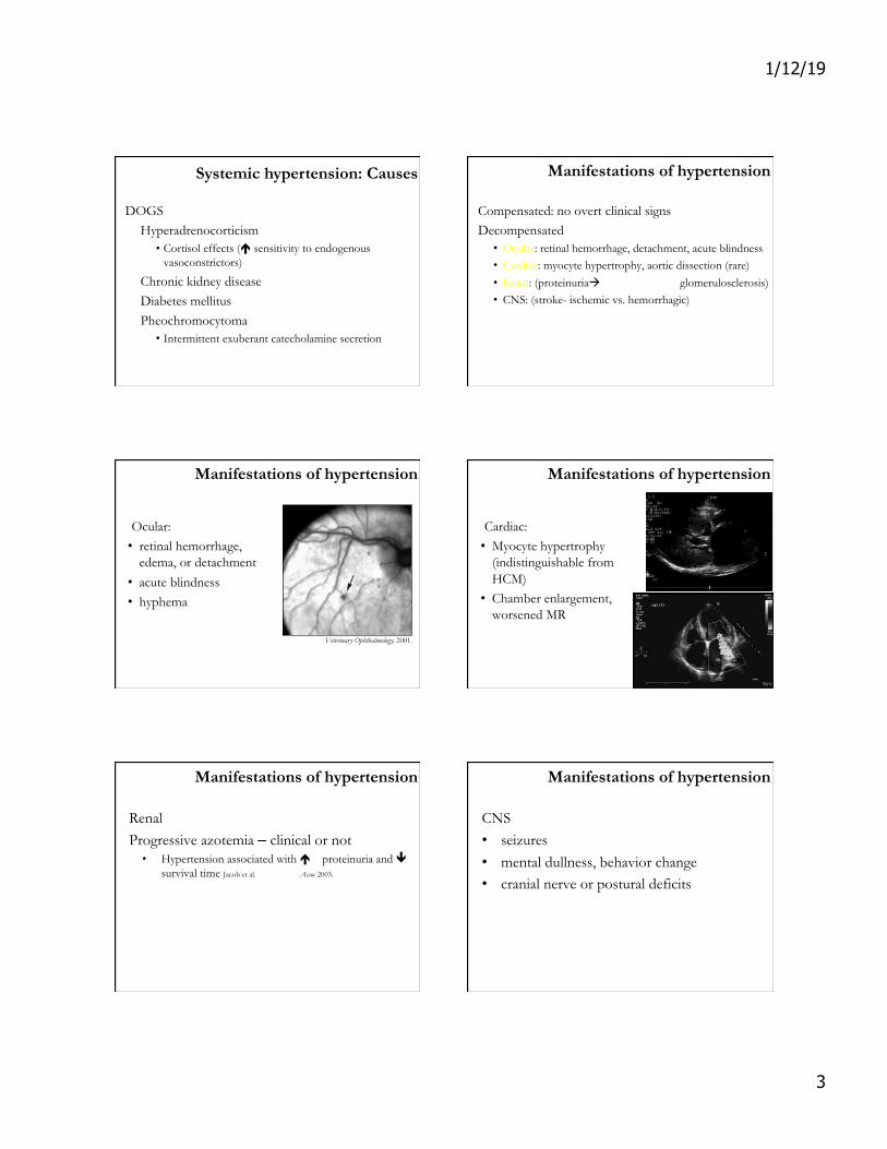
Manifestations of hypertension
Ocular: • retinal hemorrhage,
edema, or detachment • acute blindness • hyphema
Veterinary Ophthalmology, 2001.
Manifestations of hypertension
Cardiac: • Myocyte hypertrophy
(indistinguishable from HCM)
• Chamber enlargement, worsened MR
Renal Progressive azotemia – clinical or not
• Hypertension associated with é proteinuria and ê survival time Jacob et al. J Am Vet Med Assoc 2003.
Manifestations of hypertension
CNS • seizures • mental dullness, behavior change • cranial nerve or postural deficits
Manifestations of hypertension
1/12/19
4
REVIEW: Manifestations of hypertension
Target organ damageà
Treatment
Treat the underlying disease Consider primary antihypertensive tx if TOD is present or imminent
Targeted therapy
To control systemic hypertension, administer drugs that: ↓ SVR (Ca++ channel blockers) ↓ Cardiac output [via↓ SV or ↓ HR] (Ca++ channel blockers, ACEI, β-blockers, diuretics)
Treatment
Ca++ channel blocker (Amlodipine) ACE-inhibitors – evaluate renal function before and after Beta-blockers Sodium nitroprusside - emergency Diuretics- more commonly used in humans than small animals
Changes in dose or additional agents may be necessary for optimal
BP control.
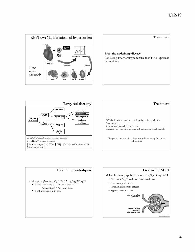
Amlodipine (Norvasc®) 0.05-0.2 mg/kg PO q 24 • Dihydropyridine Ca++ channel blocker
(vasculature>>>myocardium) • Highly efficacious in cats
Treatment: amlodipine Treatment: ACEI ACE-inhibitors (“-prils”): 0.25-0.5 mg/kg PO q 12-24
– Decreases AngII-mediated vasoconstriction – Decreases proteinuria – Potential antifibrotic effects – Typically adjunctive tx
http://www.gosh.nhs.uk
1/12/19
5
Treatment: other
Beta-blockers • ↓ CO and shows good efficacy in conjunction with
amlodipine • Renal elimination
Sodium nitroprusside • Potent vasodilator, short -lived effect, used most
commonly for emergent, severe CHF
Furosemide • ↓ CO (less effective; undesirable in CKD)
Hypertension: prognosis
Variable and dependent on: • Extent of TOD • Response to antihypertensive Rx • Ability to control underlying cause
REVIEW: systemic hypertension
§ Causes of systemic hypertension differ between cats and dogs.
§ Consequences can include ocular or CNS hemorrhage, renal effects, and myocardial hypertrophy.
§ Appropriate management requires identifying and treating underlying dz +/- anti-hypertensive agents.
How does pulmonary hypertension differ from systemic hypertension?
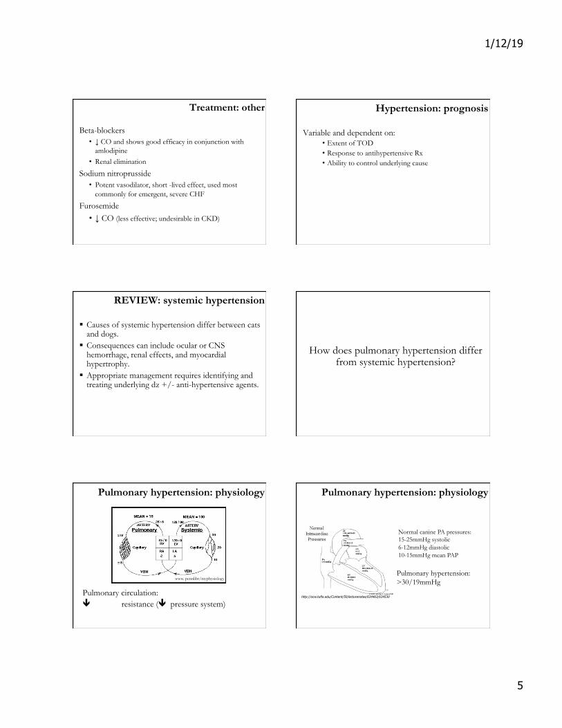
Pulmonary hypertension: physiology
Pulmonary circulation: ê vascular resistance (ê pressure system)
www. pennlibr/myphysiology
http://ocw.tufts.edu/Content/50/lecturenotes/634463/634530
Normal canine PA pressures: 15-25mmHg systolic 6-12mmHg diastolic 10-15mmHg mean PAP
Pulmonary hypertension: >30/19mmHg
Pulmonary hypertension: physiology

1/12/19
6
Pathogenesis of pulmonary hypertension
vasodilators vasoconstrictors
Endothelin (ET-b receptor)
Thromboxane A1
Serotonin
Angiotensin II
prostacyclin
O2
Endothelin (ET-a receptor)
Pathogenesis of pulmonary hypertension
vasodilators vasoconstrictors
Endothelin (ET-b receptor)
Thromboxane A1
Serotonin
Angiotensin II
prostacyclin
O2
Determinants of pulmonary arterial pressure (PAP)
MPAP = (PVR x PBF) + (PCWP)
PVR= pulmonary vascular resistance PBF= pulmonary blood flow PCWP= pulmonary capillary wedge pressure (between venules and arterioles; reflective of LA pressure)
Determinants of pulmonary arterial pressure (PAP)
MPAP = (PVR x PBF) + (PCWP)
Causes: Primary PAH (uncommon) Secondary to pulmonary dz (PTE, heartworm disease, lower airway disease) Secondary to L sided heart disease
MPAP = (PVR x PBF) + (PCWP)
(secondary to pulm dz) Interstitial lung disease Mechanical obstruction
PTE Heartworm disease
High altitudes
Determinants of pulmonary arterial pressure (PAP)
Determinants of pulmonary arterial pressure (PAP)
MPAP = (PVR x PBF) + (PCWP)
LàR shunting heart disease (PDA, VSD,
ASD)
1/12/19
7
MPAP = (PVR x PBF) + (PCWP)
L sided heart dz: Chronic valve dz DCM Congenital dz: mitral stenosis
Determinants of pulmonary arterial pressure (PAP)
Clinical presentation
Clinical signs reflect hypoxemia: • Exercise intolerance • Syncope • Cough, respiratory distress
(expiratory > inspiratory)
Clinical presentation
BAR-distressed Variable degrees of dyspnea
expiratory push +/- increased abdominal muscle tone +/- murmur (concurrent heart dz or TR) Variable HR – may be relatively low (RSA) if substantial resp dz
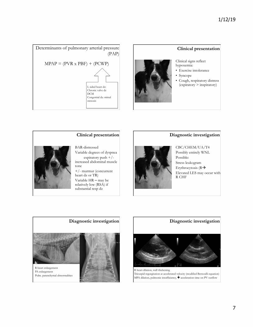
Diagnostic investigation
CBC/CHEM/UA/T4 Possibly entirely WNL Possible: Stress leukogram Erythrocytosis (RàL shunt) Elevated LES may occur with R CHF
Diagnostic investigation
R heart enlargement PA enlargement Pulm. parenchymal abnormalities
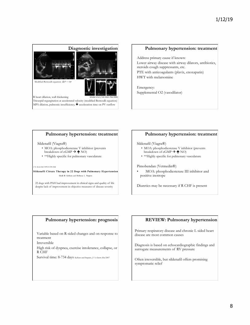
Diagnostic investigation
R heart dilation, wall thickening Tricuspid regurgitation at accelerated velocity (modified Bernoulli equation) MPA dilation, pulmonic insufficiency, ê acceleration time on PV outflow
1/12/19
8
Diagnostic investigation
R heart dilation, wall thickening Tricuspid regurgitation at accelerated velocity (modified Bernoulli equation) MPA dilation, pulmonic insufficiency, ê acceleration time on PV outflow
Schober et al (J Vet Intern Med 2006)
Modified Bernoulli equation: ΔP = 4ν2
Pulmonary hypertension: treatment
Address primary cause if known: Lower airway disease with airway dilators, antibiotics, steroids cough suppressants, etc. PTE with anticoagulants (plavix, enoxaparin) HWT with melarsomine Emergency: Supplemental O2 (vasodilator)
Pulmonary hypertension: treatment
Sildenafil (Viagra®) • MOA: phosphodiesterase V inhibitor (prevents
breakdown of cGMP à é NO) • **Highly specific for pulmonary vasculature
22 dogs with PAH had improvement in clinical signs and quality of life despite lack of improvement in objective measures of disease severity
Pulmonary hypertension: treatment
Sildenafil (Viagra®) • MOA: phosphodiesterase V inhibitor (prevents
breakdown of cGMP à é NO) • **Highly specific for pulmonary vasculature
Pimobendan (Vetmedin®) • MOA: phosphodiesterase III inhibitor and
positive inotrope
Diuretics may be necessary if R CHF is present
Pulmonary hypertension: prognosis
Variable based on R-sided changes and on response to treatment Irreversible High risk of dyspnea, exercise intolerance, collapse, or R CHF Survival time: 8-734 days Kellum and Stepien, J Vet Intern Med 2007
REVIEW: Pulmonary hypertension
Primary respiratory disease and chronic L sided heart disease are most common causes Diagnosis is based on echocardiographic findings and surrogate measurements of RV pressure Often irreversible, but sildenafil offers promising symptomatic relief
1/12/19
9
QUESTIONS? QUESTIONS?
The (word) games people play!
Bosun or boatswain (n.) member of a merchant ship who supervises the deck and the sailors
1/13/19
1
Erin Anderson, VMD, MSc, DACVIM (Cardiology)
� Morphologic and functional abnormalities of the heart and great vessels that are present at birth
� Congenital ≠ hereditary � Prevalence of 4.6-8.5/1000 clinical cases (Cote et al.
JVC 2015)
� Pediatric patients: nonpathologic (physiologic/functional) vs. pathologic murmurs
� Auscultation alone is not definitive for differentiation � Age of resolution of innocent murmurs?
• 8-13 weeks but dependent on many factors – size, breed- and some innocent murmurs never resolve
Characteristics more suggestive of pathologic murmurs: � Continuous or diastolic � R sided or PMI at the apex � Direct relative of animal with known
congenital heart dz +/- predisposed breed
� Murmur + jugular pulsation, pulse abnormalities, evidence of poor perfusion
� Pups: left basilar systolic murmurs • Physiologic
• Pulmonic stenosis
• Aortic/subaortic stenosis
P A
M
� Most common congenital defects: � PE
• audible heart murmur (If RIGHT sided, more suspicious for VSD or TR)
• +/- tachycardia
• +/- respiratory distress
• +/- abdominal distention (ascites) or pulse abnormalities
� Diagnostic evaluation • CXR: +/- cardiomegaly +/- CHF
• ECG: +/- arrhythmias or criteria of chamber enlargement
• BW: é NT-proBNP, hemoconcentration in R->L shunts
1/13/19
2
J Vet Cardiol 2015
• 5-12% of all cats presenting to a clinical cardiology service (~ 0.3% of all cats examined hospital-wide)
• Up to 21% of all dogs presenting to clinical cardiology service (~ 0.14-0.18% of all dogs examined)
Prevalence of congenital heart disease:
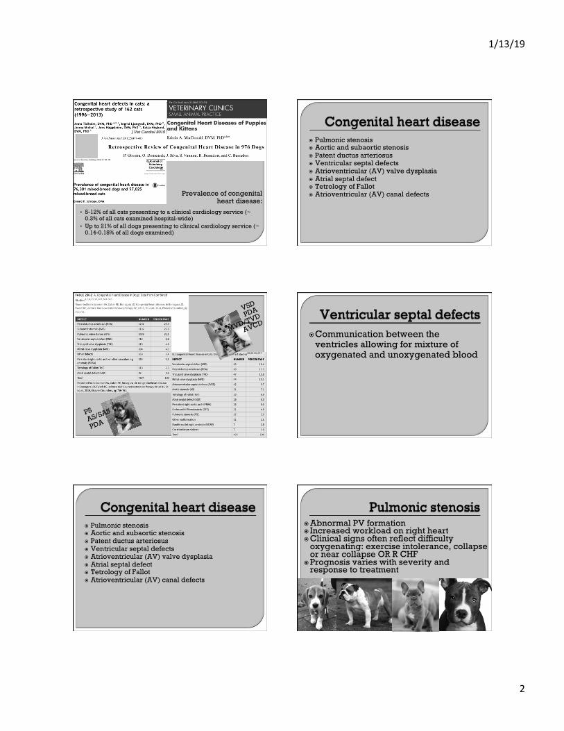
� Pulmonic stenosis � Aortic and subaortic stenosis � Patent ductus arteriosus � Ventricular septal defects � Atrioventricular (AV) valve dysplasia � Atrial septal defect � Tetrology of Fallot � Atrioventricular (AV) canal defects
PS
AS/SAS
PDA
VSD
PDA
MVD/TVD
AVCD
� Communication between the ventricles allowing for mixture of oxygenated and unoxygenated blood
� Pulmonic stenosis � Aortic and subaortic stenosis � Patent ductus arteriosus � Ventricular septal defects � Atrioventricular (AV) valve dysplasia � Atrial septal defect � Tetrology of Fallot � Atrioventricular (AV) canal defects
� Abnormal PV formation � Increased workload on right heart � Clinical signs often reflect difficulty
oxygenating: exercise intolerance, collapse or near collapse OR R CHF
� Prognosis varies with severity and response to treatment
1/13/19
3
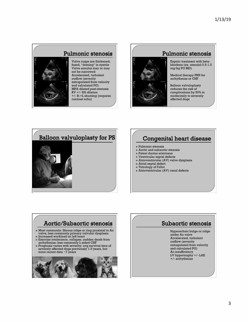
Valve cusps are thickened, fused, “doming” in systole Valve annulus may or may not be narrowed Accelerated, turbulent outflow (severity extrapolated from velocity and calculated PG) MPA dilated post-stenosis RV +/- RA dilation +/- R->L shunting (requires contrast echo)
Enpiric treatment with beta-blockers (ex. atenolol 0.5-1.5 mg/kg PO BID) Medical therapy PRN for arrhythmias or CHF Balloon valvuloplasty reduces the risk of complications by 50% in moderately to severely affected dogs
� Pulmonic stenosis � Aortic and subaortic stenosis � Patent ductus arteriosus � Ventricular septal defects � Atrioventricular (AV) valve dysplasia � Atrial septal defect � Tetrology of Fallot � Atrioventricular (AV) canal defects
� Most commonly: fibrous ridge or ring proximal to Ao valve; less commonly primary valvular dysplasia
� Increased workload on left heart � Exercise intolerance, collapse, sudden death from
arrhythmias; less commonly L sided CHF � Prognosis varies with severity; avg survival time of
severely affected dogs previously 1-3 years, but more recent data ~7 years
Hyperechoic bulge or ridge under Ao valve Accelerated, turbulent outflow (severity extrapolated from velocity and calculated PG) Ao insufficiency LV hypertrophy +/- LAE +/- arrhythmias
1/13/19
4
Enpiric treatment with beta-blockers (ex. atenolol 0.5-1.5 mg/kg PO BID) Medical therapy PRN for arrhythmias or CHF 2010: cutting and high pressure balloon valvuloplasty
Kleman et al JVC 2012
Cutting and high pressure balloon valvuloplasty: Immediate drop in LV-AO PG Long term results show tendency for PG to rise; survival benefit unclear
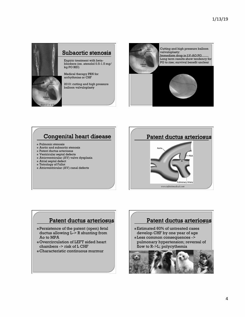
� Pulmonic stenosis � Aortic and subaortic stenosis � Patent ductus arteriosus � Ventricular septal defects � Atrioventricular (AV) valve dysplasia � Atrial septal defect � Tetrology of Fallot � Atrioventricular (AV) canal defects
www.infinitimedical.com
� Persistence of the patent (open) fetal ductus allowing L-> R shunting from Ao to MPA
� Overcirculation of LEFT sided heart chambers -> risk of L CHF
� Characteristic continuous murmur
� Estimated 60% of untreated cases develop CHF by one year of age
� Less common consequences -> pulmonary hypertension; reversal of flow to R->L; polycythemia
1/13/19
5
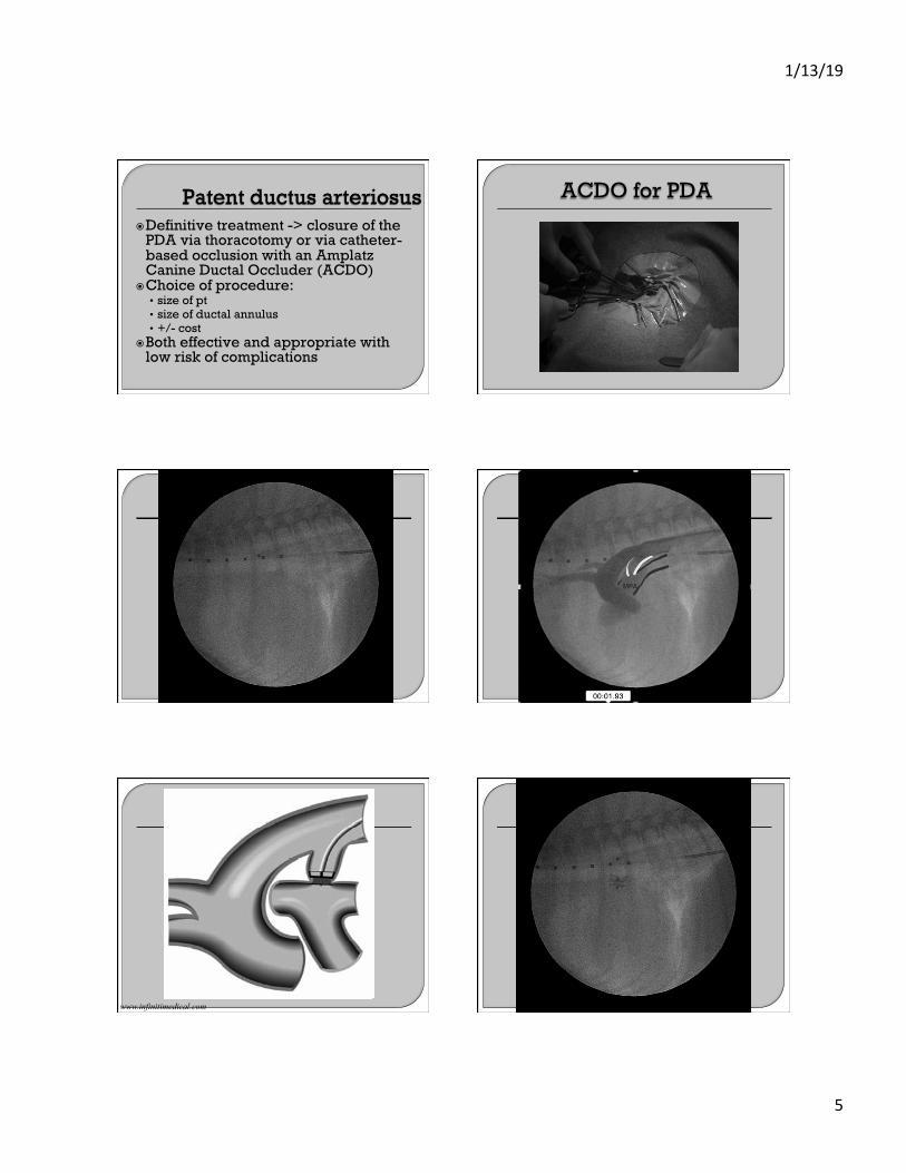
� Definitive treatment -> closure of the PDA via thoracotomy or via catheter-based occlusion with an Amplatz Canine Ductal Occluder (ACDO)
� Choice of procedure: • size of pt • size of ductal annulus • +/- cost
� Both effective and appropriate with low risk of complications
ACDO for PDA
www.infinitimedical.com
Find the continuous murmur!
www.infinitimedical.com
1/13/19
6
� Pulmonic stenosis � Aortic and subaortic stenosis � Patent ductus arteriosus � Ventricular septal defects � Atrioventricular (AV) valve dysplasia � Atrial septal defect � Tetrology of Fallot � Atrioventricular (AV) canal defects
Scansen et al. J Vet Cardiol 2015
Normal development: IVS forms from the apex up Named for affected component of IVS:
perimembranous (under AV and septal leaflet of TV) Juxtaarterial (at outlet portion under AV and PV) Muscular
Scansen et al. J Vet Cardiol 2015
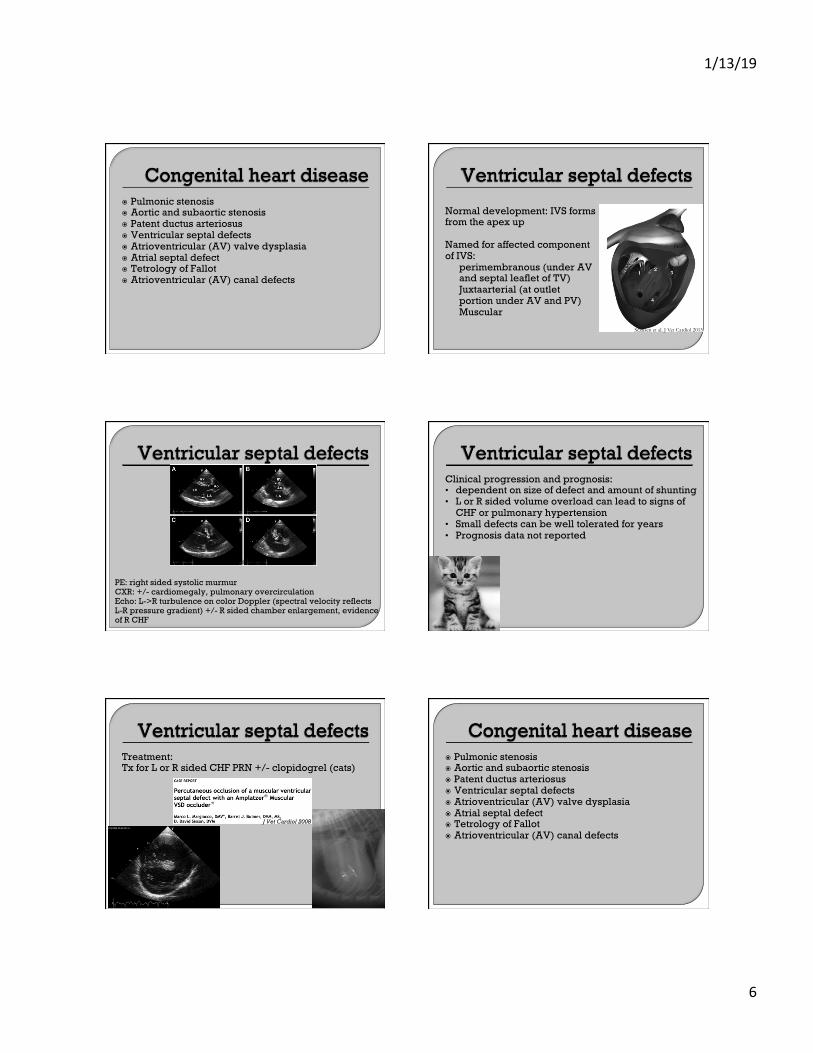
PE: right sided systolic murmur CXR: +/- cardiomegaly, pulmonary overcirculation Echo: L->R turbulence on color Doppler (spectral velocity reflects L-R pressure gradient) +/- R sided chamber enlargement, evidence of R CHF
Clinical progression and prognosis: • dependent on size of defect and amount of shunting • L or R sided volume overload can lead to signs of
CHF or pulmonary hypertension • Small defects can be well tolerated for years • Prognosis data not reported
Treatment: Tx for L or R sided CHF PRN +/- clopidogrel (cats)
J Vet Cardiol 2008
� Pulmonic stenosis � Aortic and subaortic stenosis � Patent ductus arteriosus � Ventricular septal defects � Atrioventricular (AV) valve dysplasia � Atrial septal defect � Tetrology of Fallot � Atrioventricular (AV) canal defects
1/13/19
7
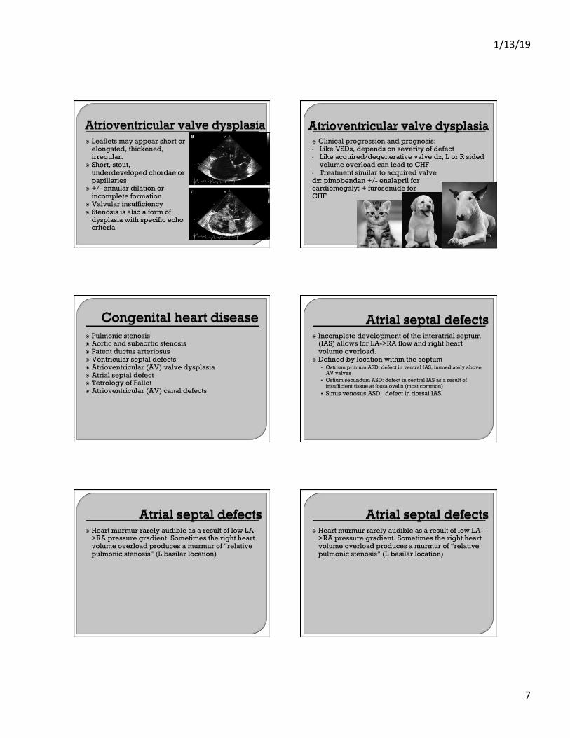
� Leaflets may appear short or elongated, thickened, irregular.
� Short, stout, underdeveloped chordae or papillaries
� +/- annular dilation or incomplete formation
� Valvular insufficiency � Stenosis is also a form of
dysplasia with specific echo criteria
Navarro-Cubas et al Open Vet J 2017
� Clinical progression and prognosis: • Like VSDs, depends on severity of defect • Like acquired/degenerative valve dz, L or R sided
volume overload can lead to CHF • Treatment similar to acquired valve dz: pimobendan +/- enalapril for cardiomegaly; + furosemide for CHF
� Pulmonic stenosis � Aortic and subaortic stenosis � Patent ductus arteriosus � Ventricular septal defects � Atrioventricular (AV) valve dysplasia � Atrial septal defect � Tetrology of Fallot � Atrioventricular (AV) canal defects
� Incomplete development of the interatrial septum (IAS) allows for LA->RA flow and right heart volume overload.
� Defined by location within the septum • Ostrium primum ASD: defect in ventral IAS, immediately above
AV valves
• Ostium secundum ASD: defect in central IAS as a result of insufficient tissue at fossa ovalis (most common)
• Sinus venosus ASD: defect in dorsal IAS.
� Heart murmur rarely audible as a result of low LA->RA pressure gradient. Sometimes the right heart volume overload produces a murmur of “relative pulmonic stenosis” (L basilar location)
� Heart murmur rarely audible as a result of low LA->RA pressure gradient. Sometimes the right heart volume overload produces a murmur of “relative pulmonic stenosis” (L basilar location)

1/13/19
8
Treatment: Medical therapy PRN for CHF +/- clopidogrel (cats)
Prognosis: varies with size of defect but generally good
prognosis. 73% of animals reported by Chetboul et al remained asymptomatic as adults
� Pulmonic stenosis � Aortic and subaortic stenosis � Patent ductus arteriosus � Ventricular septal defects � Atrioventricular (AV) valve dysplasia � Atrial septal defect � Tetrology of Fallot � Atrioventricular (AV) canal defects
� Pulmonic stenosis � Overriding aorta � Ventricular septal defect � Right ventricular hypertrophy
Result -> deoxygenated blood shunting into systemic circulation, the degree of which is dependent on severity of PS and size of VSD
Physical exam: Heart murmur over L apex +/- R side Cyanosis Clinical signs of hypoxemia likely: exercise intolerance, collapse, cyanosis, sudden death Diagnostic eval may reveal erythrocytosis, radiographic cardiomegaly +/- CHF
� Pulmonic stenosis � Aortic and subaortic stenosis � Patent ductus arteriosus � Ventricular septal defects � Atrioventricular (AV) valve dysplasia � Atrial septal defect � Tetrology of Fallot � Atrioventricular (AV) canal defects

1/13/19
9
Malformation of the AV septum, which normally connects atrial and ventricular septae thus appropriately separating the LV from the RA. A defect in this structure results in the MV and TV being on the same plane with variable AV communications1,17:
Partial AVSD: an atrial OR ventricular communication. Both AV valve annuli are isolated orifices.
Intermediate AVSD: both an atrial and ventricular communication are present but the mitral and tricuspid valve annuli remain as isolated orifices.
Complete AVSD: both an atrial and ventricular communication are present and one AV valve bridges both the LV and RV
Treatment: • Medical therapy PRN for CHF +/- clopidogrel
(cats) Progosis: • Dependent on type and severity of abnormality
but generally guarded. • Schrope Jet Vet Cardiol 2013: 9/26 cats had
concurrent congenital abnormality • 5 year survival was 53.0%, and sudden death was
recorded in 4/17 cats

Chris Rocchio, DVM [email protected]
(518) 852-8973
David Waterman [email protected]
(585) 233-4143
Contact Us Today
www.monarchbc.com
Passion. Experience. Diligence
Just a few of the qualities Monarch Business Consulting brings to every deal we do.
WE HAVE ASSOCIATE VETERINARIANS WAITING TO BUY YOUR PRACTICE!

Chris Rocchio, DVM [email protected]
(518) 852-8973
David Waterman [email protected]
(585) 233-4143
Contact Us Today
www.monarchbc.com
Practice Sales Valuations
Buy Ins-Outs Buyer Representation
Project Design
WE HAVE ASSOCIATE VETERINARIANS WAITING TO BUY YOUR PRACTICE!