Modification of gene expression of the small airway...
15
ORIGINAL ARTICLE Modification of gene expression of the small airway epithelium in response to cigarette smoking Ben-Gary Harvey & Adriana Heguy & Philip L. Leopold & Brendan J. Carolan & Barbara Ferris & Ronald G. Crystal Received: 29 March 2006 / Revised: 25 May 2006 / Accepted: 29 May 2006 / Published online: 8 November 2006 # Springer-Verlag 2006 Abstract The earliest morphologic evidence of changes in the airways associated with chronic cigarette smoking is in the small airways. To help understand how smoking modifies small airway structure and function, we developed a strategy using fiberoptic bronchoscopy and brushing to sample the human small airway (10th–12th order) bronchial epithelium to assess gene expression (Affymetrix HG- U133A and HG-133 Plus 2.0 array) in phenotypically normal smokers (n =16, 25±7 pack-years) compared to matched nonsmokers (n =17). Compared to samples from large (second to third order) bronchi, the small airway samples had a higher proportion of ciliated cells, but less basal, undifferentiated, and secretory cells, and contained Clara cells. Even though the smokers were phenotypically normal, microarray analysis of gene expression of the small airway epithelium of the smokers compared to the non- smokers demonstrated up- and downregulation of genes in multiple categories relevant to the pathogenesis of chronic obstructive lung disease (COPD), including genes coding for cytokines/innate immunity, apoptosis, mucin, response to oxidants and xenobiotics, and general cellular processes. In the context that COPD starts in the small airways, these gene expression changes in the small airway epithelium in phenotypically normal smokers are candidates for the development of therapeutic strategies to prevent the onset of COPD. J Mol Med (2007) 85:39–53 DOI 10.1007/s00109-006-0103-z Ben-Gary Harvey and Adriana Heguy contributed equally to this study. Electronic supplementary material Supplementary material is available in the online version of this article at http://dx.doi.org/ 10.1007/s00109-006-0103-z and is accessible for authorized users. B.-G. Harvey : R. G. Crystal (*) Division of Pulmonary and Critical Care Medicine, Weill Medical College of Cornell University, New York, NY, USA e-mail: [email protected] A. Heguy : P. L. Leopold : B. J. Carolan : B. Ferris : R. G. Crystal Department of Genetic Medicine, Weill Medical College of Cornell University, 515 East 71st Street, S-1000, New York, NY 10021, USA BEN-GARY HARVEY is an Associate Professor of Clinical Medicine in the Division of Pulmonary and Critical Care at the Weill Medical College of Cornell University, where he also received his fellowship training. His research interest includes gene expression profiling in the normal and diseased human lung and gene therapy for lung diseases. RONALD G. CRYSTAL obtained his M.D. degree from the University of Pennsylvania. He is professor and chairman of the Department of Genetic Medicine of the Weill Medical College of Cornell University, where he is also the Bruce Webster Professor of Internal Medicine and professor of Genetic Medicine, director of the Belfer Gene Ther- apy Core Facility, and chief of the Division of Pulmonary and Critical Care Medicine at the Weill Cornell-New York Presbyterian Hospital. His research interests are in gene and stem cell therapies for respiratory, cardiovascular, CNS, and genetic disorders; using gene transfer to develop vaccines for emerging infections and biode- fense; and defining the genetic basis of respiratory disorders.
Transcript of Modification of gene expression of the small airway...

ORIGINAL ARTICLE
Modification of gene expression of the small airwayepithelium in response to cigarette smoking
Ben-Gary Harvey & Adriana Heguy & Philip L. Leopold &
Brendan J. Carolan & Barbara Ferris & Ronald G. Crystal
Received: 29 March 2006 /Revised: 25 May 2006 /Accepted: 29 May 2006 / Published online: 8 November 2006# Springer-Verlag 2006
Abstract The earliest morphologic evidence of changes inthe airways associated with chronic cigarette smoking is inthe small airways. To help understand how smokingmodifies small airway structure and function, we developeda strategy using fiberoptic bronchoscopy and brushing tosample the human small airway (10th–12th order) bronchialepithelium to assess gene expression (Affymetrix HG-U133A and HG-133 Plus 2.0 array) in phenotypicallynormal smokers (n=16, 25±7 pack-years) compared tomatched nonsmokers (n=17). Compared to samples fromlarge (second to third order) bronchi, the small airwaysamples had a higher proportion of ciliated cells, but lessbasal, undifferentiated, and secretory cells, and containedClara cells. Even though the smokers were phenotypicallynormal, microarray analysis of gene expression of the smallairway epithelium of the smokers compared to the non-smokers demonstrated up- and downregulation of genes inmultiple categories relevant to the pathogenesis of chronicobstructive lung disease (COPD), including genes codingfor cytokines/innate immunity, apoptosis, mucin, response
to oxidants and xenobiotics, and general cellular processes.In the context that COPD starts in the small airways, thesegene expression changes in the small airway epithelium inphenotypically normal smokers are candidates for thedevelopment of therapeutic strategies to prevent the onsetof COPD.
J Mol Med (2007) 85:39–53DOI 10.1007/s00109-006-0103-z
Ben-Gary Harvey and Adriana Heguy contributed equally to this study.
Electronic supplementary material Supplementary material isavailable in the online version of this article at http://dx.doi.org/10.1007/s00109-006-0103-z and is accessible for authorized users.
B.-G. Harvey : R. G. Crystal (*)Division of Pulmonary and Critical Care Medicine,Weill Medical College of Cornell University,New York, NY, USAe-mail: [email protected]
A. Heguy : P. L. Leopold :B. J. Carolan : B. Ferris :R. G. CrystalDepartment of Genetic Medicine,Weill Medical College of Cornell University,515 East 71st Street, S-1000,New York, NY 10021, USA
BEN-GARY HARVEYis an Associate Professor ofClinical Medicine in the Divisionof Pulmonary and Critical Care atthe Weill Medical College ofCornell University, where he alsoreceived his fellowship training.His research interest includesgene expression profiling in thenormal and diseased human lungand gene therapy for lungdiseases.
RONALD G. CRYSTALobtained his M.D. degree from theUniversity of Pennsylvania. He isprofessor and chairman of theDepartment of GeneticMedicine of the Weill MedicalCollege of Cornell University,where he is also the Bruce WebsterProfessor of Internal Medicine andprofessor of Genetic Medicine,director of the Belfer Gene Ther-apy Core Facility, and chief of theDivision of Pulmonary andCritical Care Medicine at the WeillCornell-New York PresbyterianHospital. His research interests arein gene and stem cell therapies forrespiratory, cardiovascular, CNS,and genetic disorders; using genetransfer to develop vaccines foremerging infections and biode-fense; and defining the geneticbasis of respiratory disorders.

Keywords COPD . Smoking .Microarray
Introduction
Chronic obstructive pulmonary disease (COPD) associatedwith chronic cigarette smoking is characterized physiolog-ically by limitation of expiratory airflow that, unlikeasthma, is not reversed by pharmacologic intervention withbronchodilators [1–3]. The primary site of the airflowlimitation is the small airways, defined as bronchi <2 mm indiameter [4–6]. While many affected individuals withCOPD also have loss of elastic recoil and increasedcompliance secondary to destruction of central lobularalveoli, the initial site of the pathology in COPD is in thesmall airways [1–7]. Consistent with this concept, morpho-logic abnormalities are found in the small airways ofcigarette smokers who are asymptomatic and have normallung function [8–12]. Disease of the small airways is alwaysa feature of COPD, independent of stage [13, 14], and theextent of small airway disease correlates with the extent ofemphysema [13, 14].
Extensive data generated by many investigators supportsthe concept that the abnormalities of the small airways inCOPD result from a combination of the toxic elements incigarette smoke, a localized inflammatory host response,and biologic changes in the cells comprising the smallairways, initially in the epithelium [1–4, 6, 7, 10, 14–17].To help define the responses of the human airwayepithelium to the stress of cigarette smoke, we along withothers have employed microarray technology to assess theexpression of the transcriptome of the large airways, usingfiberoptic bronchoscopy and brushing to obtain purepopulations of the epithelium of the second to third orderbronchi [18–21]. While this approach has yielded valuabledata regarding the responses of large airway epithelium tothe stress of smoking, the large airways are not the initialsite of airway injury in smokers [4–6]. This fact, and theknowledge that there are differences in the relativeproportion of epithelial cell types in the small airwayscompared to the large airways (more ciliated cells, fewergoblet and basal cells) and the inclusion of a different celltype (Clara cells in the small, but not large, airways), leadsto the question: What are the gene expression responses ofthe small airway epithelium to the stress of cigarettesmoking?
To evaluate this question, we have developed methodsutilizing fiberoptic bronchoscopy and brushing to samplethe epithelium of small (10th to 12th order) airways ofhumans in high purity and in sufficient quantities to carryout microarray analysis. Assessment of the epithelial celltypes recovered by small airway sampling demonstrated acell composition consistent with small airways, including
the presence of Clara cells, a cell type not present in thelarge airways. With this technology, we compared theexpression of genes in the small airway epithelium ofnormal nonsmokers to that of phenotypically normalsmokers with an average 25±7 pack-years of smoking, asmoking history that places these individuals at the cusp ofrisk for the development of smoking-induced lung disease[22–24]. Analysis of the microarray data demonstrated alarge number of small airway epithelial genes that weresignificantly up- or downregulated in response to smoking.To place this in the context of the current concepts ofpathogenesis of COPD, we have identified classes of genespreviously implicated in the pathogenesis of COPD that ouranalysis demonstrated were significantly up- or down-regulated in the small airway epithelium of smokers. Whileby no means complete, this subset of smoking-modulatedgenes provides a working list of potential targets fortherapeutic intervention to prevent the development ofCOPD, and to assess the efficacy of therapies related toCOPD.
Materials and methods
Study population
Normal nonsmokers and normal current cigarette smokerswere recruited by posting ads in local newspapers. Therewere two groups: group A with n=11; 6 healthy smokersand 5 healthy nonsmokers, and group B with n=22; 10healthy smokers and 12 healthy nonsmokers All 33individuals were evaluated in the Weill Cornell NIHGeneral Clinical Research Center under Institutional Re-view Board approved clinical protocols. All individualswere HIV-negative and determined to be phenotypicallynormal based on standard history, physical exam, completeblood count, coagulation studies, liver function tests, urinestudies, chest X-ray, EKG, and pulmonary function tests(Supplemental Table 1). To verify smoking status, acomplete smoking history was obtained and urine sampleswere evaluated for nicotine and cotinine, and venous bloodwas evaluated for carboxyhemoglobin. All chest X-rays andpulmonary function tests (spirometry, lung volumes, anddiffusion capacity) were normal. The 16 normal smokershad a 25±7 pack-years smoking history, actively smoking1.0±0.3 pack/day. There were no differences in age(p>0.2), sex (p>0.6), or race (p>0.7) among the smokersand nonsmokers.
Sampling the airway epithelium
Fiberoptic bronchoscopy was used to collect airwayepithelial cells. After mild sedation was achieved with
40 J Mol Med (2007) 85:39–53
Demerol and Versed, and routine anesthesia of the vocalcords and bronchial airways with topical lidocaine, thefiberoptic bronchoscope (Pentax, EB-1530T3) was posi-tioned distal to the opening of the desired lobar bronchus.To obtain small airway epithelial cells, a 1.2-mm-diameterbrush was advanced approximately 7 to 10 cm distallyfrom the third order bronchial branching under fluoro-scopic guidance (Supplemental Fig. 1). The distal end ofthe brush was wedged at about the 10th to12th generationbranching of the right lower lobe, and small airwayepithelial cells were obtained by gently gliding the brushback and forth on the epithelium five to ten times in tendifferent locations in the same general area. The cellswere detached from the brush by flicking into 5 ml of ice-cold bronchial epithelial basal cell medium (BEBM,Clonetics, Walkersville, MD, USA). An aliquot of0.5 ml was used for differential cell count and to developslides for immunohistochemistry studies (typically 2×10⁴
cells per slide). The remainder (4.5 ml) was processedimmediately for RNA extraction. To compare cell typesobtained from sampling the small airways to the cell typesobtained from brushing the large airways, samples of thelarge airway epithelium were obtained in the sameindividuals using 2.0-mm disposable brushes to samplethe epithelium of second and third order bronchi in theright lower lobe as previously described [18–20]. Allindividuals tolerated the fiberoptic bronchoscopy well;radiologic and fluoroscopic evaluations after fiberopticbronchoscopy with sampling of large and small airwaysshowed no evidence of pneumothorax.
Morphology of airway epithelial cells
The total number of cells recovered by bronchial brushingwas determined by counting on a hemocytometer. Toquantify the percentage of epithelial and inflammatory cellsand the proportions of ciliated, basal, secretory, andundifferentiated epithelial cells, aliquots of 2×10⁴ cellswere prepared by centrifugation (Cytospin 11, ShandonInstruments, Pittsburgh, PA, USA) and stained with Diff-Quik (Dade Behring, Newark, NJ, USA). Aliquots werealso assessed by immunohistochemistry with antibodiesdirected against surfactant protein A (SPA, Lab Vision,Fremont, CA, USA) and Clara cell protein 10 (CC10,BioVendor, Candler, NC, USA). Cytospin preparationswere fixed with 4% paraformaldehyde in phosphate-buffered saline (PBS) at pH 7.4 for 20 min at 23°C.Incubation with anti-SPA and anti-CC10 was carried outovernight at 4°C; subsequently, cytospins were washed withPBS, followed by incubation with a secondary peroxidase-coupled antibody for 30 min at 23°C. The final step includedincubation with a 3,3′-diaminobenzidine chromogenic sub-strate detection system (Dako, Carpinteria, CA, USA),
which rendered positive cells into brown. All cytospins werecounterstained with hematoxylin. Species and subtype-matched antibodies were used as negative controls.
To assess the cell populations by transmission electronmicroscopy, the brushed airway epithelial cells were sus-pended in BEBM medium, pelleted, fixed, stained, cut, andviewed on a JSM 100 CX-II electron microscope (JEOL,Peabody, MA, USA) operated at 80 kV as previouslydescribed [25]. Images were recorded on Kodak 4489Electron Image Film (Electron Microscopy Sciences) andthen digitized on an Epson Expression 3200 Pro Scanner at800 dpi (Epson America, Long Beach, CA, USA).
RNA and microarray processing
The HG-U133A and the HG-U133 Plus 2.0 arrays (Affyme-trix, Santa Clara, CA, USA), including probes representing∼22,000 and ∼39,000 full-length human genes, respectively,were used to evaluate gene expression. Total RNA wasextracted using TRIzol (Invitrogen, Carlsbad, CA, USA),yielding 2 to 4 μg from 106 cells. Quality control included anA260/A280 ratio of 1.7 to 2.3. First and second strand cDNAwere synthesized from 6 μg (HG-U133A chip) or 3 μg (HG-U133 2.0 Plus) of RNA, in vitro transcribed, and fragmentedusing the recommended Affymetrix reagents and kits. Thequality of the RNA labeling was verified by hybridization toa test chip, and only test chips with a 3′ to 5′ ratio of <3 weredeemed satisfactory. Samples passing the quality controlcriteria were then hybridized to the HG-U133A or the HG-U133 Plus 2.0 array, processed by the fluidics station toreceive the appropriate reagents/washes, and then transferredto the scanner for duplicate scanning. The captured imagedata for HG-U133A arrays was processed using theAffymetrix Microarray Suite version 5 (MAS5) algorithm.Image data from the HG-U133 Plus 2.0 arrays was processedusing MAS5 and also by the robust multi-array average(RMA) algorithm [26], using GeneSpring version 7.2software (Agilent Technologies). MAS5 takes into accountthe perfect match and the mismatch values, while the RMAmethod utilizes only the perfect match values. MAS5-analyzed data were normalized using GeneSpring as follows:(1) per array, by dividing the raw data by the 50th percentileof all measurements; and (2) per gene, by dividing the rawdata by the median of the expression level for the gene in allsamples. RMA preprocessed data was normalized to themedian measurement for the gene across all the arrays in thedata set because the per array normalization step is includedin this method.
Microarray data analysis
To determine the normal gene expression profile (thenormal transcriptome) of the small airway epithelium in
J Mol Med (2007) 85:39–53 41

healthy nonsmokers, RNA from the small airway epitheli-um of healthy nonsmokers was assessed for gene expres-sion with the HG-U133 Plus 2.0 microarray. Expressed wasdefined as having an Affymetrix detection call of “Present”in >50% of the samples. The probe sets were grouped intofunctional categories, using the database from the Affyme-trix NetAffx Analysis Center (http://www.affymetrix.com/analysis/index.affx) by the Gene Ontology (GO) BiologicalProcesses classification.
Initial assessment of differentially expressed genes in smallairway epithelium of smokers compared to nonsmokers wascarried out in 11 healthy individuals (5 nonsmokers and 6smokers, for convenience referred to as part A of the study;see below). To identify the categories of small airwayepithelial genes up- and downregulated by smoking in theseindividuals, and to provide an overview of the relative foldchanges of these genes by gene category relevant to thepathogenesis of COPD, microarray analysis was carried outusing the Affymetrix HG-U133A microarray. Genes wereconsidered significant if p<0.05 and the fold change (up- ordown-regulation) was >2-fold between the two groups. Thefold change was calculated by dividing the geometric meanexpression value in all smoker samples by the geometricmean value in nonsmoker samples. The genes werecategorized according to the GO annotations, in categoriesrelevant to COPD pathogenesis, and additional generalcategories, such as signal transduction and transcription.Based on our assessment of patterns of gene expression insmall airways of healthy smokers vs healthy nonsmokers,and from the data in the literature regarding molecularpathways in airway epithelium previously implicated in thepathogenesis of COPD, we generated a list of categories ofgenes expressed in the small airway epithelium relevant tothe pathogenesis of COPD, including cytokines/innateimmunity, apoptosis, profibrotic, mucin, response to oxi-dants, antiproteases, and general cellular processes. From thepreliminary data comparing genes up- and downregulated inthe small airway epithelium of smokers to nonsmokers in thefirst 11 individuals studied, a total of 152 genes with knownfunction were identified and placed into various categories.From this catalog of genes, we chose examples representingthe genes with (1) the highest fold differences in each group;and (2) literature suggesting that the pathway that includesthe gene may be involved in the pathogenesis of COPD.
We independently assessed the small airway epithelialgene expression in an entirely new group of healthyindividuals (n=22, 10 healthy smokers and 12 healthynonsmokers, referred to as group B; see below) who sharedsimilar phenotypic characteristics as the initial 11 individ-uals studied in group A. Assessment of the small airwayepithelium gene expression of these new 10 healthysmokers vs 12 healthy nonsmokers (group B) was carriedout with the newest generation Affymetrix chip, the HG-
U133 Plus 2.0. Genes were considered significant if p<0.05and the fold change (up- or downregulation) was >1.5-foldbetween the two groups in both the MAS5 and the RMA-generated datasets. To limit the number of false positives,we applied the Benjamini and Hochberg false discoveryrate multiple test correction to both the MAS5 and theRMA-generated datasets [27]. Fold change was calculatedby dividing the geometric mean expression value in allsmoker samples by the geometric mean expression value innonsmoker samples. Similar to the assessment of geneexpression in group A, the genes differentially expressed ingroup B were classified according to categories relevant toCOPD pathogenesis as described above. All data wasdeposited at the Gene Expression Omnibus site (http://www.ncbi.nlm.nih.gov/geo/), a high-throughput gene ex-pression/molecular abundance data repository curated bythe National Center for Bioinformatics site [28]. Theaccession number for the HG-133A data set is GSE3320,and for the HG-U133 Plus 2.0 dataset is GSE4498.
Cluster analysis
Unsupervised classification of samples was carried out byhierarchical cluster analysis, by gene and by individualsample, using the standard correlation, with the GeneSpringsoftware (Agilent Technologies), using the expressionlevels of the expressed genes (called present in at leastone array by the MAS5 algorithm) and by genes (up- anddownregulated) modulated by smoking obtained by assess-ment of gene expression in group B. The goal of the clusterusing the significant genes was to obtain a graphicalrepresentation of general variability within this population.
TaqMan RT-PCR
TaqMan real-time reverse transcriptase (RT) PCR wascarried out for eight nonsmokers and eight smokers fromgroup B, using the same RNA samples that had been usedfor the microarray analysis. First strand cDNA wassynthesized from 2 μg of RNA in a 100-μl reactionvolume, using the TaqMan Reverse Transcriptase ReactionKit (Applied Biosystems), with random hexamers asprimers. The cDNA was diluted 1:100 or 1:50, and eachdilution was run in triplicate wells. Five microliters wereused for each TaqMan PCR reaction in 25 μl of finalreaction volume, using premade kits from Applied Bio-systems. Relative expression levels were calculated usingthe ΔΔCt method (Applied Biosystems), using ribosomalRNA as the internal control (Human Ribosomal RNA Kit,Applied Biosystems), and the average value for non-smokers, as the calibrator. The rRNA probe was labeledwith VIC and the probes for the genes of interest withFAM. The PCR reactions were run in an Applied
42 J Mol Med (2007) 85:39–53
Biosystems Sequence Detection System 7500. The relativequantity (ΔΔCt) was determined using the algorithmprovided by Applied Biosystems. For comparison purposes,the data for each individual was normalized to the medianacross all nonsmokers and smoker samples, as was donewith microarray data (see above).
Nonmicroarray-related statistical analyses
Comparison of the percentage cell types and demographicparameters in the nonsmokers and smokers was performedby two-tailed Student’s t test. A two-way ANOVA withsmoking status (smokers vs nonsmokers) and method(microarray vs TaqMan) as independent factors was carriedout using StatView version 5.0 (SAS Institute) to demon-strate that smoking was significant but the methodologywas not, therefore confirming the agreement between thetwo methodologies.
Results
Study population
The study individuals were divided into two groups (A andB). Airway epithelial samples from individuals in group A(n=11; 6 healthy smokers and 5 healthy nonsmokers) wereused to establish the morphologic differences between largeand small airway epithelium, to determine the presence ofClara cells in samples obtained from small airways, todemonstrate that airway epithelial cells from small airwaysbut not the large airways expressed surfactant apoproteins-related genes, and to carry out preliminary assessment of thedifferences in gene expression among smokers compared tononsmokers with the Affymetrix HG-U133A microarraychip. Gene expression in airway samples from individuals ingroup B (n=22; 10 healthy smokers and 12 nonsmokers)was assessed with the newest microarray chip, the Affyme-trix HG-U133 Plus 2.0. Small airway epithelium RNA fromindividuals in group B was also used for TaqMan RT-PCRconfirmation of a selected group of differentially expressedgenes among smokers vs nonsmokers.
Sampling of the small airway epithelium
From a total of 5 to 10×106 epithelial cells, more than 95%of cells recovered from small and large airways fromsmokers and nonsmokers were epithelial (Table 1). Thepercentage of inflammatory cells in the large and smallairways of smokers did not differ from that of the non-smokers (p>0.4, both comparisons). Independent of smok-ing history, albeit low (≤5%, both large and small airways),the percentage of inflammatory cells in small airways
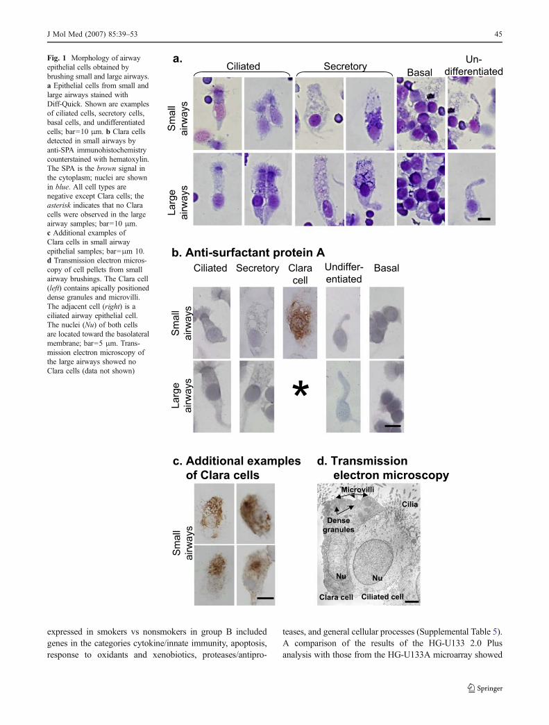
evaluated in group A was higher than in large airways (p<0.005). Less than 1% of cells recovered from large andsmall airways were squamous cells in both smokers andnonsmokers (p>0.7). Assessment with Diff-Quik stain ofthe airway epithelial cells identified the four main epithelialcell types present in the human airways [ciliated, basal,undifferentiated, and secretory [29–33] in both the largeand small airways (Table 1 and Fig. 1a)]. Evaluation byimmunohistochemistry with SPA antibody [34] demonstrat-ed the presence of Clara cells only in airway epithelial cellsobtained from the small airways, not from large airways(Fig. 1b,c).
Assessment of the large and small airway epithelial cellpopulations by transmission electron microscopy demon-strated cells typical of Clara cells only in the small airwayepithelial cell populations (Fig. 1d). These cells had 1 to2 μm dense granules in the apical cytoplasm and containedmicrovilli, but not cilia, typical of ultrastructural descrip-tions of Clara cells in human small airways [35]. BecauseClara cells are found only in airways <3 mm in diameter[35–37], the observation of Clara cells in the small airwaysamples confirms that the samples were, in fact, from thesmall airways.
Consistent with prior morphologic studies describingthe composition of airway epithelial cells in the humanlung, the small airways had a higher proportion of ciliatedcells than large airways (nonsmokers p<0.001, smokersp<0.001). In contrast, the large airways had higherproportion of basal (nonsmokers p<0.001, smokersp<0.001), undifferentiated (nonsmokers p<0.001, smok-ers p<0.001), and secretory (nonsmokers p<0.04, smok-ers p<0.04) cells than small airways (Table 1).
As further evidence that the small airway epithelium wasbeing sampled, independent of smoking status, geneexpression of airway epithelium from small airways(evaluated in the individuals in group A) revealed theexpression of surfactant apoprotein A2, surfactant apopro-tein B, and surfactant apoprotein C genes (Fig. 2).Consistent with prior studies of surfactant gene expressionin the small airway epithelium [34–36], the surfactantapoprotein genes were not expressed in the large airwayepithelial samples.
Genes expressed in the small airway epithelium of normalnonsmokers
To determine the normal small airway epithelium tran-scriptome, RNA from small airway epithelial cells from the12 healthy nonsmokers from group B was assessed with theHG-U133 Plus 2.0 microarray. In this analysis, 27,244 ofthe total 54,675 probe sets were “Present” or expressedaccording to the MAS5 algorithm in >50% of the samples.These genes were functionally grouped into 14 different
J Mol Med (2007) 85:39–53 43
categories. Forty percent, representing 10,935 probe setIDs, were classified as unknown function and were not usedto generate the data on the distribution of types of genesexpressed. The remaining genes were classified in thegeneral biological processes categories. The largest catego-ries were transcription, transport, metabolism, signal trans-duction, followed by cell cycle, apoptosis, and celladhesion; other categories included differentiation, immuneresponse, proteolysis, electron transport, cell growth, andcell signaling related genes (Fig. 3).
Differentially expressed genes in the small airwayepithelium of phenotypically normal smokers comparedto normal nonsmokers
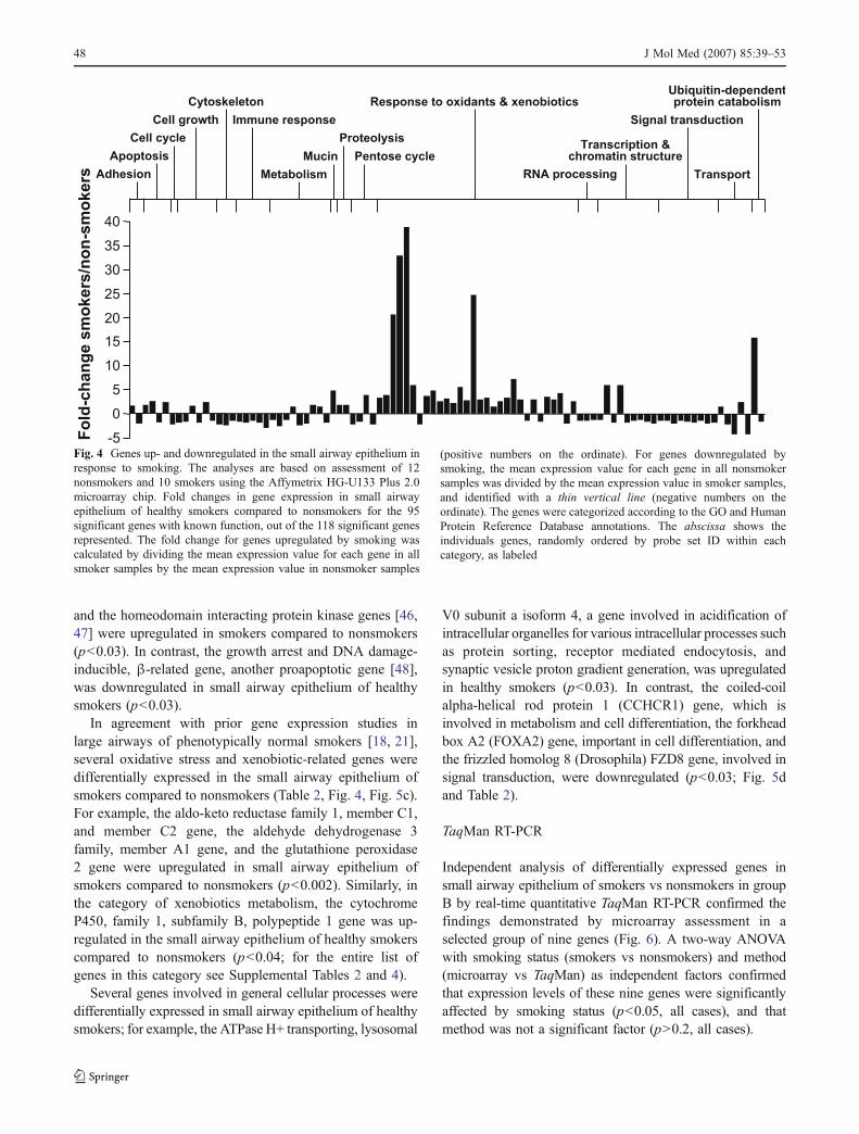
Relevant to the pathogenesis of COPD, assessment of geneexpression in the small airway epithelium of smokerscompared to nonsmokers showed a significant up- anddownregulation of several genes in various functionalcategories (Table 2, Figs. 4 and 5). Initial assessment ofgene expression in a small number of individuals (group A,n=11, 6 smokers vs 5 nonsmokers) demonstrated 152 genesdifferentially expressed, 103 genes upregulated, and 49genes downregulated in several functional categories in thesmall airway epithelium of healthy smokers compared tononsmokers (Supplemental Table 2). Of these 152 genes,
133 genes were of known function and were grouped intobiologically relevant categories. Based on the assessment ofthe small airway gene expression and a review of themolecular pathways shown in the literature to be related tothe pathogenesis of COPD, we chose the most relevant six ofthese categories, to generate a representative “small airwayepithelial smoking-induced phenotype.” These categoriesincluded cytokine/innate immunity, apoptosis, profibrotic,response to oxidants and xenobiotics, antiproteases, andgeneral cellular processes (Supplemental Table 3).
After the initial assessment of differential gene expressionin the first group of healthy individuals studied, we sought toverify these changes by studying a larger group of healthyindividuals (group B, n=22, 10 smokers vs 12 nonsmokers).It is interesting to note that consistent with the initialassessment using the HG-U133A chip (group A), genes insimilar categories were differentially expressed in smallairway epithelium of healthy smokers compared to non-smokers assessed with the HG-U133 Plus 2.0 chip (group B;Table 2). The group B assessment, which was subject to amore rigorous analysis (see “Materials and methods”),demonstrated a more restricted number of genes up- ordownregulated [118 genes, 48 upregulated and 70 down-regulated (Supplemental Table 4)] compared to the initialgene list of 152 observed in the initial analysis of group A(Supplemental Table 2). The 118 genes differentially
Table 1 Comparison of the cell types removed by brushing the airway epithelium
Group Aa Group Bb
Small airways Large airways Small airways
Nonsmokers Smokers Nonsmokers Smokers Nonsmokers Smokers
Total number of cells recovered (×106) 10±6 7±4 7±2 9±3 5±2 7±2Percentage of total cellsEpithelial 96±4 96±4 98±5 98±1 99±1 97±1Inflammatory 4±3 4±4 1±1 1±1 1±1 1±1Squamous 0 0 1±1 1±1 0 0Percentage of epithelial cellsCiliated 80±5 75±6 50±2 43±3 78±7 75±10Secretoryc 4±1 4±3 9±4 10±2 7±3 7±3Basal 5±3 8±2 20±3 27±4 7±2 9±4Undifferentiated 8±2 9±3 21±4 20±2 8±4 9±4Clarad 2±1 4±1 0 0 ND ND
ND Not determinedaLarge airway epithelial cells were collected from second to third generation bronchi and small airway epithelial cells were collected from airwaysat the 10th to 12th generation under fluoroscopic guidance by advancing the brush 7 to 10 cm beyond the third generation bronchi. Cytospinpreparations were stained with Diff-Quick to determine the percentage of epithelial vs inflammatory cells, and identification of ciliated, secretory,basal, and undifferentiated epithelial cells. The data is presented as mean±SD for each cell type in small and large airways for nonsmokers(n=5) and smokers (n=6).
bSmall airway epithelial cells were collected from airways at the 10th to 12th generation under fluoroscopic guidance as described in tablenote a. The data is presented as mean±SD for each cell type in small airways for nonsmokers (n=12) and smokers (n=10).
cSecretory cells (percentage of total epithelial cells) include Clara cells and non-Clara cells.dImmunostaining of large and small airway epithelial cells with SPA antibody with hematoxylin counterstaining was used to quantify Claracells within the secretory subset (data as percentage of total epithelial cells); percentage of Clara cells were not determined for group B.
44 J Mol Med (2007) 85:39–53
expressed in smokers vs nonsmokers in group B includedgenes in the categories cytokine/innate immunity, apoptosis,response to oxidants and xenobiotics, proteases/antipro-
teases, and general cellular processes (Supplemental Table 5).A comparison of the results of the HG-U133 2.0 Plusanalysis with those from the HG-U133A microarray showed
Ciliated SecretoryBasal
Un-
differentiated
Sm
all
airw
ays
Larg
e
airw
ays
a.
Sm
all
airw
ays
Larg
e
airw
ays
Ciliated Secretory Undiffer-
entiatedBasalClara
cell
Sm
all
airw
ays
c. Additional examples
of Clara cells
b. Anti-surfactant protein A
d. Transmission
electron microscopyMicrovilli
Dense
granules
Cilia
Nu Nu
Clara cell Ciliated cell
Fig. 1 Morphology of airwayepithelial cells obtained bybrushing small and large airways.a Epithelial cells from small andlarge airways stained withDiff-Quick. Shown are examplesof ciliated cells, secretory cells,basal cells, and undifferentiatedcells; bar=10 μm. b Clara cellsdetected in small airways byanti-SPA immunohistochemistrycounterstained with hematoxylin.The SPA is the brown signal inthe cytoplasm; nuclei are shownin blue. All cell types arenegative except Clara cells; theasterisk indicates that no Claracells were observed in the largeairway samples; bar=10 μm.c Additional examples ofClara cells in small airwayepithelial samples; bar=μm 10.d Transmission electron micros-copy of cell pellets from smallairway brushings. The Clara cell(left) contains apically positioneddense granules and microvilli.The adjacent cell (right) is aciliated airway epithelial cell.The nuclei (Nu) of both cellsare located toward the basolateralmembrane; bar=5 μm. Trans-mission electron microscopy ofthe large airways showed noClara cells (data not shown)
J Mol Med (2007) 85:39–53 45

that although there is a large degree of agreement betweenthe two data sets, there are discrepancies (SupplementalTable 6). These differences can likely be explained by thefact that these are different microarrays with differentnumber of probe sets and different hybridization conditionsbecause of array density (group A consisted of n=6 smokers
and n=5 nonsmokers, while group B consisted of n=10smokers and n=12 nonsmokers). Not only is the “n” of thegroups different, but in group B we applied the Benjamin–Hochberg correction to reduce the number of false positives,which was not applied to group A because of the low n.
Assessment of gene expression levels for the 118 genesmodulated by smoking in the small airway epithelium of thestudy individuals in group B, using an unsupervised assess-ment by hierarchical cluster analysis, showed, as expected,clustering of the samples according to smoking status. Thissuggests that taken as a group, similar changes are occurringamong all healthy smokers. Likewise, as a group, healthynonsmokers displayed a similar gene expression profile(Supplemental Fig. 2b), while cluster analysis using thecomplete list of expressed genes (30,963 probe sets, presentin at least one array) did not segregate smokers fromnonsmokers (Supplemental Fig. 2a), as noted previouslyfor large airways [18].
After assessment of gene expression in small airwayepithelium of healthy smokers vs nonsmokers in groups Aand B, we generated a list showing examples of genesdifferentially expressed in a similar fashion in smokers vsnonsmokers in both groups A and B (Table 2). Assessment ofthe list of genes in the different pathways does not representall the genes relevant for COPD; it nevertheless representsearly gene expression responses in the small airwayepithelium of individuals exposed to the insult of cigarettesmoke, the main risk factor for the development of COPD.
In the context that the assessment of differential geneexpression of healthy individuals in group B included a highernumber of individuals (n=22, 10 smokers vs 12 non-smokers), with assessment of gene expression with theAffymetrix HG-U133 Plus 2.0, and a more rigorous analysisof the gene expression data, which included independentassessment by RMA and MAS5 with Benjamini–Hochbergcorrection, the following description of differentiallyexpressed genes in the different categories relevant to thepathogenesis of COPD will focus on the results obtainedfrom small airway epithelial cells from these individuals(group B, Table 2, Figs. 4 and 5, and Supplemental Tables 4and 5).
Expression of genes potentially relevant to the pathogenesisof COPD
Independent assessment of gene expression by RMA andMAS5 demonstrated that the small airway epithelium ofsmokers vs nonsmokers downregulated several immune-related genes. These genes included the interleukin-4 (IL4)receptor gene (p<0.002), the chemokine (C-X3-C motif)ligand 1 (p<0.02), also known as fractalkine, and the spondin2 (p<0.04); these genes are involved in many inflammatoryfunctions in human airways [38–41] (Table 2 and Fig. 5a).
Transcription
Metabolism
Transport
Electron transport
Cell signaling
Cell growth
Cell cycle
Proteolysis
Immune response
Apoptosis
Differentiation
Adhesion
Signal
transduction
Other
Fig. 3 Functional categories of genes expressed in the small airwayepithelium of normal nonsmokers. The pie chart shows the differentfunctional categories of the small airway epithelium transcriptome in12 healthy nonsmokers; small airway epithelium gene expression wasassessed with the Affymetrix HG-U133 Plus 2.0 microarray chip. Thedistribution data represents the gene expression in nonsmokers withprobe sets representing genes expressed (Affymetrix detection call ofpresent) in >50% of the small airway samples. Probe sets werecategorized using the Affymetrix NetAffx Analysis Center by GOBiological Process. A total of 27,244 probe sets were grouped intofunctional categories; of these, 10,935 probe sets were classified asunknown function and were not used for the final analysis
10-1No
rmalized
exp
ressio
n levels
Surfactant B
Large Small
Surfactant C
Large Small
Surfactant A2
SmallLarge
100
101
102
103
Fig. 2 Quantitative assessment of expression of surfactant apoproteingenes in small airway vs large airway epithelial cells of healthynonsmokers and smokers. Five healthy individual nonsmokers and sixhealthy smokers (group A) were evaluated. Shown is the HG-133A-generated normalized expression level with a logarithmic scale forsurfactant apoprotein A2, surfactant apoprotein B, and surfactantapoprotein C. Each symbol represents one individual. Open symbolsrepresent nonsmokers; closed symbols represent smokers
46 J Mol Med (2007) 85:39–53

We also observed differentially expressed apoptosis-related genes: consistent with prior studies demonstratingupregulation of pirin, a proapoptotic gene, in the largeairways of smokers [20, 21, 42–45], we observed upregu-
lation of pirin in the small airway epithelium of healthysmokers compared to nonsmokers (p<0.03; Table 2 andFig. 5b). Similarly, the proapoptosis-related genes HIV-Tatinteractive protein 2, 30 kDa gene also known as TIP30,
Table 2 Examples of genes differentially expressed in the small airway epithelium in nonsmokers and smokers in functional categories that arerelevant for the pathogenesis of COPD
Category Gene Genesymbol
References in literaturethat suggested pathwaysrelevant for COPDpathogenesisa
Group A Group B
S vsNS foldchangeb
pvalueS vsNSc
S vs NSfoldchangeRMAb
pvalueS vsNSRMAd
S vs NSfoldchangeMAS5b
p valueS vsNSMAS5e
Cytokine/innateimmunity
Chemokine(C-X3-C motif)ligand 1
CX3CL1 [7, 16, 38–41] −2.97 <0.040 −2.89 <0.016 −2.96 <0.004
Apoptosis Pirin PIR [42–48, 56–61] 2.78 <0.001 2.64 <0.007 2.22 <0.028Growth arrest andDNA damage-inducible, beta
GADD45B [42–48, 56–61] −2.25 <0.043 −1.83 <0.024 −2.26 <0.014
Response tooxidantsandxenobiotics
CytochromeP450, family 1,subfamily B,polypeptide 1
CYP1B1 [7, 16, 21, 49, 62] 17.69 <0.001 20.73 <0.039 54.7 <0.004
Aldo-ketoreductase family1, member B10
AKR1B10 [7, 16, 21, 49, 62] 11.73 <0.001 24.84 <0.003 20.76 <0.002
Aldehydedehydrogenase 3family,memberA1
ALDH3A1 [7, 16, 21, 49, 62] 6.63 <0.001 75.6 <0.001 4.96 <0.001
Alcoholdehydrogenase 7
ADH7 [7, 16, 21, 49, 62] 6.24 <0.001 7.21 <0.001 6.1 <0.001
Glutathioneperoxidase 2
GPX2 [7, 16, 21, 49, 62] 5.16 <0.001 2.69 <0.009 3.73 <0.001
NAD(P)Hdehydrogenase,quinone 1
NQO1 [7, 16, 21, 49, 62] 4.41 <0.001 3.37 <0.001 3.38 <0.001
Aldo-ketoreductase family1, member C3
AKR1C3 [7, 16, 21, 49, 62] 3.09 <0.001 2.6 <0.01 2.32 <0.014
Generalcellularprocesses
Ubiquitincarboxyl-terminal esteraseL1
UCHL1 [63, 64] 11.75 <0.001 15.85 <0.002 31.07 <0.001
Group A includes 11 healthy individuals (5 healthy nonsmokers and 6 healthy smokers) in whom small airway epithelial gene expression wasassessed with the Affymetrix HG-U133A gene chip. Group B includes 22 healthy individuals (12 nonsmokers and 10 smokers) in whomsmall airway epithelial gene expression was assessed with the Affymetrix HG-U133 Plus 2.0 gene chip; for group B, expression values wereindependently generated using RMA and MAS5. Genes were considered expressed when they had Affymetrix present “P” calls in >50% ofany given group of samples (nonsmokers or smokers) in both group A and group B study individuals.
aThese references directly implicate the specific genes in some cases, or implicate pathways in which these genes are involved.bSmokers (S) vs nonsmokers (NS) fold change was calculated by dividing the average expression value in the smokers by the averageexpression value in the nonsmokers.
cp values were calculated using the Welch t test (assuming unequal variances) using the Affymetrix HG-U133A gene chip; expressionvalues were generated using MAS5.
dp values were calculated using the Welch t test (assuming unequal variances) using the Affymetrix HG-U133A Plus 2.0 gene chip;expression values were generated using RMA with Benjamini–Hochberg correction.
eSame as table note e except expression values were generated using MAS5.
J Mol Med (2007) 85:39–53 47
and the homeodomain interacting protein kinase genes [46,47] were upregulated in smokers compared to nonsmokers(p<0.03). In contrast, the growth arrest and DNA damage-inducible, β-related gene, another proapoptotic gene [48],was downregulated in small airway epithelium of healthysmokers (p<0.03).
In agreement with prior gene expression studies inlarge airways of phenotypically normal smokers [18, 21],several oxidative stress and xenobiotic-related genes weredifferentially expressed in the small airway epithelium ofsmokers compared to nonsmokers (Table 2, Fig. 4, Fig. 5c).For example, the aldo-keto reductase family 1, member C1,and member C2 gene, the aldehyde dehydrogenase 3family, member A1 gene, and the glutathione peroxidase2 gene were upregulated in small airway epithelium ofsmokers compared to nonsmokers (p<0.002). Similarly, inthe category of xenobiotics metabolism, the cytochromeP450, family 1, subfamily B, polypeptide 1 gene was up-regulated in the small airway epithelium of healthy smokerscompared to nonsmokers (p<0.04; for the entire list ofgenes in this category see Supplemental Tables 2 and 4).
Several genes involved in general cellular processes weredifferentially expressed in small airway epithelium of healthysmokers; for example, the ATPase H+ transporting, lysosomal
V0 subunit a isoform 4, a gene involved in acidification ofintracellular organelles for various intracellular processes suchas protein sorting, receptor mediated endocytosis, andsynaptic vesicle proton gradient generation, was upregulatedin healthy smokers (p<0.03). In contrast, the coiled-coilalpha-helical rod protein 1 (CCHCR1) gene, which isinvolved in metabolism and cell differentiation, the forkheadbox A2 (FOXA2) gene, important in cell differentiation, andthe frizzled homolog 8 (Drosophila) FZD8 gene, involved insignal transduction, were downregulated (p<0.03; Fig. 5dand Table 2).
TaqMan RT-PCR
Independent analysis of differentially expressed genes insmall airway epithelium of smokers vs nonsmokers in groupB by real-time quantitative TaqMan RT-PCR confirmed thefindings demonstrated by microarray assessment in aselected group of nine genes (Fig. 6). A two-way ANOVAwith smoking status (smokers vs nonsmokers) and method(microarray vs TaqMan) as independent factors confirmedthat expression levels of these nine genes were significantlyaffected by smoking status (p<0.05, all cases), and thatmethod was not a significant factor (p>0.2, all cases).
-5
0
5
10
15
20
25
30
35
40
Adhesion
Apoptosis
Cell cycle
Cell growth
Cytoskeleton
Immune response
Metabolism
Mucin Pentose cycle
Proteolysis
Response to oxidants & xenobiotics
RNA processing
Signal transduction
Transcription &chromatin structure
Transport
Ubiquitin-dependentprotein catabolism
Fo
ld-c
han
ge s
mo
kers
/no
n-s
mo
kers
Fig. 4 Genes up- and downregulated in the small airway epithelium inresponse to smoking. The analyses are based on assessment of 12nonsmokers and 10 smokers using the Affymetrix HG-U133 Plus 2.0microarray chip. Fold changes in gene expression in small airwayepithelium of healthy smokers compared to nonsmokers for the 95significant genes with known function, out of the 118 significant genesrepresented. The fold change for genes upregulated by smoking wascalculated by dividing the mean expression value for each gene in allsmoker samples by the mean expression value in nonsmoker samples
(positive numbers on the ordinate). For genes downregulated bysmoking, the mean expression value for each gene in all nonsmokersamples was divided by the mean expression value in smoker samples,and identified with a thin vertical line (negative numbers on theordinate). The genes were categorized according to the GO and HumanProtein Reference Database annotations. The abscissa shows theindividuals genes, randomly ordered by probe set ID within eachcategory, as labeled
48 J Mol Med (2007) 85:39–53

Discussion
While COPD associated with chronic cigarette smokingeventually involves all levels of the airways, the earliestsmoking-induced changes are in the small airway epithelium[4–6, 13, 14]. To begin to understand the responses of thesmall airway epithelium to the stress of cigarette smoking,we developed a strategy with fiberoptic bronchoscopy andairway brushings to obtain highly pure epithelial cells fromhuman small airways, and analyzed the epithelial cells geneexpression with microarray technology. The small airwayepithelial samples from healthy smokers and nonsmokers
differed in composition from that of large airway samples,with the small airways having more ciliated cells, and lessundifferentiated, basal, and secretory cells than the largeairways. Consistent with the known composition of thesecretory subset of airway epithelium in the small airways,epithelial cells of the small, but not large airways, demon-strated the presence of Clara cells and expression ofsurfactant apoprotein genes irrespective of smoking status.
Initial assessment of small airway epithelium geneexpression in a small group (five nonsmokers and sixsmokers, group A) of healthy individuals with the HG-133A microchip array demonstrated 152 differentially
0
1
2
3
NS S NS S NS S NS S
IL4R SPON2 SUSD4 CX3CL1p<0.009 p<0.05 p<0.04 p<0.02
Rela
tive g
en
e e
xp
ressio
na. Cytokine / innate immune-related
0
0.5
1.0
1.5
2.0
2.5
3.0
3.5
NS S NS S
HTATIP2 PIRp<0.001 p<0.008
NS S
GADD45Bp<0.03
Rela
tive g
en
e e
xp
ressio
n
b. Apoptosis-related
NS S
HIPK2p<0.01
0
1
2
3
4
5
6
20
78
60
NS S
CYP1B1
NS S
AKR1C1
NS S
AKR1C2
NS S
GPX2
p<0.02 p<0.001 p<0.001 p<0.001
Rela
tive g
en
e e
xp
ressio
n
c. Oxidative-stress and xenobiotic-related
Rela
tive g
en
e e
xp
ressio
n
d. General processes and differentiation
0.0
0.5
1.0
1.5
2.0
2.5
3.0
NS S
CCHCR1
NS S
FOXA2
NS S
FZD8
p<0.02 p<0.01 p<0.03
NS S
ATP6V0A4
p<0.02
Fig. 5 Examples of genes up- and downregulated in the small airwaysof smokers compared to nonsmokers. The data is based on 10 healthysmokers compared to12 healthy nonsmokers assessed with theAffymetrix HG-U133 Plus 2.0 microarray chip, with Benjamini–Hochberg correction for multiple comparisons. The abscissa showsthe specific genes; the ordinate shows the normalized gene expressionlevels. Each symbol represents an individual. Open symbols representnonsmokers (NS); closed symbols represent smokers (S). p values areshown below each gene symbol. a Expression of cytokine/innateimmune-related genes. The genes shown are the IL-4 receptor (IL4R)gene, the spondin 2 (SPON2) gene, the sushi domain containing four(SUSD4) genes, and the chemokine (C-X3-C motif) ligand 1 gene,also known as fractalkine (CX3CL1). b Expression of apoptosis-related genes. The genes shown are the HIV-Tat interactive protein 2,
30 kDa (HTATIP2) gene; the pirin (PIR) gene, the growth arrest andDNA damage-inducible, β-related (GADD45B) gene; and homeodo-main interacting protein kinase 2 (HIPK2) gene. c Examples ofexpression of oxidative stress and xenobiotic-related genes. Shown arethe cytochrome P450, family 1, subfamily B, polypeptide 1 (CYP1B1)gene; the aldo-keto reductase family 1, member C1 (AKR1C1) gene;the aldo-keto reductase family 1, member C2 (AKR1C2) gene; and theglutathione peroxidase 2 (GPX2) gene. d Examples of expression ofgeneral processes and differentiation genes. Shown are the ATPase H+transporting, lysosomal V0 subunit a isoform 4 (ATP6V0A4) gene; thecoiled-coil alpha-helical rod protein 1 (CCHCR1) gene, which isinvolved in metabolism and cell differentiation; the forkhead box A2(FOXA2) gene, important in cell differentiation; and the frizzledhomolog 8 (Drosophila) (FZD8) gene, involved in signal transduction
J Mol Med (2007) 85:39–53 49

expressed genes (103 upregulated, 49 downregulated) inresponse to cigarette smoking; 133 of which are of knownfunction and belong to several functional categories. Afterthese initial assessments, we carried out an independentstudy in an entirely new and larger group of healthysmokers vs nonsmokers (12 nonsmokers vs 10 smokers,group B). This analysis, assessed with the HG-U133 Plus2.0 microarray chip, with MAS5 and RMA (independent-ly), and with Benjamini–Hochberg correction for falsediscovery rate, demonstrated 118 differentially expressedgenes in healthy smokers vs nonsmokers.
Based on review of the literature on the differentmolecular pathways implicated in the pathogenesis ofCOPD involving the airway epithelium, and on the
assessment of the differentially expressed genes in thesmall airways of smokers compared to nonsmokers, wedeveloped a working list of genes divided into categoriesrelevant to the pathogenesis of COPD. The categories ofgenes generated on the assessment of group A and group Bincluded cytokine/innate immunity, apoptosis, response tooxidants and xenobiotics, proteolysis/antiproteases, andgeneral cellular processes. These genes represent a snapshotof the early molecular changes in the small airwayepithelium of healthy individuals who actively smoke, andare therefore at risk to develop COPD. These genes mayrepresent COPD susceptibility genes and protective genes,and the risk for COPD may depend on an individual’sspecific pattern of combined expression for susceptibility
0
1
2
3
4
5
NQ01 ALDH3A1 AKR1C3 ADH7 PIR HIPK2 CDKN1C FOXA2 CX3CL1
No
rma
lize
d e
xp
res
sio
n l
ev
el
Arr
ay
TaqM
an
Arr
ay
TaqM
an
Arr
ay
TaqM
an
Arr
ay
TaqM
an
Arr
ay
TaqM
an
Arr
ay
TaqM
an
Arr
ay
TaqM
an
Arr
ay
TaqM
an
Arr
ay
TaqM
an
Non-smokers
Smokers
Fig. 6 Confirmation of microarray results with TaqMan real-time RT-PCR. Expression levels of six genes upregulated by smoking and threegenes downregulated by smoking on initial assessment by microarrayanalysis (RMA-based) with the Affymetrix HG-U133 Plus 2.0 chipwere confirmed with TaqMan real-time RT-PCR. To allow directcomparisons of values obtained using the two independent methods,TaqMan expression levels were normalized by dividing individualvalues by the median expression level of all nonsmokers and smokersfor that method, as was done for microarray analysis. Relativeexpression levels (ordinate) are shown for six genes upregulated bysmoking, as follows: four genes involved in the response to oxidativestress or xenobiotics, the NAD(P)H dehydrogenase, quinone 1 (NQO1)gene; the aldehyde dehydrogenase 3 family, member A1 (ALDH3A1)
gene; the aldo-keto reductase family 1, member C3 (AKR1C3) gene; thealcohol dehydrogenase 7 (ADH7) gene; two genes involved inapoptosis, the pirin (PIR); and the homeodomain interacting proteinkinase 2 (HIPK2) genes; and for three genes downregulated bysmoking: the cyclin-dependent kinase inhibitor 1C (CDKN1C) gene,also known as p57 or Kip2, a cell cycle arrest protein; the transcriptionfactor forkhead box A2 (FOXA2) gene, involved in transcription of thesurfactant genes; and the chemokine (C-X3-C motif) ligand 1(CX3CL1). A two-way ANOVA with smoking status (smokers vsnonsmokers) and method (microarray vs TaqMan) as independentfactors confirmed that expression levels of these nine genes weresignificantly affected by smoking status (p<0.05, all cases), and thatmethod was not a significant factor (p>0.2, all cases)
50 J Mol Med (2007) 85:39–53

and protective genes. Furthermore, these genes mayrepresent only a subset of the genes underlying thepathogenesis of COPD.
Small airways, smoking, and COPD
The small airways (<2 mm) represent the main site of airwayobstruction in individuals with COPD [4–6, 13, 14].Asymptomatic smokers display evidence of small airwayinflammation; for example, Niewoehner et al. [8] studied thelungs of 19 young smokers and 20 nonsmokers, anddemonstrated that the small airways of smokers haddefinitive pathologic abnormalities with denuded epitheliumand increased number of mural inflammatory cells. Thesedata are consistent with the concept that the small airwaysrepresent the earliest site of smoking-induced structuralchanges before the development of COPD [4–6, 8–14]. Itis interesting to note that the extent of small airway diseasecorrelates with the extent of alveolar destruction [13, 14].
Small airway epithelial smoking-induced phenotype
The airway epithelium plays an important role in control-ling many airway functions and is capable of up- anddownregulating genes in several categories as well asproducing and secreting mediators important in severalaspects of airway function [16–18, 21, 33, 49–54]. Thesegenes and mediators, among others, include cytokines,chemokines, apoptosis-related, profibrotic-related, oxidativestress-related, proteolysis/antiproteases-related, mucin-related, and genes related to general processes [16–18, 21,33, 49–54]. In this context, persistent activation of the smallairway epithelium with the insult of cigarette smoke leads toan alteration of the “resting” state of the small airwayepithelium with up- and downregulation of genes in differentcategories as observed in our study population.
The cytokine, innate, and immunomodulatory res-ponses of the small airway epithelium to the insult ofcigarette smoke play an important role, over time, in theeventual development of the small airway inflammatorycomponent characteristic of individuals with establishedCOPD. The small airways from healthy smokers demon-strate inflammation, as do symptomatic smokers, andindividuals at different COPD stages [Global Initiativefor Chronic Obstructive Lung Disease (GOLD) stages 1–4][1, 2, 8–12]. It is interesting to note that although somedegree of overlap in small airway inflammation is observedamong asymptomatic smokers and individuals with COPDGOLD stages 0–3 [12], increased numbers of CD8+ Tlymphocytes are observed only in smokers who developCOPD [11]. This suggests that the cytokine/innate immuneresponse seems to be nonspecific at earlier stages of smoking,but over time, for those individuals who develop COPD, the
immune response undergoes a more specific change, whichresults in increased accumulation of CD8+ T lymphocytes.
We found downregulation of the IL4 receptor, a mediatorof several proinflammatory functions in human airways [38],and downregulation of chemokine (C-X3-C motif) ligand1 (CX3CL1), which is involved in cell adhesion andrecruitment of monocytes and T lymphocytes cells [39,40]. It is interesting to note that microarray analysis of lungtissue from individuals with COPD demonstrated upregula-tion of the CX3CL1 gene in individuals with later stageCOPD compared to individuals with early COPD [55].Although lower mRNA levels do not necessarily reflectlower protein levels, it can be speculated that the down-regulation of CX3CL1 in our study suggests that phenotyp-ically healthy smokers attempt to maintain a balance in theinflammatory response in the small airways by suppressingsignals that could potentially injure the epithelium.
The role of apoptosis in the pathogenesis of COPD iswell recognized, and increased apoptosis of airway epithe-lial cells from individuals with COPD was documentedeven after cessation of smoking [56–61]. In our study,assessment of the small airway epithelium of healthysmokers showed smoking-related modulation of severalproapoptotic genes [42–48], suggesting ongoing attempts ofan “apoptosis balance” in the small airway epithelium ofphenotypically healthy smokers.
It is well recognized that the oxidative stress of cigarettesmoking plays an important role in the pathogenesis ofCOPD [49, 62]. We observed upregulation of severaloxidative stress-related and xenobiotic genes in the smallairway epithelium. This suggests that similar to studies ofincreased expression of oxidative stress-related genes in thelarge airway epithelium of healthy smokers [18, 21], thesmall airway epithelium responds to the insult of cigarettesmoking by upregulating several oxidative stress-relatedand xenobiotic genes.
Several genes relevant to general cellular processes wereupregulated, consistent with the increased energy expendi-ture observed in healthy smokers and in individuals withCOPD [63, 64].
Implications for the understanding and treatmentof COPD
Assessment of the molecular changes of the small airwayepithelium in healthy smokers is relevant to establishingpatterns of gene expression for comparison with the geneexpression in small airways of individuals at various stagesof COPD. The differential gene expression observed in thesmall airway epithelium of healthy smokers represents theinitial deviations of gene expression observed in the mainsite of potential disease in individuals at risk for COPD.
J Mol Med (2007) 85:39–53 51
This study may help in the identification of novel genes notrelated to already known mechanisms of COPD pathogen-esis. Assessment of the expression of these genes in thesmall airway epithelium of individuals with COPD shouldbe useful in identifying mechanisms relevant to thepathogenesis of COPD and potential new therapeutictargets for intervention.
Acknowledgements We thank the Pulmonary Fellows and thenurses of the bronchoscopy suite of the Division of Pulmonary andCritical Care Medicine for helping with bronchoscopies; IgorDolgalev and Tina Raman for excellent technical assistance; T.O’Connor and N. Hackett for helpful discussions; and N. Mohamedfor help in preparing this manuscript. These studies were supported, inpart, by NIH R01 HL074326 and Weill Cornell GCRC M01RR00047and the Will Rogers Memorial Fund, Los Angeles, CA.
References
1. Pauwels RA, Buist AS, Calverley PM, Jenkins CR, Hurd SS(2001) Global strategy for the diagnosis, management, andprevention of chronic obstructive pulmonary disease. NHLBI/WHO Global Initiative for Chronic Obstructive Lung Disease(GOLD) workshop summary. Am J Respir Crit Care Med163:1256–1276
2. Celli BR, MacNee W (2004) Standards for the diagnosis andtreatment of patients with COPD: a summary of the ATS/ERSposition paper. Eur Respir J 23:932–946
3. Shapiro SD, Ingenito EP (2005) The pathogenesis of chronicobstructive pulmonary disease: advances in the past 100 years.Am J Respir Cell Mol Biol 32:367–372
4. Hogg JC, Macklem PT, Thurlbeck WM (1968) Site and nature ofairway obstruction in chronic obstructive lung disease. N Engl JMed 278:1355–1360
5. Yanai M, Sekizawa K, Ohrui T, Sasaki H, Takishima T (1992) Siteof airway obstruction in pulmonary disease: direct measurementof intrabronchial pressure. J Appl Physiol 72:1016–1023
6. Hogg JC (2004) Pathophysiology of airflow limitation in chronicobstructive pulmonary disease. Lancet 364:709–721
7. Barnes PJ, Shapiro SD, Pauwels RA (2003) Chronic obstructivepulmonary disease: molecular and cellular mechanisms. EurRespir J 22:672–688
8. Niewoehner DE, Kleinerman J, Rice DB (1974) Pathologicchanges in the peripheral airways of young cigarette smokers.N Engl J Med 291:755–758
9. Cosio MG, Hale KA, Niewoehner DE (1980) Morphologic andmorphometric effects of prolonged cigarette smoking on the smallairways. Am Rev Respir Dis 122:265–321
10. Saetta M, Finkelstein R, Cosio MG (1994) Morphological andcellular basis for airflow limitation in smokers. Eur Respir J7:1505–1515
11. Cosio Piqueras MG, Cosio MG (2001) Disease of the airways inchronic obstructive pulmonary disease. Eur Respir J Suppl34:41s–49s
12. Willemse BW, ten Hacken NH, Rutgers B, Postma DS, Timens W(2005) Association of current smoking with airway inflammationin chronic obstructive pulmonary disease and asymptomaticsmokers. Respir Res 6:38
13. Cosio M, Ghezzo H, Hogg JC, Corbin R, Loveland M, Dosman J,Macklem PT (1978) The relations between structural changes insmall airways and pulmonary-function tests. N Engl J Med298:1277–1281
14. Hogg JC, Chu F, Utokaparch S, Woods R, Elliott WM, Buzatu L,Cherniack RM, Rogers RM, Sciurba FC, Coxson HO, Pare PD(2004) The nature of small-airway obstruction in chronicobstructive pulmonary disease. N Engl J Med 350:2645–2653
15. Saetta M, Turato G, Maestrelli P, Mapp CE, Fabbri LM (2001)Cellular and structural bases of chronic obstructive pulmonarydisease. Am J Respir Crit Care Med 163:1304–1309
16. Barnes PJ (2004) Mediators of chronic obstructive pulmonarydisease. Pharmacol Rev 56:515–548
17. Spurzem JR, Rennard SI (2005) Pathogenesis of COPD. SeminRespir Crit Care Med 26:142–153
18. Hackett NR, Heguy A, Harvey BG, O’Connor TP, Luettich K,Flieder DB, Kaplan R, Crystal RG (2003) Variability ofantioxidant-related gene expression in the airway epithelium ofcigarette smokers. Am J Respir Cell Mol Biol 29:331–343
19. Heguy A, Harvey BG, O’Connor TP, Hackett NR, Crystal RG(2003) Sampling-dependent up-regulation of gene expression insequential samples of human airway epithelial cells. Mol Med9:200–208
20. Kaplan R, Luettich K, Heguy A, Hackett NR, Harvey BG, CrystalRG (2003) Monoallelic up-regulation of the imprinted H19 genein airway epithelium of phenotypically normal cigarette smokers.Cancer Res 63:1475–1482
21. Spira A, Beane J, Shah V, Liu G, Schembri F, Yang X, Palma J, BrodyJS (2004) Effects of cigarette smoke on the human airway epithelialcell transcriptome. Proc Natl Acad Sci USA 101:10143–10148
22. Burrows B, Knudson RJ, Cline MG, Lebowitz MD (1977)Quantitative relationships between cigarette smoking and ventila-tory function. Am Rev Respir Dis 115:195–205
23. Troisi RJ, Speizer FE, Rosner B, Trichopoulos D, Willett WC(1995) Cigarette smoking and incidence of chronic bronchitis andasthma in women. Chest 108:1557–1561
24. Lindstrom M, Kotaniemi J, Jonsson E, Lundback B (2001)Smoking, respiratory symptoms, and diseases: a comparativestudy between northern Sweden and northern Finland: report fromthe FinEsS study. Chest 119:852–861
25. Bailey CJ, Crystal RG, Leopold PL (2003) Association ofadenovirus with the microtubule organizing center. J Virol77:13275–13287
26. Irizarry RA, Hobbs B, Collin F, Beazer-Barclay YD, AntonellisKJ, Scherf U, Speed TP (2003) Exploration, normalization, andsummaries of high density oligonucleotide array probe level data.Biostatistics 4:249–264
27. Benjamini Y, Hochberg Y (1995) Controlling the false discoveryrate: a practical and powerful approach to multiple testing. J RStat Soc B57:289–300
28. Barrett T, Suzek TO, Troup DB, Wilhite SE, Ngau WC, Ledoux P,Rudnev D, Lash AE, Fujibuchi W, Edgar R (2005) NCBI GEO:mining millions of expression profiles-database and tools. NucleicAcids Res 33:D562–D566
29. Breeze RG, Wheeldon EB (1977) The cells of the pulmonaryairways. Am Rev Respir Dis 116:705–777
30. McDowell EM, Barrett LA, Glavin F, Harris CC, Trump BF(1978) The respiratory epithelium. I. Human bronchus. J NatlCancer Inst 61:539–549
31. Mercer RR, Russell ML, Roggli VL, Crapo JD (1994) Cellnumber and distribution in human and rat airways. Am J RespirCell Mol Biol 10:613–624
32. Danel C, Erzurum SC, McElvaney NG, Crystal RG (1996)Quantitative assessment of the epithelial and inflammatory cellpopulations in large airways of normals and individuals withcystic fibrosis. Am J Respir Crit Care Med 153:362–368
52 J Mol Med (2007) 85:39–53
33. Knight DA, Holgate ST (2003) The airway epithelium: structural andfunctional properties in health and disease. Respirology 8:432–446
34. Hermans C, Bernard A (1999) Lung epithelium-specific proteins:characteristics and potential applications as markers. Am J RespirCrit Care Med 159:646–678
35. Plopper CG, Hyde DM, Buckpitt AR (1997) Clara cells. In: RGCrystal, JB West, ER Weibel, PJ Barnes (eds) The lung: scientificfoundations. Lippincott-Raven, Philadelphia, p 535
36. Massaro GD, Singh G, Mason R, Plopper CG, Malkinson AM,Gail DB (1994) Biology of the Clara cell. Am J Physiol 266:L101–L106
37. Boers JE, Ambergen AW, Thunnissen FB (1999) Number andproliferation of Clara cells in normal human airway epithelium.Am J Respir Crit Care Med 159:1585–1591
38. Mueller TD, Zhang JL, Sebald W, Duschl A (2002) Structure,binding, and antagonists in the IL-4/IL-13 receptor system.Biochim Biophys Acta 1592:237–250
39. D’Ambrosio D, Mariani M, Panina-Bordignon P, Sinigaglia F (2001)Chemokines and their receptors guiding T lymphocyte recruitment inlung inflammation. Am J Respir Crit Care Med 164:1266–1275
40. Fujimoto K, Imaizumi T, Yoshida H, Takanashi S, Okumura K,Satoh K (2001) Interferon-gamma stimulates fractalkine expres-sion in human bronchial epithelial cells and regulates mononu-clear cell adherence. Am J Respir Cell Mol Biol 25:233–238
41. Kazanskaya O, Glinka A, del Barco Barrantes I, Stannek P, NiehrsC, Wu W (2004) R-Spondin2 is a secreted activator of Wnt/beta-catenin signaling and is required for Xenopus myogenesis. DevCell 7:525–534
42. Gelbman B, Heguy A, O’Connor TP, Zabner J, Crystal RG (2005)Adenovirus-mediated gene transfer demonstrates that pirin, atranscription factor up-regulated in the bronchial epithelium bycigarette smoke, mediates bronchial epithelial cell apoptosis. MolTher 2:A803
43. Dechend R, Hirano F, Lehmann K, Heissmeyer V, Ansieau S,Wulczyn FG, Scheidereit C, Leutz A (1999) The Bcl-3 oncopro-tein acts as a bridging factor between NF-kappaB/Rel and nuclearco-regulators. Oncogene 18:3316–3323
44. Orzaez D, de Jong AJ, Woltering EJ (2001) A tomato homologueof the human protein PIRIN is induced during programmed celldeath. Plant Mol Biol 46:459–468
45. Wendler WM, Kremmer E, Forster R, Winnacker EL (1997)Identification of pirin, a novel highly conserved nuclear protein.J Biol Chem 272:8482–8489
46. Hofmann TG, Stollberg N, Schmitz ML, Will H (2003) HIPK2regulates transforming growth factor-beta-induced c-Jun NH(2)-terminal kinase activation and apoptosis in human hepatoma cells.Cancer Res 63:8271–8277 (Dec 1)
47. Shi M, Zhang X, Wang P, Zhang HW, Zhang BH, Wu MC (2005)TIP30 regulates apoptosis-related genes in its apoptotic signaltransduction pathway. World J Gastroenterol 11:221–227
48. Takekawa M, Saito H (1998) A family of stress-inducibleGADD45-like proteins mediate activation of the stress-responsiveMTK1/MEKK4 MAPKKK. Cell 95:521–530
49. Bowler RP, Crapo JD (2002) Oxidative stress in airways: is therea role for extracellular superoxide dismutase? Am J Respir CritCare Med 166:S38–S43
50. Fischer BM, Voynow JA (2002) Neutrophil elastase inducesMUC5AC gene expression in airway epithelium via a pathwayinvolving reactive oxygen species. Am J Respir Cell Mol Biol26:447–452
51. Mio T, Romberger DJ, Thompson AB, Robbins RA, Heires A,Rennard SI (1997) Cigarette smoke induces interleukin-8 releasefrom human bronchial epithelial cells. Am J Respir Crit Care Med155:1770–1776
52. Profita M, Chiappara G, Mirabella F, Di GR, Chimenti L, CostanzoG, Riccobono L, Bellia V, Bousquet J, Vignola AM (2003) Effect ofcilomilast (Ariflo) on TNF-alpha, IL-8, and GM-CSF release byairway cells of patients with COPD. Thorax 58:573–579
53. Shao MX, Nakanaga T, Nadel JA (2004) Cigarette smoke inducesMUC5AC mucin overproduction via tumor necrosis factor-alpha-converting enzyme in human airway epithelial (NCI-H292) cells.Am J Physiol Lung Cell Mol Physiol 287:L420–L427
54. Willems LN, Kramps JA, Stijnen T, Sterk PJ, Weening JJ,Dijkman JH (1989) Antileukoprotease-containing bronchiolarcells. Relationship with morphologic disease of small airwaysand parenchyma. Am Rev Respir Dis 139:1244–1250
55. Ning W, Li CJ, Kaminski N, Feghali-Bostwick CA, Alber SM, DiYP, Otterbein SL, Song R, Hayashi S, Zhou Z, Pinsky DJ,Watkins SC, Pilewski JM, Sciurba FC, Peters DG, Hogg JC, ChoiAM (2004) Comprehensive gene expression profiles revealpathways related to the pathogenesis of chronic obstructivepulmonary disease. Proc Natl Acad Sci USA 101:14895–14900
56. Vayssier M, Banzet N, Francois D, Bellmann K, Polla BS (1998)Tobacco smoke induces both apoptosis and necrosis in mammaliancells: differential effects of HSP70. Am J Physiol 275:L771–L779
57. Li X, Shu R, Filippatos G, Uhal BD (2004) Apoptosis in lunginjury and remodeling. J Appl Physiol 97:1535–1542
58. Bartalesi B, Cavarra E, Fineschi M, Lucattelli B, Martorana PA,Lungarelia G (2005) Different lung responses to cigarette smoke intwo strains of mice sensitive to oxidants. Eur Respir J 25:15–22
59. Calabrese F, Giacometti C, Beghe B, Rea F, Loy M, Zuin R, MarulliG, Baraldo S, Saetta M, Valente M (2005) Marked alveolarapoptosis/proliferation imbalance in end-stage emphysema. RespirRes 6:14
60. Hodge S, Hodge G, Holmes M, Reynolds PN (2005) Increasedairway epithelial and T-cell apoptosis in COPD remains despitesmoking cessation. Eur Respir J 25:447–454
61. Zheng T, Kang MJ, Crothers K, Zhu Z, Liu W, Lee CG, Rabach LA,Chapman HA, Homer RJ, Aldous D, Desanctis G, Underwood S,Graupe M, Flavell RA, Schmidt JA, Elias JA (2005) Role ofcathepsin S-dependent epithelial cell apoptosis in IFN-gamma-induced alveolar remodeling and pulmonary emphysema. J Immunol174:8106–8115
62. Repine JE, Bast A, Lankhorst I (1997) Oxidative stress in chronicobstructive pulmonary disease. Oxidative Stress Study Group. AmJ Respir Crit Care Med 156:341–357
63. Perkins KA (1992) Metabolic effects of cigarette smoking. J ApplPhysiol 72:401–409
64. Goris AH, Vermeeren MA, Wouters EF, Schols AM, Westerterp KR(2003) Energy balance in depleted ambulatory patients with chronicobstructive pulmonary disease: the effect of physical activity andoral nutritional supplementation. Br J Nutr 89:725–731
J Mol Med (2007) 85:39–53 53