39 pneumothorax
15
39 Pneumothorax
-
Upload
muhammad-bin-zulfiqar -
Category
Education
-
view
136 -
download
0
Transcript of 39 pneumothorax

39 Pneumothorax

CLINICAL IMAGAGINGAN ATLAS OF DIFFERENTIAL DAIGNOSIS
EISENBERG
DR. Muhammad Bin Zulfiqar PGR-FCPS III SIMS/SHL
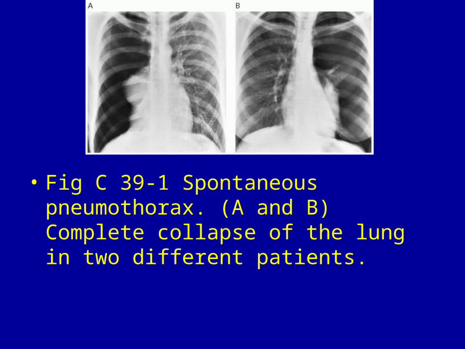
• Fig C 39-1 Spontaneous pneumothorax. (A and B) Complete collapse of the lung in two different patients.
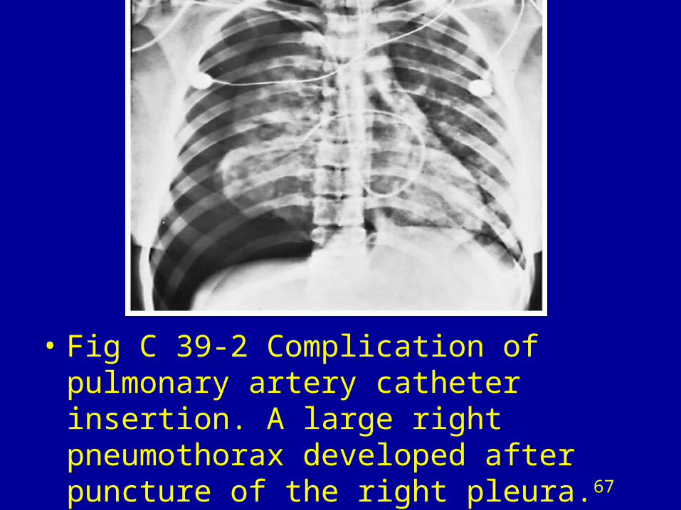
• Fig C 39-2 Complication of pulmonary artery catheter insertion. A large right pneumothorax developed after puncture of the right pleura.67
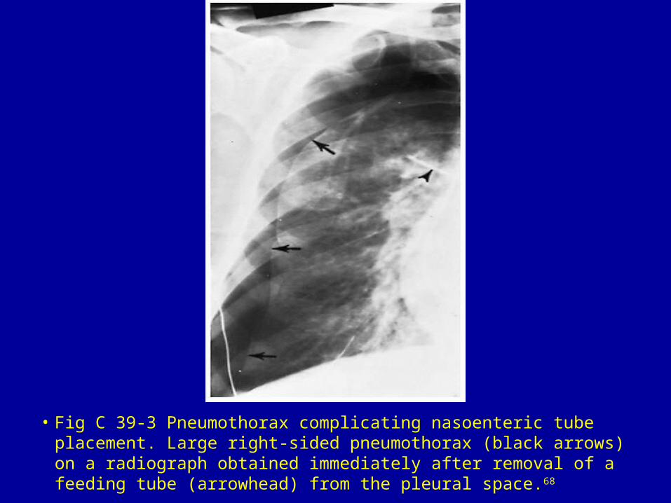
• Fig C 39-3 Pneumothorax complicating nasoenteric tube placement. Large right-sided pneumothorax (black arrows) on a radiograph obtained immediately after removal of a feeding tube (arrowhead) from the pleural space.68

• Fig C 39-4 Posttraumatic pneumothorax. Anteromedial pneumothorax (arrows) along with extensive air-space parenchymal disease.69
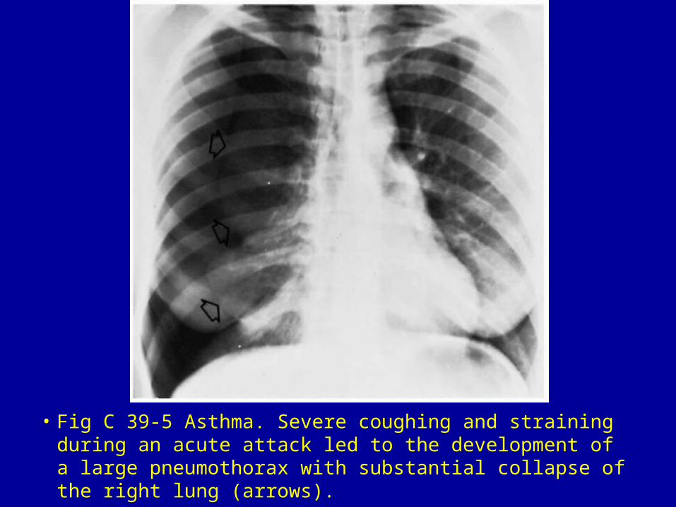
• Fig C 39-5 Asthma. Severe coughing and straining during an acute attack led to the development of a large pneumothorax with substantial collapse of the right lung (arrows).
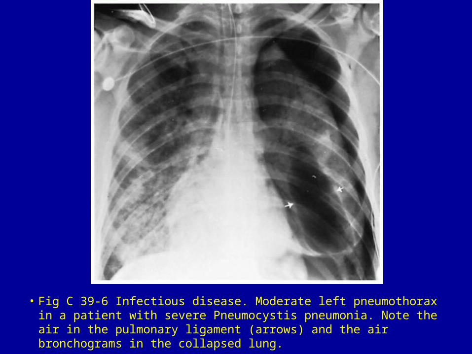
• Fig C 39-6 Infectious disease. Moderate left pneumothorax in a patient with severe Pneumocystis pneumonia. Note the air in the pulmonary ligament (arrows) and the air bronchograms in the collapsed lung.
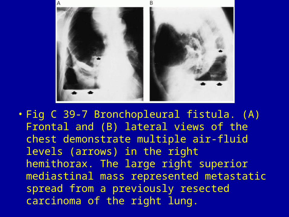
• Fig C 39-7 Bronchopleural fistula. (A) Frontal and (B) lateral views of the chest demonstrate multiple air-fluid levels (arrows) in the right hemithorax. The large right superior mediastinal mass represented metastatic spread from a previously resected carcinoma of the right lung.
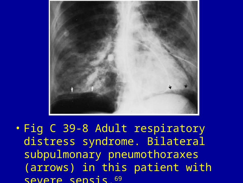
• Fig C 39-8 Adult respiratory distress syndrome. Bilateral subpulmonary pneumothoraxes (arrows) in this patient with severe sepsis.69