Modern techniques of increase the antibacterial properties...
6
49 they may be in direct contact with the product, or may migrate and cause the transfer of con- taminants into the product 3 . Some organic compounds used for desinfection purposes pose some disadvantages, including their toxicity for the human body 4 . ereofore there is an increasing of interest in using of inoarganic disenfectants, such as metal nanoparticles (NPs) 5 . Furthermore, a crucial factor related to the use of chemotherapeutics and antibiotics is the enhancement of resistant bacteria toward these decontaminant agents 6 . erefore it’s ne- cessary to develop new compounds wich would Modern techniques of increase the antibacterial properties of the instruments Kristýna Číhalová 1,2 , Dagmar Chudobová 1,2 , Vedran Milosavljevic 1,2 , Pavel Kopel 1,2 , Zbyněk Heger 1,2 , Vojtěch Adam 1,2 , René Kizek 1,2 a Department of Chemistry and Biochemistry, Mendel University in Brno, Zemedelska 1, CZ-613 00 Brno, Czech Republic - European Union b Central European Institute of Technology, Brno University of Technology, Technicka 3058/10, CZ-616 00 Brno, Czech Republic - European Union Accepted: 9. 12. 2014 Keywords: antibacterial, resistance, nanoparticles of metals, graphene oxide 1. Introduction Nowadays food and textile industries as well as health centers have employed advanced technologies and precaution measures which prevent bacteria proliferation and distribution among staff, consumers and patients. 1-3 ese technologies and measures aren’t always effecti- ve to fulfill the aseptic requirements. One of the main objectives to aseptic conditions is related to the necessity for the treatment of surfaces, tools and working materials by decontami- nants. e application of chemotherapeutics or antibiotics isn’t always a good choice since Modern techniques of increase the antibacterial properties of the instruments Complication caused by resistant bacteria require qualified technical and technological measures since bacteria proliferation can occur in operations where aseptic environment is required. Constituents used as decontaminating compositions can be either bactericidal inorganic or organic substances like chemotherapeutics and antibiotics. In research special attention is paid to the problem of bacterial resistence toward antibiotics. A possible solution to issue consists in the employment of ionts and metal complexes such as silver nitrate or new nanoparticles (silver, cooper), whose antibacterial properties are known over centuries. In the focus of this research is the preparation of a composite material, consisiting of gra- phene oxide and nanoparticles of zinc oxide (ZnO), silver phhosphate (Ag 3 PO 4 ) and silver. Graphene oxide was prepared by Hummers method, involving the oxidation of graphite flake in sulfuric acid with permanganate with subsequent addition of the necessary components for the formation of metal nanoparticles. Physicochemical methods are considered for their characterisation. e size of nanoparticles ranges from 50 – 200 nm. Subsequently, the an- tibacterial effect of composites was tested by disk method on bacterial cultures of S.aureus, E.coli, methicilin-resistant S. aureus (MRSA). Selenium nanoparticles exhibit the highest antibacterial activity from selected nanoparticles containing graphene oxide composite with an inhibitory zone with the size 5 ± 1 mm. Silver nanoparticles display also a distinguishable antibacterial effect with inhibition zones of 2 ± 1 mm. Graphene oxide which is modified by zinc oxide nanoparticles shows no inhibitory effect. e obtained results show suitability of the prepared composite materials as candidates for alternative antimicrobialmaterials. ARTICLE
Transcript of Modern techniques of increase the antibacterial properties...
49
they may be in direct contact with the product, or may migrate and cause the transfer of con-taminants into the product 3. Some organic compounds used for desinfection purposes pose some disadvantages, including their toxicity for the human body 4. Thereofore there is an increasing of interest in using of inoarganic disenfectants, such as metal nanoparticles (NPs) 5. Furthermore, a crucial factor related to the use of chemotherapeutics and antibiotics is the enhancement of resistant bacteria toward these decontaminant agents 6. Therefore it’s ne-cessary to develop new compounds wich would
Modern techniques of increase the antibacterial properties of the instruments Kristýna Číhalová1,2, Dagmar Chudobová1,2, Vedran Milosavljevic1,2, Pavel Kopel1,2, Zbyněk Heger1,2, Vojtěch Adam1,2, René Kizek1,2
a Department of Chemistry and Biochemistry, Mendel University in Brno, Zemedelska 1, CZ-613 00 Brno, Czech Republic - European Union
b Central European Institute of Technology, Brno University of Technology, Technicka 3058/10, CZ-616 00 Brno, Czech Republic - European Union
Accepted: 9. 12. 2014Keywords: antibacterial, resistance, nanoparticles of metals, graphene oxide
1. IntroductionNowadays food and textile industries as well
as health centers have employed advanced technologies and precaution measures which prevent bacteria proliferation and distribution among staff, consumers and patients.1-3 These technologies and measures aren’t always effecti-ve to fulfill the aseptic requirements. One of the main objectives to aseptic conditions is related to the necessity for the treatment of surfaces, tools and working materials by decontami-nants. The application of chemotherapeutics or antibiotics isn’t always a good choice since
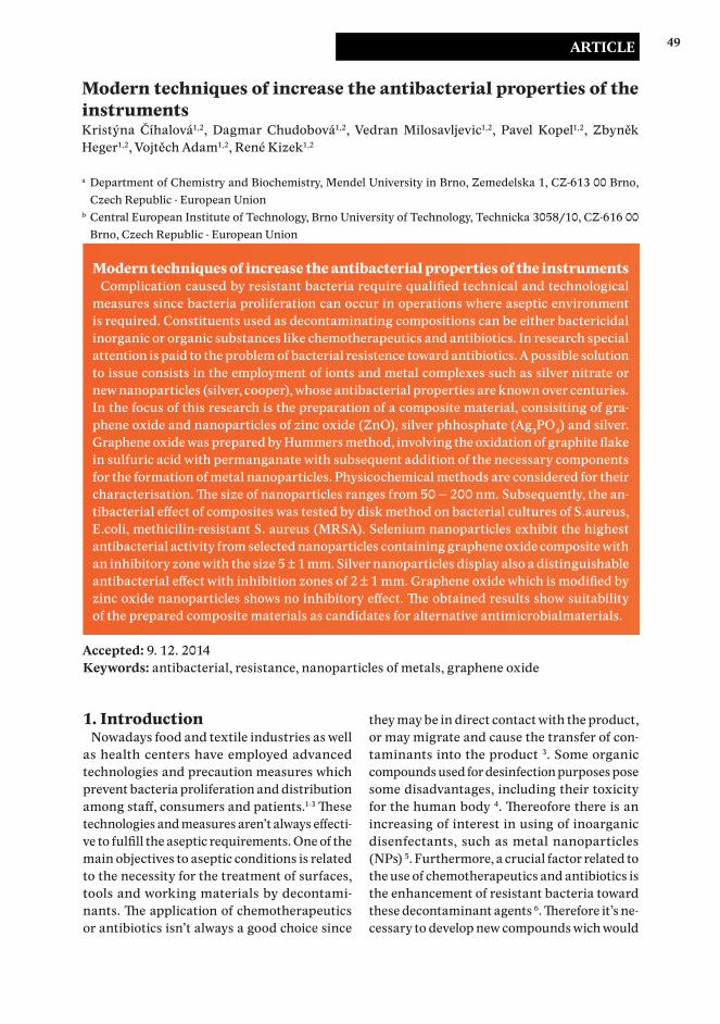
Modern techniques of increase the antibacterial properties of the instruments Complication caused by resistant bacteria require qualified technical and technological
measures since bacteria proliferation can occur in operations where aseptic environment is required. Constituents used as decontaminating compositions can be either bactericidal inorganic or organic substances like chemotherapeutics and antibiotics. In research special attention is paid to the problem of bacterial resistence toward antibiotics. A possible solution to issue consists in the employment of ionts and metal complexes such as silver nitrate or new nanoparticles (silver, cooper), whose antibacterial properties are known over centuries. In the focus of this research is the preparation of a composite material, consisiting of gra-phene oxide and nanoparticles of zinc oxide (ZnO), silver phhosphate (Ag3PO4) and silver. Graphene oxide was prepared by Hummers method, involving the oxidation of graphite flake in sulfuric acid with permanganate with subsequent addition of the necessary components for the formation of metal nanoparticles. Physicochemical methods are considered for their characterisation. The size of nanoparticles ranges from 50 – 200 nm. Subsequently, the an-tibacterial effect of composites was tested by disk method on bacterial cultures of S.aureus, E.coli, methicilin-resistant S. aureus (MRSA). Selenium nanoparticles exhibit the highest antibacterial activity from selected nanoparticles containing graphene oxide composite with an inhibitory zone with the size 5 ± 1 mm. Silver nanoparticles display also a distinguishable antibacterial effect with inhibition zones of 2 ± 1 mm. Graphene oxide which is modified by zinc oxide nanoparticles shows no inhibitory effect. The obtained results show suitability of the prepared composite materials as candidates for alternative antimicrobialmaterials.
Article

50
show baktericide properties also toward bacte-rial species immuned with special resistance or multiresistance to antibiotics 6, 7. Bacteria with enhanced resistance are often found in hospi-tal operations as a consequence of long-term antibiotics administration to patients, leading to genetic mutations and formation of various defense systems against damaging mechanism of antibiotics 8. The most resistant form which evolved from the bacteria S. aureus is methici-lin-resistant S. aureus (MRSA). Enhanced resi-stance to antibiotics or other bactericidal agents in staphylococ is present due to the variable genetic element such as pathogenicity islands genome, plasmids, transpons and others. The responsible gene for resistance enhancement in MRSA if found in the staphyloccocal chro-mosomal cassette mec (SCCmec) 9.
The integration of SCCmec into the genome of S. aureus leads to methicillin-resistant strain (MRSA) which is resistant to nearly all β-lactam antibiotics. The resistance toward methicillin is dedicated to the presence of penicillin-bin-ding protein 2a (PBP2a) and the β-lactamase enzyme that cleaves β-lactam antibiotics (pe-nicillin-based antibiotics), and is encoded by the mecA gene10. The PBP2a is an analogue of PBP transpeptidases whose presence is essen-tial for normal cell, as they play an important role in cell-wall synthesis 11. The presence of β-lactam antibiotics inhibit their function, and the cell loses its ability to produce cell wall and consequently dies. However PBP2a exhibits a low affinity toward β-lactam antibiotics, the-refore is not subject to their inhibition9. All of this lead to cost uneffective and time consu-ming therapy for hospitalized patients withMRSA 12.The treatment of surfaces, tools, fabrics or packages with antibacterial agents in the above mentioned areas is an absolute necessi-ty, and it is therefore it is necessary to ensure an effective treating agent with pronounced antibacterial effect. Such a property is prone of metal nanoparticles 13, graphene oxide (GO) or biofunctionalized graphene 14. Graphene with antimicrobial effect is used in containers for food packaging or for the coating of biomedical devices, wherein the bacterial colonization on the surface are unwanted 15. Modification of
graphene metal nanoparticles with antibacte-rial effect streamlines this effect.
2. Material and Methods2.1 Preparation of graphene oxide and metal nanoparticles
Preparation of graphene oxide (GO)Graphene oxide was prepared by Hummers
method using potassium permanganate. This occurred by transferring 46 ml of concentrated H2SO4 into an ice bathed beaker under continu-ous stirring of 2 g of graphite, 1 g NaNO3 and 6 g KMnO4. After it, the beaker was removed from the ice bath and placed at at room tempe-rature. Later on, 92 ml of water were added into the resulting solution , followed by 15 minutes of still standing, and the addition of 280 ml of warm water. Finally a 3% H2O2 was transferred into the prepared solution until until it exhibits a bright yellow color.
Preparation of zinc oxide nanoparticles (ZnO)
Zinc acetate dihydrate (assay ≥ 98%) was used as the raw material to synthesize ZnO nanostructures. According to this method 1 g of zinc acetate dihydrate was placed in an oven and then it was heated up for 2 h at 300 °C. The heating rate seemed to be a very important parameter in the amount of the product and the undesired phases which might appear du-ring the heating. So the heating rate was set to 10 °C/min and the samples were annealed at 300 °C for 2 h, and then cooled to room tempera-ture for 12 h. It was observed that two different kinds of products were obtained after sample annealing. A white porous powder covering the crucible walls was denoted as sample No. 1 a gray black powder stucking to the bottom of the crucible denoted as sample No. 2. After microwaving and naturally cooling down, the samples were transferred in eppendorf tubes and washed with ethanol. After that the pre-cipitate at the bottom of the eppendorf tube was washed 3 times with sufficient amounts of MiliQ water. The obtained sample was kept at ambient conditions 12 hours drying, yielding thereafter a total amount of 72 mg.
Journal of Metallomics and Nanotechnologies 2014, 4, 49—54

51
Preparation of silver nanoparticles (SPNPs)1 ml of AgNO3 (850 mg of AgNO3 in 10 ml
of MiliQ) was mixed with 25 ml of MiliQ and separately 1 ml of Na2HPO4 (850 mg of Na2HPO4 in 10 ml of MiliQ) was mixed with 25 ml of MiliQ. Immediately after preparation, these two solutions were mixed together and mixed a few minutes on magnetic stirrer until its color turned to light yellow. Then, 500 mg of cekol was added into this complex under continuous mixing of 2 hours.
Preparation of selenium nanoparticles (SeNPs)
5 ml of Na2SeO3 (Na2SeO3 263 mg/ 50 ml of MiliQ) was dissolved in 45 ml water followed by adding 80 µl of 3-mercaptopropionic acid (MPA). Its pH was adjusted to 9 by the addition of 1 M NaOH (0.2 ml). The reaction mixture was stirred for 2 hours yielding to a red color. Then these two solutions were mixed under magnetic stirring for 30 minutes. The color of solution was black and its pH was 6. Finally, 1 g of cekol was added into the solution under continuous mixing for two hours6.
2.2 cultivation of S. aureus, methicillin-re-sistant S. aureus and E. coli
S. aureus (NCTC 8511), methicillin-resistant S. aureus (7111 2/A8) and E. coli were obtained from the Czech Collection of Microorganisms, Faculty of Science, Masaryk University, Brno, Czech Republic. Cultivation media (LB = Luria Bertani) were inoculated with bacterial culture for 24 hours on a shaker at 130 rpm and 37°C. The bacterial culture was diluted by cultiva-tion medium to OD600 = 0.1 for the following experiments.
2.3 Determination of antimicrobial propertiesInhibition zones and growth curves
Agar surface in a Petri dish was covered with a mixture of 100 ml of 24 hour grown culture of S. aureus or E. coli and 3 ml of LB medium (Luria Bertani medium). The circles with a dia-meter of 1 cm were delivered by the factory (Vyzkumny ustav pletarsky in Brno) and mixed
with SPNPs with a concentration of 300 µM in complexes with hyaluronic acid (8.3 mM) or chitosan (9.7 mM). Petri dishes were incuba-ted in a thermostat at 37 °C for 24 hours. The antimicrobial effect of tested compounds was measured by measuring the absorbance using the apparatus Multiskan EX (Thermo Fisher Scientific, Germany). The culture was diluted with LB medium to obtain absorbance 0.1 AU at 600 nm. In the microtitration plate S. aure-us or E. coli culture was mixed with various concentrations of SPNPs (0, 10, 25, 50, 75, 150, 225 and 300 µM) and constant concentrati-on of hyaluronic acid (8.3 mM) and chitosan (9.7 mM). Total volume in the microtitration plate wells was always 300 µl.
3. Results and Discussion3.1 characterization of particles
Average size of nanoparticle and distribution curves were determined by a dynamic light sca-ttering with a device Zetasizer Nano (Malvern) (Fig. 1). The average size of the nanoparticles was GO 200 nm, ZnO 50 nm, SPNPs 100 nm and SeNPs 15 nm. The composition of the nano-particles was also demonstrated by using X-ray fluorescence spectrometry (XRF) instrument Spectro XEPOS. In the spektra were found peaks corresponding to lines of zinc, silver, phosphorus and selenium.
For using graphene oxide modified by nano-particles of zinc oxide to creating to inhibition zones around the diffusion dishes in any of the tested cultres. A similiar study Leung (2012) dealt with the GO modified by ZnO nanopar-ticles, when the researchers demonstrated the antibacterial effect of this composite applica-tions to surface materials. Diferent results are caused by the different preparation, size and surface finish of GO and ZnO nanoparticles16. Graphene oxid is modified with silver nanopar-ticles and it was used as the second potential antibacterial components. For these particles were give 2 mm of inhibition zone during incu-bation with E. coli, S. aureus was inhibited by
Číhalová et al.

52
formation of 3 mm inhibiton zone and MRSA was inhibited by formation only 1 mm inhibi-tion zone. Method of measuring the inhibition zones is useful as a primary test the inhibitory effect of substances 17. In the third composite with GO were chosen selenium nanoparticles (SeNPs). Selenium nanoparticles are new alter-native growth inhibition of bacterial cultures, the effect of which dealt with the study 6, when were compared the effect SeNPs and SPNPs. For SPNPs were measured inhibition zones of 3 mm, while SeNPs inhibition zone of size 7 mm. Graphene oxide modified by SeNPs worked on bacterial cultures in the most efficient inhibi-tion effect of the test substances. For bacterial E. coli culture was made 4 mm inhibition zone from the diffusion disk with inhibitory substan-ce. In S. aureus was measured 6 mm inhibition zone and in MRSA was measured 5 mm zone of inhibition. Graphene oxid was modified with selenium nanoparticles and it can be classified as an effective inhibitory components on G+, G- and resistant bacteria.
Journal of Metallomics and Nanotechnologies 2014, 4, 49—54
Figure 1. Results of measurment of particle size by using dynamic light scattering.
Figure 2. Testing the antibacterial action by method of inhibition zones. While we tested GO modified by ZnO avoid to the formation of inhibition zones around any of the test bacterial cultures E. coli (a), S. aureus (b) a MRSA (c). In GO modified by SPNPs have been created 2 mm inhibiti-on zone in E. coli (d), 3 mm in S. aureus (e) a 1 mm in MRSA (f). In modification GO by SeNPs particles leads to the for-mation of 4 mm inhibition in E. coli (g), 6 mm in S. aureus (h) and 5 mm in MRSA (i).

53
An inhibition effect was different for diversity of the selected bacteria. Gram negative E. coli has a stronger cell wall due to the presence of the outer membrane resulting in a poor transport of nanoparticles inside cells 18. High inhibition effect on S. aureus is due to absence of lipopo-lysaccharide and outer membrane layer in cell wall 19. The mechanism causing resistence to β-lactam antibiotics fails to protect the resi-stant bacteria to antibiotics against complex of graphene oxide and selenium nanoparticles 20.
From the above results we can conclude that the GO modified by SeNPs act by inhibitory effect in all the cultures, while the GO modified by ZnO doesn’t provide an inhibitory effect on any of these cultures. A similiar Leung study (2012) dealt with the GO modified by ZnO nano-particles, when researchers proved antibacterial effect of this composite applications on surface materials. Different results may be caused by different prepare GO and ZnO nanoparticles 16.
AcknowledgementsFinancial support from the project ANTIBAKT
TR9140017 is highly acknowledged
The authors declare they have no potential conflicts of interests concerning drugs, pro-ducts, services or another research outputs in this study.
The Editorial Board declares that the ma-nuscript met the ICMJE „uniform require-ments“ for biomedical papers.
References1. Yip, J., et al., Investigation of Antifungal and
Antibacterial Effects of Fabric Padded with Highly Stable Selenium Nanoparticles. Journal of Applied Polymer Science, 2014. 131(17).
2. Nateghi, M.R. and H. Hajimirzababa, Effect of silver nanoparticles morphologies on antimicrobial properties of cotton fabrics. Journal of the Textile Institute, 2014. 105(8): p. 806-813.
3. Olar, R., et al., Synthesis, characterisation and thermal behaviour of some thiosulfato- and sulfato copper(II) complexes - Antibacterial activity. Journal of Thermal Analysis and Calorimetry, 2008. 92(1): p. 245-251.
4. Messad, S., et al., Frequency of contamination and antimicrobial resistance of thermotolerant Campylobacter isolated from some broiler farms and slaughterhouses in the region of Algiers. Food Control, 2014. 40: p. 324-328.
5. Hajipour, M.J., et al., Antibacterial properties of nanoparticles. Trends in Biotechnology, 2012. 30(10): p. 499-511.
6. Chudobova, D., et al., Comparison of the effects of silver phosphate and selenium nanoparticles on Staphylococcus aureus growth reveals potential
Číhalová et al.
Figure 3. Growth curves of bacterial cultures after application grephene oxide with different concentrations of selenium nanoparticles. Using concentration of grephene oxide was stacionary for all different concentrations of selenium nano-particles. S. aureus (a) and MRSA (b) growth in a similar downward trend with increasing concen-trations of selenium nanoparticles in complex with graphene oxide (1 mg/ml). For E. coli was not too significant downward trend (c).

54
for selenium particles to prevent infection. Fems Microbiology Letters, 2014. 351(2): p. 195-201.
7. Panacek, A., et al., Silver colloid nanoparticles: Synthesis, characterization, and their antibacterial activity. Journal of Physical Chemistry B, 2006. 110(33): p. 16248-16253.
8. Thomas, R., et al., Antibacterial Activity and Synergistic Eff ect of Biosynthesized AgNPs with Antibiotics Against Multidrug-Resistant Biofi lm-Forming Coagulase-Negative Staphylococci Isolated from Clinical Samples. Applied Biochemistry and Biotechnology, 2014. 173(2): p. 449-460.
9. Bhargava, K. and Y.F. Zhang, Characterization of methicillin-resistant coagulase-negative staphylococci (MRCoNS) in retail meat. Food Microbiology, 2014. 42: p. 56-60.
10. Khan, S., et al., Rapid optical determination of beta-lactamase and antibiotic activity. Bmc Microbiology, 2014. 14.
11. Wan, M.T. and C.C. Chou, Spreading of beta-lactam resistance gene (mecA) and methicillin-resistant Staphylococcus aureus through municipal and swine slaughterhouse wastewaters. Water research, 2014. 64: p. 288-95.
12. Hernandez-Porto, M., et al., Risk factors for development of methicillin-resistant Staphylococcus aureus-positive clinical culture in nasal carriers aft er decolonization treatment. American Journal of Infection Control, 2014. 42(7): p. E75-E79.
13. Kanmani, P. and S.T. Lim, Synthesis and characterization of pullulan-mediated silver nanoparticles and its antimicrobial activities. Carbohydrate Polymers, 2013. 97(2): p. 421-428.
14. Sang, J.K., et al., Composition useful for exhibiting antibacterial eff ect with respect to Gram-positive bacterium e.g. Streptococcus iniae or Gram-negative bacterium e.g. Escherichia coli, comprises graphene oxide, univ cheju nat ind academic coop found (uych-non-standard).
15. Yadav, S.K., et al., Mechanically Robust, Electrically Conductive Biocomposite Films Using Antimicrobial Chitosan-Functionalized Graphenes. Particle & Particle Systems Characterization, 2013. 30(8): p. 721-727.
16. Leung, Y.H., et al., Antibacterial activity of ZnO nanoparticles with a modified surface under ambient illumination. Nanotechnology, 2012. 23(47).
17. Chudobova, D., et al., Complexes of Silver(I) Ions and Silver Phosphate Nanoparticles with Hyaluronic Acid and/or Chitosan as Promising Antimicrobial Agents for Vascular Grafts. International Journal of Molecular Sciences, 2013. 14(7): p. 13592-13614.
18. Zanzen, U., et al., Antibacterial action of copper ions on food-contaminating bacteria. Acta Biologica Szegediensis, 2013. 57(2): p. 149-151.
19. Kolar, S.L., et al., NsaRS is a Cell-Envelope-Stress Sensing Two-Component System of Staphylococcus aureus. ArrayExpress Archive, 2012.
20. Lee, S.J., et al., Electrospun chitosan nanofi bers with controlled levels of silver nanoparticles. Preparation, characterization and antibacterial
activity. Carbohydrate Polymers, 2014. 111: p. 530-537.
Journal of Metallomics and Nanotechnologies 2014, 4, 49—54
This licence allows users to download and share the article for
non-commercial purposes, so long as the article is reproduced in the whole without changes, and the original authorship is acknowledged.


![Autoclave Sterilization /Desinfection of materials [Eng].pdf · DEFINITION Autoclave-Pressurized vessel-A steam saturated environment permits obtaining temperature >100°C Autoclaving](https://static.fdocuments.in/doc/165x107/5cd75c0288c993273a8c13ff/autoclave-sterilization-desinfection-of-engpdf-definition-autoclave-pressurized.jpg)